Unit 5 Skeletal system. Skeletal system Composed of cartilage and bone.
58
Unit 5 Skeletal system
-
Upload
abigail-mould -
Category
Documents
-
view
222 -
download
0
Transcript of Unit 5 Skeletal system. Skeletal system Composed of cartilage and bone.

Unit 5 Skeletal system
Skeletal system
Composed of cartilage and bone

Functions
• Support soft tissue
• Prtects delicate organs
• Movement
• Storage area ( Calcium, phosphorus, fats
• Hemopoiesis: blood cell formation

Cartilage:
• consists of chondrocytes living in a cavity (lacuna) and is avascular with cells being nourished by diffusion
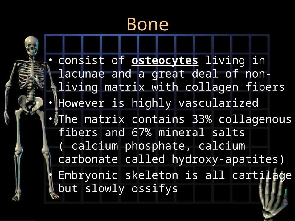
Bone
• consist of osteocytes living in lacunae and a great deal of non-living matrix with collagen fibers
• However is highly vascularized• The matrix contains 33% collagenous fibers
and 67% mineral salts ( calcium phosphate, calcium carbonate called hydroxy-apatites)
• Embryonic skeleton is all cartilage but slowly ossifys
Bone structures
• Diaphysis: the shaft ( compact bone)
• Epiphysis: ends ( thin layer of compact bone covering spongy bone
• Metaphysis: region where epiphysis and diaphysis come together, region of epiphyseal plate

Bone structures
• Articular cartilage: thin hyaline cartilage covering of epiphyseal surface at joints
• Medullary cavity (marrow cavity): space within diaphysis

Bone structures
• Periosteum: dense irregular, white fibrous layered covering of a bone.
• Is composed of 2 layers: • Fibrous layers: outer layer composed of
bibrous connective tissue containing blood vessels
• Osteogenic layer: layer closer to bone that contains elastic fibers, blood vessels, osteoclast and osteroblast
Functions of periosteum
• Growth
• Repair
• Nutrition
• Attachments for ligament and tendons

Classification of bones by shape
• Long: longer than they are wide ex: most extremities, phalanges
• Short: wider than they are long ex: carpals and tarsals
• Flat: ex: cranium, ribs, shoulder girdle• Irregular: vertebrae and facial• Sessamoid: patella• Wormian: grows between bones of
skull

2 Types of bone tissue
1. Compact:
• Covers spongy bone, is dense and provides strength
• Structural unit of compact bone is the osteon

Osteon
• The osteon is a hard bone matrix arranged in rings (lamellae) around a haversian canal
• Volkman’s canal: carry blood vessels and nerves from the periosteum into the haversian canal and on into the medullary cavity
• Osteocytes: mature osteoblasts, live in the lacunae between lamellae
• Canaliculi: are tiny canals which connect lacunae with each other and with the haversian canal.
• Inside canaliculi are cellular extensions of osteocytes creating a network for nutrients and distribution
• Areas between osteons where the lamellae are only partially circled are called interstitial lamellae.
• They are the remnants of old partially destroyed osteons.


Types of bone tissue
2. Spongy bone• Consists of a lattice work of bone called
trabeculae• In between the trabeculae are spaces filled
with red bone marrow and blood vessels• There is no network of canals because the
blood can reach the osteocytes by diffusion through marrow spaces
• Is less dense and less strong and is found in the epiphysis of long bones and the bulk of flat bones or irregular bones
Bone formation ( osteogenesis or ossification
• Embryo’s skeleton is made of cartilage and fibrous membranes
• At around 8 weeks after conception and throughout childhood ( until about age 20) this connective tissue model is slowly ossified ( turned to bone)
Intramembraneous ossification
• Results in formation of flatbones and some irregular ones by ossification of pre existing fibrous connective tissue
1. The osteoblasts begin secreting bone matrix eventually forming spongy bone …later the outer layers are reconstructed into compact bone
Intramembraneous ossification
2. The original connective tissue which encloses the “growing” bone becomes the periosteum
3. Some osteoblasts trap themselves in the matrix (lacunae) and are now called osteocytes
• Bones are destroyed and reformed many times before adulthood and final size and shape

Endochondral ossification
• Most bones formed this way• Involves replacement of hyaline cartilage with
bone• Embryonic skeletons are cartilage enclosed by
connective tissue called perichondrium• Once a blood vessel penetrates the
perichondrium of the embryonic bones the chondrocytes become osteoblasts which begin forming bone matrix
Endochondral ossification
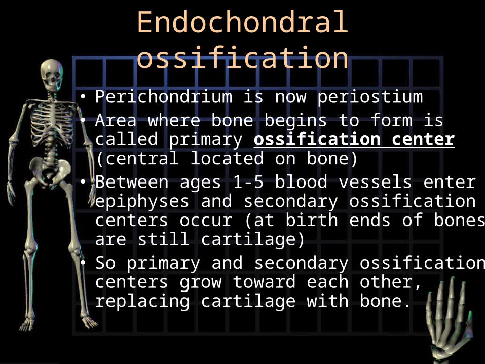
• Perichondrium is now periostium• Area where bone begins to form is called
primary ossification center (central located on bone)
• Between ages 1-5 blood vessels enter epiphyses and secondary ossification centers occur (at birth ends of bones are still cartilage)
• So primary and secondary ossification centers grow toward each other, replacing cartilage with bone.

Endochondral ossification
• The cartilage between these ossification centers is called epiphyseal plate (don’t ossify until about 18-25)
• Growth in diameter is due to osteoblasts in periosteum adding new osseous tissue to outside surface of bones
• Initially all bone growth begins with spongy bone which is later remodeled into compact bone.

Bone Remodeling
• The continuous balance between bone formation and bone reabsorption, occurs at all periosteal and endosteal surfaces
• Bone is constantly replacing itself throughout adult life.

Bone disorders
1. Rickets: childhood disease due to lack of vitamin D (sunlight) causes cartilage not to be calcified, bones are soft and malformed
2. Osteomalacia: adult rickets… too little vitamin D
• Solution:Solution: both need dietary calcium and sunlight (vitamin D)
• Both disorders can also be due to either kidney or liver disease and resultant is a poor processing of calcium

Bone disorders
3. Osteoporosis: character sized by a decrease in bone mass due to decease osteoblast activity. Generally age related ( decrease in hormone activity, females often due to menopause)
• Also due to insufficient exercise, resulting in brittle porous bones
• Bones are easly broken, decreased height, hunched back etc…
• Solution: exercise, calcium and vitamin supplements and maybe hormones

Bone disorders
4. Osteomyelitis: includes all infectious disease of bone

Divisions of skeletal system
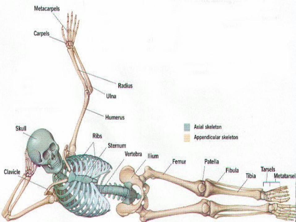
1. Axial skeleton: forms midline of skeleton
• 80 of 206 bones in human body are axial ( 28 in skull alone
2. Appendicular: remiainng 126 bones
• Found in arms, legs, pectoral and plevic girdle


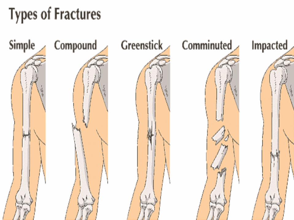
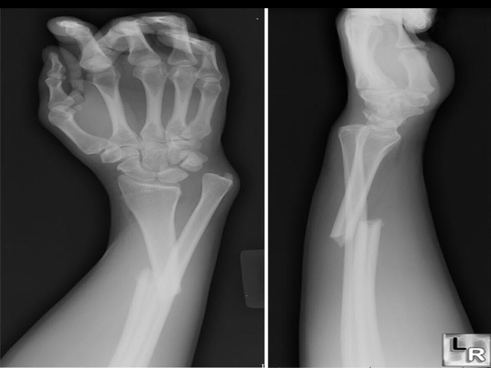
Fractures: breaks in bones
• Simple: Bone breaks cleanly, but does not penetrate skin
• Compound; broken ends of bone protrude trough skin
• Comminuted: bone fragments into many pieces
• Compression: bone is crushed
• Depression: broken bone portion is pressed inward
• Impacted: broken bone ends are forced into each other
• Spiral: ragged break occurs when excessive twisting forces are applied to a bone
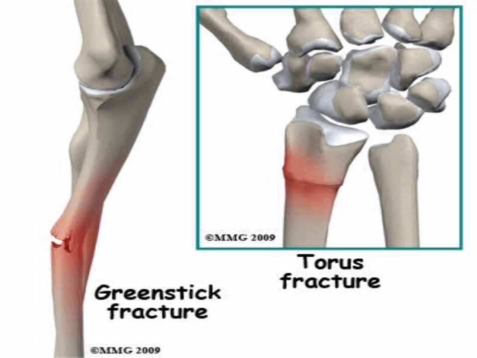
• Greenstick: bone breaks incompletely, much in the way a green twig breaks
• Colle’s fracture: fracture of distal end of lateral forearm (radius) in which distal fragment is displaced posterior
• Stress fracture: microscopic fissures in bone that form without evidence of injury to other tissue no visible break.
• Pott’s fracture: fracture at distal end of lateral legbone (fibula)) with serious injury of distal tibial articulation


Intramembraneous ossification
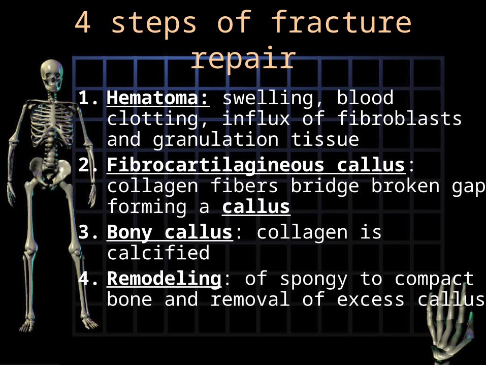
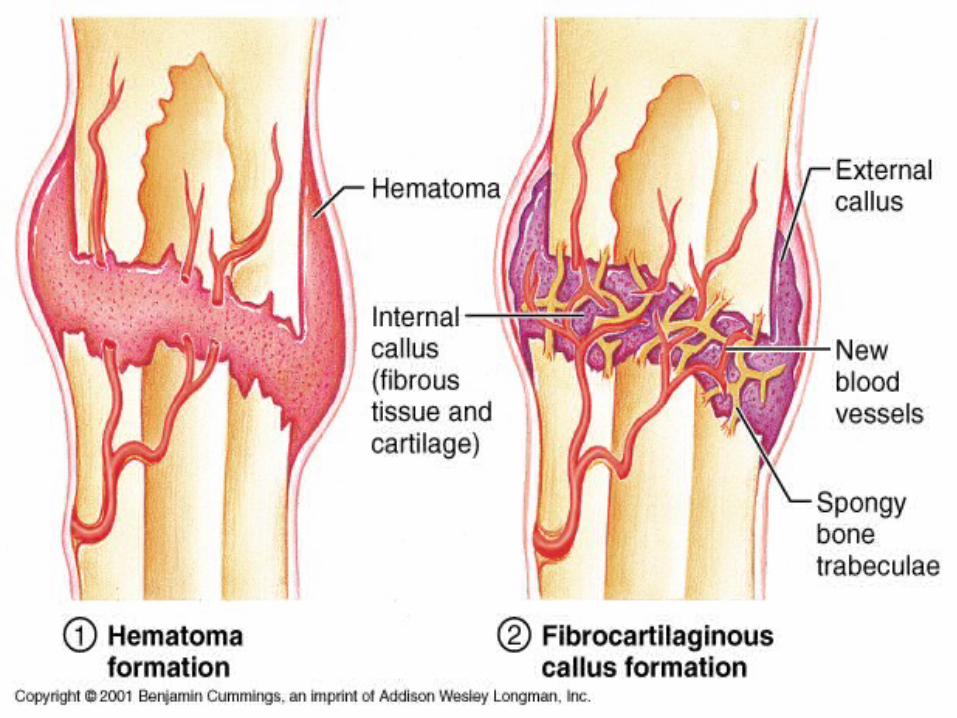
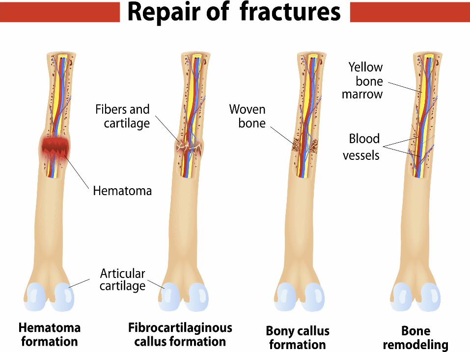
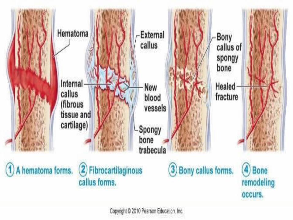
4 steps of fracture repair
1. Hematoma: swelling, blood clotting, influx of fibroblasts and granulation tissue
2. Fibrocartilagineous callus: collagen fibers bridge broken gap forming a callus
3. Bony callus: collagen is calcified4. Remodeling: of spongy to compact
bone and removal of excess callus

Articulations (joints)
• Functional classification
1. synarthroses: immovable joint (skull sutures)
2. Amphiarthroses: slightly moveable (between vertebrae or in pelvis)
3. Diarthrosis: freely moveable

Structural Classification
1.1. Fibrous joints:Fibrous joints: no join cavity, bones held together by fiberous connective tissue
2. Cartilaginous joints: still no cavity, bones held together by cartilage
3. Synovial joints: have cavities, free moving• Have articular cartilage (haline)• Have articular capsule (units articular bones)• Has synovial fluid• Has reinforcing ligaments

Types of Fibrous joints
a) Sutures: only in skull, bones touch joined by connective tissue (synathrotic)
b) Syndesmosis: bones slightly separated (amphiarthrotic) ex tibia, fibula
c) Gomphosis: peg in a socket ex teeth

Types of Cartilaginous joints
a) Synchondrosis: immovable, connective tissue is hyaline cartilage ex rib to sternum
b) Symphysis: connective tissue is fibrocartilage disk, little movement. Ex between vertebrae, pubic symphysis

Bursae and Tendon sheaths
• Little sac-like synovial membrane bags filled with synovial fluid and serving as friction reducing cushions between tendons, ligaments, muscles and bones
• It keeps these from rubbing each other ( inflammation of bursa is Bursitis)

Factors limiting synovial joint movement
1. Structure/shape of articulating bones
2. Ligament tension and arrangement
3. Muscle tone/tension
4. Apposition of soft parts
Miscellaneous
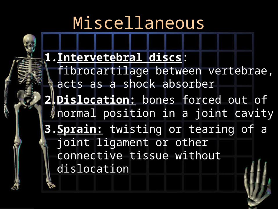
1. Intervetebral discs: fibrocartilage between vertebrae, acts as a shock absorber
2. Dislocation: bones forced out of normal position in a joint cavity
3. Sprain: twisting or tearing of a joint ligament or other connective tissue without dislocation

4. Strain: less severe, overstretched muscle
5. Sinuses: mucous membranes line cavities in skull, are open to nasal passages and thus to infections
6. Bunions: swelling of bursa in joint between metatasals and phalange (big toe)

6. Arthritis: catch all term referring to 100+ types of joint diseases
7. Rheumatoid arthritis: chronic inflammatory disorder affects women three times more often than men
• A disorder in which the body’s immune system attempts to destroy its own tissue
8. Gouty arthritis: metabolic disorder in which uric acid crystal s build up in soft tissues inflaming joints. Most common in males and typically affects a single joint
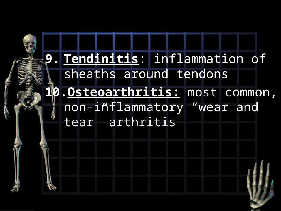
9. Tendinitis: inflammation of sheaths around tendons
10.Osteoarthritis: most common, non-inflammatory “wear and tear” arthritis

Factors in bone Physiology
• 1. sufficient dietary calcium phosphorus
• Calcium needed in nerve impulses
• Phospherous: in ATP
• 2. Vitamins:
• 3. Hormones