Human Anatomy & Physiology, Sixth Edition 6 Bones and Skeletal Tissues.
9/13/2010
1
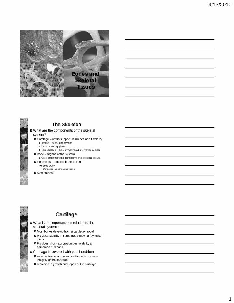
Bones andSkeletal Tissues
The SkeletonWhat are the components of the skeletal system?
Cartilage – offers support, resilience and flexibilityHyaline – nose, joint cavities Elastic – ear, epiglottisFibrocartilage – pubic symphysis & intervertebral discs
Bone – organs of the systemAlso contain nervous, connective and epithelial tissues
Ligaments – connect bone to boneTissue type?
Dense regular connective tissue
Membranes?
CartilageWhat is the importance in relation to the skeletal system?
Most bones develop from a cartilage modelProvides stability in some freely moving (synovial) jointsProvides shock absorption due to ability to compress & expand
Cartilage is covered with perichondriuma dense irregular connective tissue to preserve integrity of the cartilageAlso aids in growth and repair of the cartilage.
9/13/2010
2
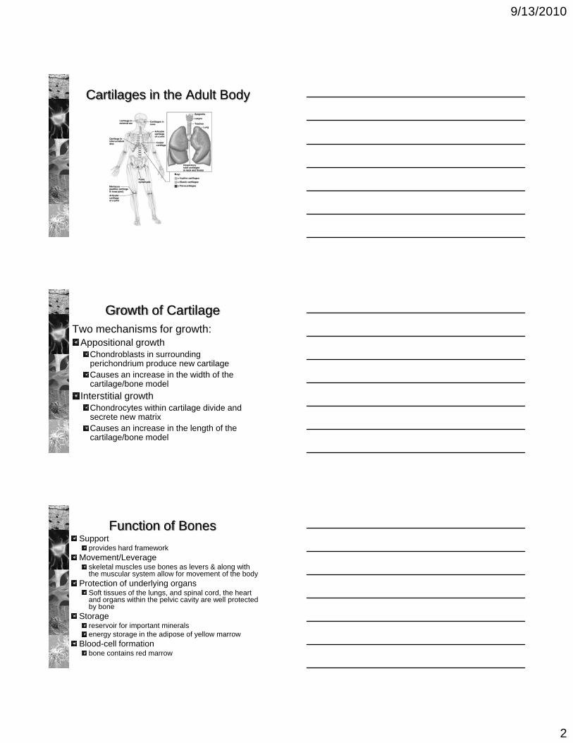
Cartilages in the Adult Body
Growth of CartilageTwo mechanisms for growth:
Appositional growth Chondroblasts in surrounding perichondrium produce new cartilageCauses an increase in the width of the cartilage/bone model
Interstitial growth Chondrocytes within cartilage divide and secrete new matrixCauses an increase in the length of the cartilage/bone model
Function of BonesSupport
provides hard frameworkMovement/Leverage
skeletal muscles use bones as levers & along with the muscular system allow for movement of the body
Protection of underlying organsSoft tissues of the lungs, and spinal cord, the heart and organs within the pelvic cavity are well protected by bone
Storagereservoir for important mineralsenergy storage in the adipose of yellow marrow
Blood-cell formationbone contains red marrow
9/13/2010
3
Classification of BonesLong bones
longer than wide containing a shaft plus endsExample: femur
Short bonesroughly cube-shapedExample: carpals & tarsals
Sutural (Wormian) bonesSmall flat bones found in the sutures of the skull
Sesamoid bonesSmall, round and usually flattened slightly and develop inside of tendonsExample: patella
Flat bonesthin and flattened, usually curvedExample: cranial bones (frontal, parietal, temporal, occipital), ribs
Irregular bonesvarious shapes, do not fit into other categoriesExample: vertebrae
Bone Structure
Bones are composed of osseous tissueMatrixCells
Outer layer of tissue on bone is the periosteumInner layer of tissue in bone is the endosteum (lining the marrow cavity)
Microscopic Structure of BoneMatrix
FibersMainly collagen
Ground substancemineralized inorganic material called hydroxyapatite
Calcium Phosphate - Ca3(PO4)2 and Calcium Hydroxide -Ca(OH)2 are the main components that interact to form hydroxyapatite
Ca10(PO4)6(OH)2
Other substances (calcium carbonate, sodium, magnesium and flouride also become incorporated in the hydroxyapatite providing strength
Both are merged in osseous tissue the collagen provides a “framework” for the inorganic salts creating a tissue that is flexible but strong

9/13/2010
4
Microscopic Structure of Bone
CellsOsteocytes – the mature bone cell
Maintain the matrix by controlling calcium salt deposits in the matrix and the release of calcium into the bloodHoused in lacunae that are embedded between the layers of matrix (lamellae)Communicate via canaliculi, allowing osteocyte processes tocommunicate with adjacent osteocytes across by diffusion or via gap junctions
Microscopic Structure of BoneCells, cont.
OsteoblastsLocated on the inner and outer surface of boneSecrete osteoid (organic portion of matrix) which later becomes mineralizedResponsible for osteogenesisOnce surrounded by matrix it becomes an osteocyte
Osteoprogenitor Cells (progenitor = ancestor)The mesenchymal cells that differentiate into osteoblastsFound on the inner lining (endosteum) and the outer lining (periosteum)
OsteoclastsLarge cells that cause osteolysissecretion of acids that dissolve the matrix
Osteoclast
Maintenance of the Matrix
Matrix maintenance is a balance between osteoclast and osteoblast activity.
Osteoclast>osteoblast = bone removal (resporption)Osteoclast<osteoblast = bone addition (deposition)
Controlled by hormones that regulate blood Ca2+ levelsCalcitonin (CT)
reduces Ca2+ plasma levelsParathyroid Hormone (PTH)
Elevates Ca2+ plasma levels
9/13/2010
5
Compact vs Spongy BoneThe matrix may be highly organized or unorganized
Compact bone = organized and relatively solidSpongy bone = unorganized and open design
Both types:present in boneshave osteocytes, canaliculi and lamellae
Compact BoneFunctional unit is the Osteon (Haversian system)Concentric lamellae surrounding a central canal, collagen fibers spiral in different directions in each layer.Also forms larger circular rings called circumferential lamellaethat surround many osteons and the matrix that “fills in” the spaces around the osteons = interstitial lamellae
Transverse Section
Compact vs Spongy BoneSpongy Bone
Matrix organized into plates of parallel lamellaeForming a lattice called trabeculaeCanaliculi open to endosteum to gain nutrientsLight weight and high strength
Locations of compact & spongy bone:Spongy in the ends of bones and in the marrow or medullary cavitiesCompact is lining all bones, thickest in areas of high stresses
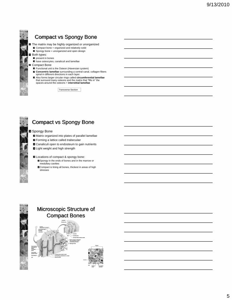
Microscopic Structure of Compact Bones
9/13/2010
6
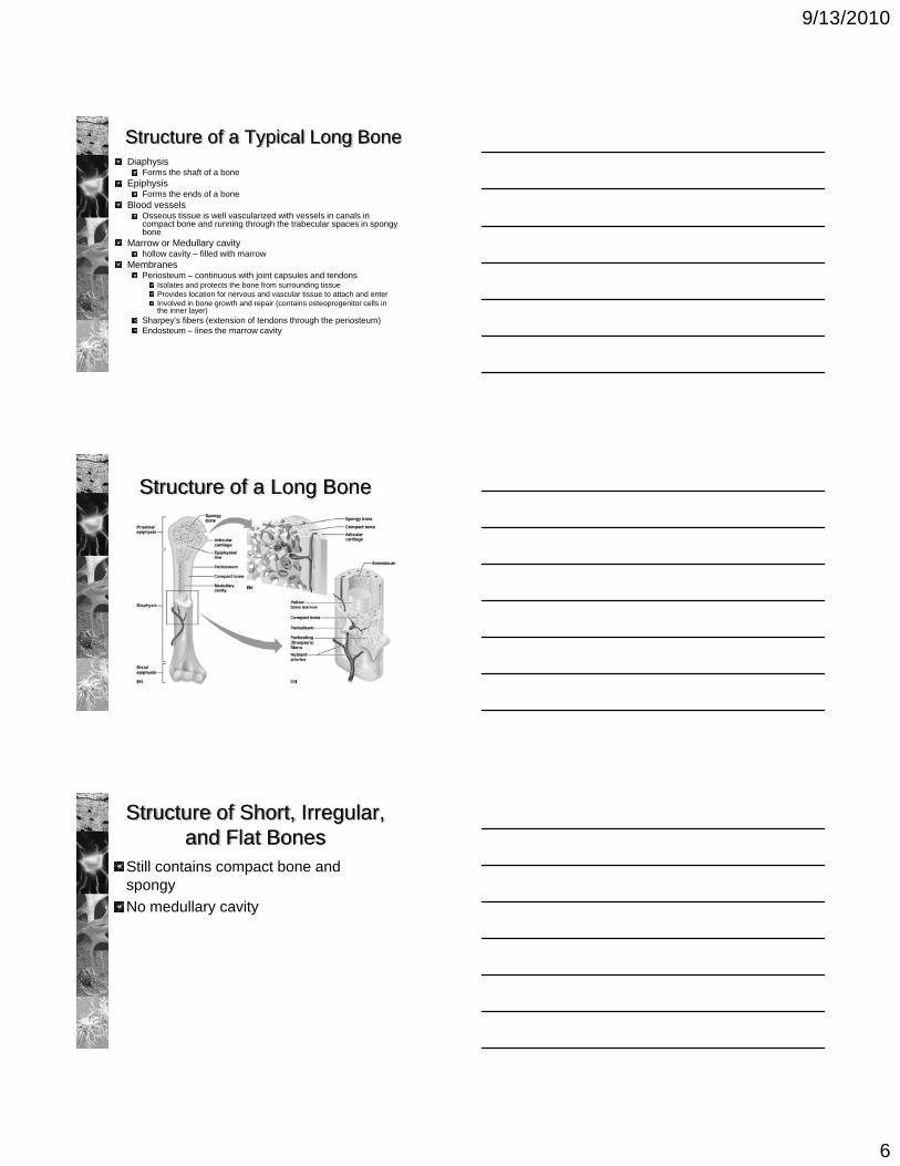
Structure of a Typical Long BoneDiaphysis
Forms the shaft of a boneEpiphysis
Forms the ends of a boneBlood vessels
Osseous tissue is well vascularized with vessels in canals in compact bone and running through the trabecular spaces in spongy bone
Marrow or Medullary cavity hollow cavity – filled with marrow
MembranesPeriosteum – continuous with joint capsules and tendons
Isolates and protects the bone from surrounding tissueProvides location for nervous and vascular tissue to attach and enterInvolved in bone growth and repair (contains osteoprogenitor cells in the inner layer)
Sharpey’s fibers (extension of tendons through the periosteum)Endosteum – lines the marrow cavity
Structure of a Long Bone
Structure of Short, Irregular, and Flat Bones
Still contains compact bone and spongyNo medullary cavity
9/13/2010
7
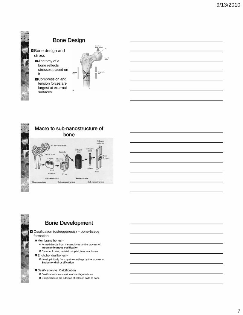
Bone Design
Bone design and stress
Anatomy of a bone reflects stresses placed on itCompression and tension forces are largest at external surfaces
Macro to sub-nanostructure of bone
Bone DevelopmentOssification (osteogenesis) – bone-tissue formation
Membrane bones –formed directly from mesenchyme by the process of: Intramembranous ossificationClavicle, frontal, parietal occipital, temporal bones
Enchchondral bones –develop initially from hyaline cartilage by the process of Endochondral ossification
Ossification vs. CalcificationOssification is conversion of cartilage to boneCalcification is the addition of calcium salts to bone
9/13/2010
8
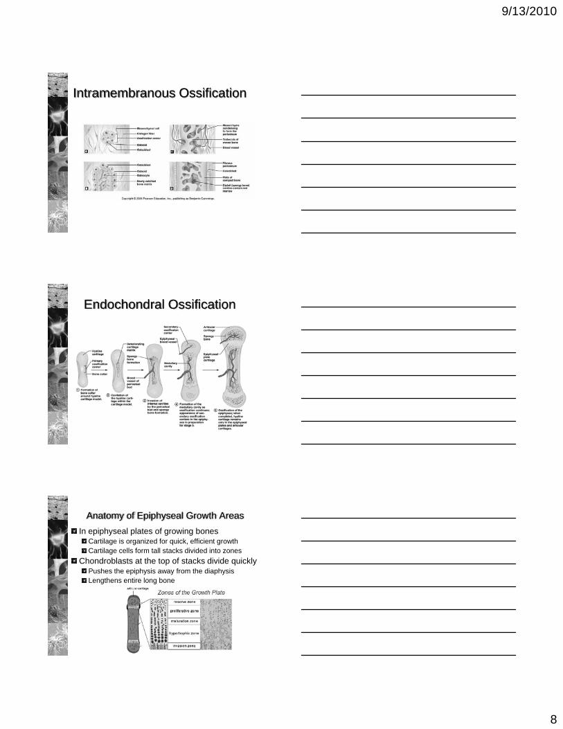
Intramembranous Ossification
Endochondral Ossification
Anatomy of Epiphyseal Growth AreasIn epiphyseal plates of growing bones
Cartilage is organized for quick, efficient growthCartilage cells form tall stacks divided into zones
Chondroblasts at the top of stacks divide quicklyPushes the epiphysis away from the diaphysisLengthens entire long bone

9/13/2010
9
Anatomy of Epiphyseal Growth Areas
Older chondrocytes signal surrounding matrix to calcifyOlder chondrocytes then die and disintegrate
Leaves long trabeculae (spicules) of calcified cartilage on diaphysis sideTrabeculae are partly eroded by osteoclastsOsteoblasts then cover trabeculae with bone tissueTrabeculae finally eaten away from their tips by osteoclasts
Growth of Other Types of Endochondral Bones
Short bones – arise from a single ossification centerIrregular bones – develop from distinct ossification centersSmall long bones
Form from a primary ossification center and a single secondary ossification center
Hormonal Regulation of Bone Growth
Growth hormoneproduced by the hypophysis (anterior pituitary gland)Stimulates epiphyseal plates
increases rate of division of chondrocytesThyroid hormone
ensures that the skeleton retains proper proportions
Sex hormonesPromote bone growth
Cartilage growth increases, butRate of osteoblast activity also increases, and
Synergistically with hGH and thyroid hormones induces closure of epiphyseal plates (between 18-25 yrs)

9/13/2010
10
Bone Remodeling
Bone deposit and removalOccurs at periosteal and endosteal surfaces
Bone remodeling Bone deposition – accomplished by osteoblastsBone reabsorption – accomplished by osteoclasts
Remodeling, Spongy Bone
Figure 6.12
Repair of Bone Fractures
Simple and compound fracturesSimple – breaks but does not penetrate skinCompound – breaks and protrudes through skin
Treatment by reductionClosed reduction – realignment by handOpen reduction – realignment by surgery
9/13/2010
11
Stages of Healing a Fracture
Figure 6.14
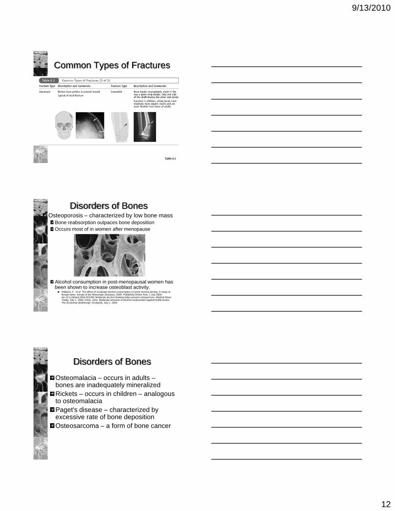
Common Types of Fractures
Table 6.1
Common Types of Fractures
Table 6.1
9/13/2010
12
Common Types of Fractures
Table 6.1
Osteoporosis – characterized by low bone massBone reabsorption outpaces bone depositionOccurs most of in women after menopause
Alcohol consumption in post-menopausal women has been shown to increase osteoblast activity.
Williams, F., et al. The effect of moderate alcohol consumption on bone mineral density: A study of female twins. Annals of the Rheumatic Diseases, 2004. Published Online First: 1 July 2004. doi:10.1136/ard.2004.022269; Moderate alcohol drinking helps prevent osteoporosis. Medical News Today, July 1, 2004; Innes, John. Moderate amounts of alcohol could protect against brittle bones. The Scotsman (Edinburgh, Scotland), July 1, 2004
Disorders of Bones
Disorders of Bones
Osteomalacia – occurs in adults –bones are inadequately mineralizedRickets – occurs in children – analogous to osteomalacia Paget's disease – characterized by excessive rate of bone deposition Osteosarcoma – a form of bone cancer