Hydrological-Microbial Interactions Controlling Landscape Phosphorus Mobility
Understanding protein–drug interactions using ionmobility–mass spectrometryClaire E Eyers1,2, Matthias Vonderach1,2, Samantha Ferries1,2,Kiani Jeacock2 and Patrick A Eyers2
Available online at www.sciencedirect.com
ScienceDirect
Ion mobility–mass spectrometry (IM–MS) is an important
addition to the analytical toolbox for the structural evaluation of
proteins, and is enhancing many areas of biophysical analysis.
Disease-associated proteins, including enzymes such as
protein kinases, transcription factors exemplified by p53, and
intrinsically disordered proteins, including those prone to
aggregation, are all amenable to structural analysis by IM–
MS. In this review we discuss how this powerful technique can
be used to understand protein conformational dynamics and
aggregation pathways, and in particular, the effect that small
molecules, including clinically-relevant drugs, play in these
processes. We also present examples of how IM–MS can be
used as a relatively rapid screening strategy to evaluate the
mechanisms and conformation-driven aspects of protein:
ligand interactions.
Addresses1Centre for Proteome Research, Institute of Integrative Biology,
University of Liverpool, Liverpool L69 7ZB, United Kingdom2Department of Biochemistry, Institute of Integrative Biology, University
of Liverpool, Liverpool L69 7ZB, United Kingdom
Corresponding author: Eyers, Claire E ([email protected])
Current Opinion in Chemical Biology 2018, 42:167–176
This review comes from a themed issue on Omics
Edited by Erin Baker and Perdita Barran
https://doi.org/10.1016/j.cbpa.2017.12.013
1367-5931/ã 2017 Published by Elsevier Ltd.
IntroductionAn important consideration during drug development is
the structural and mechanistic evaluation of the protein
target, ideally combined with a multi-level understanding
of how conformation and biological function are modu-
lated by ligand binding. Ion mobility–mass spectrometry
(IM–MS), which separates ions in the gas-phase based on
their size (mass), shape and charge [1�,2–4,5�,6,7] has
emerged as an important addition to more traditional
structural biology techniques such as NMR, X-ray crys-
tallography and Cryo-electron microscopy [8] and can be
readily exploited to help understand conformational
www.sciencedirect.com
dynamics of proteins and non-covalent protein complexes
[9–11]. Although IM–MS is unable to reveal resolution at
the atomic level, the ability to analyse heterogeneous
complexes and protein–ligand interactions in their native
conformations [11–18,19�] offers a competitive advantage
over other structural approaches, which either ‘fix’ the
conformation, for example, during crystal formation, or
are unable to handle mixtures. Indeed, the fact that
analyte mass to charge (m/z) ratio is evaluated indepen-
dently of ion mobility information means that IM–MS can
be used to analyse heterogeneous populations; it also
provides a means of analysing protein complexes that
occupy multiple conformations, whilst providing impor-
tant information on the stoichiometry of non-covalent
complexes. Moreover, application of IM–MS for struc-
tural interrogation is typically much faster than other
approaches, and only requires picomole amounts of mate-
rial for analysis. IM–MS can thus be exploited as a stand-
alone tool for protein structural interrogation, with or
without in silico molecular modelling, or to complement
high-resolution information acquired by other means [20].
For example, crystallographic evaluation of proteins (with
or without bound ligands), particular those with disor-
dered regions, often results in incomplete atomic struc-
tures [21]. Combining partial structural datasets with
experimentally derived CCS information can therefore
be used to constrain topological models through compu-
tational approaches. Coarse-grained and homology
modelling has proven useful in this regard, being applied
to structural modelling of numerous multimeric protein
complexes with distinct topologies [22–25].
Although originally the subject of some debate, a signifi-
cant body of evidence now demonstrates that in the
majority of cases, the native solution-phase structure of
a protein/protein:ligand complex can be retained in the
gas phase [26,27] when ‘native’ ESI conditions are
employed and analytical parameters are carefully con-
trolled. Once in the gas-phase, the resulting ions can be
separated based on two physical properties: their differ-
ential mobility through an inert gas in a weak electric field
[28], and by employing standard m/z-based separation
using mass spectrometry (MS). The primary purpose of
this review is to describe how IM–MS has been applied to
help understand different protein–drug interactions,
rather than to provide a background to the different forms
of this technique. Several comprehensive reviews detail-
ing the fundamentals of IM–MS are available [1�,2,6,29].Of most relevance to this article are drift tube IMS
Current Opinion in Chemical Biology 2018, 42:167–176
168 Omics
(DTIMS) [3,30], trapped ion mobility IMS (TIMS) [31]
and travelling wave IMS (TWIMS) [32], all of which are
available on commercial instruments and can therefore be
readily employed experimentally. These are discussed
briefly below.
DTIMS employs a weak homogeneous electric field to
direct ions through a drift cell, where collisions between
ions and an inert buffer gas (typically helium or nitrogen)
delay passage through the cell, allowing the rotationally
averaged collisional cross section (CCS) of different ions
to be measured directly. Unlike DTIMS, which directly
assesses the drift time of ions in a ‘stationary’ gas, in
TIMS, ions are trapped in the presence of a counter-flow
of gas, with ions ‘eluting’ selectively according to their
relative mobility through the gas. TWIMS is comparable
to DTIMS, but uses a stacked ring ion guide (SRIG)
through which ions are propelled by means of a travelling
wave DC voltage superimposed onto a radially-confining
RF voltage. Ions of high mobility will spend less time in
the travelling wave ion guide (TWIG) than ions of low
mobility as they will be more easily transported through
the gas-filled mobility cell. Calibration of the time taken
for ions of known CCS to travel through the TWIG can
then be used to compute CCS values of unknown ana-
lytes of similar chemical structure under the same buffer
and voltage conditions [33–36]. CCS reports median
protein conformation, while concurrent measurement
of the CCS distributions (CCSD) can be used to help
evaluate conformational flexibility. Moreover, when
experimental CCS information is compared with theoret-
ical calculations for a given geometry, possible candidate
structures can be proposed, ruled out or validated. Field
asymmetric IMS (FAIMS), also called differential IMS, is
an alternative method for separating gaseous ions at
atmospheric pressure [37,38]. However, due to the
non-linear effect of the applied electric field on ion
mobility and its deleterious effect on native protein
conformation, FAIMS cannot be used to determine
CCS, but is instead used to separate mixtures. However,
as with all IM–MS instrumentation, FAIMS can be
exploited to determine the dissociation constant (KD)
of protein:ligand complexes, although this type of IMS
is optimal for stronger non-covalent complexes. IM–MS
derived KD values are thought to be comparable with
values obtained using more traditional solution
approaches, such as fluorimetry, calorimetry, thermophor-
esis and Surface Plasmon Resonance (SPR).
Depending on the type of IM–MS employed, structural
information pertaining to protein:ligand complexes can
thus be obtained, including definition of KD for reversible
binding, and providing insight into whether ligands sta-
bilise (or destabilize) protein conformations (see Fig-
ure 1). KD values are determined using the titration
method followed by a nonlinear curve fit using Eq. (1)
[39]. I(PL) and I(P) define the peak area of the protein:
Current Opinion in Chemical Biology 2018, 42:167–176
ligand complex and the unbound protein respectively;
[P]0 and [L]0 are the original protein and ligand
concentrations:
IðP�LÞIðPÞ ¼1
2�1� P½ �0
KD
þ L½ �0KD
þffiffiffiffiffiffiffiffiffiffiffiffiffiffiffiffiffiffiffiffiffiffiffiffiffiffiffiffiffiffiffiffiffiffiffiffiffiffiffiffiffiffiffiffiffiffiffiffiffiffiffi4L½ �0KD
þ L½ �0KD
� P½ �0KD
�1Þ2
!vuut0@
ð1Þ
The effect of ligand binding on protein conformational
stability can also be investigated using collision-induced
unfolding (CIU) [40�,41,42]. Here, the intact protein or
protein:ligand complex (or the remaining unbound pro-
tein) is subjected to a gradual increase in collisional
activation below that required for protein or complex
dissociation, thereby inducing protein unfolding. The
(partially) unfolded protein states at different activation
energies are then analysed by IM–MS, with the resultant
‘CIU fingerprint’ being used to define the partially
unfolded transition states as a function of the applied
energy (see Figure 1). Software packages, for example,
CIUSuite, can assist with data interpretation [43] and, in
the case of ORIGAMI, also automate acquisition of CIU
fingerprints [44�].
As exemplars of the utility of IM–MS for structural
characterisation of protein–ligand interactions, we will
focus on three major classes of protein for which IM–
MS has revealed important information: amyloid, intrin-
sically disordered proteins (IDPs) and protein kinases.
AmyloidProtein aggregation and the formation of amyloid fibrils is
thought to be a causative factor in over 50 human diseases
including Alzheimer’s disease [45], Parkinson’s disease
[46], type II diabetes [47], cardiovascular disease, and
some forms of cancer [48]. IM–MS has been used to
provide important insights into the self-assembly mecha-
nisms of several distinct amyloidogenic proteins, includ-
ing b2-microglobulin, Ab40/Ab42 and a-synuclein[49�,50], helping to understand the assembly and archi-
tecture of fibrils and associated intermediates, and the
effect of small molecules on these dynamic processes
(Figure 2) [51��,52–55]. Recent work by Pagel and col-
leagues used a combination of infrared spectroscopy with
IM–MS to directly analyse the secondary structure of
individual amyloid intermediates, elegantly demonstrat-
ing that small fibril-forming 6-mer peptides yield oligo-
mers comprising an extensive b-sheet architecture [56].
By contrast to other structural techniques, IM–MS can be
used to characterise the multiple individual soluble aggre-
gate forms present during the transition from monomers
to insoluble fibrils, as opposed to an ‘average’ of proper-
ties forming an oligomer ensemble (Figure 2). One of the
www.sciencedirect.com
Understanding protein–drug interactions with IM–MS Eyers et al. 169
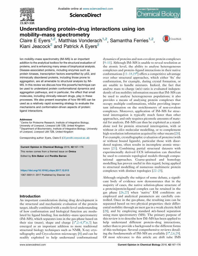
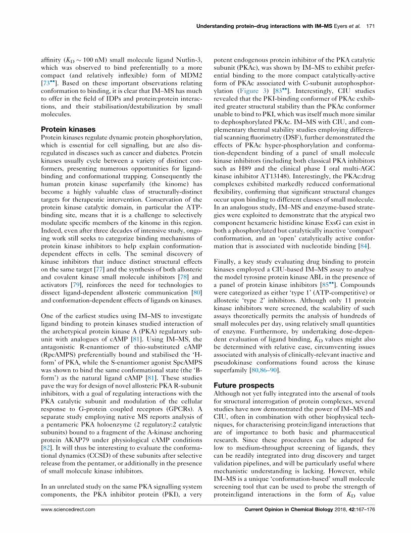
Figure 1
inh
ibit
or
con
cen
trat
ion
m/z m/z
drift time
dri
ft t
ime
dri
ft t
ime
drift time
collision energycollision energy
collisionenergy
KD
Current Opinion in Chemical Biology
Native IM–MS can yield information regarding the effect of ligand binding on protein conformation and stability. Ion mobility is used to resolve
possible conformational changes. Acquiring arrival time distributions (ATDs) at different collision energies results in a CIU fingerprint and allows
information to be obtained about ligand binding strength as well as conformational stability of the bound or unbound protein. Acquisition of mass
spectra at different small molecule or ligand concentrations can be used to determine the strength of interaction by calculation of a dissociation
constant KD.
first studies demonstrating the applicability of IM–MS to
amyloidosis was an analysis of early aggregation states of
Ab42, a putative neurotoxic species in Alzheimer’s dis-
ease [57]. The results of this investigation were in agree-
ment with labour-intensive photochemical cross-linking
experiments, and further supported by molecular model-
ling. A follow-on study revealed that the mechanism of
Ab42 aggregation is different to that of Ab40, and dem-
onstrated that the Ab42 dodecamer is likely to be the
primary toxic species in Alzheimer’s disease [51��].
Crucial for the development of therapeutic strategies for
amyloid-triggered disease is understanding the aggrega-
tion process in the presence of peptide or small molecule-
based ligands (Figure 2) [58–62,63�,64,65]. A classic
example is the study by Ashcroft, Radford and colleagues,
who demonstrated the utility of IM–MS for screening a
panel of small molecules for disrupting amyloid formation
of human islet amyloid polypeptide (hIAPP) and Ab40[66��,67]. By developing a high throughput screen, sev-
eral parameters could be assessed simultaneously: pro-
tein:ligand interactions; the protein species (monomer,
oligomer) to which the drug bound; whether binding was
specific or non-specific and the effect of ligand binding on
www.sciencedirect.com
self-assembly pathways. Ultimately, this led to the iden-
tification of novel inhibitors of in vitro Ab40 fibril
formation.
Furthermore, by combining IM–MS with complementary
strategies such as electron-transfer dissociation (ETD)
and molecular modelling, additional insight into the
mode of ligand binding and the behaviour of specific
amino acids in the target protein can be obtained for the
observed aggregates [68,69]. Indeed, in combination with
NMR and molecular dynamics, IM–MS has recently been
used to define the mechanism of binding of the small
molecule Ab42 aggregation inhibitor tramiprosate, which
is currently in phase III clinical trials for Alzheimer’s
disease [70].
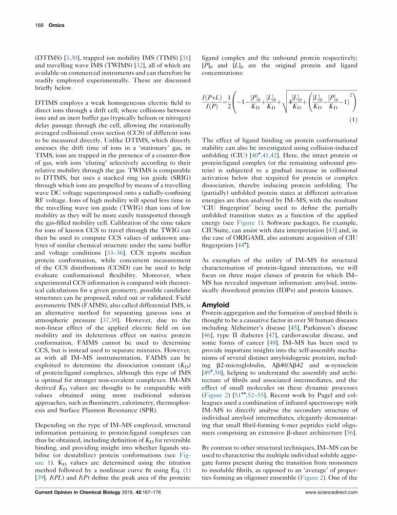
Intrinsically disordered proteinsIntrinsically disordered proteins (IDPs), which include
amyloidogenic proteins such as a-synuclein, lack a well-
defined tertiary structure and are believed to adopt a
variety of different conformations, including several
regions that are predicted to be totally unstructured
[71]. IDPs account for a significant percentage of all
proteins, with some 40% of human polypeptides
Current Opinion in Chemical Biology 2018, 42:167–176
170 Omics
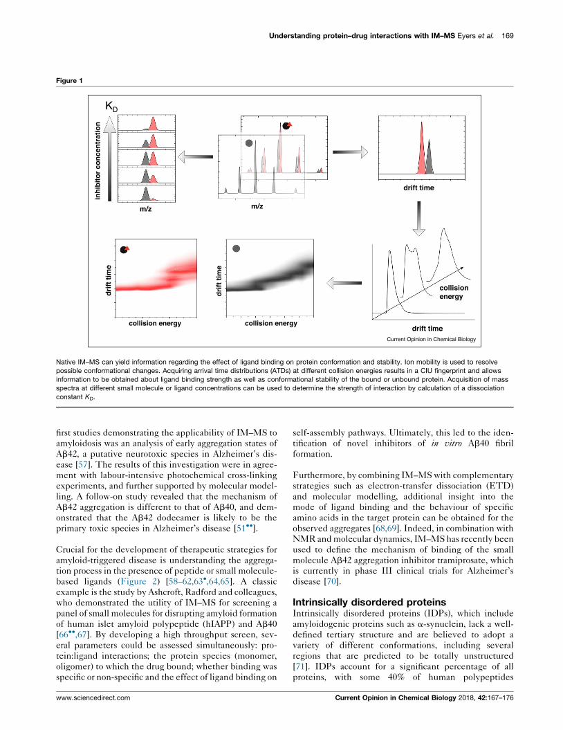
Figure 2
1 2 3 4 5 6 7 1 2 3 4 5 6 7
No Inhibitor With Inhibitor
Number of subunits, n (MS) Number of subunits, n (MS)
n = 4n = 4n = 4
n = 3
n = 2
n = 1n = 5
n = 5
n = 6
n = 6
n = 3
n = 2
n = 1 n = 7
n = 7
Conformationaltransition
Co
llisi
on
Cro
ssS
ecti
on
(IM
S)
Current Opinion in Chemical Biology
Schematic illustrating IM–MS monitoring of the self-assembly process of different amyloid structures in the absence and presence of an inhibitor.
(Top panels) Cartoon mobilograms of amyloid proteins in the absence (left) and presence of an inhibitor of amyloidosis (right). The number of
amyloid subunits, n, is determined by MS analysis, while the collision cross section (conformation) of the oligomers is determined using IMS.
(Bottom panels) The mechanism proposed by Bleiholder et al. [52] suggests that monomers self-assemble leading to the formation of soluble
oligomers (n = 2–4), at which point there is a transition from a globular conformation to a presumed toxic linear b-sheet structure. In the presence
of inhibitors of amyloidosis, aggregates either do not form (not shown) or are thought to maintain an isotropic globular conformation (n � 4, bottom
right).
predicted to contain at least one disordered region. More-
over, ordered:disordered transitions commonly occur dur-
ing endogenous protein folding subsequent to translation
[72]. Defining the structured regions and degree of (dis)
order of this important class of proteins is currently
restricted, due to the limitations of traditional techniques
such as X-ray crystallography, which require a fixed,
relatively stable structural protein disposition in order
to form crystals. Due to their roles in forming protein
complexes implicated in disease, IDPs have become
valuable new drug targets. Consequently, understanding
changes in structure and conformation that occur upon
drug binding can reveal important information about the
mode of action of potential therapeutic compounds. The
ability of IM–MS to evaluate conformational changes in
heterogeneous mixtures with very poorly defined (or
absent) higher-order structure thus makes it an ideal
technique for IDP interrogation, although its application
in this important area is currently underexploited. An
important example is the intrinsically disordered tran-
scription factor p53 [73��], a tumour suppressor with a
short half-life in cells due to its interaction with the
Current Opinion in Chemical Biology 2018, 42:167–176
ubiquitin E3-ligase MDM2, which targets p53 for degra-
dation [74,75]. The dysregulation of p53 in many cancers
(and stabilisation upon DNA damage) means that under-
standing, and potentially disrupting, the p53:MDM2
complex, is a desirable chemotherapeutic strategy that
has become an exemplar in the protein:protein interac-
tion field [76]. Using native IM–MS, Dickinson et al.evaluated the effect of two small molecules, RITA and
Nutelin-3, which were designed to interfere with the
formation of the p53:MDM2 complex by binding to
either p53 or MDM2 respectively [73��]. The non-cova-
lent RITA/p53 complex was unstable under the relatively
benign conditions used in these analyses, suggesting a
lower KD than previously measured in solution, or a
requirement for hydrophobic interactions that are lost
upon transfer to the gas-phase. However, by evaluating
the variance in charge state distribution and CCSD in the
absence or presence of RITA, a compound-dependent
conformation change in p53 with a modest decrease in
protein disorder and compaction, was observed. Similarly,
conformational changes were also reported in the N-
terminal region of MDM2 in the presence of the high-
www.sciencedirect.com
Understanding protein–drug interactions with IM–MS Eyers et al. 171
affinity (KD � 100 nM) small molecule ligand Nutlin-3,
which was observed to bind preferentially to a more
compact (and relatively inflexible) form of MDM2
[73��]. Based on these important observations relating
conformation to binding, it is clear that IM–MS has much
to offer in the field of IDPs and protein:protein interac-
tions, and their stabilisation/destabilization by small
molecules.
Protein kinasesProtein kinases regulate dynamic protein phosphorylation,
which is essential for cell signalling, but are also dis-
regulated in diseases such as cancer and diabetes. Protein
kinases usually cycle between a variety of distinct con-
formers, presenting numerous opportunities for ligand-
binding and conformational trapping. Consequently the
human protein kinase superfamily (the kinome) has
become a highly valuable class of structurally-distinct
targets for therapeutic intervention. Conservation of the
protein kinase catalytic domain, in particular the ATP-
binding site, means that it is a challenge to selectively
modulate specific members of the kinome in this region.
Indeed, even after three decades of intensive study, ongo-
ing work still seeks to categorize binding mechanisms of
protein kinase inhibitors to help explain conformation-
dependent effects in cells. The seminal discovery of
kinase inhibitors that induce distinct structural effects
on the same target [77] and the synthesis of both allosteric
and covalent kinase small molecule inhibitors [78] and
activators [79], reinforces the need for technologies to
dissect ligand-dependent allosteric communication [80]
and conformation-dependent effects of ligands on kinases.
One of the earliest studies using IM–MS to investigate
ligand binding to protein kinases studied interaction of
the archetypical protein kinase A (PKA) regulatory sub-
unit with analogues of cAMP [81]. Using IM–MS, the
antagonistic R-enantiomer of thio-substituted cAMP
(RpcAMPS) preferentially bound and stabilised the ‘H-
form’ of PKA, while the S-enantiomer agonist SpcAMPS
was shown to bind the same conformational state (the ‘B-
form’) as the natural ligand cAMP [81]. These studies
pave the way for design of novel allosteric PKA R-subunit
inhibitors, with a goal of regulating interactions with the
PKA catalytic subunit and modulation of the cellular
response to G-protein coupled receptors (GPCRs). A
separate study employing native MS reports analysis of
a pentameric PKA holoenzyme (2 regulatory:2 catalytic
subunits) bound to a fragment of the A-kinase anchoring
protein AKAP79 under physiological cAMP conditions
[82]. It will thus be interesting to evaluate the conforma-
tional dynamics (CCSD) of these subunits after selective
release from the pentamer, or additionally in the presence
of small molecule kinase inhibitors.
In an unrelated study on the same PKA signalling system
components, the PKA inhibitor protein (PKI), a very
www.sciencedirect.com
potent endogenous protein inhibitor of the PKA catalytic
subunit (PKAc), was shown by IM–MS to exhibit prefer-
ential binding to the more compact catalytically-active
form of PKAc associated with C-subunit autophosphor-
ylation (Figure 3) [83��]. Interestingly, CIU studies
revealed that the PKI-binding conformer of PKAc exhib-
ited greater structural stability than the PKAc conformer
unable to bind to PKI, which was itself much more similar
to dephosphorylated PKAc. IM–MS with CIU, and com-
plementary thermal stability studies employing differen-
tial scanning fluorimetry (DSF), further demonstrated the
effects of PKAc hyper-phosphorylation and conforma-
tion-dependent binding of a panel of small molecule
kinase inhibitors (including both classical PKA inhibitors
such as H89 and the clinical phase I oral multi-AGC
kinase inhibitor AT13148). Interestingly, the PKAc:drug
complexes exhibited markedly reduced conformational
flexibility, confirming that significant structural changes
occur upon binding to different classes of small molecule.
In an analogous study, IM–MS and enzyme-based strate-
gies were exploited to demonstrate that the atypical two
component hexameric histidine kinase ExsG can exist in
both a phosphorylated but catalytically inactive ‘compact’
conformation, and an ‘open’ catalytically active confor-
mation that is associated with nucleotide binding [84].
Finally, a key study evaluating drug binding to protein
kinases employed a CIU-based IM–MS assay to analyse
the model tyrosine protein kinase ABL in the presence of
a panel of protein kinase inhibitors [85��]. Compounds
were categorized as either ‘type 1’ (ATP-competitive) or
allosteric ‘type 2’ inhibitors. Although only 11 protein
kinase inhibitors were screened, the scalability of such
assays theoretically permits the analysis of hundreds of
small molecules per day, using relatively small quantities
of enzyme. Furthermore, by undertaking dose-depen-
dent evaluation of ligand binding, KD values might also
be determined with relative ease, circumventing issues
associated with analysis of clinically-relevant inactive and
pseudokinase conformations found across the kinase
superfamily [80,86–90].
Future prospectsAlthough not yet fully integrated into the arsenal of tools
for structural interrogation of protein complexes, several
studies have now demonstrated the power of IM–MS and
CIU, often in combination with other biophysical tech-
niques, for characterising protein:ligand interactions that
are of importance to both basic and pharmaceutical
research. Since these procedures can be adapted for
low to medium-throughput screening of ligands, they
can be readily integrated into drug discovery and target
validation pipelines, and will be particularly useful where
mechanistic understanding is lacking. However, while
IM–MS is a unique ‘conformation-based’ small molecule
screening tool that can be used to probe the strength of
protein:ligand interactions in the form of KD value
Current Opinion in Chemical Biology 2018, 42:167–176
172 Omics
Figure 3
‘Native’ MS ‘Native’ IM-MSCollision-Induced
Unfolding
PKAc PKAc PKA c
PKAc + PKI PKAc(PKI) PKAc(PKI)PKAc/PKI16+
PKAc/PKI17+
PKAc/PKI15+
14+
14+
13+
13+
12+15+
2750 3000 3250 3500
m/z3750 4000 4250 25 25 353030
15+
Rel
ativ
e In
ten
sity
Rel
ativ
e In
ten
sity
TWCCSN2 He / nm2
50
45
40
35
30
2050
45
40
35
30
25
2026 28 30 32
Collision Voltage / V
Cro
ss S
ecti
on
/ n
m2
Cro
ss S
ecti
on
/ n
m2
34 36 38 40
25
Current Opinion in Chemical Biology
Conformation-dependent binding of PKI to PKA catalytic domain (PKAc). Mass spectra (left), ion mobility spectra (middle) and CIU plots (right) of
PKAc in the absence (top) and presence (bottom) of full length PKI protein. PKAc/PKI in the mass spectrum (bottom left) refers to the non-covalent
protein kinase:inhibitor complex. IM–MS of PKAc demonstrates co-existence of two primary conformations: a more compact conformer able to
bind PKI (shaded red), and a more elongated conformer that is unable to bind PKI, termed PKAc(PKI) (shaded green). CIU plots (right)
demonstrate a significant difference in the relative stability of the total PKAc conformer population compared to the non PKI-binding kinase
conformation [PKAc(PKI)] as demonstrated by the collision voltage required to induce protein unfolding (white dotted line). See Ref. [83��] for
further information.
determination, it fails to provide the empirical atomic
level detail essential for true target-based drug-design.
In this review, we have focused on clinically relevant
protein classes, where better understanding of small
molecule binding on protein stability and conformation
can reveal insights into therapeutic target development.
However, the principles of structural analysis using IM–
MS are relevant to protein-ligand binding in any field. It is
worth noting that the resolving power of ion mobility
separation is still a potential limiting factor, with minor
conformational differences (<1%) arising upon small
molecule binding especially challenging to define. The
commercialisation of a cyclic drift tube in which ions can
travel for a variable (increasing) number of cycles before
they are ejected, could circumvent this issue
[5�,91,92,93�]. Indeed, this type of experimental setup
has been demonstrated to increase resolving power up to
20-fold, albeit not yet with proteins, which could in the
future permit evaluation of extremely small ligand-driven
protein conformational changes.
Current Opinion in Chemical Biology 2018, 42:167–176
As with any assay, consideration should also be given to
the possibility of non-specific ligand binding, which can
be assessed by varying buffer conditions used for ‘native’
electrospray ionisation (ESI). Excess ligand or the addi-
tion of essential buffer components (e.g. salt) can also
suppress ESI, resulting in MS spectra of reduced reso-
lution, with poor signal to noise [94,95]. Differential
protein ionisation in the absence or presence of ligand,
which can sometimes arise due to preferential ionisation
of the small molecule, can induce a shift in the ESI
charge state envelope, making comparison of the CCS/
CCSD for analogous charge states of the bound and
unbound protein forms problematic. Dissociation of
some weak non-covalent binders that are dependent
on hydrophobic interactions may also be lost during
transfer to the gas-phase, potentially leading to under-
estimation of KD [96]. Here, even more gentle evapora-
tion processes like cold spray ionisation [97], in conjunc-
tion with a low temperature drift tube [98,99] could
possibly improve confidence in structural elucidation
and KD determination.
www.sciencedirect.com
Understanding protein–drug interactions with IM–MS Eyers et al. 173
IM–MS has enormous potential for the development and
screening of ligands, from low molecular weight com-
pounds to therapeutic antibodies, which is only now
beginning to be realised. Furthermore, since it is also
possible to evaluate binding to differentially modified
protein forms (proteoforms) and membrane proteins [19�]that might otherwise be intractable to structural interro-
gation, IM–MS opens up the possibility of understanding
the intricacies of small molecule binding to complex
proteoforms, such as differentially phosphorylated or
glycosylated protein targets. As well as the small ligands
discussed here, IM–MS has demonstrated utility in the
evaluation of structural differences in antibodies follow-
ing drug conjugation, a growth area in the pharmaceutical
industry [100]. Structural interrogation of protein:DNA
and protein:RNA complexes, to ascertain sequence-spe-
cific binding affinity, is also likely to benefit from IM–MS
analysis [101,102]. Moreover, with the development of
robotic chip based injection systems [103] combined with
already available automated open-source software for data
extraction, processing and visualisation (e.g. Pulsar [19�],Amphitrite [104], ORIGAMI [44�] and UniDec [105]),
the age of high throughput IM–MS-based ligand screen-
ing appears to have arrived.
AcknowledgementsThe authors acknowledge support from the Biotechnology and BiologicalSciences Research Council (BBSRC) (BB/L009501/1, BB/M012557/1, BB/R000182/1 and a BBSRC DTP studentship to SF), North West CancerResearch (CR1037 and CR1088), and the Institute of Integrative Biology,University of Liverpool in the form of a summer internship for KJ.
References and recommended readingPapers of particular interest, published within the period of review,have been highlighted as:
� of special interest�� of outstanding interest
1.�
Lanucara F, Holman SW, Gray CJ, Eyers CE: The power of ionmobility–mass spectrometry for structural characterizationand the study of conformational dynamics. Nat Chem 2014,6:281-294.
This review provides an excellent overview of the different types of IM–MSand how these can be used to elucidate conformation dynamics ofdifferent types of analyte.
2. May JC, McLean JA: Ion mobility–mass spectrometry: time-dispersive instrumentation. Anal Chem 2015, 87:1422-1436.
3. Wyttenbach T, Kemper PR, Bowers MT: Design of a newelectrospray ion mobility mass spectrometer. Int J MassSpectrom 2001, 212:13-23.
4. Bowers MT: Ion mobility spectrometry: a personal view of itsdevelopment at UCSB. Int J Mass Spectrom 2014, 370:75-95.
5.�
Merenbloom SI, Glaskin RS, Henson ZB, Clemmer DE: High-resolution ion cyclotron mobility spectrometry. Anal Chem2009, 81:1482-1487.
First presentation of a cyclic ion mobility drift tube for IM–MS, whichpermits resolving power of >300 to be achieved.
6. Ewing MA, Glover MS, Clemmer DE: Hybrid ion mobility andmass spectrometry as a separation tool. J Chromatogr A 2016,1439:3-25.
7. Kanu AB, Dwivedi P, Tam M, Matz L, Hill HH Jr: Ion mobility–mass spectrometry. J Mass Spectrom 2008, 43:1-22.
www.sciencedirect.com
8. Renaud JP, Chung CW, Danielson UH, Egner U, Hennig M,Hubbard RE, Nar H: Biophysics in drug discovery: impact,challenges and opportunities. Nat Rev Drug Discov 2016,15:679-698.
9. van Duijn E, Barendregt A, Synowsky S, Versluis C, Heck AJ:Chaperonin complexes monitored by ion mobility massspectrometry. J Am Chem Soc 2009, 131:1452-1459.
10. Uetrecht C, Rose RJ, van Duijn E, Lorenzen K, Heck AJ: Ionmobility mass spectrometry of proteins and proteinassemblies. Chem Soc Rev 2010, 39:1633-1655.
11. Ruotolo BT, Benesch JL, Sandercock AM, Hyung SJ,Robinson CV: Ion mobility–mass spectrometry analysis of largeprotein complexes. Nat Protoc 2008, 3:1139-1152.
12. Cubrilovic D, Barylyuk K, Hofmann D, Walczak MJ, Graeber M,Berg T, Widerb G, Zenobi R: Direct monitoring of protein–protein inhibition using nano electrospray ionization massspectrometry. Chem Sci 2014, 5:2794-2803.
13. Atmanene C, Petiot-Becard S, Zeyer D, Van Dorsselaer A, VivatHannah V, Sanglier-Cianferani S: Exploring key parameters todetect subtle ligand-induced protein conformational changesusing traveling wave ion mobility mass spectrometry. AnalChem 2012, 84:4703-4710.
14. Stojko J, Fieulaine S, Petiot-Becard S, Van Dorsselaer A,Meinnel T, Giglione C, Cianferani S: Ion mobility coupled tonative mass spectrometry as a relevant tool to investigateextremely small ligand-induced conformational changes.Analyst 2015, 140:7234-7245.
15. El-Hawiet A, Kitova EN, Arutyunov D, Simpson DJ, Szymanski CM,Klassen JS: Quantifying ligand binding to large proteincomplexes using electrospray ionization mass spectrometry.Anal Chem 2012, 84:3867-3870.
16. Lin H, Kitova EN, Klassen JS: Quantifying protein–ligandinteractions by direct electrospray ionization–MS analysis:evidence of nonuniform response factors induced by highmolecular weight molecules and complexes. Anal Chem 2013,85:8919-8922.
17. Niu S, Rabuck JN, Ruotolo BT: Ion mobility–mass spectrometryof intact protein–ligand complexes for pharmaceutical drugdiscovery and development. Curr Opin Chem Biol 2013, 17:809-817.
18. Mehmood S, Allison TM, Robinson CV: Mass spectrometry ofprotein complexes: from origins to applications. Annu Rev PhysChem 2015, 66:453-474.
19.�
Allison TM, Reading E, Liko I, Baldwin AJ, Laganowsky A,Robinson CV: Quantifying the stabilizing effects of protein–ligand interactions in the gas phase. Nat Commun 2015, 6:8551.
The authors describe the application of IM–MS and CIU to investigateconformational and stability effects of ligand binding to membraneproteins. They also decribe software that permits quantifiation of ligandbinding effects.
20. Ahdash Z, Pyle E, Politis A: Hybrid mass spectrometry: towardscharacterization of protein conformational states. TrendsBiochem Sci 2016, 41:650-653.
21. Le Gall T, Romero PR, Cortese MS, Uversky VN, Dunker AK:Intrinsic disorder in the protein data bank. J Biomol Struct Dyn2007, 24:325-342.
22. Politis A, Park AY, Hyung SJ, Barsky D, Ruotolo BT, Robinson CV:Integrating ion mobility mass spectrometry with molecularmodelling to determine the architecture of multiproteincomplexes. PLoS One 2010, 5:e12080.
23. Politis A, Stengel F, Hall Z, Hernandez H, Leitner A, Walzthoeni T,Robinson CV, Aebersold R: A mass spectrometry-based hybridmethod for structural modeling of protein complexes. NatMethods 2014, 11:403-406.
24. D’Urzo A, Konijnenberg A, Rossetti G, Habchi J, Li J, Carloni P,Sobott F, Longhi S, Grandori R: Molecular basis for structuralheterogeneity of an intrinsically disordered protein bound to apartner by combined ESI–IM–MS and modeling. J Am Soc MassSpectrom 2015, 26:472-481.
Current Opinion in Chemical Biology 2018, 42:167–176
174 Omics
25. Politis A, Park AY, Hall Z, Ruotolo BT, Robinson CV: Integrativemodelling coupled with ion mobility mass spectrometryreveals structural features of the clamp loader in complex withsingle-stranded DNA binding protein. J Mol Biol 2013,425:4790-4801.
26. Leney AC, Heck AJ: Native mass spectrometry: what is in thename? J Am Soc Mass Spectrom 2017, 28:5-13.
27. Jurneczko E, Barran PE: How useful is ion mobility massspectrometry for structural biology? The relationship betweenprotein crystal structures and their collision cross sections inthe gas phase. Analyst 2011, 136:20-28.
28. Mason EA, McDaniel EW: Transport Properties of Ion Gases. Wiley;1988.
29. Cumeras R, Figueras E, Davis CE, Baumbach JI, Gracia I: Reviewon ion mobility spectrometry. Part 1: current instrumentation.Analyst 2015, 140:1376-1390.
30. Mason EA, Schamp HW Jr: Mobility of gaseous ions in weakelectric fields. Ann Phys 1958, 4:233-270.
31. Michelmann K, Silveira JA, Ridgeway ME, Park MA:Fundamentals of trapped ion mobility spectrometry. J Am SocMass Spectrom 2015, 26:14-24.
32. Giles K, Pringle SD, Worthington KR, Little D, Wildgoose JL,Bateman RH: Applications of a travelling wave-based radio-frequency-only stacked ring ion guide. Rapid Commun MassSpectrom 2004, 18:2401-2414.
33. Chawner R, McCullough B, Giles K, Barran PE, Gaskell SJ,Eyers CE: QconCAT standard for calibration of ion mobility–mass spectrometry systems. J Proteome Res 2012, 11:5564-5572.
34. Sun Y, Vahidi S, Sowole MA, Konermann L: Protein structuralstudies by traveling wave ion mobility spectrometry: a criticallook at electrospray sources and calibration issues. J Am SocMass Spectrom 2016, 27:31-40.
35. Allison TM, Landreh M, Benesch JLP, Robinson CV: Low chargeand reduced mobility of membrane protein complexes hasimplications for calibration of collision cross sectionmeasurements. Anal Chem 2016, 88:5879-5884.
36. Bush MF, Hall Z, Giles K, Hoyes J, Robinson CV, Ruotolo BT:Collision cross sections of proteins and their complexes: acalibration framework and database for gas-phase structuralbiology. Anal Chem 2010, 82:9557-9565.
37. Guevremont R: High-field asymmetric waveform ion mobilityspectrometry: a new tool for mass spectrometry. J ChromatogrA 2004, 1058:3-19.
38. Swearingen KE, Moritz RL: High-field asymmetric waveform ionmobility spectrometry for mass spectrometry-basedproteomics. Exp Rev Proteomics 2012, 9:505-517.
39. Daniel JM, McCombie G, Wendt S, Zenobi R: Massspectrometric determination of association constants ofadenylate kinase with two noncovalent inhibitors. J Am SocMass Spectrom 2003, 14:442-448.
40.�
Hopper JT, Oldham NJ: Collision induced unfolding of proteinions in the gas phase studied by ion mobility–massspectrometry: the effect of ligand binding on conformationalstability. J Am Soc Mass Spectrom 2009, 20:1851-1858.
One of the earliest studies applying elevated collision energy to under-stand the effects of ligand binding on protein unfolding pathways, con-firmating that gas-phase CIU studies are similar to solution-phaseobservations.
41. Freeke J, Bush MF, Robinson CV, Ruotolo BT: Gas-phase proteinassemblies: unfolding landscapes and preserving native-likestructures using noncovalent adducts. Chem Phys Lett 2012,524:1-9.
42. Bornschein RE, Niu S, Eschweiler J, Ruotolo BT: Ion mobility–mass spectrometry reveals highly-compact intermediates inthe collision induced dissociation of charge-reduced proteincomplexes. J Am Soc Mass Spectrom 2016, 27:41-49.
43. Eschweiler JD, Rabuck-Gibbons JN, Tian YW, Ruotolo BT:CIUSuite: a quantitative analysis package for collision induced
Current Opinion in Chemical Biology 2018, 42:167–176
unfolding measurements of gas-phase protein ions. AnalChem 2015, 87:11516-11522.
44.�
Migas LG, France AP, Bellina B, Barran PE: ORIGAMI: a softwaresuite for activated ion mobility mass spectrometry (aIM–MS)applied to multimeric protein assemblies. Int J Mass Spectrom2017. in press.
Describes a software suite that facilitates the rapid acquisition, analysisand visualisation of CIU IM–MS data on a Waters Synapt instrument.
45. Kirkitadze MD, Bitan G, Teplow DB: Paradigm shifts inAlzheimer’s disease and other neurodegenerative disorders:the emerging role of oligomeric assemblies. J Neurosci Res2002, 69:567-577.
46. Conway KA, Lee SJ, Rochet JC, Ding TT, Williamson RE, LansburyPT Jr: Acceleration of oligomerization, not fibrillization, is ashared property of both alpha-synuclein mutations linked toearly-onset Parkinson’s disease: implications forpathogenesis and therapy. Proc Natl Acad Sci U S A 2000,97:571-576.
47. Meier JJ, Kayed R, Lin CY, Gurlo T, Haataja L, Jayasinghe S,Langen R, Glabe CG, Butler PC: Inhibition of human IAPP fibrilformation does not prevent beta-cell death: evidence fordistinct actions of oligomers and fibrils of human IAPP. Am JPhysiol Endocrinol Metab 2006, 291:E1317-E1324.
48. Eisenberg DS, Sawaya MR: Structural studies of amyloidproteins at the molecular level. Annu Rev Biochem 2017, 86:69-95.
49.�
Hoffmann W, von Helden G, Pagel K: Ion mobility–massspectrometry and orthogonal gas-phase techniques to studyamyloid formation and inhibition. Curr Opin Struct Biol 2017,46:7-15.
This review highlights advances in the characterisation of amyloid aggre-gation and their assembly pathways using IM–MS and how this technol-ogy can be used to screen for molecules that disrupt oligomer formation.
50. Sipe JD, Benson MD, Buxbaum JN, Ikeda S, Merlini G, Saraiva MJ,Westermark P, Nomenclature Committee of the InternationalSociety of A: Amyloid fibril protein nomenclature:2012 recommendations from the Nomenclature Committee ofthe International Society of Amyloidosis. Amyloid 2012, 19:167-170.
51.��
Bernstein SL, Dupuis NF, Lazo ND, Wyttenbach T, Condron MM,Bitan G, Teplow DB, Shea JE, Ruotolo BT, Robinson CV,Bowers MT: Amyloid-beta protein oligomerization and theimportance of tetramers and dodecamers in the aetiology ofAlzheimer’s disease. Nat Chem 2009, 1:326-331.
Using IM–MS, the authors were able to study the self-assembly mechan-isms of different amyloids and suggest a possible candidate oligomer, theAb42 dodecamer, as the causative agent responsible for Alzheimer’sdisease.
52. Bleiholder C, Dupuis NF, Wyttenbach T, Bowers MT: Ion mobility–mass spectrometry reveals a conformational conversion fromrandom assembly to beta-sheet in amyloid fibril formation. NatChem 2011, 3:172-177.
53. Susa AC, Wu C, Bernstein SL, Dupuis NF, Wang H, Raleigh DP,Shea JE, Bowers MT: Defining the molecular basis of amyloidinhibitors: human islet amyloid polypeptide–insulininteractions. J Am Chem Soc 2014, 136:12912-12919.
54. Liu YQ, Graetz M, Ho L, Pukala TL: Ion mobility–massspectrometry-based screening for inhibition of alpha-synuclein aggregation. Eur J Mass Spectrom 2015, 21:255-264.
55. Woods LA, Radford SE, Ashcroft AE: Advances in ion mobilityspectrometry–mass spectrometry reveal key insights intoamyloid assembly. Biochim Biophys Acta 2013, 1834:1257-1268.
56. Seo J, Hoffmann W, Warnke S, Huang X, Gewinner S,Schollkopf W, Bowers MT, von Helden G, Pagel K: Aninfrared spectroscopy approach to follow beta-sheetformation in peptide amyloid assemblies. Nat Chem 2017,9:39-44.
57. Bernstein SL, Wyttenbach T, Baumketner A, Shea JE, Bitan G,Teplow DB, Bowers MT: Amyloid beta-protein: monomerstructure and early aggregation states of Abeta42 and itsPro19 alloform. J Am Chem Soc 2005, 127:2075-2084.
www.sciencedirect.com

Understanding protein–drug interactions with IM–MS Eyers et al. 175
58. Amijee H, Bate C, Williams A, Virdee J, Jeggo R, Spanswick D,Scopes DI, Treherne JM, Mazzitelli S, Chawner R, Eyers CE,Doig AJ: The N-methylated peptide SEN304 powerfully inhibitsAbeta(1–42) toxicity by perturbing oligomer formation.Biochemistry 2012, 51:8338-8352.
59. Gessel MM, Wu C, Li H, Bitan G, Shea JE, Bowers MT: Abeta(39–42) modulates Abeta oligomerization but not fibril formation.Biochemistry 2012, 51:108-117.
60. Zheng X, Liu D, Klarner FG, Schrader T, Bitan G, Bowers MT:Amyloid beta–protein assembly: the effect of moleculartweezers CLR01 and CLR03. J Phys Chem B 2015, 119:4831-4841.
61. Soper MT, DeToma AS, Hyung SJ, Lim MH, Ruotolo BT: Amyloid-beta–neuropeptide interactions assessed by ion mobility–mass spectrometry. Phys Chem Chem Phys 2013, 15:8952-8961.
62. Murray MM, Bernstein SL, Nyugen V, Condron MM, Teplow DB,Bowers MT: Amyloid beta protein: Abeta40 inhibits Abeta42oligomerization. J Am Chem Soc 2009, 131:6316-6317.
63.�
Young LM, Cao P, Raleigh DP, Ashcroft AE, Radford SE: Ionmobility spectrometry–mass spectrometry defines theoligomeric intermediates in amylin amyloid formation and themode of action of inhibitors. J Am Chem Soc 2014, 136:660-670.
Provides novel insights into the formation of amylin oligomers which arefound in the pancreatic islets of people with type II diabetes and identifiesthe specific conformers to which two known inhibitors bind. In addition,the authors use IMS–MS to reveal the distinct modes of action of theinhibitors in preventing oligomer formation.
64. Zheng X, Wu C, Liu D, Li H, Bitan G, Shea JE, Bowers MT:Mechanism of C-terminal fragments of amyloid beta-proteinas Abeta inhibitors: do C-terminal interactions play a key rolein their inhibitory activity? J Phys Chem B 2016, 120:1615-1623.
65. de Almeida NE, Do TD, Tro M, LaPointe NE, Feinstein SC, Shea JE,Bowers MT: Opposing effects of Cucurbit[7]uril and 1,2,3,4,6-penta-O-galloyl-beta-D-glucopyranose on amyloid beta25-35assembly. ACS Chem Neurosci 2016, 7:218-226.
66.��
Young LM, Saunders JC, Mahood RA, Revill CH, Foster RJ, Tu LH,Raleigh DP, Radford SE, Ashcroft AE: Screening and classifyingsmall-molecule inhibitors of amyloid formation using ionmobility spectrometry–mass spectrometry. Nat Chem 2015,7:73-81.
This study exemplifies the ability of IM–MS to be used as a tool to rapidlydetermine the conformation effects and mode of binding of small mole-cules to amyloids, and describes a potential lead compound for disrupt-ing pathogenic Ab40 fibril formation.
67. Young LM, Saunders JC, Mahood RA, Revill CH, Foster RJ,Ashcroft AE, Radford SE: ESI–IMS–MS: a method for rapidanalysis of protein aggregation and its inhibition by smallmolecules. Methods 2016, 95:62-69.
68. Cole H, Porrini M, Morris R, Smith T, Kalapothakis J, Weidt S,Mackay CL, MacPhee CE, Barran PE: Early stages of insulinfibrillogenesis examined with ion mobility mass spectrometryand molecular modelling. Analyst 2015, 140:7000-7011.
69. Konijnenberg A, Ranica S, Narkiewicz J, Legname G, Grandori R,Sobott F, Natalello A: Opposite structural effects ofepigallocatechin-3-gallate and dopamine binding to alpha-synuclein. Anal Chem 2016, 88:8468-8475.
70. Kocis P, Tolar M, Yu J, Sinko W, Ray S, Blennow K, Fillit H, Hey JA:Elucidating the Abeta42 anti-aggregation mechanism ofaction of tramiprosate in Alzheimer’s disease: integratingmolecular analytical methods. Pharmacokin Clin Data CNSDrugs 2017, 31:495-509.
71. Dunker AK, Lawson JD, Brown CJ, Williams RM, Romero P, Oh JS,Oldfield CJ, Campen AM, Ratliff CM, Hipps KW et al.: Intrinsicallydisordered protein. J Mol Graph Model 2001, 19:26-59.
72. Uversky VN, Dunker AK: Understanding protein non-folding.Biochim Biophys Acta 2010, 1804:1231-1264.
73.��
Dickinson ER, Jurneczko E, Nicholson J, Hupp TR, Zawacka-Pankau J, Selivanova G, Barran PE: The use of ion mobility massspectrometry to probe modulation of the structure of p53 and
www.sciencedirect.com
of MDM2 by small molecule inhibitors. Front Mol Biosci 2015,2:39.
Highlights how IM–MS can be used to investigate ligand binding tointrisincally disordered proteins. Demonstrates conformer specific smallmolecule binding and stabilisation of protein structure.
74. Lane DP: Cancer. p53, guardian of the genome. Nature 1992,358:15-16.
75. Wu X, Bayle JH, Olson D, Levine AJ: The p53–mdm-2autoregulatory feedback loop. Genes Dev 1993, 7:1126-1132.
76. Wang S, Zhao Y, Aguilar A, Bernard D, Yang CY: Targeting theMDM2–p53 protein–protein interaction for new cancertherapy: progress and challenges. Cold Spring Harb PerspectMed 2017:7.
77. Nagar B, Bornmann WG, Pellicena P, Schindler T, Veach DR,Miller WT, Clarkson B, Kuriyan J: Crystal structures of the kinasedomain of c-Abl in complex with the small molecule inhibitorsPD173955 and imatinib (STI-571). Cancer Res 2002, 62:4236-4243.
78. Byrne DP, Foulkes DM, Eyers PA: Pseudokinases: update ontheir functions and evaluation as new drug targets. Future MedChem 2017, 9:245-265.
79. Leroux AE, Schulze JO, Biondi RM: AGC kinases, mechanismsof regulation and innovative drug development. Semin CancerBiol 2017, 17 30148-7.
80. Mohanty S, Oruganty K, Kwon A, Byrne DP, Ferries S, Ruan Z,Hanold LE, Katiyar S, Kennedy EJ, Eyers PA, Kannan N:Hydrophobic core variations provide a structural frameworkfor tyrosine kinase evolution and functional specialization.PLoS Genet 2016, 12:e1005885.
81. Badireddy S, Yunfeng G, Ritchie M, Akamine P, Wu J, Kim CW,Taylor SS, Qingsong L, Swaminathan K, Anand GS: Cyclic AMPanalog blocks kinase activation by stabilizing inactiveconformation: conformational selection highlights a newconcept in allosteric inhibitor design. Mol Cell Proteomics 2011,10 M110 004390.
82. Smith FD, Esseltine JL, Nygren PJ, Veesler D, Byrne DP,Vonderach M, Strashnov I, Eyers CE, Eyers PA, Langeberg LK,Scott JD: Local protein kinase A action proceeds throughintact holoenzymes. Science 2017, 356:1288-1293.
83.��
Byrne DP, Vonderach M, Ferries S, Brownridge PJ, Eyers CE,Eyers PA: cAMP-dependent protein kinase (PKA) complexesprobed by complementary differential scanning fluorimetryand ion mobility–mass spectrometry. Biochem J 2016,473:3159-3175.
By using IM–MS the authors were able to demonstrate dynamic, con-formationally distinct populations of the PKA catalytic subunit with dis-tinct structural stability and susceptibility to both the inhibitor protein PKIand small molecule drugs.
84. Wojnowska M, Yan J, Sivalingam GN, Cryar A, Gor J,Thalassinos K, Djordjevic S: Autophosphorylation activity of asoluble hexameric histidine kinase correlates with the shift inprotein conformational equilibrium. Chem Biol 2013, 20:1411-1420.
85.��
Rabuck JN, Hyung SJ, Ko KS, Fox CC, Soellner MB, Ruotolo BT:Activation state-selective kinase inhibitor assay based on ionmobility–mass spectrometry. Anal Chem 2013, 85:6995-7002.
This study demonstrates the power of IM–MS for screening small mole-cule kinase inhibitors and defining the mode of binding as either direct orallosteric, independent of catalytic activity.
86. Murphy JM, Mace PD, Eyers PA: Live and let die: insights intopseudoenzyme mechanisms from structure. Curr Opin StructBiol 2017, 47:95-104.
87. Milani M, Byrne DP, Greaves G, Butterworth M, Cohen GM,Eyers PA, Varadarajan S: DRP-1 is required for BH3 mimetic-mediated mitochondrial fragmentation and apoptosis. CellDeath Dis 2017, 8:e2552.
88. Bailey FP, Byrne DP, Oruganty K, Eyers CE, Novotny CJ,Shokat KM, Kannan N, Eyers PA: The Tribbles 2 (TRB2)pseudokinase binds to ATP and autophosphorylates in ametal-independent manner. Biochem J 2015, 467:47-62.
Current Opinion in Chemical Biology 2018, 42:167–176
176 Omics
89. Bailey FP, Byrne DP, McSkimming D, Kannan N, Eyers PA: Goingfor broke: targeting the human cancer pseudokinome.Biochem J 2015, 465:195-211.
90. Murphy JM, Zhang Q, Young SN, Reese ML, Bailey FP, Eyers PA,Ungureanu D, Hammaren H, Silvennoinen O, Varghese LN et al.: Arobust methodology to subclassify pseudokinases based ontheir nucleotide-binding properties. Biochem J 2014, 457:323-334.
91. Glaskin RS, Valentine SJ, Clemmer DE: A scanning frequencymode for ion cyclotron mobility spectrometry. Anal Chem 2010,82:8266-8271.
92. Glaskin RS, Ewing MA, Clemmer DE: Ion trapping for ion mobilityspectrometry measurements in a cyclical drift tube. Anal Chem2013, 85:7003-7008.
93.�
Haler JRN, Far J, Aqil A, Claereboudt J, Tomczyk N, Giles K,Jerome C, De Pauw E: Multiple gas-phase conformations of asynthetic linear poly(acrylamide) polymer observed using ionmobility–mass spectrometry. J Am Soc Mass Spectrom 2017,28:2492-2499.
Demonstration of a prototype commercial high resolution multi-passcyclic travelling-wave ion mobility MS instrument.
94. Wang W, Kitova EN, Klassen JS: Nonspecific protein–carbohydrate complexes produced by nanoelectrosprayionization. Factors influencing their formation and stability.Anal Chem 2005, 77:3060-3071.
95. Guan S, Trnka MJ, Bushnell DA, Robinson PJ, Gestwicki JE,Burlingame AL: Deconvolution method for specific andnonspecific binding of ligand to multiprotein complex bynative mass spectrometry. Anal Chem 2015, 87:8541-8546.
96. Bich C, Baer S, Jecklin MC, Zenobi R: Probing the hydrophobiceffect of noncovalent complexes by mass spectrometry. J AmSoc Mass Spectrom 2010, 21:286-289.
97. Yamaguchi K: Cold-spray ionization mass spectrometry:principle and applications. J Mass Spectrom 2003, 38:473-490.
Current Opinion in Chemical Biology 2018, 42:167–176
98. May JC, Russell DH: A mass-selective variable-temperaturedrift tube ion mobility–mass spectrometer for temperaturedependent ion mobility studies. J Am Soc Mass Spectrom 2011,22:1134-1145.
99. Ujma J, Giles K, Morris M, Barran PE: New high resolution ionmobility mass spectrometer capable of measurements ofcollision cross sections from 150 to 520 K. Anal Chem 2016,88:9469-9478.
100. Debaene F, Boeuf A, Wagner-Rousset E, Colas O, Ayoub D,Corvaia N, Van Dorsselaer A, Beck A, Cianferani S: Innovativenative MS methodologies for antibody drug conjugatecharacterization: high resolution native MS and IM–MS foraverage DAR and DAR distribution assessment. Anal Chem2014, 86:10674-10683.
101. Atmanene C, Chaix D, Bessin Y, Declerck N, Van Dorsselaer A,Sanglier-Cianferani S: Combination of noncovalent massspectrometry and traveling wave ion mobility spectrometryreveals sugar-induced conformational changes of centralglycolytic genes repressor/DNA complex. Anal Chem 2010,82:3597-3605.
102. Ma X, Shah S, Zhou M, Park CK, Wysocki VH, Horton NC:Structural analysis of activated SgrAI-DNA oligomers usingion mobility mass spectrometry. Biochemistry 2013, 52:4373-4381.
103. Dethy JM, Ackermann BL, Delatour C, Henion JD, Schultz GA:Demonstration of direct bioanalysis of drugs in plasma usingnanoelectrospray infusion from a silicon chip coupled withtandem mass spectrometry. Anal Chem 2003, 75:805-811.
104. Sivalingam GN, Yan J, Sahota H, Thalassinos K: Amphitrite: aprogram for processing travelling wave ion mobility massspectrometry data. Int J Mass Spectrom 2013, 345:54-62.
105. Marty MT, Baldwin AJ, Marklund EG, Hochberg GK, Benesch JL,Robinson CV: Bayesian deconvolution of mass and ion mobilityspectra: from binary interactions to polydisperse ensembles.Anal Chem 2015, 87:4370-4376.
www.sciencedirect.com