
Tunneled HD Catheters Dr. Zaghloul Gouda
34
Egyptian Society of Nephrology and Transplantation 4 th Interventional Nephrology Workshop Mansoura International Hospital Tunneled HD Catheters Zaghloul Gouda Nephrology Department, Damanhour Medical National Institute ww.hd-cath.com www.telekidney.co
-
Upload
nephro-mih -
Category
Healthcare
-
view
87 -
download
1
Transcript of Tunneled HD Catheters Dr. Zaghloul Gouda
Egyptian Society of Nephrology and Transplantation4th Interventional Nephrology Workshop
Mansoura International Hospital
Tunneled HD Catheters
Zaghloul GoudaNephrology Department,
Damanhour Medical National Institute
www.hd-cath.com www.telekidney.com

• My Participant colleagues• All members of Mansoura International Hospital• Nephrology Department• Committee of ESNT interventional Group
www.hd-cath.com www.telekidney.com

Anatomy of venous system
www.hd-cath.com www.telekidney.com
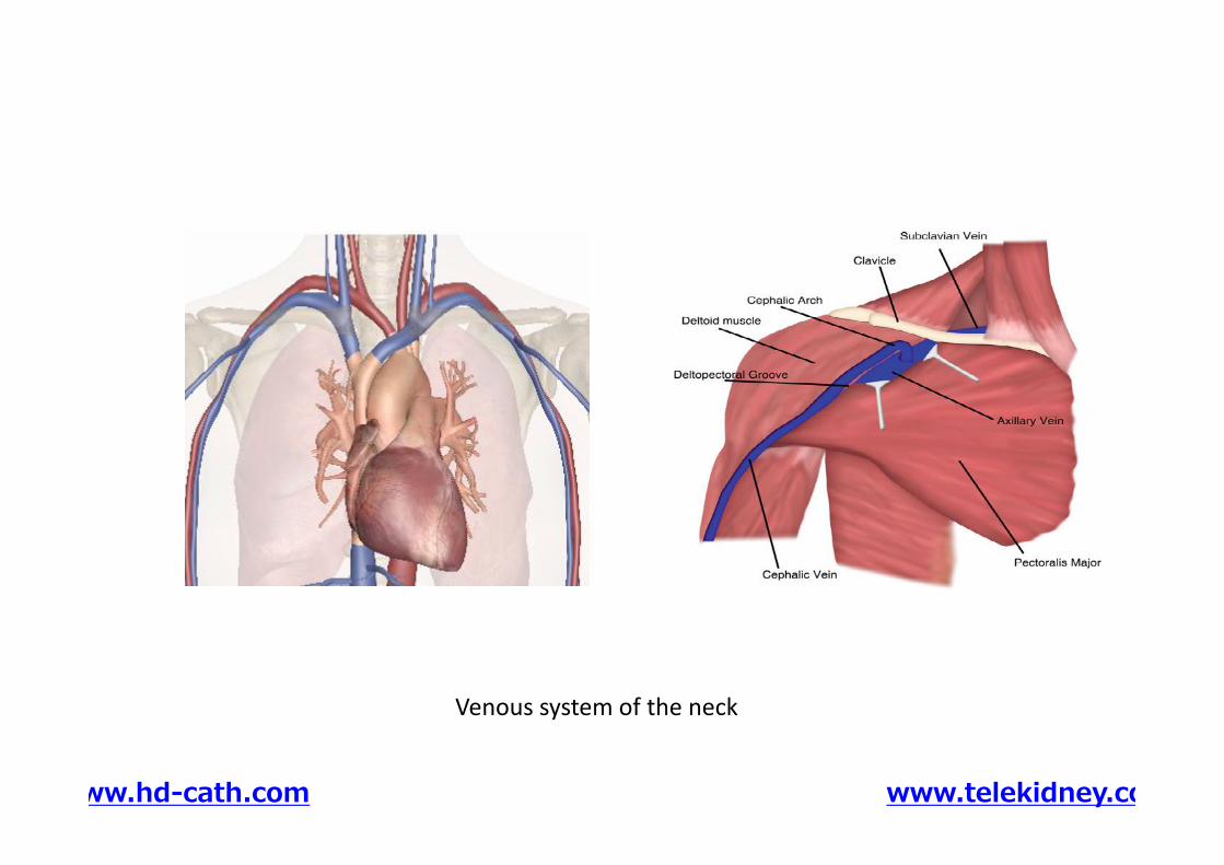
Venous system of the neck
www.hd-cath.com www.telekidney.com
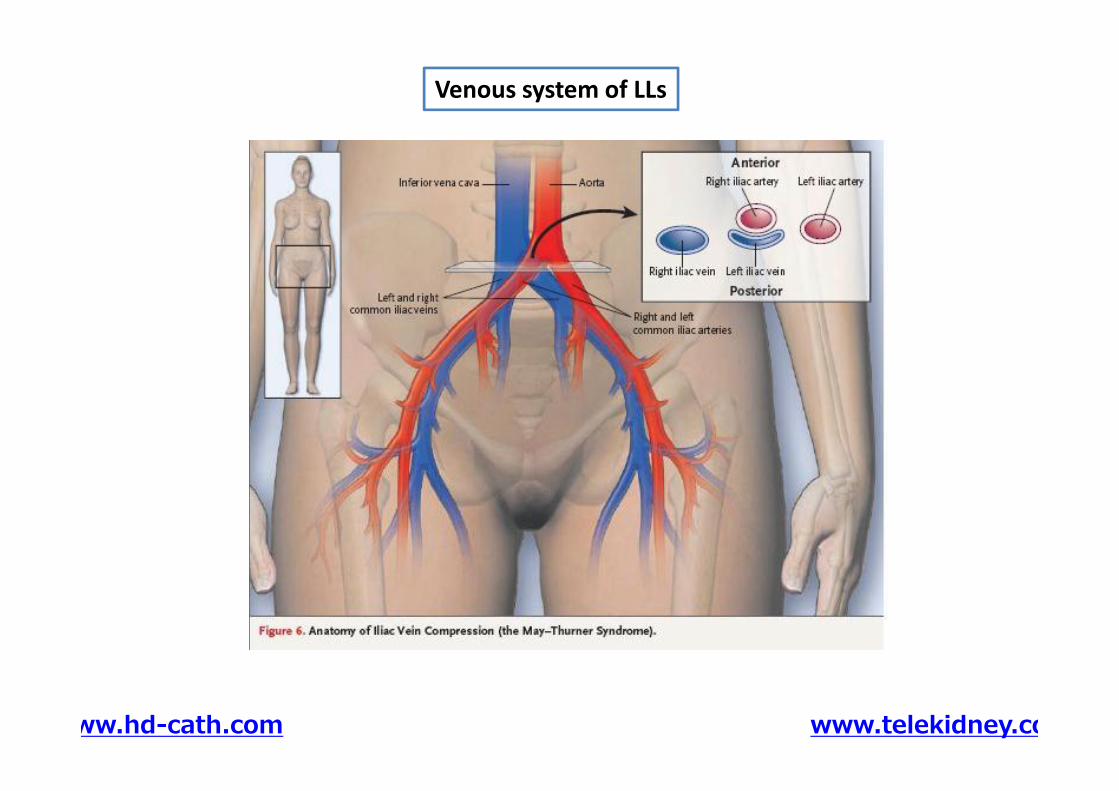
Venous system of LLs
www.hd-cath.com www.telekidney.com
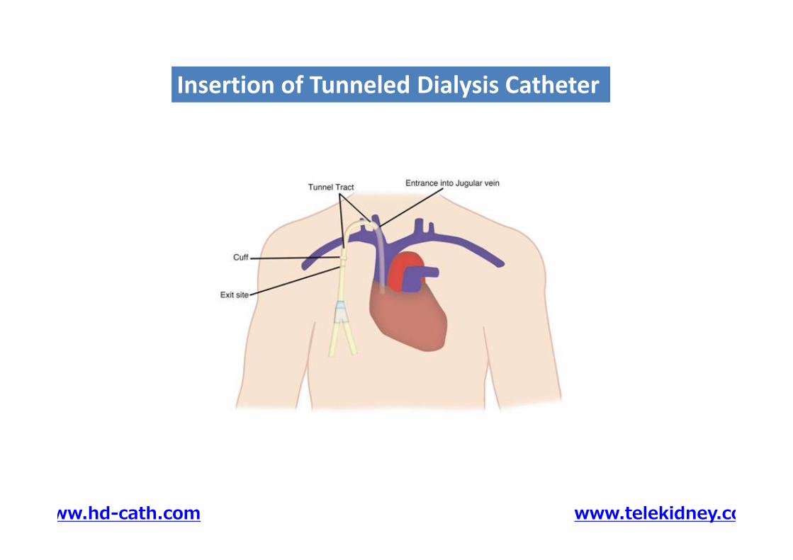
Insertion of Tunneled Dialysis CatheterInsertion of Tunneled Dialysis Catheter
www.hd-cath.com www.telekidney.com
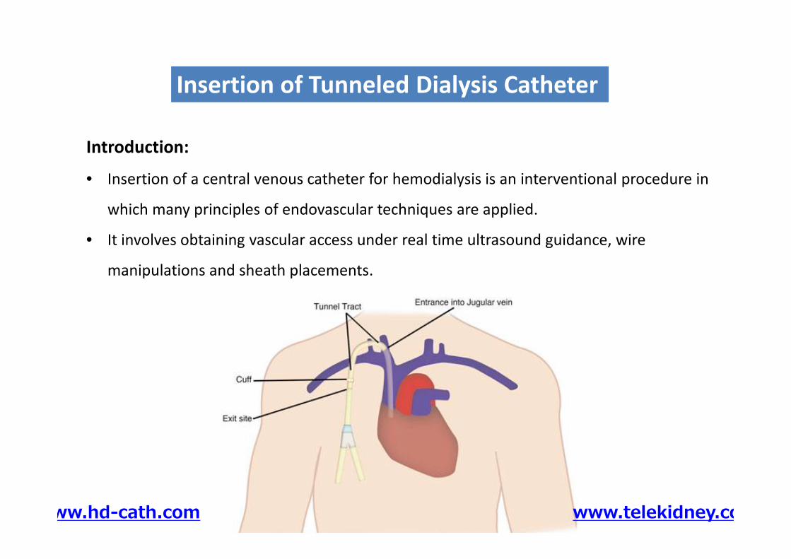
Introduction:
• Insertion of a central venous catheter for hemodialysis is an interventional procedure in
which many principles of endovascular techniques are applied.
• It involves obtaining vascular access under real time ultrasound guidance, wire
manipulations and sheath placements.
Insertion of Tunneled Dialysis CatheterInsertion of Tunneled Dialysis Catheter
Introduction:
• Insertion of a central venous catheter for hemodialysis is an interventional procedure in
which many principles of endovascular techniques are applied.
• It involves obtaining vascular access under real time ultrasound guidance, wire
manipulations and sheath placements.
www.hd-cath.com www.telekidney.com
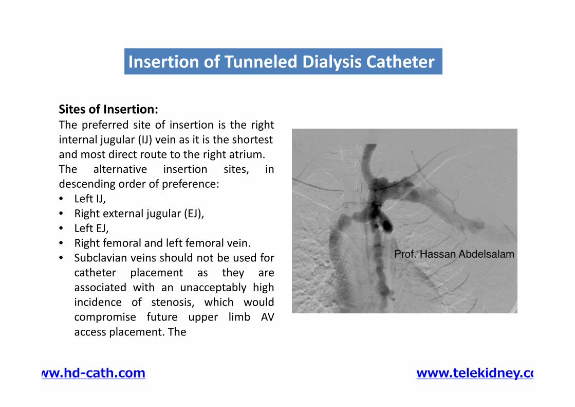
Sites of Insertion:The preferred site of insertion is the rightinternal jugular (IJ) vein as it is the shortestand most direct route to the right atrium.The alternative insertion sites, indescending order of preference:• Left IJ,• Right external jugular (EJ),• Left EJ,• Right femoral and left femoral vein.• Subclavian veins should not be used for
catheter placement as they areassociated with an unacceptably highincidence of stenosis, which wouldcompromise future upper limb AVaccess placement. The
Insertion of Tunneled Dialysis CatheterInsertion of Tunneled Dialysis Catheter
Sites of Insertion:The preferred site of insertion is the rightinternal jugular (IJ) vein as it is the shortestand most direct route to the right atrium.The alternative insertion sites, indescending order of preference:• Left IJ,• Right external jugular (EJ),• Left EJ,• Right femoral and left femoral vein.• Subclavian veins should not be used for
catheter placement as they areassociated with an unacceptably highincidence of stenosis, which wouldcompromise future upper limb AVaccess placement. The
www.hd-cath.com www.telekidney.com
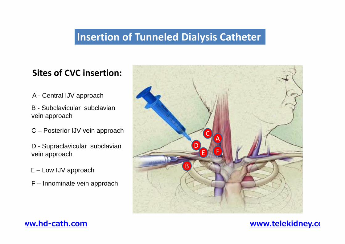
Sites of CVC insertion:
A - Central IJV approach
B - Subclavicular subclavianvein approach
Insertion of Tunneled Dialysis CatheterInsertion of Tunneled Dialysis Catheter
B
AC
DE F
C – Posterior IJV vein approach
D - Supraclavicular subclavianvein approach
E – Low IJV approach
F – Innominate vein approach
www.hd-cath.com www.telekidney.com
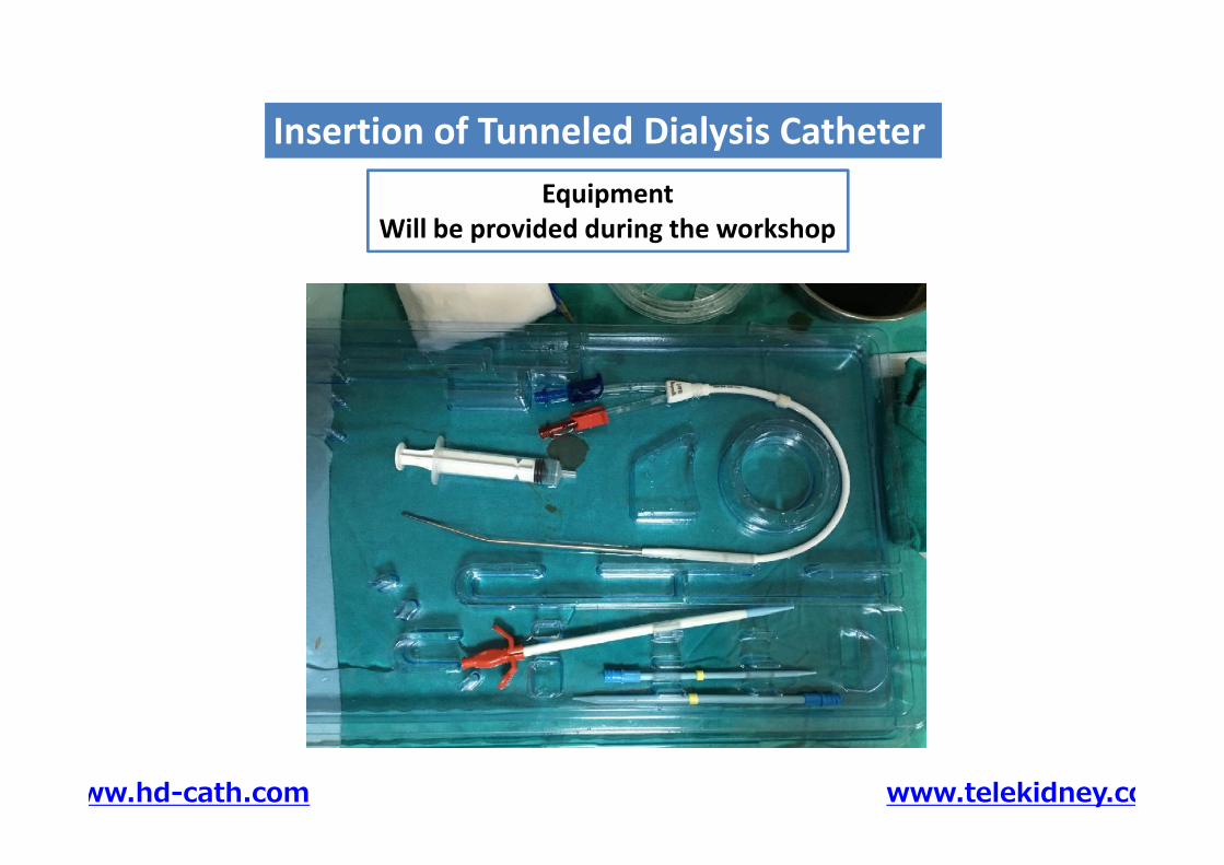
EquipmentWill be provided during the workshop
Insertion of Tunneled Dialysis CatheterInsertion of Tunneled Dialysis Catheter
www.hd-cath.com www.telekidney.com
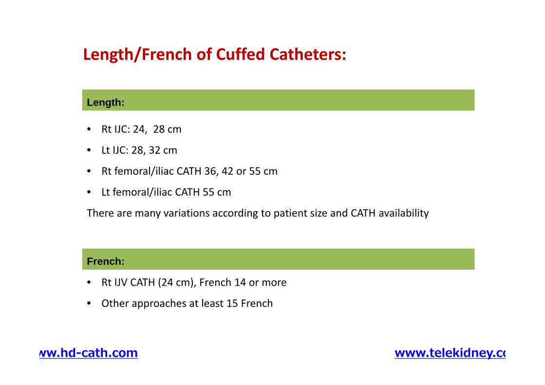
Length/French of Cuffed Catheters:
Length:
• Rt IJC: 24, 28 cm
• Lt IJC: 28, 32 cm
• Rt femoral/iliac CATH 36, 42 or 55 cm
• Lt femoral/iliac CATH 55 cm
There are many variations according to patient size and CATH availability
• Rt IJC: 24, 28 cm
• Lt IJC: 28, 32 cm
• Rt femoral/iliac CATH 36, 42 or 55 cm
• Lt femoral/iliac CATH 55 cm
There are many variations according to patient size and CATH availability
• Rt IJV CATH (24 cm), French 14 or more
• Other approaches at least 15 French
French:
www.hd-cath.com www.telekidney.com
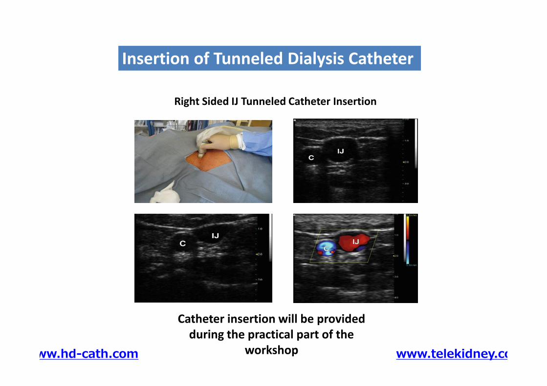
Right Sided IJ Tunneled Catheter Insertion
Insertion of Tunneled Dialysis CatheterInsertion of Tunneled Dialysis Catheter
Catheter insertion will be providedduring the practical part of the
workshopwww.hd-cath.com www.telekidney.com
Complications ofTunneled Dialysis Catheter Insertion
Insertion of Tunneled Dialysis CatheterInsertion of Tunneled Dialysis Catheter
Complications ofTunneled Dialysis Catheter Insertion
www.hd-cath.com www.telekidney.com
• Regardless of how “minor” or “simple” the procedure, never underestimate thecomplications that may arise during the procedure.
• Obeying the “rules” and developing good habits during training can go a long way todecrease procedure related complications.
Acute Complications of Tunneled Dialysis Catheter InsertionAcute Complications of Tunneled Dialysis Catheter Insertion
The following are some of the complications that one may encounter during dialysiscatheter placement, and the precautions and steps to treat them if they occur:The following are some of the complications that one may encounter during dialysiscatheter placement, and the precautions and steps to treat them if they occur:
www.hd-cath.com www.telekidney.com
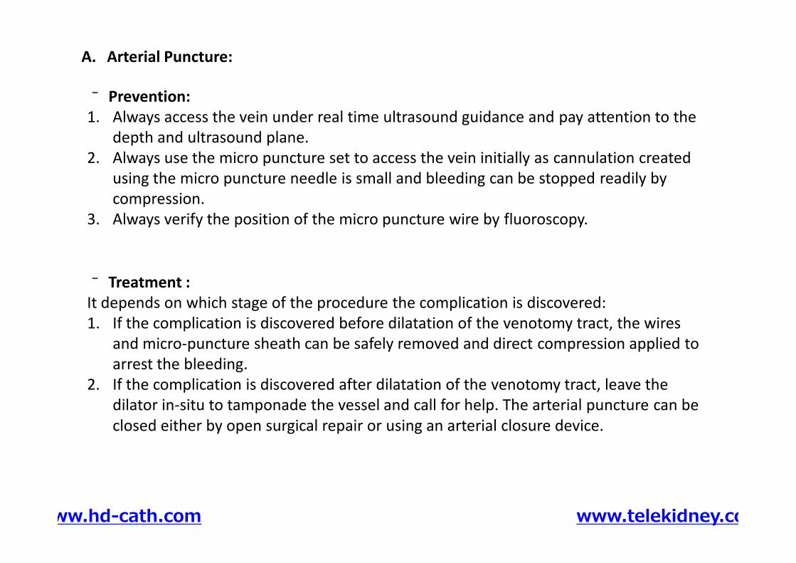
Prevention:1. Always access the vein under real time ultrasound guidance and pay attention to the
depth and ultrasound plane.2. Always use the micro puncture set to access the vein initially as cannulation created
using the micro puncture needle is small and bleeding can be stopped readily bycompression.
3. Always verify the position of the micro puncture wire by fluoroscopy.
A. Arterial Puncture:
Treatment :It depends on which stage of the procedure the complication is discovered:1. If the complication is discovered before dilatation of the venotomy tract, the wires
and micro-puncture sheath can be safely removed and direct compression applied toarrest the bleeding.
2. If the complication is discovered after dilatation of the venotomy tract, leave thedilator in-situ to tamponade the vessel and call for help. The arterial puncture can beclosed either by open surgical repair or using an arterial closure device.
www.hd-cath.com www.telekidney.com
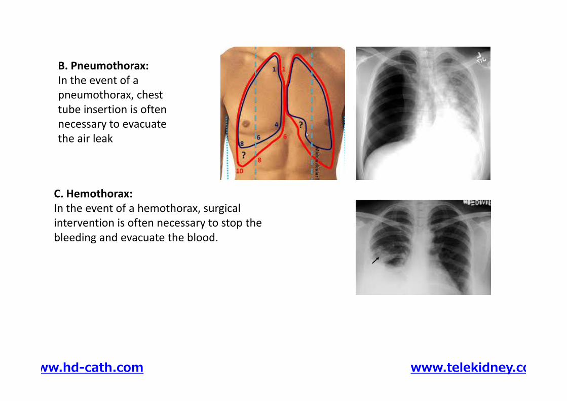
C. Hemothorax:In the event of a hemothorax, surgicalintervention is often necessary to stop thebleeding and evacuate the blood.
B. Pneumothorax:In the event of apneumothorax, chesttube insertion is oftennecessary to evacuatethe air leak
C. Hemothorax:In the event of a hemothorax, surgicalintervention is often necessary to stop thebleeding and evacuate the blood.
www.hd-cath.com www.telekidney.com
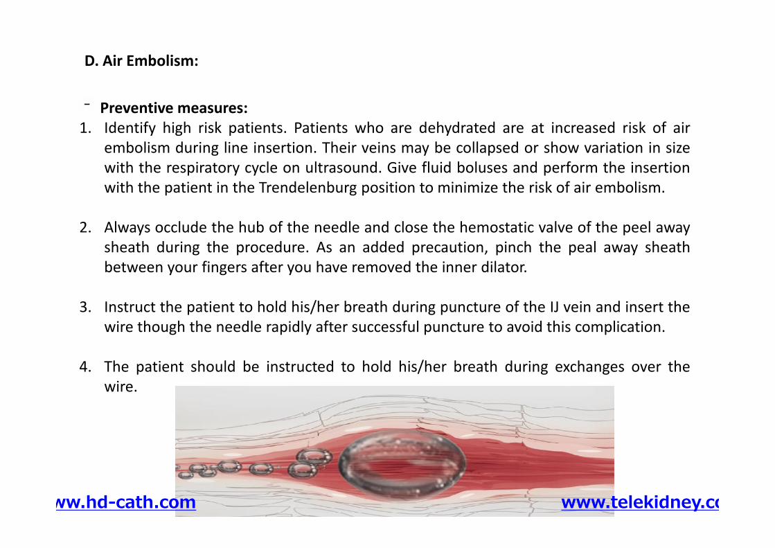
Preventive measures:1. Identify high risk patients. Patients who are dehydrated are at increased risk of air
embolism during line insertion. Their veins may be collapsed or show variation in sizewith the respiratory cycle on ultrasound. Give fluid boluses and perform the insertionwith the patient in the Trendelenburg position to minimize the risk of air embolism.
2. Always occlude the hub of the needle and close the hemostatic valve of the peel awaysheath during the procedure. As an added precaution, pinch the peal away sheathbetween your fingers after you have removed the inner dilator.
3. Instruct the patient to hold his/her breath during puncture of the IJ vein and insert thewire though the needle rapidly after successful puncture to avoid this complication.
4. The patient should be instructed to hold his/her breath during exchanges over thewire.
D. Air Embolism:
Preventive measures:1. Identify high risk patients. Patients who are dehydrated are at increased risk of air
embolism during line insertion. Their veins may be collapsed or show variation in sizewith the respiratory cycle on ultrasound. Give fluid boluses and perform the insertionwith the patient in the Trendelenburg position to minimize the risk of air embolism.
2. Always occlude the hub of the needle and close the hemostatic valve of the peel awaysheath during the procedure. As an added precaution, pinch the peal away sheathbetween your fingers after you have removed the inner dilator.
3. Instruct the patient to hold his/her breath during puncture of the IJ vein and insert thewire though the needle rapidly after successful puncture to avoid this complication.
4. The patient should be instructed to hold his/her breath during exchanges over thewire.
www.hd-cath.com www.telekidney.com
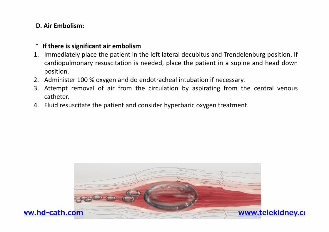
If there is significant air embolism1. Immediately place the patient in the left lateral decubitus and Trendelenburg position. If
cardiopulmonary resuscitation is needed, place the patient in a supine and head downposition.
2. Administer 100 % oxygen and do endotracheal intubation if necessary.3. Attempt removal of air from the circulation by aspirating from the central venous
catheter.4. Fluid resuscitate the patient and consider hyperbaric oxygen treatment.
D. Air Embolism:
www.hd-cath.com www.telekidney.com
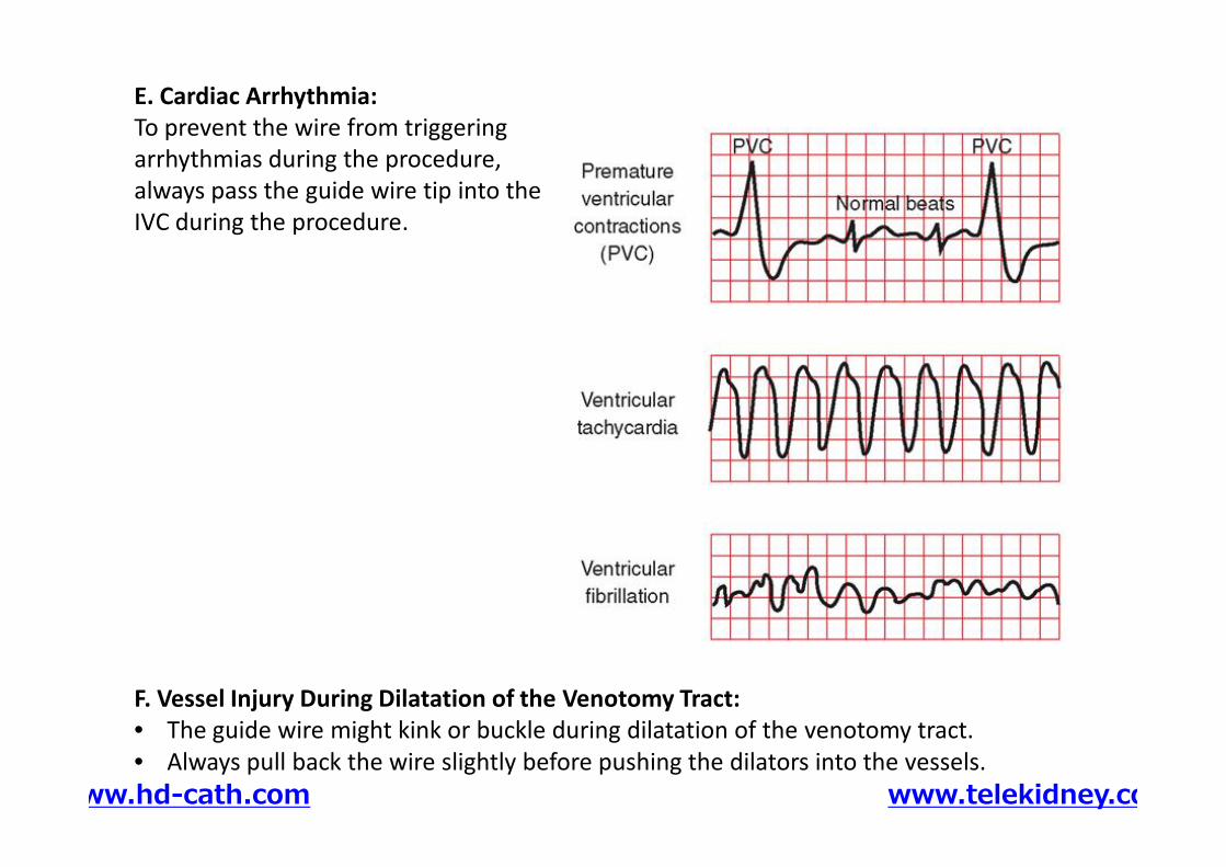
E. Cardiac Arrhythmia:To prevent the wire from triggeringarrhythmias during the procedure,always pass the guide wire tip into theIVC during the procedure.
F. Vessel Injury During Dilatation of the Venotomy Tract:• The guide wire might kink or buckle during dilatation of the venotomy tract.• Always pull back the wire slightly before pushing the dilators into the vessels.
www.hd-cath.com www.telekidney.com
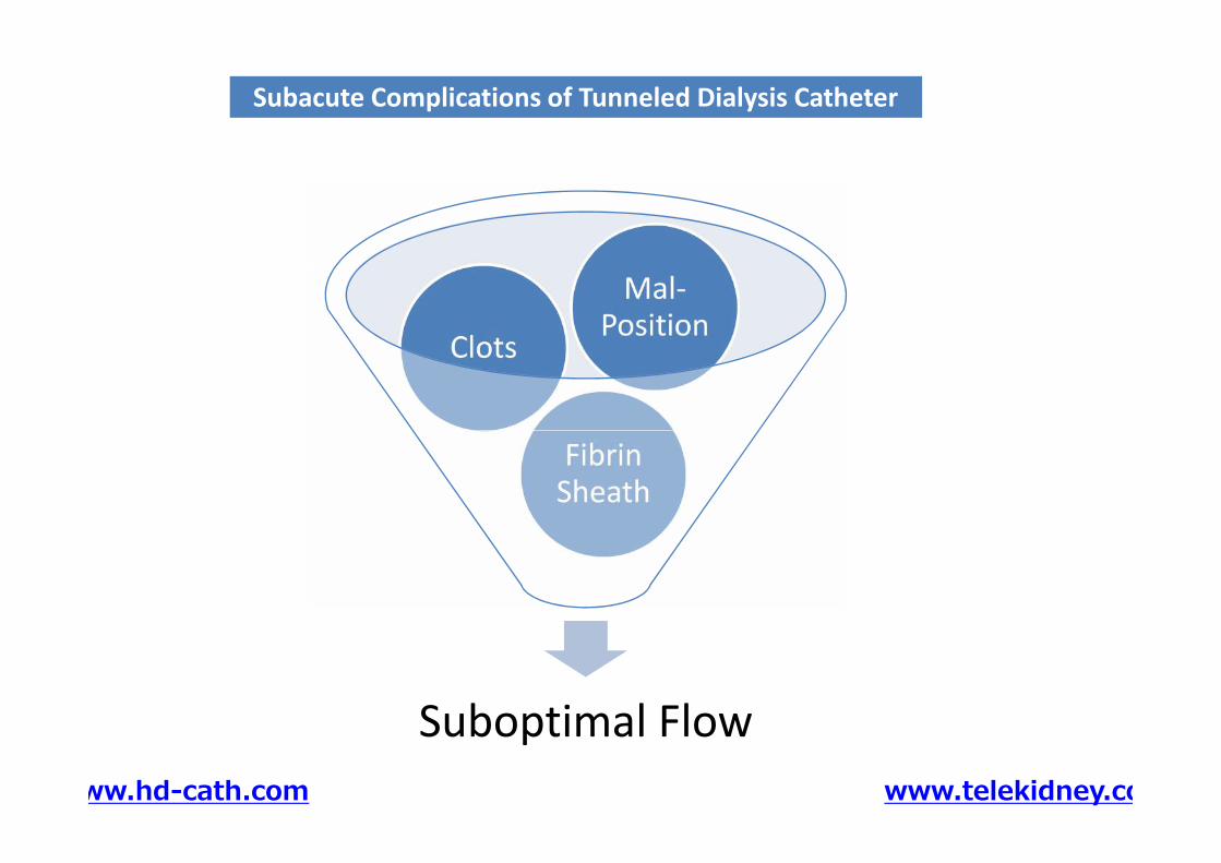
Subacute Complications of Tunneled Dialysis CatheterSubacute Complications of Tunneled Dialysis Catheter
FibrinSheath
Clots
Mal-Position
Suboptimal Flow
FibrinSheath
www.hd-cath.com www.telekidney.com
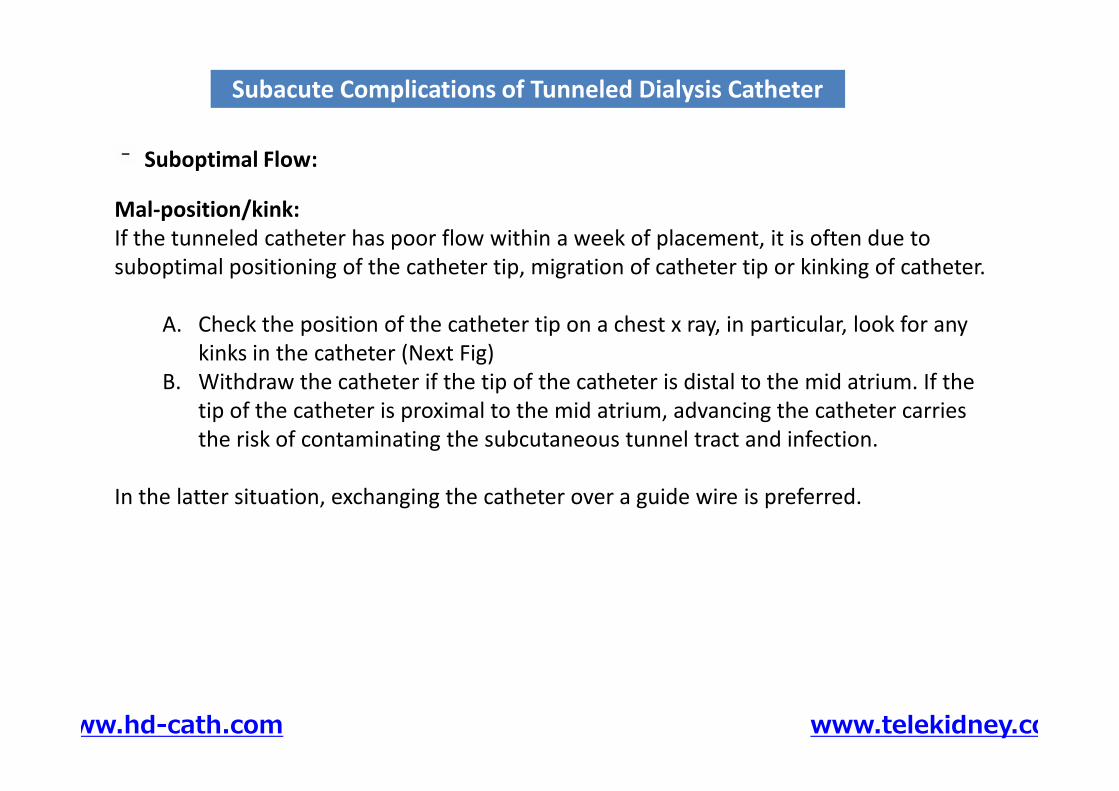
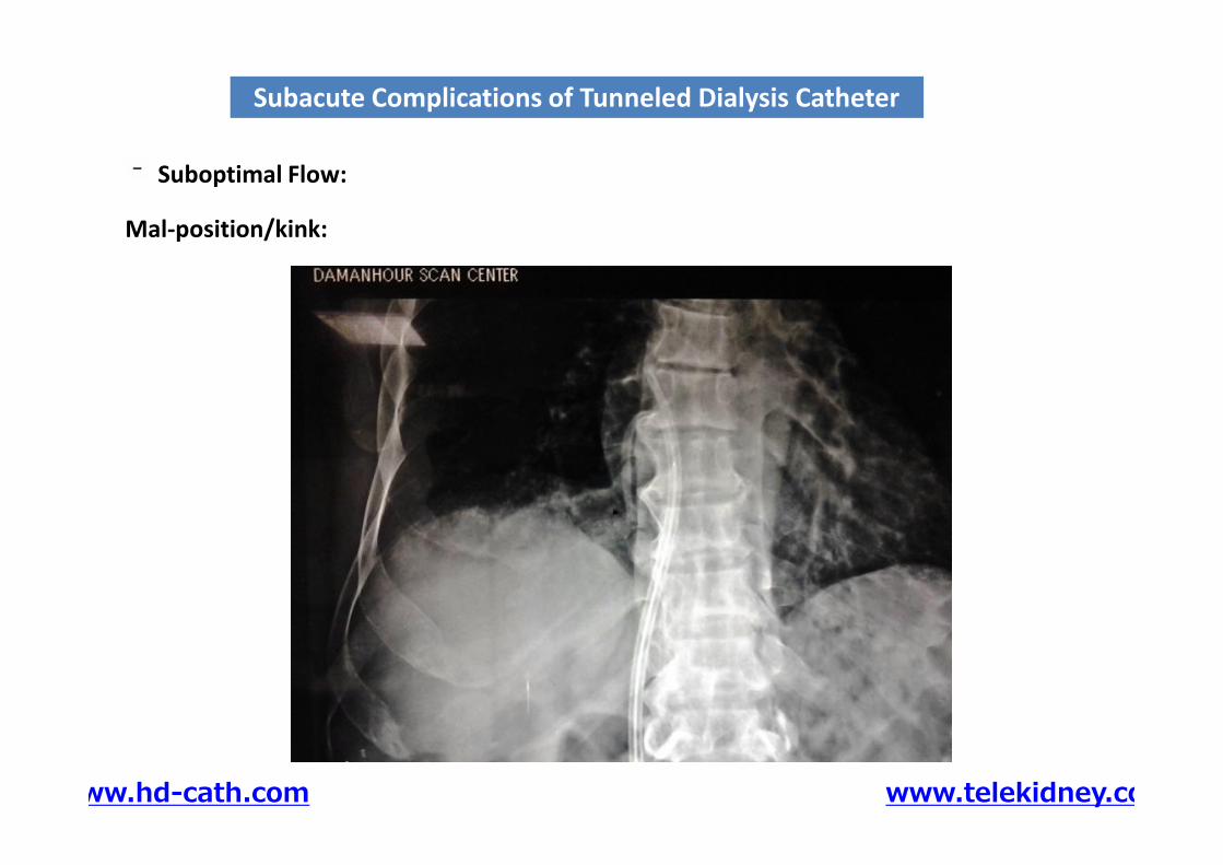
Mal-position/kink:If the tunneled catheter has poor flow within a week of placement, it is often due tosuboptimal positioning of the catheter tip, migration of catheter tip or kinking of catheter.
A. Check the position of the catheter tip on a chest x ray, in particular, look for anykinks in the catheter (Next Fig)
B. Withdraw the catheter if the tip of the catheter is distal to the mid atrium. If thetip of the catheter is proximal to the mid atrium, advancing the catheter carriesthe risk of contaminating the subcutaneous tunnel tract and infection.
In the latter situation, exchanging the catheter over a guide wire is preferred.
Subacute Complications of Tunneled Dialysis CatheterSubacute Complications of Tunneled Dialysis Catheter
Suboptimal Flow:
Mal-position/kink:If the tunneled catheter has poor flow within a week of placement, it is often due tosuboptimal positioning of the catheter tip, migration of catheter tip or kinking of catheter.
A. Check the position of the catheter tip on a chest x ray, in particular, look for anykinks in the catheter (Next Fig)
B. Withdraw the catheter if the tip of the catheter is distal to the mid atrium. If thetip of the catheter is proximal to the mid atrium, advancing the catheter carriesthe risk of contaminating the subcutaneous tunnel tract and infection.
In the latter situation, exchanging the catheter over a guide wire is preferred.
www.hd-cath.com www.telekidney.com
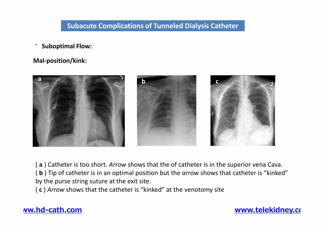
ba c
Subacute Complications of Tunneled Dialysis CatheterSubacute Complications of Tunneled Dialysis Catheter
Suboptimal Flow:
Mal-position/kink:
c
( a ) Catheter is too short. Arrow shows that the of catheter is in the superior vena Cava.( b ) Tip of catheter is in an optimal position but the arrow shows that catheter is “kinked”by the purse string suture at the exit site.( c ) Arrow shows that the catheter is “kinked” at the venotomy site
www.hd-cath.com www.telekidney.com
Mal-position/kink:
Subacute Complications of Tunneled Dialysis CatheterSubacute Complications of Tunneled Dialysis Catheter
Suboptimal Flow:
www.hd-cath.com www.telekidney.com

Clots:If the catheter tip is in the correct position, a trial of a thrombolytic agent may beattempted.
A. The procedure should be carried out in a sterile manner. Clean and drape thepatient.
B. Remove the caps of the catheter ports and aspirate 5 ml of blood from each lumento remove the locking agent.
C. Instill 2 ml of TPA (1 mg/ml) into each lumen and allow it to dwell for half an hour.D. Aspirate both catheter ports and discard the initial 5 ml of blood.E. Test catheter flow with a 20 ml syringe. If the flow remains suboptimal, schedule
for catheter exchange over a guide wire.
Subacute Complications of Tunneled Dialysis CatheterSubacute Complications of Tunneled Dialysis Catheter
Suboptimal Flow:
Clots:If the catheter tip is in the correct position, a trial of a thrombolytic agent may beattempted.
A. The procedure should be carried out in a sterile manner. Clean and drape thepatient.
B. Remove the caps of the catheter ports and aspirate 5 ml of blood from each lumento remove the locking agent.
C. Instill 2 ml of TPA (1 mg/ml) into each lumen and allow it to dwell for half an hour.D. Aspirate both catheter ports and discard the initial 5 ml of blood.E. Test catheter flow with a 20 ml syringe. If the flow remains suboptimal, schedule
for catheter exchange over a guide wire.
www.hd-cath.com www.telekidney.com
Fibrin Sheath:If the catheter develops poor flow more than a month after placement, it is probablysecondary to obstruction from fibrin sheath formation around the tip of the catheter.
A trial of tPA may be attempted. If unsuccessful, exchanging the tunneled catheter over aguide wire with or without disruption of the fibrin sheath is the treatment of choice.
A. Check the position of the catheter tip on chest x ray.B. Aspirate both catheter ports and discard the initial 5 ml of blood which contains the
locking agentC. Insert a 0.035 in. angled stiff guide wire through the venous port of the catheter into
the inferior vena cava.D. Free the preexisting catheter cuff by blunt dissection and withdraw the catheter gently
by approximately 3 cm. Gently inject 10–15 ml of contrast material into the arterialport to visualize the fibrin sheath.
E. Remove the preexisting catheter and insert the 12–14 mm angioplasty ballooncatheter over the wire via the subcutaneous tunnel tract, and inflate the balloon in theSVC to disrupt the fibrin sheath.
Subacute Complications of Tunneled Dialysis CatheterSubacute Complications of Tunneled Dialysis Catheter
Suboptimal Flow:
Fibrin Sheath:If the catheter develops poor flow more than a month after placement, it is probablysecondary to obstruction from fibrin sheath formation around the tip of the catheter.
A trial of tPA may be attempted. If unsuccessful, exchanging the tunneled catheter over aguide wire with or without disruption of the fibrin sheath is the treatment of choice.
A. Check the position of the catheter tip on chest x ray.B. Aspirate both catheter ports and discard the initial 5 ml of blood which contains the
locking agentC. Insert a 0.035 in. angled stiff guide wire through the venous port of the catheter into
the inferior vena cava.D. Free the preexisting catheter cuff by blunt dissection and withdraw the catheter gently
by approximately 3 cm. Gently inject 10–15 ml of contrast material into the arterialport to visualize the fibrin sheath.
E. Remove the preexisting catheter and insert the 12–14 mm angioplasty ballooncatheter over the wire via the subcutaneous tunnel tract, and inflate the balloon in theSVC to disrupt the fibrin sheath.
www.hd-cath.com www.telekidney.com
F. Exchange a new-tunneled dialysis catheter over the guide wire and place the tipwithin the proximal SVC. Inject 10–15 ml of contrast via the arterial port to check forresidual fibrin sheath. If fibrin sheath is still present, repeat the angioplasty. If there isno residual fibrin sheath, advance the catheter tip to the desired position in the midatrium.
Subacute Complications of Tunneled Dialysis CatheterSubacute Complications of Tunneled Dialysis Catheter
Suboptimal Flow:
Stripping of Fibrin SheathWill be provided during the workshop
www.hd-cath.com www.telekidney.com
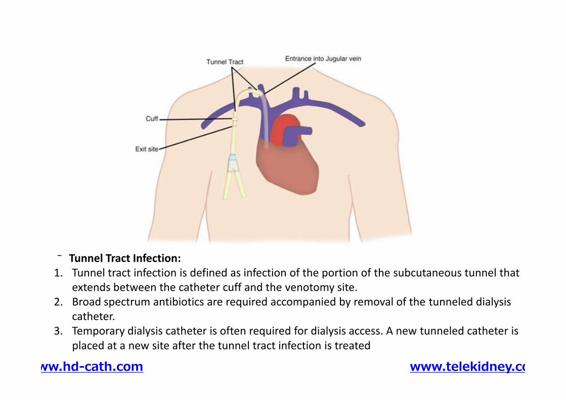
Tunnel Tract Infection:1. Tunnel tract infection is defined as infection of the portion of the subcutaneous tunnel that
extends between the catheter cuff and the venotomy site.2. Broad spectrum antibiotics are required accompanied by removal of the tunneled dialysis
catheter.3. Temporary dialysis catheter is often required for dialysis access. A new tunneled catheter is
placed at a new site after the tunnel tract infection is treated
www.hd-cath.com www.telekidney.com
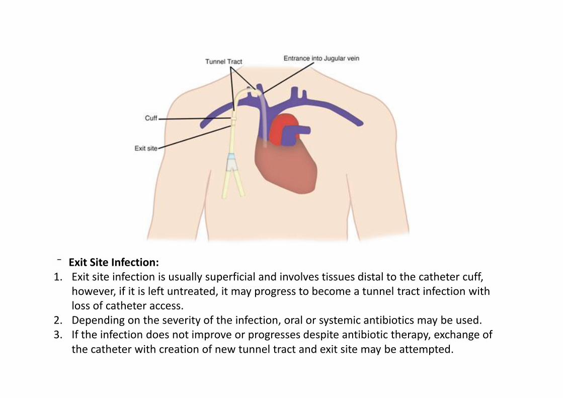
Exit Site Infection:1. Exit site infection is usually superficial and involves tissues distal to the catheter cuff,
however, if it is left untreated, it may progress to become a tunnel tract infection withloss of catheter access.
2. Depending on the severity of the infection, oral or systemic antibiotics may be used.3. If the infection does not improve or progresses despite antibiotic therapy, exchange of
the catheter with creation of new tunnel tract and exit site may be attempted.
Removal of Tunneled Dialysis CatheterRemoval of Tunneled Dialysis Catheter
www.hd-cath.com www.telekidney.com
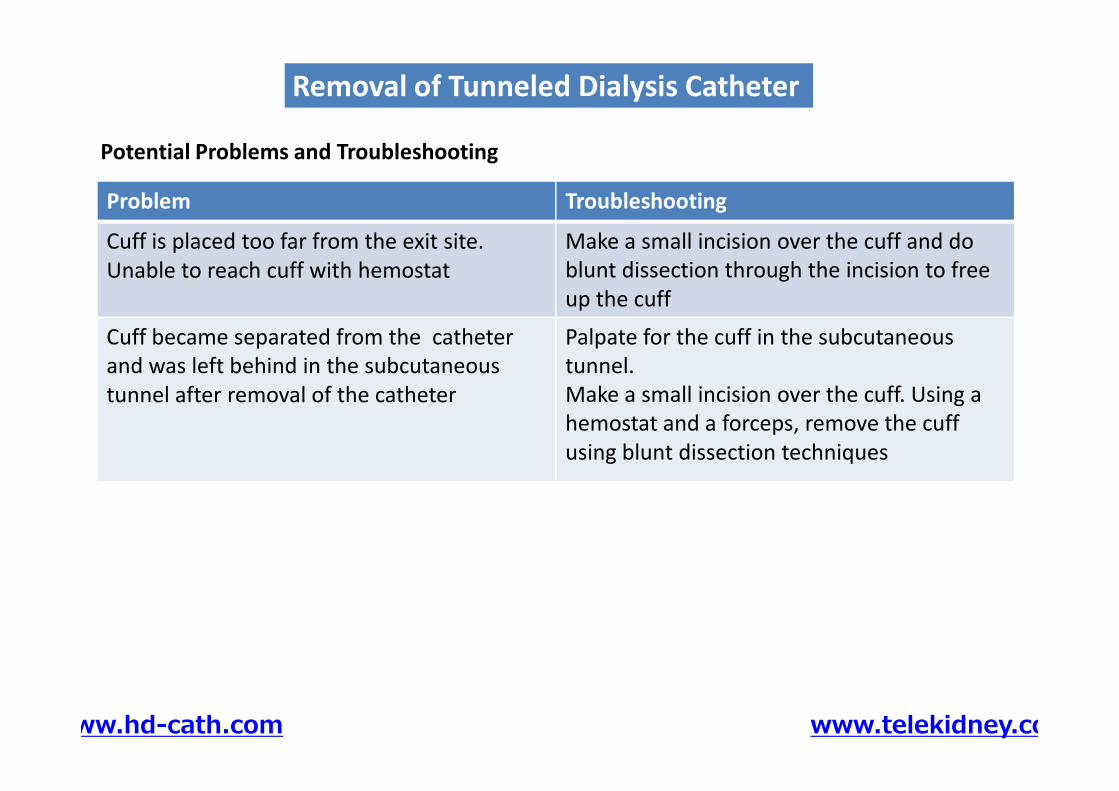
TroubleshootingProblem
Make a small incision over the cuff and doblunt dissection through the incision to freeup the cuff
Cuff is placed too far from the exit site.Unable to reach cuff with hemostat
Palpate for the cuff in the subcutaneoustunnel.Make a small incision over the cuff. Using ahemostat and a forceps, remove the cuffusing blunt dissection techniques
Cuff became separated from the catheterand was left behind in the subcutaneoustunnel after removal of the catheter
Potential Problems and Troubleshooting
Removal of Tunneled Dialysis CatheterRemoval of Tunneled Dialysis Catheter
Palpate for the cuff in the subcutaneoustunnel.Make a small incision over the cuff. Using ahemostat and a forceps, remove the cuffusing blunt dissection techniques
Cuff became separated from the catheterand was left behind in the subcutaneoustunnel after removal of the catheter
www.hd-cath.com www.telekidney.com

Stage 1&2
Stage3a,b
Stage 5
Stage 4
30 ml/minInform
1- You have reduced kidneyfunction and you may needkidney replacement therapy(KRT) to save your life.
2- Describe modalities of KRT.
3- Spare forearm and upperarm veins from injections.Dorsum of hands is thepreferred site for injections andcannulation. This will savethese veins for creation of AVF.
٣١
Stage 1&2Stage 5
30 ml/minInform
1- You have reduced kidneyfunction and you may needkidney replacement therapy(KRT) to save your life.
2- Describe modalities of KRT.
3- Spare forearm and upperarm veins from injections.Dorsum of hands is thepreferred site for injections andcannulation. This will savethese veins for creation of AVF.
20 ml/min ……. CreateThis is the optimal time for creation of AVF/
preparation for preemptive kidneytransplantation
www.hd-cath.com www.telekidney.com
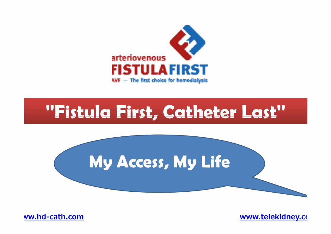
''Fistula First, Catheter Last''''Fistula First, Catheter Last''
www.hd-cath.com www.telekidney.com
''Fistula First, Catheter Last''''Fistula First, Catheter Last''
My Access, My Life

www.hd-cath.comRegister to receive your
FREE trial version
www.hd-cath.com www.telekidney.com

THANK YOU


![Focus on peripherally inserted central catheters in ... · as “medium term VADs”, while non-tunneled CICCs are regarded as “short term VADs”[8]. This review will not discuss](https://static.fdocuments.net/doc/165x107/5f4224d35277916ab3231c55/focus-on-peripherally-inserted-central-catheters-in-as-aoemedium-term-vadsa.jpg)


![Tunneled Peritoneal Catheter Placement in …downloads.hindawi.com/journals/bmri/2019/4132396.pdfof life []. Treatments with tunneled peritoneal catheter insertion, peritoneovenous](https://static.fdocuments.net/doc/165x107/5f8b6407216452101b766e47/tunneled-peritoneal-catheter-placement-in-of-life-treatments-with-tunneled-peritoneal.jpg)


