Tumors of intestine
40
DR. MOHANAD R. ALWAN TUMORS OF THE SMALL AND LARGE INTESTINES SMS 2044
-
Upload
mohanad-aljashamy -
Category
Education
-
view
370 -
download
0
Transcript of Tumors of intestine

DR. MOHANAD R. ALWAN
TUMORS OF THE SMALL AND LARGE INTESTINES
SMS 2044

Tumors of the small and large intestineClassification
Non-neoplastic polyps Hyperplastic polyps Hamartomatous polyps (Juvenile & Peutz-Jeghers polyps) Inflammatory polyps Lymphoid polyps
Neoplastic ( epithelial) polyps Benign polyps (adenoma) Malignant lesions
AdenocarcinomaCarcinoidAnal zone carcinoma
Mesenchymal lesions GI stromal tumors (benign & malignant) Others ( lipoma, neuroma, angioma, Kaposi sarcoma)
Lymphoma• Diffuse large cell lymphoma. MALT cell lymphoma.

Intestinal NeoplasmsSmall intestine= neoplasms are rareBenign tumors MC (membranous cells) = Adenoma MC site - ampulla of Vater (the union of the pancreatic duct and the
common bile duct) Complaints= occult blood loss/ rarely-obstruction or intussusceptions Associated with familial polyposis Rx =surgical excision difficult Clinical Course=premalignantMalignant tumors MC= Adenocarcinoma MC site - Duodenum clinical -intestinal obstruction occult blood loss -only sign if involves ampulla of Vater -cause fluctuating obstructive jaundice
Risk factorsMost tumors -no identifiable factor, Crohn’s, celiac disease
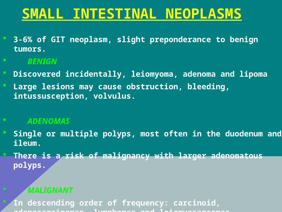
SMALL INTESTINAL NEOPLASMS 3-6% of GIT neoplasm, slight preponderance to benign tumors. BENIGN Discovered incidentally, leiomyoma, adenoma and lipoma Large lesions may cause obstruction, bleeding, intussusception,
volvulus.
ADENOMAS Single or multiple polyps, most often in the duodenum and ileum. There is a risk of malignancy with larger adenomatous polyps.
MALIGNANT In descending order of frequency: carcinoid, adenocarcinomas,
lymphomas and leiomyosarcomas. Leiomyosarcomas have tyrosine kinase receptors, can be treated by
STI-571(gleevec)

Adenoma in duodenum
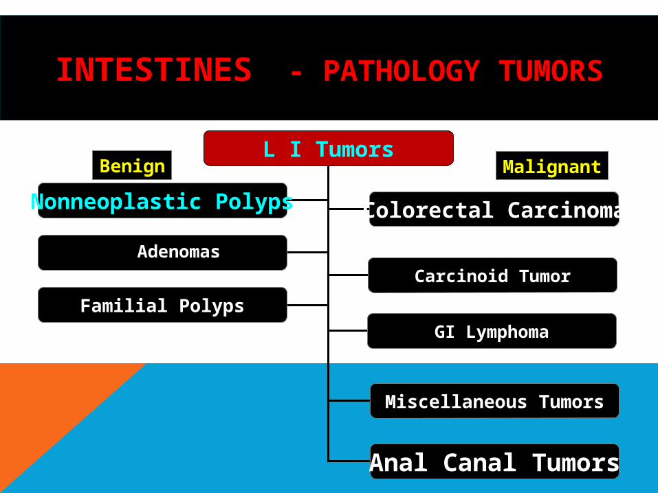
INTESTINES - PATHOLOGY TUMORS
L I Tumors
GI Lymphoma
Nonneoplastic Polyps
Adenomas
Colorectal Carcinoma
Familial PolypsCarcinoid Tumor
Miscellaneous Tumors
Anal Canal Tumors
Benign Malignant

Intestinal NeoplasmsLarge intestine= neoplasms are commonBenign tumors MC = Adenoma= PolypMC site of GI Polyps= ColonTypes Non-neoplastic (Hyperplastic, inflammatory, hamartomatous) Neoplastic or adenomatous( Tubular, Villous, Tubulo-villous) Malignant risk of adenomatous polyps correlates with:1) polyp size ( > 4cm)2) degree of dysplasia 3) extent of villous component (More villous= more cancerous)Feature Non-neoplastic Neoplastic
Frequency & Age
MC (90%) young peopleMostly Hyperplastic,
Only 10%, elderly peopleMostly Tubular
Dysplasia without dysplasia With dysplasiaMechanism excess production of epithelial cells
than their lossMutations in genes
Complication No risk of malignancy premalignant

Tumours of the small and large intestine
The most common:Epithelial tumours are a major cause of morbidity and mortality worldwide.
The colorectal is the GIT most commonly affected by primary tumors
Adenocarcinoma in colorectum represent 70% of all malignancy of GIT
Benign tumors, primarily epithelial, are present in 25 to 50% of older adults.

Hamartomatous polyp

N…P. All adenomatous lesions arise as the result of epithelial
proliferation and dysplasia, which may range from mild to so severe as to represent transformation to carcinoma.

Tubular adenoma
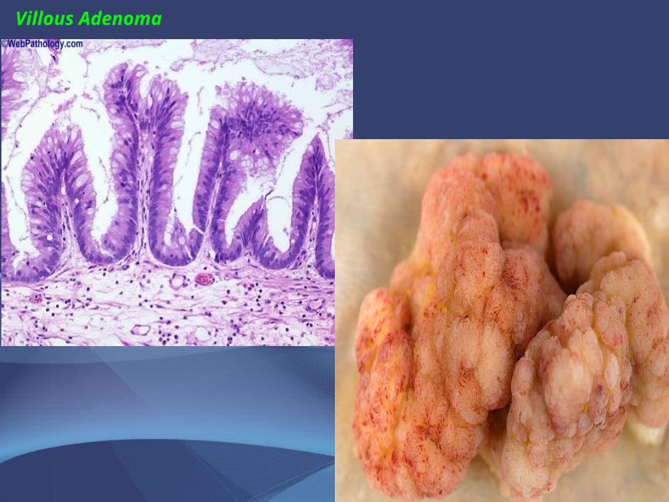
Villous Adenoma

Malignant Tumors of Large Intestine
Adenocarcinoma
Constitutes 98% of all cancers in the large intestine.
Worldwide distribution, highest incidence in west.Peak incidence: 60 to 70 years of age< 20% cases before age of 50adenomas – presumed precursor lesions for most
tumorsmales affected ≈ 20% more often than females

• Incidence rates for colorectal cancer differ by sub-sites
Anatomic Distribution

Carcinogenesis• Chromosome instability pathway
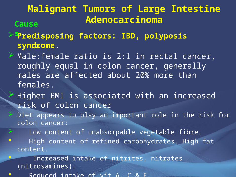
Malignant Tumors of Large IntestineAdenocarcinoma
Predisposing factors: IBD, polyposis syndrome. Male:female ratio is 2:1 in rectal cancer, roughly equal in
colon cancer, generally males are affected about 20% more than females.
Higher BMI is associated with an increased risk of colon cancer
Diet appears to play an important role in the risk for colon cancer: Low content of unabsorpable vegetable fibre. High content of refined carbohydrates. High fat content. Increased intake of nitrites, nitrates (nitrosamines). Reduced intake of vit A, C & E. The use of aspirin and NSAID (cyclooxygenase-2 inhibitors)
exerts a protective effect against colon cancer Alcohol, Tobacco
Causes

Colorectal ad.CarcinomaMorphology
Sixty to 70% of colorectal carcinomas are in the rectum, rectosigmoid and sigmoid colon.
The carcinomas tend to be annular, encircling lesions with early symptoms of obstruction.
Neoplasms start superficially, slowly invading the deeper layers with ulceration and eventually metastasis.


Early carcinomas, i.e., tumors limited to the submucosa, are mostly
• Polypoid • Pedunculated • Semipedunculated• Sessile • Flat lesions• Flat with slight elevation with light central depression
Advanced carcinomas (invading beyond the submucosa) are four types, similar to the Borrmann categories of gastric carcinoma:
• Polypoid (protuberant)• Ulcerated, with sharply demarcated margins• Ulcerated without definite borders• Diffusely infiltrating

There is a solitary mass attached via a long stalk to the colonic mucosa. It is discreet and does not involve the wall of the colon. The surface is dark red (hemorrhagic). The stool guaiac was positive


Histological Description Most lesions consist of gland formation lining of tall columnar
cells with palisading large oval or pencil shaped nuclei with increased coarse chromatin, easily found.
The epithelium bears a clear resemblance to the dysplastic epithelium which covers adenomas.
Tumor has well defined margins The center is depressed due to tumor necrosis Mucin lakes are found in 10-15% of tumors and these
patients are often those with hereditary non-polyposis syndromes or ulcerative colitis
Bleeding from the tumor is often apparent as the tumor tends to grow blood vessel


• Tumor has well defined margins • The center is depressed due to tumor necrosis • The thin margin of intact tumor is raised (arrow) • Adjoing mucosa is normal with delicate mucosal folds

MUCINOUS ADENOCARCINOMA
>

Colorectal Carcinoma
Clinical Features
May remain asymptomatic for years and the symptoms develop insidiously
Abdominal pain and tenderness in the lower abdomen Blood in the stool Diarrhea, constipation, or other change in bowel habits Narrow stools Weight loss with no known reason Weakness, malaise, weight loss, unexplained anemia
(secondary to early bleeding).

Colorectal Carcinoma Spread: - Direct extension.
- Metastasis through: - Lymphatic - Blood vessels
- Favored sites are regional lymph node, liver, lungs, bones.
Serum level of carcinoembryonic antigen (CEA) is related to tumor size and extent of spread.
It is helpful in monitoring for recurrence of tumor after resection.
Overall 5-year survival is 35 to 49% in the United States.

Staging Tx: No description of the tumor's. Tis: In situ carcinoma; the tumor involves only the muscularis
mucosa T1: The cancer has grown through the muscularis mucosa
and extends into the submucosa T2: The cancer has grown through the submucosa and
extends into the muscularis propria T3: The cancer has grown through the muscularis propria and
into the outermost layers of the colon but not through them; it has not reached any nearby organs or tissues
T4a: The cancer has grown through the serosa (visceral peritoneum)
T4b: The cancer has grown through the wall of the colon and is attached to or invades nearby tissues or organs


Colorectal NeoplasmOther Tumors
Malignant spindle cell (mesenchymal) tumors and lymphomas.
Grossly and microscopically resemble those arising elsewhere in the GI tract. Carcinoid tumors may arise anywhere in the colon,
especially the rectum. Squamous cell carcinomas are largely limited to the anal
canal. Initially present as plaque-like lesions, later becoming ulcerated or fungating.
Malignant melanoma at the anal verge.
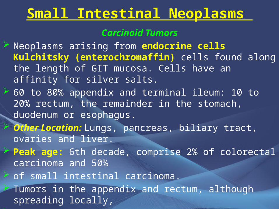
Small Intestinal Neoplasms Carcinoid Tumors
Neoplasms arising from endocrine cells Kulchitsky (enterochromaffin) cells found along the length of GIT mucosa. Cells have an affinity for silver salts.
60 to 80% appendix and terminal ileum: 10 to 20% rectum, the remainder in the stomach, duodenum or esophagus.
Other Location: Lungs, pancreas, biliary tract, ovaries and liver. Peak age: 6th decade, comprise 2% of colorectal carcinoma and 50% of small intestinal carcinoma. Tumors in the appendix and rectum, although spreading locally, seldom metastasize. Ileal, gastric, and colonic carcinoids are frequently malignant.

Carcinoid TumorsPathological Lesion
Round submucosal elevations that are bright yellow or yellow-gray, may be deeply infiltrative and penetrate muscle to the serosa.
Gastric and ileal carcinoids are frequently multiple. Tumor cells arranged in trabecular, insular, glandular or
undifferentiated patterns are monotonously similar to each other with regular round nuclei
Ultrastructral features: neurosecretory electron dense bodies in the cytoplasm

Small Intestinal NeoplasmsCarcinoid Tumor
Clinical features Asymptomatic May cause obstruction, intussusception or bleeding. May elaborate hormones: Zollinger-Ellison , Cushing’s ,
carcinoid or other syndromes. 5 years survival rate is 90%, small bowel Carcinoid with liver metastasis the 5 years
survival rate is better than 50%

Small Intestinal Neoplasms
LymphomaUp to 40% of lymphomas arise in sites other than lymph
nodes, gut is the most.1% to 4% of all gastrointestinal malignancies are
lymphomas. Primary GIT lymphomas exhibit no evidence of liver,
spleen, or bone marrow involvement at the time of diagnosis.

Small Intestinal NeoplasmsLymphoma
Sporadic lymphoma arise from the B cells of mucosa-associated lymphoid tissue (MALT).
This usually affects adults, lacks a sex predilection, and may arise anywhere in the gut: stomach - 55% to 60%
small intestine - 25% to 30% proximal colon - 10% to 15% distal colon - up to 10%

Small Intestinal Neoplasms
Lymphoma• Gastric MALT lymphomas arise in the
setting of mucosal lymphoid activation, as a result of Helicobacter associated chronic gastritis.
• Celiac disease is associated with a higher than normal risk of T-cell lymphomas.

Small Intestinal Neoplasms
LymphomaPrimary GIT lymphomas have a better
prognosis than do those arising in other sites.
Treatment: combined surgery, chemotherapy, and radiation therapy.

Therapy
• Chemotherapy• Radiotherapy• Photodynamic therapy• Radical surgery• Gene therapy

Question?????