Treg and Th17 T cell Immunology Tools Cat. #’s TCLxxx series
10
User Manual A limited-use label license covers this product. By use of this product, you accept the terms and conditions outlined in the Licensing and Warranty Statement contained in this user manual. Store Cell lines at -80ºC immediately upon receipt Store Vectors at -20ºC immediately upon receipt Treg and Th17 T cell Immunology Tools Cat. #’s TCLxxx series
Transcript of Treg and Th17 T cell Immunology Tools Cat. #’s TCLxxx series
User Manual
A limited-use label license covers thisproduct. By use of this product, youaccept the terms and conditions outlinedin the Licensing and Warranty Statementcontained in this user manual.
Store Cell lines at -80ºC immediately upon receiptStore Vectors at -20ºC immediately upon receipt
Treg and Th17 T cell
Immunology Tools
Cat. #’s TCLxxx series

Treg and Th17 T cell Immunology Tools Cat. #’s TCLxxx
888-266-5066 (Toll Free) 650-968-2200 (outside US) Page 1
Contents I. Introduction and Background
A. Overview 2 B. Lentivectors expressing Foxp3 and RORt 2 C. PiggyBac vectors expressing Foxp3 and RORt 4 D. Reporter constructs for Foxp3, IL-17, RORt promoters 4 E. Product Handling Guidelines 6
II. Protocols A. Packaging lentivectors into virus information 6 B. Spinoculation of virus and Jurkat T cells 6 C. Transfecting PiggyBac transposons information 6 III. T cell Technical References 7
IV. Technical Support 8
V. Licensing and Warranty Statement 9

System Biosciences (SBI) User Manual
Page 2 ver.1-042412 www.systembio.com
I. Introduction and Background
A. Overview Regulatory T cells (Treg), known as suppressor T cells maintain tolerance to self-antigens, and downregulate autoimmune responses and diseases such as transplant graft rejection, and reduce inflammatory bowel disease, autoimmune diseases like rheumatoid arthritis and allergy conditions. Human clinical trials have been attempted to treat autoimmune disease such as type I diabetes, and facilitate organ transplantation using Treg cell infusion therapy. Although high activity of Treg cells can prevent autoimmune disease, prevalence of Treg cells contributes to tumor growth. Foxp3, a member of the forkhead/winged-helix family of transcription factors, is a master regulator of Treg development and suppressive function. Foxp3 expression can reprograms normal T cells into Treg-like cells by positively and negatively regulate downstream gene expression. The sustained high level of Foxp3 expression is required for suppressive function of Tregs. So called “Ex-Tregs” that have lost both Foxp3 expression and suppressive function are detected in inflamed tissues. Therefore Foxp3 expression level is often used for the index of suppressive function of Tregs. Th17 cells are a subset of T helper cells producing interleukin 17 (IL-17). Th17 cells serve a very important function in anti-microbial immunity at epithelial / mucosal barriers to clear out certain types of microbe (such as Candida and Staphylococcus). Thus, a severe lack of Th17 cells may leave the host susceptible to opportunistic infections. However excessive amounts of the Th17 cell can lead to the development of autoimmune disease. Similar to Treg cells, Th17 cells express a master regulator of transcription factor, RORt for mouse and RORC for human, members of the retinoic acid receptor-related orphan nuclear hormone. RORt positively regulate IL-17 gene expression upon activation of Th17 cells. Immunosuppressive Tregs and pro-inflammatory Th17 cells functionally antagonize each other. Interestingly, Treg cells can be re-differentiated into Th17 cells under pro-inflammatory environment. Because Treg and Th17 cells’ immune function and differentiation programs are interconnected, it is emerging concept to study both Treg cells and Th17 cells at any given experimental model. Because of clinical implications and the promising therapeutic potential, Treg and Th17 cell fields are rapidly growing and expanding among immunology-related research. SBI has developed immunological tools for Treg and Th17 cells to facilitate these research fields.
This manual provides details and information necessary to use the overexpression constructs, reporters and cell lines to study T cell dynamics. To ensure optimal results, please read the entire manual before using the reagents and material supplied with this kit.
B. Lentivectors expressing Foxp3 and RORC/RORt Overexpression of Foxp3 and RORt in cells can be achieved by transduction of cells by lentivirus. We cloned cDNAs for Foxp3 and RORt (mouse and human) into lentivector (mouse Foxp3, TCL100A-1; human FOXP3, TCL200A-1; mouse RORt, TCL300A-1; human RORC, TCL400A-1). Foxp3 and RORt expression is driven by MSCV promoter. EF1 promoter controls GFP and puromycin resistant gene (Figure 1). Packaged virus can be used to transduce difficult-transfected cells such as Jurkat T cells and primary cells. Using this system, Foxp3 and RORC/RORt expression in targeted cells can be monitored either by microscopy and flow cytometry of GFP in live cells or by western blotting analysis of cell lysates.
Figure 1: Plasmid maps for lentivectors expressing Human or Mouse Foxp3 and Human RORC or Mouse RORt.

Treg and Th17 T cell Immunology Tools Cat. #’s TCLxxx
888-266-5066 (Toll Free) 650-968-2200 (outside US) Page 3
To test lentivectors expressing Foxp3 and RORt, virus was packaged by transfection of TCLxxxA-vectors along with pPACK (Cat# LV500A-1) in 293 FT cells followed by instructions of SBI’s lentivector expression system manual. 72 h after transfection, viral supernatant was collected and used for spinoculation of Jurkat T cells (detailed protocol for spinoculation, please see page 11). Infected cells were selected with puromycin (2.5 g/ml) for 7 days. Flow cytometric analysis showed that over 99% (for Foxp3) and 97% (RORt and RORC) stable cells express GFP. Foxp3 and RORt protein were detected from cell lysate of stable cells by western blotting analysis. Data are shown in Figure 2 below.
Figure 2: Flow cytometric analysis (the upper panel) and western blotting analysis (the lower panel) of Jurkat stable cell line expressing Foxp3 and RORC/RORt.
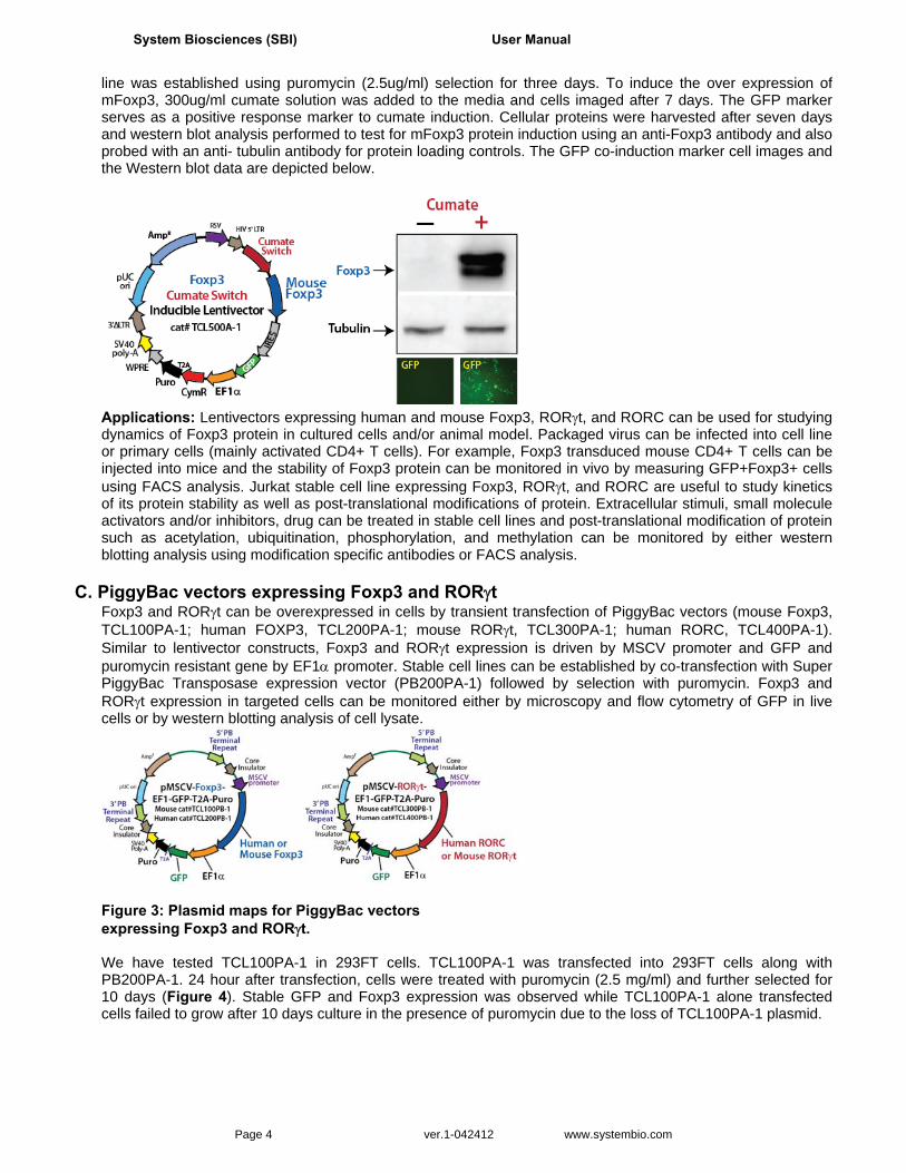
Inducible Foxp3 cumate switch lentivector The all-in-one cumate switch lentivector can be used to establish stable cell lines that can be induced to overexpress Foxp3 using cumate. The mouse Foxp3 was cloned upstream of the IRES GFP cassette and was placed under the control of the upstream cumate switch promoter. The EF1 alpha promoter drives the expression of the CymR repressor-T2A-Puro cassette. The TCL500A-1 CuO-Mouse Foxp3-IRES-GFP-EF1-CymR-T2A-Puro lentivector plasmid was packaged into lentivirus and transduced into 293FT cells. A stable cell
System Biosciences (SBI) User Manual
Page 4 ver.1-042412 www.systembio.com
line was established using puromycin (2.5ug/ml) selection for three days. To induce the over expression of mFoxp3, 300ug/ml cumate solution was added to the media and cells imaged after 7 days. The GFP marker serves as a positive response marker to cumate induction. Cellular proteins were harvested after seven days and western blot analysis performed to test for mFoxp3 protein induction using an anti-Foxp3 antibody and also probed with an anti- tubulin antibody for protein loading controls. The GFP co-induction marker cell images and the Western blot data are depicted below.
Applications: Lentivectors expressing human and mouse Foxp3, RORt, and RORC can be used for studying dynamics of Foxp3 protein in cultured cells and/or animal model. Packaged virus can be infected into cell line or primary cells (mainly activated CD4+ T cells). For example, Foxp3 transduced mouse CD4+ T cells can be injected into mice and the stability of Foxp3 protein can be monitored in vivo by measuring GFP+Foxp3+ cells using FACS analysis. Jurkat stable cell line expressing Foxp3, RORt, and RORC are useful to study kinetics of its protein stability as well as post-translational modifications of protein. Extracellular stimuli, small molecule activators and/or inhibitors, drug can be treated in stable cell lines and post-translational modification of protein such as acetylation, ubiquitination, phosphorylation, and methylation can be monitored by either western blotting analysis using modification specific antibodies or FACS analysis.
C. PiggyBac vectors expressing Foxp3 and RORt Foxp3 and RORt can be overexpressed in cells by transient transfection of PiggyBac vectors (mouse Foxp3, TCL100PA-1; human FOXP3, TCL200PA-1; mouse RORt, TCL300PA-1; human RORC, TCL400PA-1). Similar to lentivector constructs, Foxp3 and RORt expression is driven by MSCV promoter and GFP and puromycin resistant gene by EF1 promoter. Stable cell lines can be established by co-transfection with Super PiggyBac Transposase expression vector (PB200PA-1) followed by selection with puromycin. Foxp3 and RORt expression in targeted cells can be monitored either by microscopy and flow cytometry of GFP in live cells or by western blotting analysis of cell lysate.
Figure 3: Plasmid maps for PiggyBac vectors expressing Foxp3 and RORt. We have tested TCL100PA-1 in 293FT cells. TCL100PA-1 was transfected into 293FT cells along with PB200PA-1. 24 hour after transfection, cells were treated with puromycin (2.5 mg/ml) and further selected for 10 days (Figure 4). Stable GFP and Foxp3 expression was observed while TCL100PA-1 alone transfected cells failed to grow after 10 days culture in the presence of puromycin due to the loss of TCL100PA-1 plasmid.

Treg and Th17 T cell Immunology Tools Cat. #’s TCLxxx
888-266-5066 (Toll Free) 650-968-2200 (outside US) Page 5
Figure 4: 293FT stable cells established by cotransfection of TCL100PA-1 and PB200PA-1 and puromycin selection. GFP expression was visualized by microscopy and Foxp3 expression from total cell lysate was detected by western blotting analysis with anti-mFoxp3 and anti--Tubulin antibodies.
Applications: PiggyBac vectors expressing human and mouse Foxp3, RORgt, and RORC can be used for studying dynamics of its protein in cultured cells and/or animal models. PiggyBac vectors can be introduced into cell lines or primary cells (mainly activated CD4+ T cells) by highly efficient transfection method such as nucleofection or electroporation. 293FT stable cell line expressing Foxp3, RORt, and RORC are useful to study kinetics of its protein stability as well as post-translational modifications of protein. Extracellular stimuli, small molecule activators and/or inhibitors, drug can be treated in stable cell lines and post-translational modification of protein such as acetylation, ubiquitinylation, phosphorylation, and methylation can be monitored by either western blotting using modification specific antibodies or FACS analysis.
D. Reporter constructs for Foxp3, IL-17, RORt promoters
In addition to naturally occurring Thymus-derived Treg (nTreg), induced Treg (iTregs) can be developed in the peripheral tissues. Th17 cells are differentiated during inflammatory response in the periphery. Since expression levels of Foxp3, IL-17, RORt are direct indicators of numbers and activities of Tregs and Th17 cells, mechanisms responsible for regulating these genes are actively searched. Differentiation of Tregs and Th17 cells are elaborated processes coupled with extracellular stimuli (TCR mediated activation) and cytokine signaling which negatively or positively regulate downstream transcription factors. Differentiation of iTregs can be recapitulated in vitro by culturing naïve CD4+ T cells in the presence of suboptimal costimulation of TCR-
mediated signals (anti-CD3/CD28 antibodies) and TGF-Small molecules such as TSA, classI HDAC inhibitor and Ex-527, SIRT1 inhibitor have been tested to modulate Treg differentiation using in vitro system. Th17 cells
can be differentiated in vitro as well by culturing naïve CD4+ T cells with TCR-mediated signals, TGF-and IL-6 cytokines. Several important transcription factors such as RORt, Runx1, Stat3, Stat4, Samd3, and HIF1 have identified as key factors for Treg and Th17 cell differentiation. Detailed mapping of promoter regions, cross interactions, and searching for new transcription factors are actively investigated in the field. We have built reporter constructs for mouse Foxp3, IL-17, RORt promoter in which promoter activity can be measured both GFP expression and luciferase activity. The promoter sequences are carefully designed based on the recent literatures. Promoter sequences and structure designs are based on the following publications: Foxp3 promoter: Tone M, Greene MI, 2011, Zheng Y, et al., 2011 and Burgler S, et al., 2010. RORγt promoter: Dang EV, et al., 2011, Lazarevic V, et al., 2011 and Ruan Q,et al., 2011. IL-17 promoter: Zhang F, Meng G, Strober W, 2008.

System Biosciences (SBI) User Manual
Page 6 ver.1-042412 www.systembio.com
Figure 5: The schematic structures of mouse Foxp3, RORt, and IL-17 promoter are shown. Upon PMA/Ionomycin stimulation, luciferase activity is increased in transduced Jurkat T cells with lentivirus containing the Foxp3, RORt, and IL-17 promoter reporters. Applications: The specific transcription factor activity to regulate mouse Foxp3, IL-17, RORt gene expression can be tested using the reporter constructs of Foxp3, IL-17, RORt promoter. For example, 293T cells or Jurkat T cells can be transduced with lentivirus carrying reporter constructs. Expression vectors for various transcription factors can be transfected for activity on the promoters. GFP expression and luciferase activity can be measured. Reporter constructs for mouse Foxp3, RORt, and IL-17 promoter can be used to monitor Treg and Th17 cell differentiation from homogenous such as CD4+ T cells or heterogeneous population such as PBMC (peripheral blood mononuclear cells). Transduced cells can be treated with extracellular stimuli (like
TCR activation by anti-CD3/CD28) with combination of cytokines (TGF-, IL-6) to promote differentiation of Tregs and Th17 cells. Double positive populations (Foxp3+GFP+, IL17+GFP+, RORt+GFP+) can be monitored as differentiated Tregs and Th17 cells using FACS analysis. Small molecule activators and/or inhibitors, drug can be screened for regulating Treg and Th17 cell differentiation.
E. Product Handling Guidelines Bacterial culture of plasmids All lentivector and PiggyBac plasmids for expressing Foxp3, RORt, and RORC are recommended to transform into Stbl-2 bacterial strain (Invitrogen) that minimize the chance for recombination within HIV LTR region and PiggyBac ITR region. Packaging Lentivirus The detailed procedure of packing of lentivectors (TCL100A-1, TCL200A-1, TCL300A-1, and TCL400A-1) can be found in the manual of SBI’s lentivector expression system. Propagating Cell lines Jurkat stable cells should culture in RPMI1650 with 10% FBS and 1% penicillin/streptomycin. Please maintain the cells at a density of about 250,000-2,000,000 cells/ml. For long term storage of Jurkat stable cell line, cells can be stored in liquid nitrogen in freezing media (90% FBS and 10% DMSO) followed by standard cell freezing method.
II. Protocols
A. Packaging lentivectors into virus SBI has all the reagents needed to produce high titer, concentrated lentivirus.

Treg and Th17 T cell Immunology Tools Cat. #’s TCLxxx
888-266-5066 (Toll Free) 650-968-2200 (outside US) Page 7
See this PDF file for details on the protocol. Guide to Lentivector Packaging and Transduction of Target Cells (PDF) » http://www.systembio.com/downloads/web_manual_lentivector_exp_sys_071510.pdf
B. Spinoculation of virus and Jurkat T cells
Spinoculation Protocol (PDF) » http://www.systembio.com/downloads/Spinoculation_protocol.pdf
C. Transfecting PiggyBac transposons
SBI has all the reagents needed to stably transpose your target cells using the piggyBac transposon system. See this PDF file for details on the protocol. PiggyBac Transposon Vector System User Manual (PDF) » http://www.systembio.com/downloads/Manual_PiggyBac_Web.pdf
III. T cell Technical References (selected)
Lazarevic V, Chen X, Shim JH, Hwang ES, Jang E, Bolm AN, Oukka M, Kuchroo VK, Glimcher LH. T-bet represses T(H)17 differentiation by preventing Runx1-mediated activation of the gene encoding RORyt. Nat Immunol. 2011 Jan;12(1):96- 104.
Tone M, Greene MI. Cooperative regulatory events and Foxp3 expression. Nat Immunol. 2011 Jan;12(1):14-6.
Kwon HS, Lim HW, Wu J, Schnölzer M, Verdin E, Ott M. Three novel acetylation sites in the Foxp3 transcription factor regulate the suppressive activity of regulatory T cells. J Immunol. 2012 Mar 15;188(6):2712-21.
Burgler S, Mantel PY, Bassin C, Ouaked N, Akdis CA, Schmidt-Weber CB. RORC2 is involved in T cell polarization through interaction with the FOXP3 promoter. J Immunol. 2010 Jun 1;184(11):6161-9.
Zheng Y, Josefowicz S, Chaudhry A, Peng XP, Forbush K, Rudensky AY. Role of conserved non-coding DNA elements in the Foxp3 gene in regulatory T-cell fate. Nature. 2010 Feb 11;463(7282):808-12.
Ruan Q, Kameswaran V, Zhang Y, Zheng S, Sun J, Wang J, DeVirgiliis J, Liou HC, Beg AA, Chen YH. The Th17 immune response is controlled by the Rel-RORy-RORyT transcriptional axis. J Exp Med. 2011 Oct 24;208(11):2321-33.
Ruan Q, Chen YH. Nuclear factor-kB in immunity and inflammation: the Treg and Th17 connection. Adv Exp Med Biol. 2012;946:207-21.
Eisenstein EM, Williams CB. The T(reg)/Th17 cell balance: a new paradigm for autoimmunity. Pediatr Res. 2009 May;65(5 Pt 2):26R-31R.
Dang EV, Barbi J, Yang HY, Jinasena D, Yu H, Zheng Y, Bordman Z, Fu J, Kim Y, Yen HR, Luo W, Zeller K, Shimoda L, Topalian SL, Semenza GL, Dang CV, Pardoll DM, Pan F. Control of T(H)17/T(reg) balance by hypoxia-inducible factor 1. Cell. 2011 Sep 2;146 (5):772-84.
Zhang F, Meng G, Strober W. Interactions among the transcription factors Runx1, RORgammat and Foxp3 regulate the differentiation of interleukin 17-producing T cells. Nat Immunol. 2008 Nov;9(11):1297-306.
System Biosciences (SBI) User Manual
Page 8 ver.1-042412 www.systembio.com
Tone Y, Furuuchi K, Kojima Y, Tykocinski ML, Greene MI, Tone M. Smad3 and NFAT cooperate to induce Foxp3 expression through its enhancer. Nat Immunol. 2008 Feb;9(2):194-202.
Zheng Y, Rudensky AY. Foxp3 in control of the regulatory T cell lineage. Nat Immunol. 2007 May;8(5):457-62.
IV. Technical Support For more information about SBI products and to download manuals in PDF format, please visit our web site: http://www.systembio.com
For additional information or technical assistance, please call or email us at:
System Biosciences (SBI) 265 North Whisman Rd. Mountain View, CA 94043
Phone: (650) 968-2200 (888) 266-5066 (Toll Free)
Fax: (650) 968-2277
E-mail: General Information: [email protected] Technical Support: [email protected] Ordering Information: [email protected]

Treg and Th17 T cell Immunology Tools Cat. #’s TCLxxx
888-266-5066 (Toll Free) 650-968-2200 (outside US) Page 9
V. Licensing and Warranty Statement
Limited Use License
Use of the T cell vectors, reporters and cell lines (i.e., the “Product”) is subject to the following terms and conditions. If the terms and conditions are not acceptable, return all components of the Product to System Biosciences (SBI) within 7 calendar days. Purchase and use of any part of the Product constitutes acceptance of the above terms.
Purchase of the product does not grant any rights or license for use other than those explicitly listed in this Licensing and Warranty Statement. Use of the Product for any use other than described expressly herein may be covered by patents or subject to rights other than those mentioned. SBI disclaims any and all responsibility for injury or damage which may be caused by the failure of the buyer or any other person to use the Product in accordance with the terms and conditions outlined herein.
SBI has pending patent applications related to the Product. For information concerning licenses for commercial use, contact SBI.
Limited Warranty
SBI warrants that the Product meets the specifications described in the accompanying Product Analysis Certificate. If it is proven to the satisfaction of SBI that the Product fails to meet these specifications, SBI will replace the Product or provide the purchaser with a refund. This limited warranty shall not extend to anyone other than the original purchaser of the Product. Notice of nonconforming products must be made to SBI within 30 days of receipt of the Product.
SBI’s liability is expressly limited to replacement of Product or a refund limited to the actual purchase price. SBI’s liability does not extend to any damages arising from use or improper use of the Product, or losses associated with the use of additional materials or reagents. This limited warranty is the sole and exclusive warranty. SBI does not provide any other warranties of any kind, expressed or implied, including the merchantability or fitness of the Product for a particular purpose.
SBI is committed to providing our customers with high-quality products. If you should have any questions or concerns about any SBI products, please contact us at (888) 266-5066.
© 2012 System Biosciences (SBI).