Epileptic Seizures Related to Cerebral Venous Sinus Thrombosis ...

Treatment and Management of Venous Sinus Thrombosis
Sebastian Pollandt, MD
Neurocritical Care/Epilepsy
Rush University Medical Center
04/29/2016
Disclosures
• No actual or potential conflict of interest in regards to this presentation
• The planners, editors, faculty and reviewers of this activity have no relevant financial relationships to disclose.
• This presentation was created without any commercial support.

Learning Objectives
At the conclusion of this course participants will be able to
• Identify the epidemiology, pathophysiology and clinical features of cerebral venous sinus thrombosis (CVST)
• Analyze diagnostic modalities of CVST
• Evaluate principles of treatment of CVST
Case
• 35 year old healthy Female delivered a healthy infant via C-section
• 3-4 days later, she developed headaches
• Another 3 days later, friends found her confused on the floor with her baby next to her
• Later in the day had a witnessed episode of generalized shaking
Case
• Head CT at OSH shows cerebral sinus venous thrombosis
• Admitted, heparin drip started, transitioned to subcutaneous enoxaparin
• Had several odd episodes of generalized shaking while maintaining consciousness
• “Screaming in pain” with one of those episodes
• Repeat head imaging showed progression of CVST thrombosis despite anticoagulation
• Transfer to Rush

• Extensive dural venous sinus thrombosis involving the entire superior sagittal sinus, right transverse and sigmoid sinuses
• Partial thrombosis of the left transverse/sigmoid sinuses and several parasagittal cortical veins

Upon arrival to Rush: • Somnolent, but easily arousable • Complains of severe headache • Fully oriented • Intact cranial nerves
• Fundoscopy shows blurred disc margins • Full motor strength throughout •No sensory deficit
• Heparin gtt restarted
Case

Next day:
• More somnolent, difficult to arouse, inattentive, non-verbal • CN intact • All 4 extremities drift to bed when she becomes inattentive, improves when stimulated
• Consulted endovascular team
– Severe thrombosis of all the intracranial sinuses
– Undergoes mechanical thrombectomy and partial recanalization of all sinuses
Case
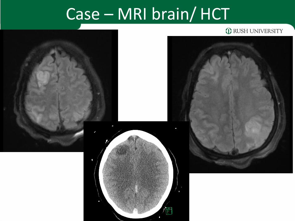
Case – MRI brain/ HCT

Case - Hospital Course
• Mental status and speech slowly improve over days • Transitioned to coumadin • Discharged to acute rehab
3 months later: • Doing very well • Neurologic exam is normal • Headaches subsided 2-3 weeks after discharge
• Continues on warfarin, INR is therapeutic • Levetiracetam for seizures, which have not recurred

Learning Objectives
Epidemiology, pathophysiology and clinical features of CVST
CVST - Epidemiology
• Cerebral venous thrombosis
– Venous sinus thrombosis
– Cortical vein thrombosis
• Relatively rare cause of stroke (<1%)
• Annual incidence estimated 3 - 7 cases per million
• Comparable incidence to acute bacterial meningitis in adults
• More common among young women and children
• Can cause devastating injury to the brain, but most patients have a good prognosis if it is recognized and treated early
Ferro & Canhao, Curr Cardiol Rep (2014) 16:523 Samuels & Webb, NCS Practice Update 2013

CVST - Pathophysiology
Saposnik et al., Stroke. 2011;42:1158-1192
% location of CVST (International Study on Cerebral Venous and Dural Sinuses Thrombosis, n= 624)

CVST - Pathophysiology
Thrombosis of the cortical veins:
– Localized vasogenic edema
– Venous infarction with cytotoxic edema
– Hemorrhage
• Symptoms:
– Seizures
– focal neurologic symptoms
Samuels & Webb, NCS Practice Update 2013

CVST - Pathophysiology
Thrombosis of the large venous sinuses:
– Obstructs venous drainage
– Impaired CSF absorption through arachnoid villi
– Intracranial hypertension without hydrocephalus
• Symptoms:
– Elevated intracranial pressure
– Bi-hemispheric symptoms (stupor, coma)
Samuels & Webb, NCS Practice Update 2013

CVST - Pathophysiology
• Multiple etiologic factors
• Usually one or more predisposing risk factors plus one inciting factor
• Thrombosis develops through common pathways of:
• Hypercoagulability
• Hemoconcentration
• Direct injury or inflammation of the vessel
• Venous stasis
• Transient and/or permanent risk factors raise suspicion for CVST and influence treatment duration
Samuels & Webb, NCS Practice Update 2013

CVST - Transient Risk Factors
• Infections – Central nervous system (empyema, meningitis)
– Ear, sinus, mouth, face and neck (otitis, mastoiditis, tonsillitis, stomatitis, sinusitis, cellulitis)
– Systemic infections (sepsis, endocarditis, tuberculosis, HIV, malaria)
• Pregnancy and puerperium
• Physical precipitants
– Head trauma
– Lumbar puncture, myelography, intrathecal medications, spinal anesthesia
– Radical neck surgery
– Neurosurgical procedures
– Jugular and subclavian catheters
Ferro & Canhao, Curr Cardiol Rep (2014) 16:523

CVST - Transient Risk Factors
• Drugs with prothrombotic action
– Oral contraceptives
– Hormone replacement therapy
– Androgens
– Medroxyprogesterone acetate
– L- asparaginase
– Cyclosporine
– Tamoxifen
– Steroids
– Lithium
– Thalidomide
– Ecstasy
– Sildenafil
• Other conditions – Dehydration
– Diabetic ketoacidosis
Ferro & Canhao, Curr Cardiol Rep (2014) 16:523
CVST - Permanent Risk Factors
Genetic
• Protein S, C, antithrombin deficiencies
• Factor V Leiden
• Prothrombin mutations
Acquired
• Antiphospholipid AB syndrome
• Nephrotic syndrome
• Cyanotic congenital heart disease
Ferro & Canhao, Curr Cardiol Rep (2014) 16:523
Prothrombotic conditions

CVST - Permanent Risk Factors
• Malignancy – Central nervous system (meningioma)
– Solid tumors outside central nervous system
– Hematological (leukemias, lymphomas)
• Hematological condition
– Anemias (sickle cell disease and trait, iron deficiency, folic acid deficiency)
– Paroxysmal nocturnal hemoglobinuria
– Polycythemia (primary or secondary)
– Thrombocythemia (primary or secondary)
Ferro & Canhao, Curr Cardiol Rep (2014) 16:523
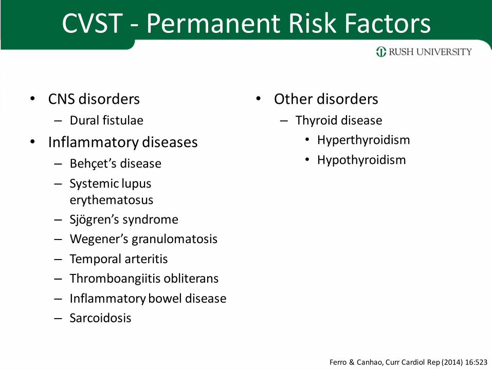
CVST - Permanent Risk Factors
• CNS disorders – Dural fistulae
• Inflammatory diseases – Behçet’s disease
– Systemic lupus erythematosus
– Sjögren’s syndrome
– Wegener’s granulomatosis
– Temporal arteritis
– Thromboangiitis obliterans
– Inflammatory bowel disease
– Sarcoidosis
• Other disorders – Thyroid disease
• Hyperthyroidism
• Hypothyroidism
Ferro & Canhao, Curr Cardiol Rep (2014) 16:523

CVST - Clinical Features
• Onset acute, subacute or chronic
• Headache is most common, nearly 90% of patients
• Other common presenting symptoms:
– Focal or generalized seizure (40%)
– Focal motor weakness (37%)
– Encephalopathy or change in mental status (22%)
– Vision loss (13%)
– Diplopia (13%)
– Stupor or coma (13%)
Samuels & Webb, NCS Practice Update 2013
CVST - Clinical Features
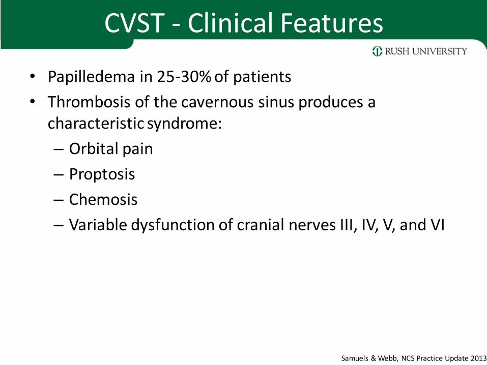
• Papilledema in 25-30% of patients
• Thrombosis of the cavernous sinus produces a characteristic syndrome:
– Orbital pain
– Proptosis
– Chemosis
– Variable dysfunction of cranial nerves III, IV, V, and VI
Samuels & Webb, NCS Practice Update 2013

Learning Objectives
Diagnosis of CVST

CVST - Diagnosis
High degree of clinical suspicion is key to the diagnosis
– Head CT
– CT Venography (CTV)
– MRI/MRV
– Catheter Angiography (DSA)
Samuels & Webb, NCS Practice Update 2013

CVST Diagnosis
Head CT
• Non-contrast head CT may be normal
• Cannot exclude a diagnosis of CVST
• Suspicious findings include:
– Cerebral edema
– Bilateral infarction
– Infarction in a non-arterial distribution
– Lobar intracerebral or subarachnoid hemorrhage
– Hyperdense thrombosed cortical veins
– Hyperdensities within the venous sinuses
Samuels & Webb, NCS Practice Update 2013
CVST - Diagnosis
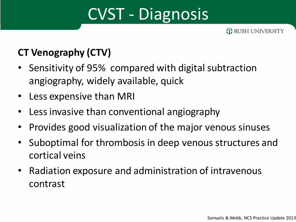
CT Venography (CTV)
• Sensitivity of 95% compared with digital subtraction angiography, widely available, quick
• Less expensive than MRI
• Less invasive than conventional angiography
• Provides good visualization of the major venous sinuses
• Suboptimal for thrombosis in deep venous structures and cortical veins
• Radiation exposure and administration of intravenous contrast
Samuels & Webb, NCS Practice Update 2013

CVST - Diagnosis
MRI/MRV
• MRI in combination with time-of-flight or contrast enhanced MR venography (MRV) highly sensitive for the diagnosis CVST
• Abnormal T1 and/T2 signal within the venous sinus and absence of normal flow through the venous sinus on MRV confirms the diagnosis
• Age of the thrombus determines T1 and T2 signal characteristics
Samuels & Webb, NCS Practice Update 2013
CVST - Diagnosis
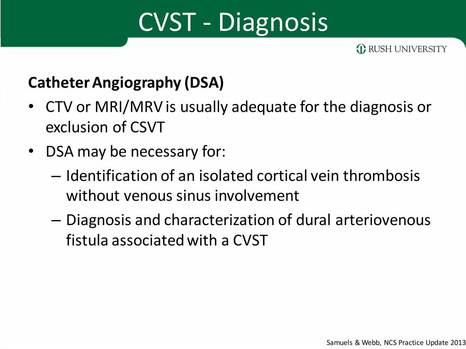
Catheter Angiography (DSA)
• CTV or MRI/MRV is usually adequate for the diagnosis or exclusion of CSVT
• DSA may be necessary for:
– Identification of an isolated cortical vein thrombosis without venous sinus involvement
– Diagnosis and characterization of dural arteriovenous fistula associated with a CVST
Samuels & Webb, NCS Practice Update 2013

Learning Objectives
Treatment of CVST
CVST - Treatment
Anticoagulation
• Cornerstone of treatment for CVST
• Prevent extension of the thrombosis and support spontaneous thrombus resolution
• Indicated even in the presence of intracranial hemorrhage
Samuels & Webb, NCS Practice Update 2013

CVST - Treatment
“no new symptomatic cerebral hemorrhages. Anticoagulation proved safe, even in patients with cerebral hemorrhage”
Patients with ICH and CVST: 27 treated with IV heparin, 4 died (mortality 15%) 13 not treated with heparin, 9 died (mortality 69%) “ICH is not a contraindication to heparin treatment ”
Einhäupl et al., Lancet. 1991 Sep 7;338(8767):597-600.
de Bruijn & Stam, Stroke 1999;30:484-8.
CVST - Treatment
• American Heart Association recommendations:
“…initial anticoagulation with adjusted-dose UFH or weight-based LMWH in full anticoagulant doses is reasonable, followed by vitamin K antagonists, regardless of the presence of ICH”
(Class IIa; Level of Evidence B)
“Continuation of oral anticoagulation with vitamin-K antagonists is reasonable for 3-6 months followed by antiplatelet therapy“
(Class IIa, Level B)
• Essentially identical recommendations from (now defunct) European Federation of Neurological Societies
Saposnik et al., Stroke. 2011;42:1158-1192

CVST - Treatment
Thrombolytics and Endovascular Treatment Options • Numerous case reports using localized thrombolytics and mechanical clot
disruption
• No controlled trials to establish efficacy or safety of these therapies
• Appropriate agent, dose, route of administration and clinical situation have yet to be defined
• Increased risk of intracranial hemorrhage is most commonly reported complication
• Thrombolytic and endovascular treatment should be limited to select patients who decline despite anticoagulation
• Should be performed only in centers with sufficient expertise in neuro-endovascular interventions
Samuels & Webb, NCS Practice Update 2013

CVST - Treatment
Seizures
• Most common in patients with
– Focal edema
– Venous infarcts
– Intracranial hemorrhage
• Prophylactic anticonvulsants may be considered
• Duration of treatment depends on: – Seizure recurrence (unprovoked, 5% to 32% of patients)
– EEG findings in follow-up
– Tolerability of antiseizure drugs
Samuels & Webb, NCS Practice Update 2013

CVST - Treatment
Intracranial Pressure • Intracranial hemorrhage, edema and infarction lead to localized mass effect
• Venous outflow impairment causes decreased CSF reabsorption, communicating hydrocephalus and intracranial hypertension
• Hyperosmolar therapy (mannitol, hypertonic saline) should be administered to patients at risk for cerebral herniation
• Acetazolamide is reasonable to reduce CSF production
• CSF diversion (lumbar puncture, ventriculostomy) or optic nerve decompression can be effective if there is progressive visual loss
• Resection of hemorrhagic infarction or decompressive craniectomy may be required
• Anticoagulation should be resumed as soon as possible following surgical intervention
Samuels & Webb, NCS Practice Update 2013 Saposnik et al., Stroke. 2011;42:1158-1192
CVST - Outcome
• Recanalization
– At 3 months 84%
– At 1 year 85%
– Highest recanalization rates in deep cerebral veins and cavernous sinus thrombosis, lowest in lateral sinus thrombosis
• In adults, recanalization of the occluded sinus is not related to outcome
Saposnik et al., Stroke. 2011;42:1158-1192

CVST - Outcome
• 3% to 15% of patients die in the acute phase
• Patients at risk:
– Depressed consciousness
– Altered mental status
– Thrombosis of the deep venous system
– Right hemisphere hemorrhage
– Posterior fossa lesions.
• Main cause of acute death with CVT is transtentorial herniation due to large hemorrhagic lesion
• Second is herniation due to multiple lesions or to diffuse brain edema.
• Status epilepticus, medical complications, and PE are other causes
Saposnik et al., Stroke. 2011;42:1158-1192

CVST - Outcome
• 79% of patients will have complete recovery
• 9.7% are functionally dependent (mRS 3 or greater)
• 50% of survivors feel depressed or anxious, minor cognitive or language deficits may preclude them from resuming previous jobs
• Abulia, executive deficits, and amnesia result from thrombosis of the deep venous system, with bilateral panthalamic infarcts
• Memory deficits, behavioral problems, or executive deficits may persist
Saposnik et al., Stroke. 2011;42:1158-1192

CVST
Questions?