Epileptic Seizures Related to Cerebral Venous Sinus Thrombosis ...
6
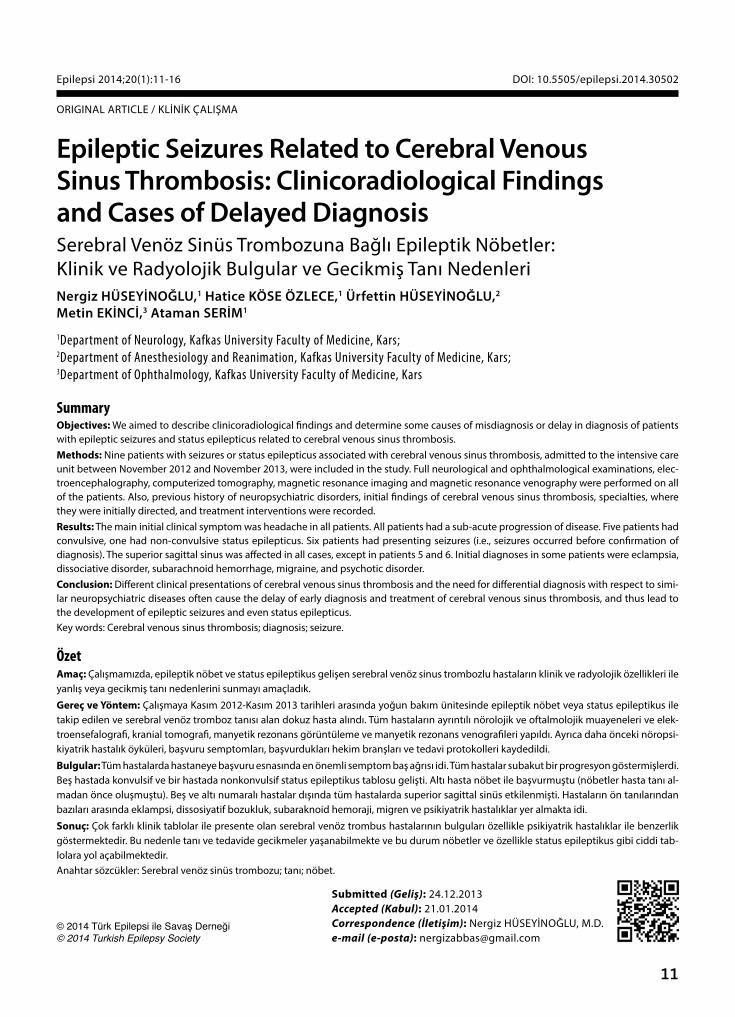
Epileptic Seizures Related to Cerebral Venous Sinus Thrombosis: Clinicoradiological Findings and Cases of Delayed Diagnosis Özet Amaç: Çalışmamızda, epileptik nöbet ve status epileptikus gelişen serebral venöz sinus trombozlu hastaların klinik ve radyolojik özellikleri ile yanlış veya gecikmiş tanı nedenlerini sunmayı amaçladık. Gereç ve Yöntem: Çalışmaya Kasım 2012-Kasım 2013 tarihleri arasında yoğun bakım ünitesinde epileptik nöbet veya status epileptikus ile takip edilen ve serebral venöz tromboz tanısı alan dokuz hasta alındı. Tüm hastaların ayrıntılı nörolojik ve oftalmolojik muayeneleri ve elek- troensefalografi, kranial tomografi, manyetik rezonans görüntüleme ve manyetik rezonans venografileri yapıldı. Ayrıca daha önceki nöropsi- kiyatrik hastalık öyküleri, başvuru semptomları, başvurdukları hekim branşları ve tedavi protokolleri kaydedildi. Bulgular: Tüm hastalarda hastaneye başvuru esnasında en önemli semptom baş ağrısı idi. Tüm hastalar subakut bir progresyon göstermişlerdi. Beş hastada konvulsif ve bir hastada nonkonvulsif status epileptikus tablosu gelişti. Altı hasta nöbet ile başvurmuştu (nöbetler hasta tanı al- madan önce oluşmuştu). Beş ve altı numaralı hastalar dışında tüm hastalarda superior sagittal sinüs etkilenmişti. Hastaların ön tanılarından bazıları arasında eklampsi, dissosiyatif bozukluk, subaraknoid hemoraji, migren ve psikiyatrik hastalıklar yer almakta idi. Sonuç: Çok farklı klinik tablolar ile presente olan serebral venöz trombus hastalarının bulguları özellikle psikiyatrik hastalıklar ile benzerlik göstermektedir. Bu nedenle tanı ve tedavide gecikmeler yaşanabilmekte ve bu durum nöbetler ve özellikle status epileptikus gibi ciddi tab- lolara yol açabilmektedir. Anahtar sözcükler: Serebral venöz sinüs trombozu; tanı; nöbet. Epilepsi 2014;20(1):11-16 DOI: 10.5505/epilepsi.2014.30502 11 Serebral Venöz Sinüs Trombozuna Bağlı Epileptik Nöbetler: Klinik ve Radyolojik Bulgular ve Gecikmiş Tanı Nedenleri Nergiz HÜSEYİNOĞLU, 1 Hatice KÖSE ÖZLECE, 1 Ürfettin HÜSEYİNOĞLU, 2 Metin EKİNCİ, 3 Ataman SERİM 1 Summary Objectives: We aimed to describe clinicoradiological findings and determine some causes of misdiagnosis or delay in diagnosis of patients with epileptic seizures and status epilepticus related to cerebral venous sinus thrombosis. Methods: Nine patients with seizures or status epilepticus associated with cerebral venous sinus thrombosis, admitted to the intensive care unit between November 2012 and November 2013, were included in the study. Full neurological and ophthalmological examinations, elec- troencephalography, computerized tomography, magnetic resonance imaging and magnetic resonance venography were performed on all of the patients. Also, previous history of neuropsychiatric disorders, initial findings of cerebral venous sinus thrombosis, specialties, where they were initially directed, and treatment interventions were recorded. Results: The main initial clinical symptom was headache in all patients. All patients had a sub-acute progression of disease. Five patients had convulsive, one had non-convulsive status epilepticus. Six patients had presenting seizures (i.e., seizures occurred before confirmation of diagnosis). The superior sagittal sinus was affected in all cases, except in patients 5 and 6. Initial diagnoses in some patients were eclampsia, dissociative disorder, subarachnoid hemorrhage, migraine, and psychotic disorder. Conclusion: Different clinical presentations of cerebral venous sinus thrombosis and the need for differential diagnosis with respect to simi- lar neuropsychiatric diseases often cause the delay of early diagnosis and treatment of cerebral venous sinus thrombosis, and thus lead to the development of epileptic seizures and even status epilepticus. Key words: Cerebral venous sinus thrombosis; diagnosis; seizure. 1 Department of Neurology, Kafkas University Faculty of Medicine, Kars; 2 Department of Anesthesiology and Reanimation, Kafkas University Faculty of Medicine, Kars; 3 Department of Ophthalmology, Kafkas University Faculty of Medicine, Kars © 2014 Türk Epilepsi ile Savaş Derneği © 2014 Turkish Epilepsy Society Submitted (Geliş) : 24.12.2013 Accepted (Kabul): 21.01.2014 Correspondence (İletişim): Nergiz HÜSEYİNOĞLU, M.D. e-mail (e-posta): [email protected] ORIGINAL ARTICLE / KLİNİK ÇALIŞMA
Transcript of Epileptic Seizures Related to Cerebral Venous Sinus Thrombosis ...
Epileptic Seizures Related to Cerebral VenousSinus Thrombosis: Clinicoradiological Findingsand Cases of Delayed Diagnosis
ÖzetAmaç: Çalışmamızda, epileptik nöbet ve status epileptikus gelişen serebral venöz sinus trombozlu hastaların klinik ve radyolojik özellikleri ile yanlış veya gecikmiş tanı nedenlerini sunmayı amaçladık.
Gereç ve Yöntem: Çalışmaya Kasım 2012-Kasım 2013 tarihleri arasında yoğun bakım ünitesinde epileptik nöbet veya status epileptikus ile takip edilen ve serebral venöz tromboz tanısı alan dokuz hasta alındı. Tüm hastaların ayrıntılı nörolojik ve oftalmolojik muayeneleri ve elek-troensefalografi, kranial tomografi, manyetik rezonans görüntüleme ve manyetik rezonans venografileri yapıldı. Ayrıca daha önceki nöropsi-kiyatrik hastalık öyküleri, başvuru semptomları, başvurdukları hekim branşları ve tedavi protokolleri kaydedildi.
Bulgular: Tüm hastalarda hastaneye başvuru esnasında en önemli semptom baş ağrısı idi. Tüm hastalar subakut bir progresyon göstermişlerdi. Beş hastada konvulsif ve bir hastada nonkonvulsif status epileptikus tablosu gelişti. Altı hasta nöbet ile başvurmuştu (nöbetler hasta tanı al-madan önce oluşmuştu). Beş ve altı numaralı hastalar dışında tüm hastalarda superior sagittal sinüs etkilenmişti. Hastaların ön tanılarından bazıları arasında eklampsi, dissosiyatif bozukluk, subaraknoid hemoraji, migren ve psikiyatrik hastalıklar yer almakta idi.
Sonuç: Çok farklı klinik tablolar ile presente olan serebral venöz trombus hastalarının bulguları özellikle psikiyatrik hastalıklar ile benzerlik göstermektedir. Bu nedenle tanı ve tedavide gecikmeler yaşanabilmekte ve bu durum nöbetler ve özellikle status epileptikus gibi ciddi tab-lolara yol açabilmektedir.Anahtar sözcükler: Serebral venöz sinüs trombozu; tanı; nöbet.
Epilepsi 2014;20(1):11-16 DOI: 10.5505/epilepsi.2014.30502
11
Serebral Venöz Sinüs Trombozuna Bağlı Epileptik Nöbetler:Klinik ve Radyolojik Bulgular ve Gecikmiş Tanı NedenleriNergiz HÜSEYİNOĞLU,1 Hatice KÖSE ÖZLECE,1 Ürfettin HÜSEYİNOĞLU,2
Metin EKİNCİ,3 Ataman SERİM1
SummaryObjectives: We aimed to describe clinicoradiological findings and determine some causes of misdiagnosis or delay in diagnosis of patients with epileptic seizures and status epilepticus related to cerebral venous sinus thrombosis.Methods: Nine patients with seizures or status epilepticus associated with cerebral venous sinus thrombosis, admitted to the intensive care unit between November 2012 and November 2013, were included in the study. Full neurological and ophthalmological examinations, elec-troencephalography, computerized tomography, magnetic resonance imaging and magnetic resonance venography were performed on all of the patients. Also, previous history of neuropsychiatric disorders, initial findings of cerebral venous sinus thrombosis, specialties, where they were initially directed, and treatment interventions were recorded.Results: The main initial clinical symptom was headache in all patients. All patients had a sub-acute progression of disease. Five patients had convulsive, one had non-convulsive status epilepticus. Six patients had presenting seizures (i.e., seizures occurred before confirmation of diagnosis). The superior sagittal sinus was affected in all cases, except in patients 5 and 6. Initial diagnoses in some patients were eclampsia, dissociative disorder, subarachnoid hemorrhage, migraine, and psychotic disorder.Conclusion: Different clinical presentations of cerebral venous sinus thrombosis and the need for differential diagnosis with respect to simi-lar neuropsychiatric diseases often cause the delay of early diagnosis and treatment of cerebral venous sinus thrombosis, and thus lead to the development of epileptic seizures and even status epilepticus.Key words: Cerebral venous sinus thrombosis; diagnosis; seizure.
1Department of Neurology, Kafkas University Faculty of Medicine, Kars;2Department of Anesthesiology and Reanimation, Kafkas University Faculty of Medicine, Kars;3Department of Ophthalmology, Kafkas University Faculty of Medicine, Kars
© 2014 Türk Epilepsi ile Savaş Derneği© 2014 Turkish Epilepsy Society
Submitted (Geliş) : 24.12.2013Accepted (Kabul): 21.01.2014Correspondence (İletişim): Nergiz HÜSEYİNOĞLU, M.D.e-mail (e-posta): [email protected]
ORIGINAL ARTICLE / KLİNİK ÇALIŞMA
Introduction
Cerebral venous sinus thrombosis (CVST) is a neurological emergency characterized by thrombosis of the intracranial venous sinuses and/or cerebral veins, which leads to reten-tion of venous drainage, increase in intracranial pressure and/or venous infarcts.[1] CSVT is responsible for about 1% of strokes.[2] CSVT has acute, sub-acute and chronic speeds of onset and may present with a wide spectrum of clinical findings. Clinical presentations of CVST range from head-ache and vomiting to focal neurological signs and loss of consciousness. A frequent feature of CVST is epileptic sei-zures or even status epilepticus (SE) that usually develops in the early stages of the disease.[3,4] The results of previ-ous studies have shown that one third of CVST patients experience focal or generalized epileptic seizures before confirmation of the diagnosis.[5] Different clinical presenta-tions and the need for a differential diagnosis from other similar diseases are the usual causes of delay of early di-agnosis and treatment of CVST. Thus, delayed diagnosis of CVST might be one cause of the development of refractory seizures of epileptic status, which is a serious emergency condition with high mortality and morbidity.[6,7] For these reasons, we focused on presenting epileptic seizures (i.e. seizures that occur before confirmation of a diagnosis of CVST) and, especially, on presenting status epilepticus, and explored some causes of misdiagnosis or delay of diagno-sis in patients with cerebral venous sinus thrombosis. We also aimed to describe the clinico-radiological findings of patients with CVST with seizures, who were treated in our intensive care unit (ICU).
Materials and Methods
Patients admitted to the Kafkas University Medical Faculty NICU between November 2012 and November 2013 were included in this study. In this period, 11 adult patients with CSVT were admitted to the ICU. We selected only 9 patients with presenting and early epileptic seizures.
CVST was diagnosed according to two diagnostic criteria: clinical (seizures or encephalopathy) and radiological (mag-netic resonance venography demonstrating absence of flow in venous channels).[8] For the exact diagnosis of CVST, we performed full neurological examinations and all three computerized cerebral tomography (CT), magnetic reso-nance imaging (MRI) and magnetic resonance venography (MRV). CTs were not performed on pregnant women. All pa-
tients underwent a wide hematological, biochemical and, if necessary, genetic work-up, aimed at investigating predis-posing factors for CVST and seizures.
We recorded the main clinico-radiological findings and treatment interventions, and calculated the mean dura-tion of treatment in the NICU, mean time from the onset of symptoms to development of epileptic seizures and the mean time from the onset of seizures to confirmation of the final diagnosis in patients with presenting seizures. Also, pa-tients’ neurological status on admission and discharge were assessed with the modified Rankin Scale (mRS).[9]
We excluded patients with a previous history of epilepsy and patients with status epilepticus provoked by metabolic derangement, alcohol or drug intoxication or withdrawal, and those who used medications with a relative risk for de-veloping epilepsy (such as some selective serotonin reup-take inhibitors, serotonin and norepinephrine reuptake inhibitors and sympathomimetics). Subjects under 18 years of age, and patients with subarachnoid, subdural, and epi-dural hemorrhages were also excluded.
DefinitionsEpileptic status: The definition of generalized SE refers to >30 minutes of a single epileptic seizure or 2 or more dis-crete seizures between which there is incomplete recovery of consciousness. Status epilepticus was subgrouped as convulsive and nonconvulsive according to clinical symp-toms and electroencephalography (EEG).[10,11]
Convulsive SE is defined as tonic-clonic (generalized con-vulsive status epilepticus (GCSE)) and myoclonic.
Nonconvulsive SE (NCSE) is defined as impaired conscious-ness lasting at least 30 min with persistent or continuous epileptiform abnormality in the EEG.[12] Nonconvulsive sta-tus epilepticus (NCSE) encompasses all forms of SE in which the motor manifestations are absent or minimal, and as such can be difficult to diagnose without an EEG.
Presenting seizures have been defined as occurring before the confirmation of a diagnosis of CVST.[13,14]
Early seizures have been defined as those first occurring within 2 weeks after stroke.[13,14]
Epilepsi 2014;20(1):11-16
12

Hüseyinoğlu et al., Epileptic Seizures Related to Cerebral Venous Sinus Thrombosis
13
tonic NaCl solutions. Epileptic seizures were managed with intravenous diazepam, phenytoin sodium and valproic acid according to the guidelines.[16] Patient 3 underwent a cae-sarean section under general anesthesia and was intubated for 4 days because of insufficient spontaneous breathing after cardiopulmonary arrest at the time of admission. Dur-ing the intubation period this patient received propofol and midazolam infusions to control her epileptic seizures.
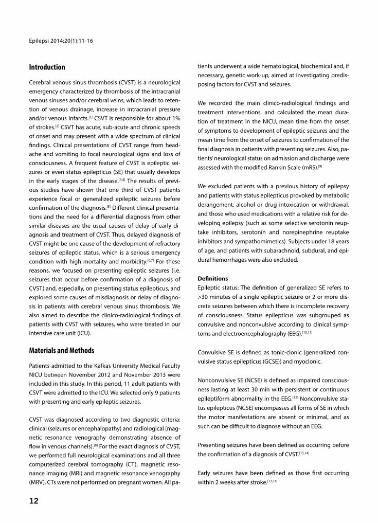
The superior sagittal sinus (SSS) was affected in all cases, except patients 5 and 6. The both transverse and sigmoid sinuses were affected in 5 and 6 cases, respectively. Sample radiological imagings of patients are presented in Figure 1.
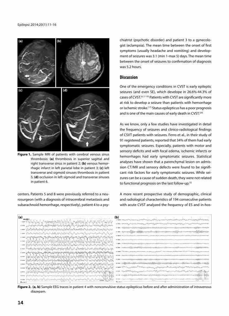
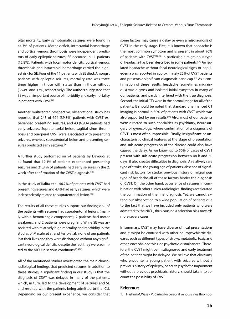
EEG (10-20 system) was performed on all of the patients for final confirmation of diagnosis and follow-up of the course of the disease. Sample EEG sequences of patient 4 with NCSE are shown in Figure 2.
All patients with initial complaints as headache, vomiting and psychotic symptoms, were initially admitted to out-patient clinics or emergency units of secondary medical
Results
In the period between November 2012 and November 2013, a total of 11 patients with a diagnosis of CVST were admitted to the NICU. We evaluated nine patients (81.8%) who had epileptic seizures. Eight of those were female and 1 male with a mean age 36.1 (min 25-max 44) years. Six (54.5%) had SE, one of which had NCSE. The other 5 patients had GCSE. Todd’s paresis was present in a single patient (pa-tient 2). The demographic, clinical, and radiological charac-teristics of patients are summarized in Table 1.
The course of the CVST in all patients was sub-acute (i.e. the symptoms developed between 48 h and 30 days).[15] The mean hospitalization period in the NICU was 7.6 days (min 4-max 18 days). None of the patients died during treatment in the NICU or in hospital.
All patients received heparin or low molecular weight hepa-rin and later warfarin, despite the presence of hemorrhagic infarct in two of the patients. Raised intracranial pressure was treated by intravenous mannitol, and sometimes hyper-
Table 1. Demographic, clinicoradiological findings and initial diagnosis of patients
Patients 1 2 3 4 5 6 7 8 9
Age/gender 42/F 29/M 25/F 41/F 39/F 37/F 31/F 37/F 44/FInitial symptoms
Headache + + + –/+ + + + + + Vomiting – + – – + – + + + Encephalo-pathy + – + + – + – – – Papilledema – – + – + + – – – Hemiparesia +/– +/– – – – + – – –Site of thrombosis SSS SSS SSS SSS TS SS SSS SSS SSS TS TS TS SS TS SS TS SSSeizures Presenting + + + + + + Early + + +SE + + + + + + Initial diagnosis DD Migr Eclmp Psyc. disord. CNS met. Stroke Migr SAH MigrPredisp. factors DH Hom Preg, OCS, Smok. CA OCS Puerp OCS OCS HT, MTHFR mRS in admission 5 4 5 5 4 4 4 5 4mRS in discharge 0 0 0 0 0 0 0 0 0
CA: Gastric cancer; CNS: Met-central nervous system metastasis; Delir: Delirium; DD: Dissociative (conversion) disorder; DH: Dehidratation; Eclmp: Eclampsia; Hom: Hyperhomocysteinemia; HT: Arterial hypertension; Migr: Migraine; mRS: Modified Rankin Scale; MTHFR: Methylenetetrahydrofolate reductase gene mutation; OCS: Oral contraception; Preg: Pregnancy; Puerp: Puerperium; Psyc. Disord.: Psychotic disorder; SAH: Subarachnoid he-morrhage; SE: Status epilepticus; SS: Sigmoid sinus; Smok.: Smoking; SSS: Superior sagittal sinus; TS: Transvers sinus; +: Present; –: Absent.
14
Epilepsi 2014;20(1):11-16
centers. Patients 5 and 8 were previously referred to a neu-rosurgeon (with a diagnosis of intracerebral metastasis and subarachnoid hemorrhage, respectively), patient 4 to a psy-
chiatrist (psychotic disorder) and patient 3 to a gynecolo-gist (eclampsia). The mean time between the onset of first symptoms (usually headache and vomiting) and develop-ment of seizures was 3.1 (min 1-max 5) days. The mean time between the onset of seizures to confirmation of diagnosis was 5.2 hours.
Discussion
One of the emergency conditions in CVST is early epileptic seizures (and even SE), which develope in 26.6%-44.3% of cases of CVST.[4,17-19] Patients with CVST are significantly more at risk to develop a seizure than patients with hemorrhage or ischemic stroke.[17] Status epilepticus has a poor prognosis and is one of the main causes of early death in CVST.[20]
As we know, only a few studies have investigated in detail the frequency of seizures and clinico-radiological findings of CSVT patients with seizures. Ferro et al., in their study of 91 registered patients, reported that 34% of them had early symptomatic seizures. Especially, patients with motor and sensory deficits and with focal edema, ischemic infarcts or hemorrhages had early symptomatic seizures. Statistical analyses have shown that a parenchymal lesion on admis-sion CT/MR and sensory defects were found to be signifi-cant risk factors for early symptomatic seizures. While sei-zures can be a cause of sudden death, they were not related to functional prognosis on the last follow-up.[3]
A more recent prospective study of demographic, clinical and radiological characteristics of 194 consecutive patients with acute CVST analyzed the frequency of ES and in-hos-
Figure 1. Sample MRI of patients with cerebral venous sinus thrombosis: (a) thrombosis in superior sagittal and right transverse sinus in patient 2; (b) venous hemor-rhagic infarct in left parietal lobe in patient 3; (c) left transverse and sigmoid sinuses thrombosis in patient 5; (d) occlusion in left sigmoid and transverse sinuses in patient 6.
(a)
(c)
(b)
(d)
Figure 2. (a, b) Sample EEG traces in patient 4 with nonconvulsive status epilepticus before and after administration of intravenous diazepam.
(a) (b)
Hüseyinoğlu et al., Epileptic Seizures Related to Cerebral Venous Sinus Thrombosis
15
pital mortality. Early symptomatic seizures were found in 44.3% of patients. Motor deficit, intracranial hemorrhage and cortical venous thrombosis were independent predic-tors of early epileptic seizures. SE occurred in 11 patients (12.8%). Patients with focal motor deficits, cortical venous thrombosis and intracranial hemorrhage carried the high-est risk for SE. Four of the 11 patients with SE died. Amongst patients with epileptic seizures, mortality rate was three times higher in those with status than in those without (36.4% and 12%, respectively). The authors suggested that SE was an important source of morbidity and early mortality in patients with CVST.[4]
Another multicenter, prospective, observational study has reported that 245 of 624 (39.3%) patients with CVST ex-perienced presenting seizures, and 43 (6.9%) patients had early seizures. Supratentorial lesion, sagittal sinus throm-bosis and puerperal CVST were associated with presenting seizures, whereas supratentorial lesion and presenting sei-zures predicted early seizures.[5]
A further study performed on 94 patients by Davoudi et al. found that 19.1% of patients experienced presenting seizures and 21.3 % of patients had early seizures in the 2. week after confirmation of the CVST diagnosis.[19]
In the study of Kalita et al. 46.7% of patients with CVST had presenting seizures and 4.4% had early seizures, which were independently related to supratentorial lesion.[21]
The results of all these studies support our findings: all of the patients with seizures had supratentorial lesions (main-ly with a hemorrhagic component), 2 patients had motor weakness, and 2 patients were pregnant. While SE was as-sociated with relatively high mortality and morbidity in the studies of Masuhr et al. and Ferro et al., none of our patients lost their lives and they were discharged without any signifi-cant neurological deficits, despite the fact they were admit-ted to the NICU in serious conditions.[3,4,20]
All of the mentioned studies investigated the main clinico-radiological findings that predicted seizures. In addition to these studies, a significant finding in our study is that the diagnosis of CSVT was delayed in many of the patients, which, in turn, led to the development of seizures and SE and resulted with the patients being admitted to the ICU. Depending on our present experience, we consider that
some factors may cause a delay or even a misdiagnosis of CVST in the early stage. First, it is known that headache is the most common symptom and is present in about 90% of patients with CVST.[22,23] In particular, a migrainous type of headache has been described in some patients.[24] An iso-lated headache without focal neurological signs or papill-edema was reported in approximately 25% of CVST patients and presents a significant diagnostic handicap.[25] As a con-firmation of these results, headache (sometimes migrain-ous) was a gross and isolated initial symptom in many of our patients, and partly interfered with the true diagnosis. Second, the initial CTs were in the normal range for all of the patients. It should be noted that standard unenhanced CT imaging is normal in 30% of patients with CVST which was also supported by our results.[26] Also, most of our patients were directed to such specialties as psychiatry, neurosur-gery or gynecology, where confirmation of a diagnosis of CSVT is most often impossible. Finally, insignificant or un-characteristic clinical features at the stage of presentation and sub-acute progression of the disease could also have caused the delay. As we know, up to 50% of cases of CSVT present with sub-acute progression between 48 h and 30 days; it also creates difficulties in diagnosis. A relatively rare type of stroke, the young age of patients, absence of signifi-cant risk factors for stroke, previous history of migrainous type of headache-all of these factors hinder the diagnosis of CVST. On the other hand, occurrence of seizures in com-bination with other clinico-radiological findings accelerated the confirmation of the final diagnosis. Yet, we cannot ex-tend our observation to a wide population of patients due to the fact that we have included only patients who were admitted to the NICU, thus causing a selection bias towards more severe cases.
In summary, CVST may have diverse clinical presentations and it might be confused with other neuropsychiatric dis-eases such as different types of stroke, metabolic, toxic and other encephalopathies or psychotic disturbances. There-fore, the CVST might be misdiagnosed and early treatment of the patient might be delayed. We believe that clinicians, who encounter a young patient with seizures without a previous history of epilepsy, or acute psychotic impairment without a previous psychiatric history, should take into ac-count the possibility of CVST.
References1. Hashmi M, Wasay M. Caring for cerebral venous sinus thrombo-
16
Epilepsi 2014;20(1):11-16
sis in children. J Emerg Trauma Shock 2011;4(3):389-94.
2. Stam J. Thrombosis of the cerebral veins and sinuses. N Engl J Med 2005;352(17):1791-8.
3. Ferro JM, Correia M, Rosas MJ, Pinto AN, Neves G; Cerebral Ve-nous Thrombosis Portuguese Collaborative Study Group [Ve-noport]. Seizures in cerebral vein and dural sinus thrombosis. Cerebrovasc Dis 2003;15(1-2):78-83.
4. Masuhr F, Busch M, Amberger N, Ortwein H, Weih M, Neu-mann K, et al. Risk and predictors of early epileptic seizures in acute cerebral venous and sinus thrombosis. Eur J Neurol 2006;13(8):852-6.
5. Ferro JM, Canhão P, Bousser MG, Stam J, Barinagarrementeria F; ISCVT Investigators. Early seizures in cerebral vein and dural sinus thrombosis: risk factors and role of antiepileptics. Stroke 2008;39(4):1152-8.
6. Myint PK, Staufenberg EF, Sabanathan K. Post-stroke seizure and post-stroke epilepsy. Postgrad Med J 2006;82(971):568-72.
7. Rumbach L, Sablot D, Berger E, Tatu L, Vuillier F, Moulin T. Status epilepticus in stroke: report on a hospital-based stroke cohort. Neurology 2000;54(2):350-4.
8. Moharir MD, Shroff M, Pontigon AM, Askalan R, Yau I, Mac-gregor D, et al. A prospective outcome study of neonatal cere-bral sinovenous thrombosis. J Child Neurol 2011;26(9):1137-44.
9. van Swieten JC, Koudstaal PJ, Visser MC, Schouten HJ, van Gijn J. Interobserver agreement for the assessment of handicap in stroke patients. Stroke 1988;19(5):604-7.
10. ILAE Commission Report. The epidemiology of the epilepsies: future directions. International League Against Epilepsy. Epi-lepsia 1997;38(5):614-8.
11. Thurman DJ, Beghi E, Begley CE, Berg AT, Buchhalter JR, Ding D, et al. Standards for epidemiologic studies and surveillance of epilepsy. Epilepsia 2011;52 Suppl 7:2-26.
12. Afsar N, Kaya D, Aktan S, Sykut-Bingol C. Stroke and status epi-lepticus: stroke type, type of status epilepticus, and prognosis. Seizure 2003;12(1):23-7.
13. Bladin CF, Alexandrov AV, Bellavance A, Bornstein N, Chambers B, Coté R, et al. Seizures after stroke: a prospective multicenter study. Arch Neurol 2000;57(11):1617-22.
14. Berges S, Moulin T, Berger E, Tatu L, Sablot D, Challier B, et al. Seizures and epilepsy following strokes: recurrence factors. Eur
Neurol 2000;43(1):3-8.
15. Guenther G, Arauz A. Cerebral venous thrombosis: a diagnostic and treatment update. [Article in English, Spanish] Neurologia 2011;26(8):488-98.
16. Shearer P, Riviello J. Generalized convulsive status epilepticus in adults and children: treatment guidelines and protocols. Emerg Med Clin North Am 2011;29(1):51-64.
17. Conrad J, Pawlowski M, Dogan M, Kovac S, Ritter MA, Evers S. Seizures after cerebrovascular events: risk factors and clinical features. Seizure 2013;22(4):275-82.
18. Benbir G, Ince B, Bozluolcay M. The epidemiology of post-stroke epilepsy according to stroke subtypes. Acta Neurol Scand 2006;114(1):8-12.
19. Davoudi V, Keyhanian K, Saadatnia M. Risk factors for remote sei-zure development in patients with cerebral vein and dural sinus thrombosis. Seizure 2013 Nov 4. pii: S1059-1311(13)00305-1.
20. Ferro JM, Correia M, Pontes C, Baptista MV, Pita F; Cerebral Ve-nous Thrombosis Portuguese Collaborative Study Group (Ve-noport). Cerebral vein and dural sinus thrombosis in Portugal: 1980-1998. Cerebrovasc Dis 2001;11(3):177-82.
21. Kalita J, Chandra S, Misra UK. Significance of seizure in cerebral venous sinus thrombosis. Seizure 2012;21(8):639-42.
22. Ferro JM, Canhão P, Stam J, Bousser MG, Barinagarrementeria F; ISCVT Investigators. Prognosis of cerebral vein and dural sinus thrombosis: results of the International Study on Cerebral Vein and Dural Sinus Thrombosis (ISCVT). Stroke 2004;35(3):664-70.
23. Demir CF, Inci MF, Özkan F, Yıldız M, Özdemir H. Clinical and radiological management and outcome of pregnancies com-plicated by cerebral venous thrombosis: a review of 19 cases. J Stroke Cerebrovasc Dis 2013;22(8):1252-7.
24. Cumurciuc R, Crassard I, Sarov M, Valade D, Bousser MG. Head-ache as the only neurological sign of cerebral venous throm-bosis: a series of 17 cases. J Neurol Neurosurg Psychiatry 2005;76(8):1084-7.
25. Crassard I, Bousser MG. Headache in patients with cerebral venous thrombosis. [Article in French] Rev Neurol (Paris) 2005;161(6-7):706-8. [Abstract]
26. Kim BS, Do HM, Marks MP. Diagnosis and management of ce-rebral venous and sinus thrombosis. Semin Cerebrovasc Dis Stroke 2004;4(4):205-16.