Trauma-induced heme release increases susceptibility to ...
49
Trauma-induced heme release increases susceptibility to bacterial infection Ghee Rye Lee, … , Carl J. Hauser, Leo E. Otterbein JCI Insight. 2021. https://doi.org/10.1172/jci.insight.150813. In-Press Preview Graphical abstract Research Infectious disease Inflammation Find the latest version: https://jci.me/150813/pdf
Transcript of Trauma-induced heme release increases susceptibility to ...

Trauma-induced heme release increases susceptibility tobacterial infection
Ghee Rye Lee, … , Carl J. Hauser, Leo E. Otterbein
JCI Insight. 2021. https://doi.org/10.1172/jci.insight.150813.
In-Press Preview
Graphical abstract
Research Infectious disease Inflammation
Find the latest version:
https://jci.me/150813/pdf

1
Trauma-Induced Heme Release Increases Susceptibility to Bacterial Infection 1 2 3
By 4 5
1Ghee Rye Lee, 1David Gallo, 1Rodrigo W. Alves de Souza, 1Shilpa Tiwari-Heckler 6 1Eva Csizmadia, 1James D. Harbison, 1Sidharth Shankar, 2Valerie Banner-Goodspeed 7
1,3Michael B. Yaffe, 2Maria Serena Longhi, 1Carl J. Hauser & 1*Leo E. Otterbein 8 9
Harvard Medical School, 1Department of Surgery and 2Anesthesia, Beth Israel 10 Deaconess Medical Center, Boston, MA 02115.3Koch Institute for Integrative 11
Cancer Research, Massachusetts Institute of Technology, Cambridge, MA 02139. 12 13 14 15 16 17 18 19 20 21 22 23 24 25 26
27 28 *For correspondence 29 Leo E. Otterbein, PhD 30 Professor of Surgery 31 Harvard Medical School 32 Beth Israel Deaconess Medical Center 33 Center for Life Science 34 3 Blackfan Circle, EC/CLS 603 35 Boston, MA 02115 36 (O): 617-735-2851 37 Email: [email protected] 38 39
40
41
42
43

2
Abstract 44
Infection is a common complication of major trauma that causes significantly increased 45
morbidity and mortality. The mechanisms however, linking tissue injury to increased 46
susceptibility to infection remain poorly understood. To study this relationship, we 47
present a novel murine model where a major liver crush injury is followed by bacterial 48
inoculation into the lung. We find that such tissue trauma both impaired bacterial 49
clearance and was associated with significant elevations in plasma heme levels. While 50
neutrophil (PMN) recruitment to the lung in response to Staphylococcus aureus was 51
unchanged after trauma, PMN cleared bacteria poorly. Moreover, PMN show >50% less 52
expression of TLR2, which is responsible, in part, for bacterial recognition. 53
Administration of heme effectively substituted for trauma. Last, day 1 trauma patients 54
(n=9) showed similar elevations in free heme to that seen after murine liver injury and 55
circulating PMN showed similar TLR2 reduction compared to volunteers (n=6). These 56
findings correlate to high infection rates. 57
3
Introduction 58
Trauma-related injuries are the third leading cause of death in the United States 59
the most common cause of death in persons younger than 45 (1). Moreover, these rates 60
are continuing to rise: from 2000 to 2010, the number of trauma deaths increased by 61
23% (2) while death rates for cancer and heart disease declined by 20% between 1991 62
and 2009 (3) and 31% between 2000-2010 (4), respectively. Importantly, nosocomial 63
infection and multiple organ failure (MOF) remain the major causes of late mortality in 64
trauma patients (5, 6). While brain injury and hemorrhage lead to early deaths, with a 65
median time of less than 24 hours, nosocomial infections and MOF contribute to deaths 66
that occur days or weeks later (7, 8). Thus, trauma patients who survive their initial 67
injuries have a significantly heightened susceptibility to infection at sites remote from the 68
primary injury that place them at risk for mortality (9). In efforts to align human pathology 69
of trauma and increased infection rates, we developed a two-hit model in mice that is 70
comprised of a liver crush injury followed by a bacterial inoculation of the lung. 71
The dominant mechanism by which the injured host is thought to become 72
susceptible to infection involves the effects of Damage Associated Molecular Pattern 73
molecules (DAMPs) released and accumulated as a result of tissue injury. These 74
include purine metabolites, S100 family members, HMGB1, and heme (10–12). In 75
addition, as evolutionary endosymbionts, mitochondria bear close molecular similarities 76
to bacterial Pathogen Associated Molecular Patterns (PAMPs) that, like DAMPs, can 77
trigger powerful innate immune responses (13). Collectively, DAMPs and PAMPs are 78
potent immunomodulators and cells have evolved elegant cognate receptors and 79
signaling cascades by which these molecules modulate cell function. DAMPs and 80
4
PAMPs induce the prototypical cytokine storm, oxidant radical generation, transcription 81
factor mobilization and resulting stress response gene expression (12). Paradoxically, 82
this early sequence of events can also lead to immunosuppressive events and can 83
place the host at an increased risk of infection by bacteria that would be successfully 84
cleared by an archetypical myeloid cell response absent the traumatic injury. 85
Heme is a complex of iron and protoporphyrin IX that serves multiple essential 86
functions in aerobic organisms as the prosthetic group of hemoproteins. Key examples 87
include hemoglobin and myoglobin, the oxidases of the mitochondrial electron transport 88
chain, cytochrome p450 and other signaling proteins like guanylate cyclase and the 89
nitric oxide synthases(14). Under normal conditions heme is critical to normal enzymatic 90
function. In contrast, elevated levels of free heme occurring at times of cell and tissue 91
injury or during hemolysis can be toxic due to their ability to elicit oxidative stress and 92
inflammation. Fortunately, robust systems are in place that clear extracellular free 93
heme. These begin with a battery of serum heme-binding proteins that include 94
hemopexin, haptoglobin, and albumin. But heme can also bind to members of the Toll 95
family of receptors, including Toll-Like Receptor-2 and 4 (TLR2,4) (15–17). When these 96
receptors bind heme, they can activate NF-kB-dependent pro-inflammatory signaling 97
events. Free heme is ultimately cleared by transport into the cell where it is rapidly 98
metabolized by Heme Oxygenases 1 and 2 (HO; gene Hmox) (16, 18, 19). 99
One of the common bacterial strains causing nosocomial infection after trauma is 100
the Gram-positive bacterium Staphylococcus aureus (S. aureus) (20–22), which is 101
normally an innocuous colonizer of the nose and skin in healthy humans. When the host 102
immune system is compromised by injury however, S. aureus can become an important 103
5
infective organism (23, 24). We developed this post-traumatic pneumonia model as a 104
translational research tool to study the pathophysiology and mechanisms of immune-105
susceptibility to opportunistic infection following traumatic organ injury. Our objective 106
was to better understand how an initiating tissue injury-induced release of heme results 107
in alterations in the hosts response to a second challenge of bacterial inoculation in the 108
lung. We focused on the neutrophil as the primary cell that infiltrates to sites of infection 109
and identify a population of neutrophils (PMN) that, while recruited normally, are poor at 110
clearing bacteria. 111

6
Results 112
113
Trauma to the liver impairs subsequent bacterial clearance in the lung. 114
Trauma patients have a higher susceptibility to develop infection (9) and little is 115
known about the mechanism by which sterile injuries can heighten a subjects’ 116
susceptibility to pathogens that are usually benign in tissues distant from the injured 117
site. Since liver is the solid organ that is damaged most commonly as a result of blunt 118
trauma(25–27), we developed and standardized a model of non-lethal blunt liver trauma 119
in mice. One hour after liver crush, clotting was observed both macroscopically (not 120
shown) and microscopically (Figure 1A). Four hours after liver crush, PMN rise in the 121
circulation and accumulate in the injured liver (Figure 1A, Supplemental Figure 1A-C). 122
Standardization of the severity of the crush injury was confirmed by measuring serum 123
alanine aminotransferase (ALT) levels over time (Figure. 1B). 124
To test whether liver injury alters host bacterial clearance from the lung, mice 125
were inoculated with 106-107 colony forming units (CFU) of S. aureus into the lungs 4 126
hours after liver crush injury as depicted in Figure 1C. S. aureus is a clinically relevant 127
pathogen that commonly causes early post-traumatic pneumonia in at-risk trauma 128
subjects (6). Twenty-four hours after inoculation, animals were euthanized and a 129
bronchoalveolar lavage (BAL) was performed to measure cell and bacteria counts. 130
Mice without surgery and mice with laparotomy only, cleared bacteria effectively from 131
the lung at 24h. Mice subjected to liver crush however, showed 100-fold more bacteria 132
in the BAL and the lung tissues at 24h when compared to sham controls (Figure 1D-E). 133
Similarly, higher S. aureus counts were detected in the blood of infected mice with liver 134
crush injury (Figure 1F). Lung infected mice showed increased accumulation of protein 135

7
in the BAL as a marker of lung injury, but no significant differences were observed 136
between infected mice with or without liver injury (Figure 1G). However, a greater level 137
of inflammation was observed by blinded histological analysis in crush plus infection 138
compared to infection alone (Supplemental Figure 2A). Mice with liver injury alone 139
showed no translocation of endogenous bacteria into the blood, lung tissue or BAL fluid 140
(data not shown). All mice survived liver crush injury plus infection, with bacterial 141
clearance observed 48 hours after inoculation in both groups (Supplemental Figure 142
2B). However, mice challenged with a higher dose of S. aureus in the presence of liver 143
crush all died while all survived the high dose inoculation in the absence of liver crush 144
(Figure 1H). 145
When bacterial inoculation was delayed 24h post-trauma we found similar 146
deficiencies in animals’ ability to clear bacteria as compared with controls (Figure 1I). 147
We also noted that liver injury had the same effect on Gram-negative E. coli clearance 148
as S. aureus (Figure 1J). Taken together, the results show that an increased 149
susceptibility to bacterial infection similar to that observed in human trauma can be 150
simulated by liver trauma in mice. 151
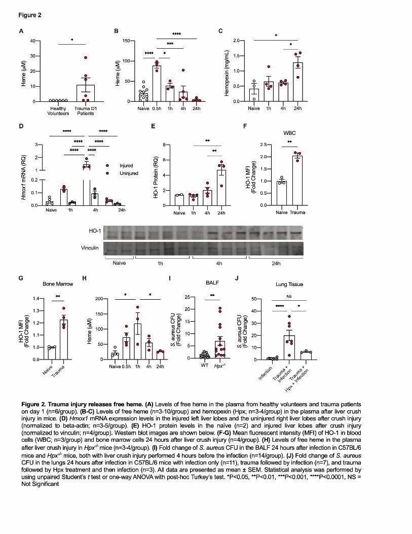
152 Trauma injury results in a transient release of free heme in mice and humans 153
Traumatic injury can lead to bleeding with release of heme from extravascular 154
blood as well as from the intracellular contents of damaged or dying cells. Under these 155
circumstances, heme can act as a potent DAMP and can contribute to further cellular 156
injury (12). Healthy humans have very low or undetectable levels of free heme in the 157
plasma (<1µM, Figure 2A). In contrast, plasma samples from trauma patients collected 158
one day after traumatic injury showed a 10-fold rise in free serum heme (Figure 2A). 159
8
Variability may reflect the time the first blood sample was collected relative to the time 160
the trauma occurred. Similarly, mice subjected to a liver crush injury showed a time-161
dependent rise in circulating free heme from a baseline of 25µM that peaked at >75µM 162
within 30 minutes after injury and returned to baseline by 4h (Figure 2B). The 163
difference in peak heme levels between human and mouse may be attributed to the 164
type of injury and/or differences in physiological and metabolic response times between 165
human and mice. Given the unpredictability of human trauma, it is challenging to 166
generate a specific kinetic of heme release in trauma patients and thus we grouped 167
plasma collected from trauma patients into batches. It is likely that peak plasma heme 168
levels occur early after injury depending on the severity and cause. Hemopexin (Hpx) is 169
a serum protein whose principal role is to scavenge and bind free heme. After liver 170
crush, Hpx levels in plasma increased and peaked at 24h (Figure 2C). Elevated Hpx 171
levels most likely reflect an acute phase mechanism to clear free heme and thus limit its 172
activity as a DAMP. Measurement of plasma Hpx in patients collected on day 0, 1, and 173
3 after trauma showed no significant differences compared to healthy volunteers (data 174
not shown). Explanations might include timing of the collection of the blood samples, the 175
small sample size, or a difference between trauma in mice and trauma in humans. 176
Heme is well known to be metabolized by heme oxygenases. So next we 177
measured inducible HO-1 as a response to the increase in free heme elicited by liver 178
crush injury. HO-1 expression increased in both the injured and uninjured liver lobes at 179
4h at the mRNA level, but was significantly higher in tissue from the injured lobe (Figure 180
2D). HO-1 protein expression peaked at 24h in the injured liver lobes. (Figure 2E). 181
Additionally, both circulating WBC and bone marrow (BM) PMN showed increased HO-182

9
1 expression 24h after liver crush (Figure 2F-G). These data suggest that heme, rapidly 183
released as a result of tissue injury, is taken up and processed at multiple sites based 184
on HO-1 expression. Of note, acute inflammation will also increase HO-1 independent 185
of free heme(28). 186
Based on the serum heme data, we next tested the hypothesis that free heme 187
might be responsible, at least in part, for the immune dysfunction observed in the lung in 188
response to bacteria after trauma. To test this, we performed liver crush injury plus 189
infection in Hpx-/- mice. Hpx-/- animals showed a similar rise in plasma heme levels, but 190
heme clearance was delayed, peaking at 1h compared to 30 min in wild-type controls. 191
(Figure 2B & 2H). Free heme is eventually cleared from the circulation after trauma in 192
Hpx-/- animals, likely by the secondary scavengers haptoglobin and albumin. Notably 193
however, Hpx-/- mice were far worse at clearing bacteria from the lung after liver crush 194
than were wild-type injury plus infection mice (Figure 2I). 195
We also tested whether scavenging free heme released after liver crush by Hpx 196
would reverse the effect of liver crush injury on the increased host susceptibility to 197
bacterial infection. To test this, Hpx was administered to mice twice after liver crush 198
injury; shortly after the injury and again 45 minutes after the injury (50mg/kg, i.p. in 199
PBS). Injured mice, inoculated with bacteria and treated with Hpx showed that mice 200
cleared bacteria to levels equivalent to infected mice without a liver crush injury (Figure 201
2J). Collectively the data support a significant causative role for heme released by 202
traumatic injury in the observed impairment of bacterial clearance. 203
204
Traumatic injury does not affect recruitment of PMN into the lung in response to 205
bacterial infection 206

10
A significant increase in the number of circulating PMN was noted 4h after liver 207
crush injury many of which are Ly6G+ (Figure 3A and Supplemental Figure 1C). Of 208
note, no PMN were observed in the BAL in mice with liver crush injury alone (Figure 209
3B). Since PMN are also the first cells recruited to sites of infection, we posited that the 210
lack of bacterial clearance after trauma might simply be due to insufficient PMN 211
recruitment into the airway to kill bacteria. We found however, that PMN migration was 212
unaffected by crush injury (Figure 3B). Analysis of circulating and BM PMN showed a 213
comparable percentage of PMN in peripheral blood and a significantly lower percentage 214
of PMN in the BM when compared to PMN from infection without trauma (Figure 3C-D). 215
Remarkably, PMN recruited to the airways after infection exhibited much lower Ly6G 216
expression levels in the mice with liver crush injury compared to the mice without the 217
injury (Figure 3E-F). Profiling of the Ly6G+ CD11b+ populations in peripheral blood and 218
BM showed that both circulating and BM PMN exhibited low Ly6G expression (Figure 219
3G-H), which was similar to that observed in the BAL. These data suggest that more 220
PMN migrated from the BM into the circulation and then into the lung in mice subjected 221
to liver crush injury followed by bacterial infection. PMN populations that are Ly6Glow or 222
Ly6Gint have been characterized as immature PMN by others(29, 30). It is possible that 223
the more mature PMN populations are recruited to the liver in response to injury, as 224
PMN with higher Ly6G expression, indicative of more mature PMN, observed in the 225
circulation 4h after liver injury alone (Supplemental Figure 1C), and thus a concurrent 226
immune challenge such as bacterial inoculation at a remote site may show a greater 227
propensity to recruit immature PMN to the site since there would be insufficient mature 228
PMN available. Again, this might contribute to infective risk. 229

11
The levels of keratinocyte-derived chemokine (KC; CXCL1), a potent 230
chemoattractant for PMN to the sites of infection, were significantly higher in the BAL 231
fluid in mice with liver crush injury compared to naïve and mice with infection (Figure 232
3I), which may explain, in part the enhanced migration of PMN into the lung in mice 233
subjected to trauma before bacterial inoculation. Similar upregulation of KC after 234
traumatic injuries has been reported by others (31). Additionally, we measured the 235
levels of interferon gamma-induced protein 10 (IP-10; CXCL10) and macrophage 236
inflammatory protein-1 alpha (MIP-1α; CCL3) in BAL fluid after lung infection in the 237
presence and absence of liver crush injury. These cytokines are known to be released 238
by PMN and are implicated in bacterial killing and clearance (32, 33). Interestingly, a 239
significantly lower amount of IP-10 and MIP-1α were detected in infected mice after liver 240
crush injury (Figure 3J-K). Despite the enhanced recruitment of PMN to the site of lung 241
infection, lower levels of IP-10 and MIP-1α suggest that immature PMN are 242
dysfunctional and unable to contribute to an innate inflammatory response against a 243
bacterial challenge. 244
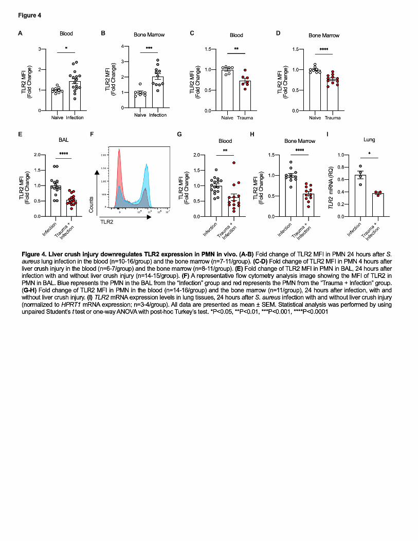
245 Trauma decreases TLR2 expression in PMN and Macrophages 246
Toll-Like Receptor-2 (TLR2) contributes to appropriate recognition of S. aureus 247
by binding lipoteichoic acid, a component of the cell wall that functions as a PAMP(34). 248
When this pathogen’s molecules bind TLR2, a signaling cascade results in translocation 249
of NF-κB and cellular activation(35, 36). As predicted, inoculation of the lung with S. 250
aureus increased TLR2 expression in circulating PMN and BM PMN (Figure 4A-B). We 251
then tested whether liver crush injury alone altered expression of TLR2. Trauma 252
induced downregulation of TLR2 in PMN in both the blood and BM 4 hours after liver 253

12
crush (Figure 4C-D). TLR2 expression was significantly lower in PMN in the BAL after 254
liver crush than laparotomy only plus infection (Figure 4E-F). This reduction in TLR2 255
expression after trauma plus infection was also observed in PMN from lung, peripheral 256
blood and BM (Figure 4G-I). 257
PMN that migrate into the airway in response to bacterial infection express lower 258
TLR2 receptor and therefore are likely be at a disadvantage in recognizing S. aureus. 259
Collectively, these data suggest that DAMPs released from crushed liver modify the 260
PMN pool such that the more mature PMN are recruited to the injury site in the liver 261
(Supplemental Figure 1) leaving more immature PMN populations in the circulation 262
and BM that are less capable of clearing bacteria when recruited to the lung. 263
Alveolar macrophages are also crucial for host defense against pathogen 264
invasion of the lung. The decreased levels of TLR2 after liver crush injury followed by 265
infection are not limited to the PMN as alveolar macrophages also showed decreased 266
TLR2 expression in infected mice after liver crush when compared to infection in the 267
absence of trauma (Supplemental Figure 3C-D). 268
269
Heme impairs bacterial clearance in the lung by decreasing TLR2 levels on PMN 270
Heme as a DAMP is known to activate innate immunity both by increasing 271
oxidative stress and by direct binding of TLR4 (12). Based on elevated plasma heme 272
levels after trauma, we next asked whether the downregulation of TLR2 following liver 273
crush involved free heme. To test this, mice were challenged with a sublethal dose of 274
heme (50 mg/kg, i.p.) to mimic release of heme by injured tissues. As seen after liver 275
trauma, mice treated with heme exhibited lower TLR2 expression in circulating PMN 276
and BM PMN (Figure 5A-B). Furthermore, mice treated with heme followed 4h later by 277
13
S. aureus inoculation in the lung were unable to effectively clear S. aureus (Figure 5C). 278
PMN recruited to the lung as well as peripheral blood and BM PMN exhibited 279
significantly lower expression of TLR2 after liver crush (Figure 5D-G). 280
The mechanism by which TLR2 is regulated in response to heme is not known, 281
but heme is a known ligand for TLR4 (16, 17). Thus we also examined TLR4 expression 282
after heme and in response to liver crush. Similar to TLR2, we observed a significant 283
reduction in TLR4 expression in circulating PMN after liver crush and heme challenge 284
(Supplemental Figure 3E-F). This decrease in TLR4 expression may also explain, in 285
part the observed impairment in E. coli clearance in the lung after liver crush (Figure 286
1J). These data suggest that liver crush injury, presumably through release of heme, 287
increases susceptibility to bacterial infection after trauma and that this effect is 288
explained, in part, by reduced expression of TLR2 and TLR4. In addition to TLR2/4, we 289
observed that both TLR1 and TLR5 expression were reduced in BAL PMN 290
(Supplemental Figure 3A-B). TLR1 is known to form a heterodimer with TLR2 (37). 291
The role of TLR5 in S. aureus infection is not well-known, thus these findings warrant 292
further investigation. Collectively, these data further support our findings. 293
Similar to the observation made in mice with lung infection after liver crush injury, 294
heme-challenged mice also exhibited significantly higher levels of KC in the BAL fluid 295
after lung infection (Figure 5H). Furthermore, the levels of IP-10 and MIP-1α were 296
significantly lower in the BAL fluid of infected mice in the presence of heme (Figure 5I-297
J). Like the dysfunction of PMN observed after liver crush, these data implicate heme as 298
the DAMP that impairs release of IP-10 and MIP-1α ultimately preventing bacterial 299
clearance. 300
14
We next tested whether the serum collected after liver crush injury when plasma 301
heme levels are peaking would influence PMN antimicrobial function by measuring their 302
ability to generate Reactive Oxygen Species (ROS). To test this, trauma serum was 303
collected 30 minutes after liver crush injury when heme levels peak (Figure 2B). PMN, 304
isolated from the BM of naïve mice negatively selected against Ly6G+ cells were then 305
cultured with naïve mouse serum or trauma serum for 90 minutes. PMN were then 306
exposed to S. aureus and ROS were measured by chemiluminescence as previously 307
reported (69). PMN incubated with trauma serum showed reduced ROS generation 308
compared to naïve-serum treated PMN suggesting on potential mechanism that would 309
explain, in part, the lack of bacterial clearance (Figure 5K) 310
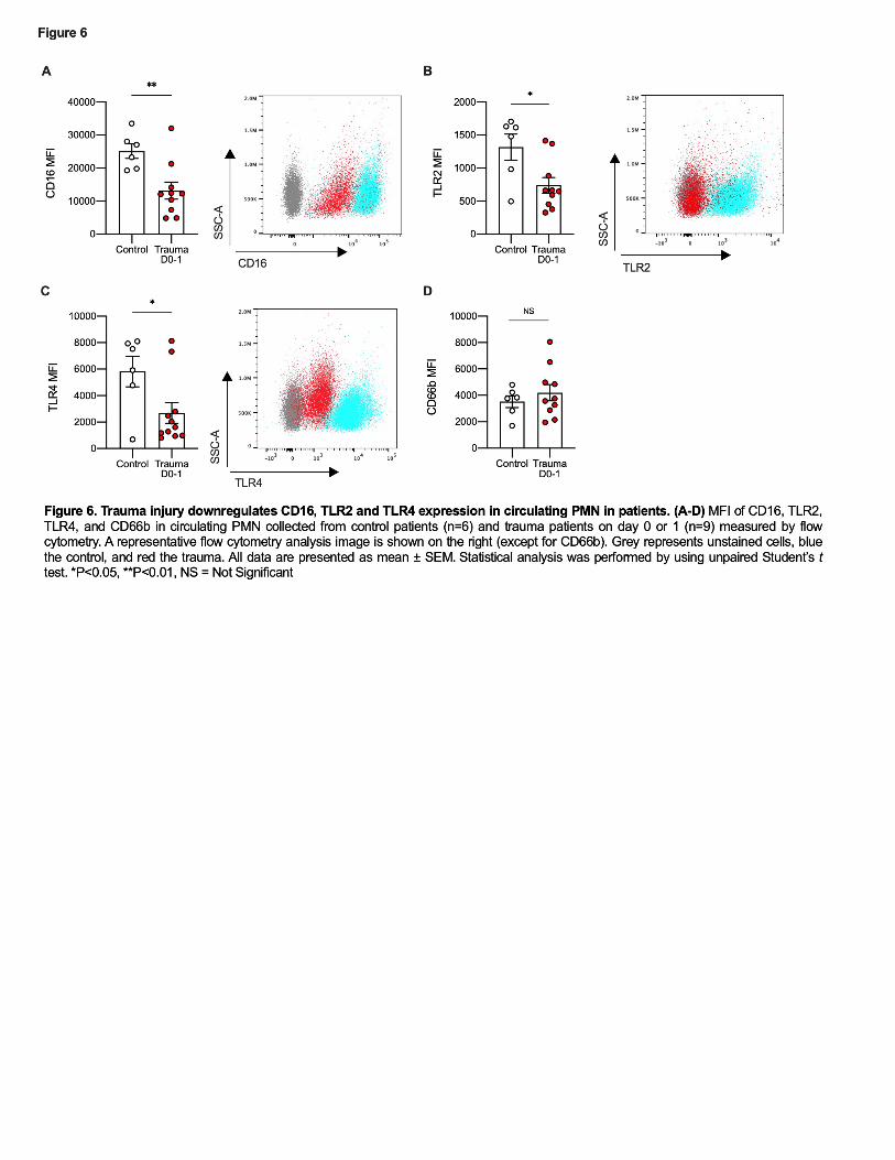
311 PMN from trauma patients show reduced expression of CD16, TLR2 and TLR4. 312
We next characterized circulating PMN purified from blood of trauma patients 313
one day after major trauma by measuring expression of CD16, CD66b, TLR2 and TLR4. 314
Like Ly6Glow cells in mice, nearly all PMN isolated from peripheral blood in trauma 315
patients were CD16low or CD16- (Figure 6A) and so would generally be considered 316
“immature” PMN (38, 39). These findings are consistent with prior reports that showed 317
significantly lower PMN expression of CD16 after major trauma (40). Low expression of 318
Ly6G or CD16 are both considered characteristic of immature PMN(41, 42). 319
In addition to decreased CD16, circulating PMN from trauma subjects showed 320
lower expression of both TLR2 and TLR4 shortly after injury (Figure 6B-C). Again, 321
these parallel findings observed in mice. Human PMN do not express Ly6G, but notably 322
expression of CD66b, another PMN activation marker, showed no differences between 323
PMN from trauma and control subjects (Figure 6D). This shows that the decreases in 324

15
TLR2, 4 and CD16 seen do not reflect global receptor downregulation. Of note, 5 out of 325
9 trauma patients developed infections during their hospital stay (Table 1). Thus again, 326
the human data correlate with the mouse model and collectively the data suggest that 327
downregulation of TLR2 and/or TLR4 as a result of heme released by traumatic injuries 328
may be an important cause of increased clinical susceptibility to bacterial infection. 329
330
16
Discussion 331
Contrary to historic beliefs, the airway is not sterile. Rather, constant low-level 332
inoculation from the digestive tract is cleared by innate immune responses that prevent 333
establishment of the inoculum (43–47). After injury, this homeostasis is disturbed and 334
invasive infection becomes more common. The murine model of post-traumatic 335
pneumonia we developed and characterized for this study closely mimics that aspect of 336
human injury. Inoculation of the lung with a sub-effective dose of bacteria in the 337
presence of a liver crush injury recapitulates human pathology, showing how trauma 338
can predispose both animals and humans to bacterial infection. Moreover, we show 339
here for the first time that the mechanism involves, in part, downregulation of PMN 340
TLR2 and TLR4 in both species. Thus, trauma suppresses a primary receptor 341
mechanism used by the host to sense the presence of pathogens, effectively creating 342
an immunosuppressive environment at barrier sites like the lung, that may be distant 343
from physical injuries. Cahill et al., employed a multiplex mediator signature to 344
differentiate sepsis from sterile SIRS in humans and concluded that biomarker release 345
patterns can be used as powerful diagnostics that, in turn, direct personalized treatment 346
strategies (48). Applied here, patterns of TLR expression in conjunction with specific 347
cytokine/chemokine expression could be used to determine susceptibility to infection. 348
In fact, we see here that trauma causes multiple phenotypic changes in innate 349
immune cells, particularly PMN, as those cells are sequentially mobilized into the 350
circulation and then to injury sites. Those events appear to markedly alter the 351
phenotype of cells available to respond to distant infections. In response to a liver crush 352
injury, a significant number of PMN are mobilized to the liver as a prototypic response to 353
17
tissue injury. Thus, we initially speculated that impaired lung bacterial clearance after 354
injury might reflect either a transient neutropenia that limited the number of cells 355
available for recruitment, or reflect a defect in PMN migration. Trauma did not however, 356
impair PMN migration to sites of bacterial infection in this model, but did modify alveolar 357
macrophages. Rather we observed a modest but significant increase in PMN presence 358
in the airway in response to bacteria. The enhanced PMN recruitment to the site of 359
infection is likely due, in part, to the greater amounts of KC in the BAL fluid (Figure 3I). 360
What is intriguing is that this does not ensure that infiltrating PMN are in fact functional. 361
The PMN that migrated exhibited low Ly6G expression, a phenotype of immature PMN 362
(or ‘bands’) (29, 30, 41, 42). This population of cells is thought to be sequestered from 363
the BM prematurely, but still transmigrate into the airway in response to chemokine 364
gradients generated in response to bacteria inoculation (49). Circulating human PMN 365
after injury showed low CD16 expression; similarly considered a marker of immature 366
PMN (40). Emergence of immature granulocytes in trauma patients has been 367
documented by others and perhaps these populations of PMN are phenotypically and 368
functionally different (40, 50, 51). Indeed, there are N1 and N2 PMN that exhibit 369
different functionality, N1 being pro-inflammatory and N2 being anti-inflammatory (52, 370
53). Neely et al. have demonstrated that in a murine model of burn injury with infection, 371
N1 PMN (IL-10-IL-12+) are more effective than N2 PMN (IL-10+IL-12-) at clearing 372
Pseudomonas aeruginosa (52). Emerging data and data presented here suggest the 373
existence of subpopulations of PMN with different functionality that can be polarized by 374
their particular environment. In two-hit scenario’s, the immune system is challenged 375
across two fronts. As presented here, liver trauma elicits a primary response, which if 376
18
left alone would heal appropriately over a predictable and well-orchestrated kinetic of 377
pro-inflammatory sequelae followed by a battery of anti-inflammatory responses (54). 378
Moreover, these environments are well-characterized by the presence or absence of 379
specific cell populations that appear and disappear as damage control and healing 380
progress. But, if a second threat occurs during the late proinflammatory or anti-381
inflammatory phase of the initial injury, the host mounts an inappropriate response with 382
recruitment of PMN that are designed more for damage control and healing versus an 383
aggressive, pathogen killing phenotype. Therefore, this heterogeneity of PMN and 384
corresponding functionality needs to be further investigated and understood in the 385
setting of trauma. 386
Toll-like receptors (TLR) are a class of evolutionarily conserved pattern 387
recognition receptors that detect PAMPs derived from invading pathogens, in turn 388
inducing and modulating innate immune responses to those pathogens. TLR2 389
recognizes Gram-positive PAMPs like lipoprotein and peptidoglycan (34, 55, 56) and 390
several studies have shown the importance of TLR2 in Staphylococcal infection. TLR2-391
deficient mice are much more susceptible to S. aureus infection than controls (34–36). 392
Perhaps, this explains why S. aureus appear more aggressive during trauma care (20). 393
Here, mice subjected to liver crush injury were more susceptible to S. aureus infection 394
and this was associated with down-regulation of TLR2 protein expression in BM and 395
circulating PMN. These data suggest either that this population of PMN expressed lower 396
TLR2 levels or that trauma and heme induce down-regulation of both receptors. 397
In addition to TLR2, trauma and heme downregulated TLR4 in circulating PMN in 398
mice (Supplemental Figure 3E-F) as well as circulating PMN in trauma patients 399

19
(Figure 6C). Skinner et al. also reported reductions in TLR2 and TLR4 expression in 400
human monocytes after trauma. Again, this was correlated with increased risk for sepsis 401
(57). One weakness of our study was the small number of patient samples. This was 402
limited, in part, because of patient care and the need for informed consent. Our goal 403
was to assess any overlapping findings with the mouse model in the samples available. 404
Despite the relatively low numbers however, we observed clear statistical significance in 405
comparing samples from trauma patients and volunteer controls. Collectively, these 406
data on the effects of tissue injury on TLR2 and TLR4 expression in humans and mice 407
support a global concept that pattern recognition receptors have redundant functions, 408
and that their modulation by injury-derived DAMPs may help explain the higher rate of 409
both Gram-negative and Gram-positive infections in trauma patients. 410
Furthermore, we observed that the levels of IP-10 and MIP-1α were significantly 411
lower in BAL fluid in mice with lung infection after liver crush injury. Zeng et al. showed 412
that IP-10 is a critical cytokine in antibacterial host defense in the lung and that 413
neutralization of IP-10 not only enhanced the number of recruited PMN to the infection 414
but also impaired bacterial killing and clearance in the lung (32). This is completely 415
consistent with our observations here. PMN actively secrete MIP-1α in response to 416
pathogens and lack of MIP-1α increases the host’s susceptibility to bacterial infection 417
(33, 58). The observed reduction in MIP-1α levels in the BAL of infected mice after liver 418
crush injury or heme is consistent with the observed impairment in bacterial clearance. 419
While we acknowledge that different components of DAMPs and PAMPs are involved in 420
altering the extent to which various cytokines are released, these data point to heme as 421
a major contributing factor to the observed PMN dysfunction after trauma. 422
20
While heme is an indispensable prosthetic group of many hemoproteins, free 423
heme, especially when excessive amounts overwhelm scavenging systems, can be 424
detrimental to the host. Historically, extravascular hemorrhage has been thought to 425
favor infection by iron acting as a bacterial nutrient (59–63). More recently though, 426
heme has been found to be a potent DAMP that can contribute to immune activation 427
through NF-kB signaling (12). Heme is a widely accepted ligand for TLR4 (16) and 428
more recently, it has been reported that heme is a potent agonist of TLR2 in astrocytes 429
(15). To date though, the regulation of TLR2 and TLR4 by trauma and heme in PMN is 430
unreported. The effects may also involve TLR1 and TLR5 whose expression was also 431
reduced. Alveolar macrophages also showed a phenotypic switch in response to trauma 432
and likely influence the host response to bacteria. 433
The pathophysiology of bacterial infection following trauma injury is complex. 434
Trauma patients are typically immunocompetent and the term “opportunistic” usually 435
refers to microorganisms that are normally harmless but become pathogenic when host 436
resistance is impaired. We see here clear mechanistic evidence of how injury can 437
create immune suppression. S. aureus typically exists as part of the host microbiome on 438
the skin and in the upper respiratory tract. While our animal model only tested liver as 439
the injury site and the lung as the site of infection, trauma patients present with injuries 440
to multiple organs with a range of severity and have a wide range of infectious events 441
occurring at multiple barrier sites (9, 64, 65). Our cohort of trauma patients developed 442
urinary tract infections (11%), respiratory infections (33%) and in some cases, infections 443
at both sites (11%). While our animal model only studied lung infection, our findings 444
strongly support that this model is a potent and generalizable research tool that is both 445
21
clinically relevant and can shed light on the complicated dynamics of the immune 446
response to infection after injury. The model of liver crush that we describe here is 447
comparable and adapted in part from others in terms of methodology and severity (66, 448
67). Taken together our data highlight the utility of this animal model and importantly, 449
identify new therapeutic targets including heme and Toll receptors that may be 450
important targets in the care of trauma patients by reducing the heightened incidence of 451
infection. 452
453
454
22
Methods 455
456
Animals 457
Male C57BL/6 (H-2b) mice (Charles River Laboratories), Hemopexin null (Hpx-/-, The 458
Jackson Laboratory, #029380) were used at the age of 8 to 10 weeks. All mice were 459
maintained in a specific pathogen free facility. All procedures involving animals were 460
approved by the Beth Israel Deaconess Medical Center Institutional Animal Care and 461
Use Committee and the Department of Defense, USAMRMC Animal Care and Use 462
Review Office. 463
464
Human blood PMN 465
Human blood samples were collected from healthy volunteers, control patients and 466
trauma patients who were enrolled at Beth Israel Deaconess Medical Center. Control 467
patients were enrolled from patients scheduled for elective surgical procedures. 468
Samples from trauma patients with Injury Severity Score with a range from 14 to 75 469
were collected the day after admission into the hospital. Patients with pre-existing 470
immunosuppressive conditions were excluded from the study. Signed consent was 471
obtained from all subjects. 472
PMN isolation: PMN were isolated fresh from peripheral blood as described by Itagaki et 473
al(68). Briefly, PMN were isolated from minimally heparinized whole blood by gradient 474
centrifugation using 1-Step Polymorph (Accurate Chemical & Scientific). Isolated PMN 475
were stored frozen in CryoStor CS10 (Stemcell) at -80°C. The frozen samples were 476
thawed in a 37°C water bath for 2 minutes and washed with cold PBS two times prior to 477
further analysis. Of note, all samples were frozen after isolation. We show in 478
23
Supplemental Figure 4 that the freezing/thawing affects specific surface maker 479
expression, but since it likely affected all cells the same, comparisons could be made. It 480
is logistically impossible to simultaneously obtain multiple trauma PMN samples in order 481
to study them concurrently with control patient PMN. Therefore, the best option for 482
measuring expression was to use frozen preparations that were batched and run 483
together in a standardized fashion. 484
485 Liver crush injury in mice 486
To simulate blunt force liver trauma, a Crile needle-holder (Integra Lifesciences) was 487
modified by adding a stem and screw to the locking ratchet. This prevents the tips from 488
closing completely and provides reproducible compression. Mice were anesthetized 489
with isoflurane using a calibrated vaporizer (3%v/v) during the procedure. Fifteen 490
minutes prior to the liver crush procedure, 1.2mg/kg of sustained release buprenorphine 491
was administered subcutaneously. A laparotomy was performed and the left liver lobe 492
was crushed 10 times uniformly. Animals were allowed to recover under controlled 493
heat. Shams underwent laparotomy only without liver crush. 494
495 Bacterial lung infection and counts 496
Staphylococcus Aureus (S. aureus; ATCC) or Escherichia Coli (E. coli; ATCC) was 497
grown to saturation overnight at 37°C in 100ml Tryptic Soy Broth (TSB; BD 25923). 498
Next day, 2ml of the overnight culture was grown in fresh 100ml TSB at 37°C for 2 499
hours to reach log phase growth. The bacterial culture was then centrifuged at 4000 500
rpm for 10 min at 4°C and diluted in sterile saline to an OD600 of 0.3 (or 0.6 for the 501

24
survival study). To determine the number of bacteria, 10µl of serially diluted bacterial 502
suspensions were streaked on TSA plates (BD 90002) and counted the following day. 503
504
Mice (liver crush or sham controls) were anesthetized with ketamine (10mg/kg; i.p.) and 505
xylazine (4mg/kg; i.p.). A small incision was made through the dermis in the neck. The 506
sublingual glands and the sternothyroid and sternocleidomastoid were gently dissected 507
to expose the trachea. A 30-gauge needle was carefully inserted between the cartilage 508
rings and 50µl of bacterial suspension (106-107 CFU) was slowly infused during 509
inspirations. After inoculation, the neck wound was closed with a surgical staple. Mice 510
that reached a body condition score of 2, which includes lethargy, lack of movement, 511
and a drop in body temperature designated as moribund and therefore were humanely 512
euthanized. Twenty-four hours later, mice were overdosed with anesthesia and the 513
heart transected, the trachea was then intubated and BAL was performed 3x with 1ml of 514
PBS. Combined BAL fluid was separated from the cell pellet by centrifugation (1500rpm 515
for 5min at 4°C). The number of colony forming units (CFU) of bacteria in the BAL fluid 516
was measured by plating 10µl of the fluid or serially diluted fluid on TSA plates and 517
counting the following day. Protein levels in BAL fluid were determined using a Pierce 518
BCA Protein Assay Kit (Thermo Scientific). Residual red blood cells in the cell pellet 519
were lysed (AKC Lysing Buffer, Thermofisher) and BAL leukocytes were processed for 520
further analysis. Recovered cells were counted on a hemocytometer and cytospin slides 521
were prepared by cytocentrifugation onto positively charged microslides at 1800 rpm for 522
7 minutes and stained using Hema3 Staining Kit (Fisher Scientific). Cell differentials 523
were analyzed by morphological criteria of 300 cells. To determine bacteria CFU in lung 524
tissue, the lung tissue was homogenized in 1ml of PBS, centrifuged at 1500rpm for 10 525

25
minutes and 10µl of the supernatant was diluted 1:50 or 1:500, and plated on TSA 526
plates. CFU were counted the following day. For the number of CFU of bacteria in the 527
blood, whole blood was collected by cardiac puncture in EDTA-coated tubes and the 528
plasma was collected by centrifuging at 1500rpm for 10 minutes. 50µl of the plasma 529
was plated on TSA plates and CFU was counted the following day. 530
531
Hemopexin Treatment 532
Hemopexin (Sigma-Aldrich, SRP6514) was prepared in PBS and mice were dosed two 533
times at 50mg/kg in 200µl immediately after (i.v.) and again 45 minutes after liver crush 534
injury (i.p.). Control mice received PBS in the same volume and at the same intervals. 535
536
Heme challenge 537
Heme (Sigma-Aldrich, 52180) was prepared by adjusting the pH to 6.9-7.2 using 1N 538
NaOH (Sigma-Aldrich) and 6N HCl (Sigma-Aldrich) solutions in PBS and then to a final 539
concentration of 5 mg/ml in PBS. Mice were dosed at 50mg/kg, i.p. 540
541 ALT, heme and hemopexin measurements 542
Whole blood was collected by cardiac puncture in EDTA-coated tubes. Plasma was 543
collected by centrifugation at 5500rpm at 4°C for 15 minutes. Plasma was kept on ice or 544
at -20°C for longer storage. ALT in the plasma samples was measured by using an 545
IDEXX Catalyst DX analyzer (IDEXX Laboratories). Heme concentration in plasma was 546
measured by the hemin colorimetric assay kit (GWB-AXR320, Genway Biotech). 547
Hemopexin in plasma was measured by ELISA (NBP2-60633, Novus Biologicals). 548
549

26
PMN Isolation from BM and ROS Measurement 550
Femurs were removed from mice and BM cells were collected by centrifugation (12000 551
rpm for 15 minutes). Red blood cells were lysed (ACK Lysing Buffer, Thermofisher). BM 552
cells were washed with PBS and PMN was isolated using Miltenyi’s Neutrophil Isolation 553
Kit (130-097-658, Miltenyi). 1.25x10⁶ PMN were incubated in 10% RPMI+10% Fetal 554
Bovine Serum and 90% naïve or trauma serum for 90 minutes. Trauma serum was 555
prepared from whole blood 30 minutes after liver crush injury. After incubation with 556
serum, PMN were washed with PBS and ROS release was measured for 2 hours after 557
being challenged with 106 CFU of S. aureus. ROS generation was performed as 558
described by Konecna et al (69). 559
560
Histologic analyses 561
The left liver lobe was collected from mice with or without liver crush injury and 5 µm 562
sections were stained with hematoxylin and eosin. Lung tissue, was collected 24 hours 563
after infection with or without prior liver crush injury. The left lung was fixed by instilling 564
500µl of 10% buffered formalin. After processing 5 µm sections, were stained with 565
hematoxylin and eosin and blindly evaluated for inflammatory cells, hemorrhage, edema 566
and changes in architecture. 567
568
Gene expression – mRNA levels 569
Total RNA was extracted from tissues using TRizol reagent (Invitrogen) following the 570
manufacturer’s instruction. Reverse transcription was performed using High Capacity 571
cDNA synthesis kit (Thermo Scientific). After cDNA synthesis, quantitative real-time 572
27
PCR (RT-qPCR) was performed on QuantStudio 3 (Applied Biosystems) using Maxima 573
SYBR Green Master Mix (ThermoScientific). 574
For samples from mice, the following primers (written in a 5’-3’ direction) were used: 575
Actb (F: CCTTCTTGGGTATGGAATCCTGT, R: GAGGTCTTTACGGATGTCAACG), 576
Hmox1 (F: CAGAAGAGGCTAAGACCGCC, R: AGCTCCTCAAACAGCTCAATGT), 577
Hprt1 (F: CAGTCCCAGCGTCGTGATT, R: GCAAGTCTTTCAGTCCTGTCCAT), 578
Tlr2 (F: ACCTCAGACAAAGCGTCAAA, R: ACAGCGTTTGCTGAAGAGGA), 579
Fold-change in mRNA expression was normalized to Actb or Hprt1 by using the DDCt 580
method. 581
582
Immunoblotting 583
200mg of the injured liver lobes, uninjured liver lobes, and lungs from the euthanized 584
mice were homogenized in RIPA lysis buffer containing halt protease inhibitors (100:1) 585
at 4°C. The tissue homogenates were vortexed every 5 minutes for 30 minutes on ice 586
and centrifuged at 12000g for 15 minutes at 4°C. The supernatant containing the tissue 587
protein was collected and the concentration of protein was determined by the Pierce 588
BCA Protein Assay Kit (Thermo Scientific). Equal amounts of proteins (20 or 30ug) were 589
loaded and separated by 4-15% polyacrylamide gel (Bio-Rad) and transferred onto 590
nitrocellulose membranes at 110 mA and 18V for 70 min. Membranes were blocked 591
with 5% nonfat milk in tris-buffered saline (TBS) and Tween 20 (TBS-T) buffer overnight 592
at 4°C with gentle rocking. The membranes were then incubated with primary antibody 593
against HO-1 (1:1000, ab52947) and vinculin (1:1000, MAB6896, R&D Systems) for 2 594
hours at room temperature with gentle rocking. The membranes were washed with 595
28
TBS-T buffer for 5 minutes for 3 times and then with TBS buffer for 5 minutes with 596
gentle rocking at room temperature. Then, the membranes were incubated with IRDye 597
680RD goat anti-rabbit (1:15000, LI-COR 925-68071) or IRDye 800RD goat anti-rabbit 598
(1:15000, LI-COR 800925-32211) for 90 minutes at room temperature and then washed 599
with TBS-T buffer for 5 minutes for 3 times and with TBS buffer for 5 minutes. The 600
images were visualized on the Odyssey CLx Imaging System and the band intensities 601
were quantified using Image Studio Lite software and normalized to vinculin as a 602
loading control. 603
604
Flow cytometry 605
Mouse: peripheral blood was collected by cardiac puncture into EDTA tubes. Bone 606
marrow (BM) cells were isolated by centrifuging femurs (12K for 15 min). Liver tissues 607
were homogenized gently using a 3ml-syringe plunger. Red blood cells were lysed 608
(ACK Lysing Buffer, Thermofisher). Any clumps of cells were removed by filtration using 609
70µm strainers (Fisher Scientific). Cells were washed and kept in PBS supplemented 610
with 2% Fetal Bovine Serum and were stained with the following fluorochrome-611
conjugated antibodies: CD45 (Alexa Fluor 700, Biolegend 103128, 1:300), CD11b 612
(Brilliant Violet 421, Biolegend 101235, 1:500), Ly6G (APC, 127614, 1:300, or FITC, 613
127605, 1:500), CD11c (Alexa Fluor 488, Biolegend 117313, 1:100 or PE-Cyanine7, 614
Biolegend 117318, 1:100), TLR2 (PE, Biolegend 148604, 1:200), TLR4 (PE-Cyanine7, 615
117610, 1:200), TLR1 (PE, Thermo Fisher 12-9011-80, 1:100), and TLR5 (APC, 616
Biolegend 148105, 1:100) at 4ºC and washed twice. Intracellular staining for HO-1 was 617
performed by permeabilizing the cells using the BD Cytofix/Cytoperm 618
Fixation/Permeabilization Kit (BD Biosciences) and staining the cells with antibody 619

29
against HO-1 (ab52947, 1:200) followed by donkey anti-rabbit secondary antibody 620
(Biolegend 406414, 1:500). 621
622
Human: cells were stained with the following fluorochrome-conjugated antibodies: TLR2 623
(APC, Biolegend 309719, 1:100 or FITC, Biolegend 309705, 1:100), TLR4 (PE, 624
Biolegend 312805, 1:100), CD15 (FITC, Biolegend 301903, 1:100), CD16 (Alexa Fluor 625
700, Biolegend 302025, 1:100), CD11b (Brilliant Violet 421, Biolegend 101235, 1:100), 626
CD66b (PE-Cyanine7, 305115, 1:100). The stained cells were run on CytoFLEX Flow 627
Cytometer (Beckman Coulter). Flow cytometry data was analyzed by Flow Jo software. 628
Flow cytometry gating strategy is shown in Supplemental Figure 5. 629
630
Cytokine/Chemokine Determination 631
BAL fluid was analyzed on a 32-plex array by Eve Technologies (Calgary, Canada). 632
633
Statistics 634
All data that represent means ± standard error of the mean (mean ± SEM). Data were 635
compared by unpaired Student’s t test, or by one-way ANOVA with post-hoc Tukey’s 636
test using Graphpad Prism 8. A p-value less than 0.05 was considered significant. 637
638
Study Approval 639
Animal studies: All procedures involving animals were approved by the Beth Israel 640
Deaconess Medical Center Institutional Animal Care and Use Committee and the 641
Department of Defense, USAMRMC Animal Care and Use Review Office. 642

30
Human studies: The Code of Ethics of the World Medical Association (Declaration of 643
Helsinki) was followed and the study was approved by the Institutional Review Board of 644
Beth Israel Deaconess Medical Center. A written informed consent form was obtained 645
from all individuals. 646
647

31
Author Contributions 648
G.R.L., D.G. and L.E.O. conceived and designed the study. G.R.L., D.G., R.W.A.S., 649
S.T.H., E.C., acquired data. J.H. and S.S. acquired and processed human samples. 650
G.R.L. and V.B. analyzed clinical data. G.R.L. analyzed data. G.R.L. and L.E.O. wrote 651
the paper. R.W.A.S., M.B.Y., M.S.L., C.J.H. provided critical revision of the paper. 652
653
Conflict of Interest Statement 654
Leo E. Otterbein is a scientific consultant for Hillhurst Biopharmaceuticals. All other 655
authors have declared that no conflict of interest exists. 656

32
Acknowledgements 657
We thank Mike Schylijuk for engineering the modified needle holder to provide 658
consistent and highly reproducible liver crush injury in mice. 659
660
Sources of Funding 661
This work was largely supported by Department of Defense award W81XWH-16-0464 662
and the National Football League Players Association to L.E.O. 663
664
665
666
667 668 669 670

33
References 671
1. Web-based injury statistics query and reporting system (WISQARS), Leading Causes 672
of Death Reports, United States, 1999-2019 673
[Internet]https://webappa.cdc.gov/sasweb/ncipc/leadcause.html. cited 674
2. Rhee P et al. Increasing trauma deaths in the United States.. Ann Surg 675
2014;260(1):13–21. 676
3. Siegel R, Naishadham D, Jemal A. Cancer statistics, 2013.. CA Cancer J Clin 677
2013;63(1):11–30. 678
4. Go AS et al. Heart disease and stroke statistics--2014 update: a report from the 679
American Heart Association.. Circulation 2014;129(3):e28–e292. 680
5. Dutton RP et al. Trauma mortality in mature trauma systems: are we doing better? An 681
analysis of trauma mortality patterns, 1997-2008.. J Trauma 2010;69(3):620–626. 682
6. Fraser DR, Dombrovskiy VY, Vogel TR. Infectious complications after vehicular 683
trauma in the United States.. Surg Infect (Larchmt) 2011;12(4):291–296. 684
7. Coccolini F et al. Risk Factors for Infections in Trauma Patients. Current Trauma 685
Reports 2017;3(4):285–291. 686
8. Oyeniyi BT et al. Trends in 1029 trauma deaths at a level 1 trauma center: Impact of 687
a bleeding control bundle of care.. Injury 2017;48(1):5–12. 688
9. Papia G et al. Infection in hospitalized trauma patients: incidence, risk factors, and 689
complications.. J Trauma 1999;47(5):923–927. 690
34
10. Roh JS, Sohn DH. Damage-Associated Molecular Patterns in Inflammatory 691
Diseases.. Immune Netw 2018;18(4):e27. 692
11. Hauser CJ, Otterbein LE. Danger signals from mitochondrial DAMPS in trauma and 693
post-injury sepsis.. Eur J Trauma Emerg Surg 2018;44(3):317–324. 694
12. Wegiel B, Hauser CJ, Otterbein LE. Heme as a danger molecule in pathogen 695
recognition.. Free Radic Biol Med 2015;89:651–661. 696
13. Zhang Q et al. Circulating mitochondrial DAMPs cause inflammatory responses to 697
injury.. Nature 2010;464(7285):104–107. 698
14. Lee GR, Shaefi S, Otterbein LE. HO-1 and CD39: It Takes Two to Protect the 699
Realm.. Front Immunol 2019;10:1765. 700
15. Min H, Choi B, Jang YH, Cho I-H, Lee SJ. Heme molecule functions as an 701
endogenous agonist of astrocyte TLR2 to contribute to secondary brain damage after 702
intracerebral hemorrhage.. Mol Brain 2017;10(1):27. 703
16. Figueiredo RT et al. Characterization of heme as activator of Toll-like receptor 4.. J 704
Biol Chem 2007;282(28):20221–20229. 705
17. Belcher JD et al. Identification of a Heme Activation Site on the MD-2/TLR4 706
Complex.. Front Immunol 2020;11:1370. 707
18. Hvidberg V et al. Identification of the receptor scavenging hemopexin-heme 708
complexes.. Blood 2005;106(7):2572–2579. 709
35
19. Ascenzi P et al. Hemoglobin and heme scavenging.. IUBMB Life 2005;57(11):749–710
759. 711
20. Villavicencio RT, Wall MJJ. The pathogenesis of Staphylococcus aureus in the 712
trauma patient and potential future therapies.. Am J Surg 1996;172(3):291–296. 713
21. Weber DJ et al. Healthcare-associated infections among patients in a large burn 714
intensive care unit: incidence and pathogens, 2008-2012.. Infect Control Hosp 715
Epidemiol 2014;35(10):1304–1306. 716
22. Jones RN. Microbial etiologies of hospital-acquired bacterial pneumonia and 717
ventilator-associated bacterial pneumonia.. Clin Infect Dis 2010;51 Suppl 1:S81-87. 718
23. Otto M. Staphylococcus colonization of the skin and antimicrobial peptides.. Expert 719
Rev Dermatol 2010;5(2):183–195. 720
24. Krismer B, Weidenmaier C, Zipperer A, Peschel A. The commensal lifestyle of 721
Staphylococcus aureus and its interactions with the nasal microbiota.. Nat Rev Microbiol 722
2017;15(11):675–687. 723
25. Clancy TV, Gary Maxwell J, Covington DL, Brinker CC, Blackman D. A statewide 724
analysis of level I and II trauma centers for patients with major injuries.. J Trauma 725
2001;51(2):346–351. 726
26. Matthes G et al. Blunt liver injuries in polytrauma: results from a cohort study with 727
the regular use of whole-body helical computed tomography.. World J Surg 728
2003;27(10):1124–1130. 729

36
27. Afifi I et al. Blunt liver trauma: a descriptive analysis from a level I trauma center.. 730
BMC Surg 2018;18(1):42. 731
28. Alcaraz MJ, Fernández P, Guillén MI. Anti-inflammatory actions of the heme 732
oxygenase-1 pathway.. Curr Pharm Des 2003;9(30):2541–2551. 733
29. Sánchez Á et al. Map3k8 controls granulocyte colony-stimulating factor production 734
and neutrophil precursor proliferation in lipopolysaccharide-induced emergency 735
granulopoiesis.. Sci Rep 2017;7(1):5010. 736
30. Evrard M et al. Developmental Analysis of Bone Marrow Neutrophils Reveals 737
Populations Specialized in Expansion, Trafficking, and Effector Functions.. Immunity 738
2018;48(2):364-379.e8. 739
31. Frink M et al. Keratinocyte-derived chemokine plays a critical role in the induction of 740
systemic inflammation and tissue damage after trauma-hemorrhage.. Shock 741
2007;28(5):576–581. 742
32. Zeng X et al. Interferon-inducible protein 10, but not monokine induced by gamma 743
interferon, promotes protective type 1 immunity in murine Klebsiella pneumoniae 744
pneumonia.. Infect Immun 2005;73(12):8226–8236. 745
33. Tecchio C, Cassatella MA. Neutrophil-derived chemokines on the road to immunity.. 746
Semin Immunol 2016;28(2):119–128. 747
34. Gillrie MR et al. Divergent roles of Toll-like receptor 2 in response to lipoteichoic 748
acid and Staphylococcus aureus in vivo.. Eur J Immunol 2010;40(6):1639–1650. 749

37
35. Takeuchi O, Hoshino K, Akira S. Cutting edge: TLR2-deficient and MyD88-deficient 750
mice are highly susceptible to Staphylococcus aureus infection.. J Immunol 751
2000;165(10):5392–5396. 752
36. Yimin et al. Contribution of toll-like receptor 2 to the innate response against 753
Staphylococcus aureus infection in mice.. PLoS One 2013;8(9):e74287. 754
37. Jin MS et al. Crystal structure of the TLR1-TLR2 heterodimer induced by binding of 755
a tri-acylated lipopeptide.. Cell 2007;130(6):1071–1082. 756
38. Lu Y et al. CD16 expression on neutrophils predicts treatment efficacy of 757
capecitabine in colorectal cancer patients.. BMC Immunol 2020;21(1):46. 758
39. van Grinsven E et al. Immature Neutrophils Released in Acute Inflammation Exhibit 759
Efficient Migration despite Incomplete Segmentation of the Nucleus.. J Immunol 760
2019;202(1):207–217. 761
40. Visser T, Hietbrink F, Groeneveld KM, Koenderman L, Leenen LPH. Isolated blunt 762
chest injury leads to transient activation of circulating neutrophils.. Eur J Trauma Emerg 763
Surg 2011;37(2):177–184. 764
41. Zhu YP et al. Identification of an Early Unipotent Neutrophil Progenitor with Pro-765
tumoral Activity in Mouse and Human Bone Marrow.. Cell Rep 2018;24(9):2329-766
2341.e8. 767
42. Boivin G et al. Durable and controlled depletion of neutrophils in mice.. Nat 768
Commun 2020;11(1):2762. 769

38
43. Martin-Loeches I et al. The importance of airway and lung microbiome in the 770
critically ill.. Crit Care 2020;24(1):537. 771
44. Lynch SV. The Lung Microbiome and Airway Disease.. Ann Am Thorac Soc 2016;13 772
Suppl 2(Suppl 5):S462–S465. 773
45. Dumas A, Bernard L, Poquet Y, Lugo-Villarino G, Neyrolles O. The role of the lung 774
microbiota and the gut-lung axis in respiratory infectious diseases.. Cell Microbiol 775
2018;20(12):e12966. 776
46. Marsland BJ, Yadava K, Nicod LP. The airway microbiome and disease.. Chest 777
2013;144(2):632–637. 778
47. Tan J-Y, Tang Y-C, Huang J. Gut Microbiota and Lung Injury.. Adv Exp Med Biol 779
2020;1238:55–72. 780
48. Cahill LA et al. Multiplexed Plasma Immune Mediator Signatures Can Differentiate 781
Sepsis From NonInfective SIRS: American Surgical Association 2020 Annual Meeting 782
Paper.. Ann Surg 2020;272(4):604–610. 783
49. Hesselink L et al. Neutrophil heterogeneity and its role in infectious complications 784
after severe trauma.. World J Emerg Surg 2019;14:24. 785
50. Hampson P et al. Neutrophil Dysfunction, Immature Granulocytes, and Cell-free 786
DNA are Early Biomarkers of Sepsis in Burn-injured Patients: A Prospective 787
Observational Cohort Study.. Ann Surg 2017;265(6):1241–1249. 788
39
51. Guérin E et al. Circulating immature granulocytes with T-cell killing functions predict 789
sepsis deterioration*.. Crit Care Med 2014;42(9):2007–2018. 790
52. Neely CJ et al. Flagellin treatment prevents increased susceptibility to systemic 791
bacterial infection after injury by inhibiting anti-inflammatory IL-10+ IL-12- neutrophil 792
polarization.. PLoS One 2014;9(1):e85623. 793
53. Tsuda Y et al. Three different neutrophil subsets exhibited in mice with different 794
susceptibilities to infection by methicillin-resistant Staphylococcus aureus.. Immunity 795
2004;21(2):215–226. 796
54. Robinson MW, Harmon C, O’Farrelly C. Liver immunology and its role in 797
inflammation and homeostasis.. Cell Mol Immunol 2016;13(3):267–276. 798
55. Hashimoto M et al. Not lipoteichoic acid but lipoproteins appear to be the dominant 799
immunobiologically active compounds in Staphylococcus aureus.. J Immunol 800
2006;177(5):3162–3169. 801
56. Fournier B. The function of TLR2 during staphylococcal diseases.. Front Cell Infect 802
Microbiol 2012;2:167. 803
57. Skinner NA, MacIsaac CM, Hamilton JA, Visvanathan K. Regulation of Toll-like 804
receptor (TLR)2 and TLR4 on CD14dimCD16+ monocytes in response to sepsis-related 805
antigens.. Clin Exp Immunol 2005;141(2):270–278. 806

40
58. Lindell DM, Standiford TJ, Mancuso P, Leshen ZJ, Huffnagle GB. Macrophage 807
inflammatory protein 1alpha/CCL3 is required for clearance of an acute Klebsiella 808
pneumoniae pulmonary infection.. Infect Immun 2001;69(10):6364–6369. 809
59. Torres VJ et al. A Staphylococcus aureus regulatory system that responds to host 810
heme and modulates virulence.. Cell Host Microbe 2007;1(2):109–119. 811
60. Rosa L, Cutone A, Lepanto MS, Paesano R, Valenti P. Lactoferrin: A Natural 812
Glycoprotein Involved in Iron and Inflammatory Homeostasis.. Int J Mol Sci 2017;18(9). 813
doi:10.3390/ijms18091985 814
61. Brown JS, Holden DW. Iron acquisition by Gram-positive bacterial pathogens.. 815
Microbes Infect 2002;4(11):1149–1156. 816
62. Parrow NL, Fleming RE, Minnick MF. Sequestration and scavenging of iron in 817
infection.. Infect Immun 2013;81(10):3503–3514. 818
63. Bullen JJ. The significance of iron in infection.. Rev Infect Dis 1981;3(6):1127–1138. 819
64. Giamberardino HIG, Cesário EP, Carmes ER, Mulinari RA. Risk factors for 820
nosocomial infection in trauma patients.. Braz J Infect Dis 2007;11(2):285–289. 821
65. Pories SE et al. The epidemiologic features of nosocomial infections in patients with 822
trauma.. Arch Surg 1991;126(1):97–99. 823
66. Tsukamoto T, Pape HC. Animal models for trauma research: what are the options?. 824
Shock 2009;31(1):3–10. 825

41
67. Cox JM, Kalns JE. Development and characterization of a rat model of 826
nonpenetrating liver trauma.. Comp Med 2010;60(3):218–224. 827
68. Itagaki K et al. Formyl Peptide Receptor-1 Blockade Prevents Receptor Regulation 828
by Mitochondrial Danger-Associated Molecular Patterns and Preserves Neutrophil 829
Function After Trauma.. Crit Care Med 2020;48(2):e123–e132. 830
69. Konecna B et al. Monocyte exocytosis of mitochondrial danger-associated 831
molecular patterns in sepsis suppresses neutrophil chemotaxis.. J Trauma Acute Care 832
Surg 2021;90(1):46–53. 833
834
835


47
Table I. Demographic and Clinical Data of 9 Trauma Patients 936
Age Mechanism of Injury
Injured Body System Infection Injury Severity Score
76 Fall Spine, Head/Brain, Lung/Chest Wall
Urinary Tract Infection,
Respiratory Tract Infection
75
28 Motor vehicle accident
Spine, Lung/Chest wall, Abdominal Injury
Urinary Tract Infection
38
39 Fall Spine, Lung/Chest Wall, Abdominal Injury,
Pelvis/Extremities Injury
Respiratory Tract Infection
22
20 Motor vehicle accident
Head/Brain, Lung/Chest Wall, Pelvis/Extremities
Injury
Respiratory Tract Infection
43
67 Pedestrian hit Spine, Lung/Chest wall, Pelvis/Extremities
Injury
Respiratory Tract Infection
17
22 Fall Spine, Head/Brain, Lung/Chest Wall, Pelvis/Extremities
Injury
None 43
49 Motor vehicle accident
Head/Brain None 75
57 Motor vehicle accident
Lung/Chest Wall, Abdominal Injury
None 45
18 Motor vehicle accident
Lung/Chest Wall, Pelvis/Extremities
Injury
None 14
Table I. Demographic and clinical data of 9 trauma patients admitted to the Trauma 937
Center of Beth Israel Deaconess Medical Center, Boston, MA, whose peripheral blood 938
samples were collected and analyzed in this study. 939