1 RCAN1 (DSCR1) increases neuronal susceptibility to oxidative ...
12
RCAN1 (DSCR1) increases neuronal susceptibility to oxidative stress: a potential pathogenic process in neurodegeneration Sı ´lvia Porta 1,{ , Selma A. Serra 3,{ , Meritxell Huch 1 , Miguel A. Valverde 3 , Franc Llorens 2 , Xavier Estivill 1 , Maria L. Arbone ´s 1 and Eula `lia Martı ´ 1, * 1 Genes and Disease Program, 2 Bioinformatics and Genomics Program, Center for Genomic Regulation (CRG-UPF), Biomedical Research Park Building, E-08003 Barcelona, Catalonia, Spain and 3 Molecular Physiology and Channelopathies, Universitat Pompeu Fabra, Barcelona, Catalonia, Spain Received November 27, 2006; Revised February 19, 2007; Accepted February 28, 2007 Oxidative stress (OS) underlies neuronal dysfunction in many neurodegenerative disorders. Regulator of Calcineurin 1 (RCAN1 or DSCR1) is a dose-sensitive gene whose overexpression has been linked to Down syndrome (DS) and Alzheimer’s disease (AD) neuropathology and to the response of cells to stress stimuli. Here, we show that RCAN1 mRNA and protein expression are sensitive to OS in primary neurons, and we evaluate the involvement of RCAN1 dosage in neuronal death induced by OS. We find that Rcan1 2/2 neurons display an increased resistance to damage by H 2 O 2 , which can be reverted by RCAN1 overexpression or by exogenous inhibitors of calcineurin. Although increased intracellular Ca 21 concentration is an important factor in OS-mediated cell death, our results show that Ca 21 loading after exposure to H 2 O 2 was similar in Rcan1 1/1 and Rcan1 2/2 neurons. Our data further suggest that CaN and NFAT signaling protect against OS in both Rcan1 1/1 and Rcan1 2/2 neurons. To explain the observed differential vulnerability, we therefore propose a mechanism downstream of H 2 O 2 -mediated Ca 21 entry, involving calcineurin-NFAT signaling. These findings highlight the importance of RCAN1 gene dosage in the modulation of cell survival and death pathways and suggest that changes in the amount of RCAN1 could represent an important mechanism for regulating susceptibility to neurodegeneration, especially in DS and AD. INTRODUCTION RCAN1 (regulator of calcineurin 1, also known as CALP1, MCIP1 and Adapt78) belongs to a highly conserved family of proteins that modulate the activity of calcineurin (CaN) (1–4). CaN is a serine/threonine phosphatase which is extre- mely abundant in the brain and is regulated by Ca 2þ and cal- modulin. CaN modulates many Ca 2þ -mediated responses, including neurotransmitter release, cytoskeletal stabilization, long-term memory, neurite extension and apoptosis [see (5) for a review]. The endogenous CaN regulator RCAN1 is thus relevant to the control of physiological and pathological CaN-dependent processes. The gene encoding RCAN1, RCAN1—originally named DSCR1 (Down Syndrome Candidate Region gene 1)—is located on human chromosome 21 (6). RCAN1 is mostly expressed in brain and skeletal and cardiac muscle (7,8). The RCAN1 gene contains seven exons, and differential promotor usage and first exon choice can generate several transcripts (7,9). The different mRNAs contain one of four possible initial exons (E1 through E4) and the three exons (E5 through E7) common to all forms of RCAN1 mRNA (7). There are two main protein isoforms of RCAN1, which differ in their N-terminal regions (7): the long isoform (RCAN1L), with 252 amino acids, is encoded by the RCAN1.1 mRNA (10); and the 197 amino-acid short isoform (RCAN1S) is encoded by the RCAN1.4 transcript. RCAN1.4 expression is induced by Ca 2þ via a mechanism that depends on CaN activity. This finding suggested that RCAN1 might participate in a negative feedback loop to # The Author 2007. Published by Oxford University Press. All rights reserved. For Permissions, please email: [email protected] { The authors wish it to be known that, in their opinion, the first two authors should be regarded as joint First Authors. *To whom correspondence should be addressed at: Genes and Disease Program, Genomic Regulation Center (CRG-UPF), Biomedical Research Park, C/Dr Aiguader 88, E-08003 Barcelona, Catalonia, Spain. Tel: þ34 933160201; Fax: þ34 933160099; Email: [email protected] Human Molecular Genetics, 2007, Vol. 16, No. 9 1039–1050 doi:10.1093/hmg/ddm049 Advance Access published on March 6, 2007 Downloaded from https://academic.oup.com/hmg/article-abstract/16/9/1039/687534 by guest on 07 February 2018
Transcript of 1 RCAN1 (DSCR1) increases neuronal susceptibility to oxidative ...

RCAN1 (DSCR1) increases neuronal susceptibilityto oxidative stress: a potential pathogenic processin neurodegeneration
Sılvia Porta1,{, Selma A. Serra3,{, Meritxell Huch1, Miguel A. Valverde3, Franc Llorens2,
Xavier Estivill1, Maria L. Arbones1 and Eulalia Martı1,*
1Genes and Disease Program, 2Bioinformatics and Genomics Program, Center for Genomic Regulation (CRG-UPF),
Biomedical Research Park Building, E-08003 Barcelona, Catalonia, Spain and 3Molecular Physiology and
Channelopathies, Universitat Pompeu Fabra, Barcelona, Catalonia, Spain
Received November 27, 2006; Revised February 19, 2007; Accepted February 28, 2007
Oxidative stress (OS) underlies neuronal dysfunction in many neurodegenerative disorders. Regulator ofCalcineurin 1 (RCAN1 or DSCR1) is a dose-sensitive gene whose overexpression has been linked to Downsyndrome (DS) and Alzheimer’s disease (AD) neuropathology and to the response of cells to stress stimuli.Here, we show that RCAN1 mRNA and protein expression are sensitive to OS in primary neurons, and weevaluate the involvement of RCAN1 dosage in neuronal death induced by OS. We find that Rcan12/2 neuronsdisplay an increased resistance to damage by H2O2, which can be reverted by RCAN1 overexpression or byexogenous inhibitors of calcineurin. Although increased intracellular Ca21 concentration is an importantfactor in OS-mediated cell death, our results show that Ca21 loading after exposure to H2O2 was similar inRcan11/1 and Rcan12/2 neurons. Our data further suggest that CaN and NFAT signaling protect againstOS in both Rcan11/1 and Rcan12/2 neurons. To explain the observed differential vulnerability, we thereforepropose a mechanism downstream of H2O2-mediated Ca21 entry, involving calcineurin-NFAT signaling.These findings highlight the importance of RCAN1 gene dosage in the modulation of cell survival anddeath pathways and suggest that changes in the amount of RCAN1 could represent an important mechanismfor regulating susceptibility to neurodegeneration, especially in DS and AD.
INTRODUCTION
RCAN1 (regulator of calcineurin 1, also known as CALP1,MCIP1 and Adapt78) belongs to a highly conserved familyof proteins that modulate the activity of calcineurin (CaN)(1–4). CaN is a serine/threonine phosphatase which is extre-mely abundant in the brain and is regulated by Ca2þ and cal-modulin. CaN modulates many Ca2þ-mediated responses,including neurotransmitter release, cytoskeletal stabilization,long-term memory, neurite extension and apoptosis [see (5)for a review]. The endogenous CaN regulator RCAN1 isthus relevant to the control of physiological and pathologicalCaN-dependent processes.
The gene encoding RCAN1, RCAN1—originally namedDSCR1 (Down Syndrome Candidate Region gene 1)—is
located on human chromosome 21 (6). RCAN1 is mostlyexpressed in brain and skeletal and cardiac muscle (7,8).The RCAN1 gene contains seven exons, and differentialpromotor usage and first exon choice can generate severaltranscripts (7,9). The different mRNAs contain one of fourpossible initial exons (E1 through E4) and the three exons(E5 through E7) common to all forms of RCAN1 mRNA(7). There are two main protein isoforms of RCAN1, whichdiffer in their N-terminal regions (7): the long isoform(RCAN1L), with 252 amino acids, is encoded by theRCAN1.1 mRNA (10); and the 197 amino-acid short isoform(RCAN1S) is encoded by the RCAN1.4 transcript. RCAN1.4expression is induced by Ca2þ via a mechanism thatdepends on CaN activity. This finding suggested thatRCAN1 might participate in a negative feedback loop to
# The Author 2007. Published by Oxford University Press. All rights reserved.For Permissions, please email: [email protected]
{The authors wish it to be known that, in their opinion, the first two authors should be regarded as joint First Authors.
*To whom correspondence should be addressed at: Genes and Disease Program, Genomic Regulation Center (CRG-UPF), Biomedical Research Park,C/Dr Aiguader 88, E-08003 Barcelona, Catalonia, Spain. Tel: þ34 933160201; Fax: þ34 933160099; Email: [email protected]
Human Molecular Genetics, 2007, Vol. 16, No. 9 1039–1050doi:10.1093/hmg/ddm049Advance Access published on March 6, 2007
Downloaded from https://academic.oup.com/hmg/article-abstract/16/9/1039/687534by gueston 07 February 2018

control CaN activity (9). In addition, RCAN1.1 expressionhas been recently shown to be up-regulated by glucocorti-coids (11) and down-regulated by Notch signaling (12). Themouse ortholog maps to chromosome 16 and has a similargenomic structure (13). Transcripts containing the mouseequivalents of human exons 1 and 4 respectively give rise tothe expression of the long and short mouse RCAN1 isoforms.
The first indication that RCAN1 might play a central rolein pathological processes was the finding that mammaliancells increase RCAN1 expression after exposure to damagingstimuli associated with Ca2þ overloading, such as oxidativestress (OS) (14–16). Interest in RCAN1 was reinforced bythe discoveries that RCAN1 transcripts are overexpressed inthe brains of Down syndrome (DS) fetuses (2) and thatRCAN1 transcript and protein are both increased in thebrains of individuals with Alzheimer’s disease (AD) (17,18).It has been proposed that long-term or chronic induction ofRCAN1 might be associated with neurodegeneration in ADand DS, whereas short-term induction might protect cellsagainst stress-induced damage (19,20). The detrimental conse-quences of RCAN1 overexpression have been analyzed incultured primary neurons, and include the formation ofmicrotubule-dependent aggregosome-like inclusion bodies,as well as synaptic impairment (21). Altered synaptic trans-mission also underlies the severe learning defects seen withmodifications in the gene dosage of nebula, the Drosophilaortholog of RCAN1 (22). Moreover, the correct dosage ofnebula is essential for the maintenance of mitochondrialfunction, which has important implications in the context ofOS (23).
The possible involvement of RCAN1 in OS-related signal-ing mechanisms is especially relevant in the context of neuro-degenerative diseases, where OS is the common hallmarkof progression toward loss of neuronal function. Althoughthe published data clearly indicate that increased RCAN1gene dose underlies neuronal dysfunction, little is knownabout the involvement of RCAN1 in OS-mediated neuronaldeath. In the present study, we have analyzed susceptibilityto OS and related intracellular signaling mechanisms inprimary neuronal cell cultures expressing different amountsof RCAN1. Our results show a mechanistic link betweenRCAN1 dose and neuronal viability in response to OS, thussuggesting that the amount of RCAN1 has a significant rolein modulating signaling pathways underlying the balancebetween neuronal death and survival.
RESULTS
Expression of RCAN1 by primary neurons issensitive to OS
Primary neurons obtained from mouse cerebellar cortex atearly postnatal time-points were chosen for the presentstudy, since they are a very homogeneous population of neur-onal granule cells, and have been widely used as a model todissect mechanisms of neuronal death. Moreover, Rcan1 tran-scripts are abundantly expressed in the cerebellar cortex inadult and early postnatal mice (8).
Since previous studies in established human cell lines haveshown increased RCAN1 mRNA expression in response to
reactive oxygen species (ROS) (14,16), we first examinedwhether OS could induce Rcan1 transcription in mouseprimary neurons. Exposure of cells to H2O2 specificallyinduced expression of Rcan1.4, but not Rcan1.1, 4 h aftertreatment (Fig. 1A). No significant changes in Rcan1.4 mRNAexpression were detected at earlier time points (data notshown). Previous reports have shown that Rcan1.4 induction inother experimental situations occurs via a Ca2þ/CaN-dependentmechanism involving Rcan1.4 promoter regions that containNFAT transcription-factor binding motifs (2,9,24). To determinethe involvement of CaN in Rcan1.4 mRNA induction in ourexperimental system, neurons were pre-incubated with theCaN inhibitors cyclosporine A (CsA) and FK506. The inhibitorswere used in combination because both drugs are required inprimary neurons to form enough inhibitory complexes to neutral-ize CaN (25,26). Pre-incubation with CsA and FK506 largelyprevented Rcan1.4 induction, suggesting that this process ishighly dependent on CaN activity (Fig. 1A).
Protein expression was analyzed with antibodies recognizingthe C-terminal region common to RCAN1L and RCAN1S iso-forms. Indirect immunofluorecence revealed a major cyto-plasmic accumulation of RCAN1 in primary granular cells,with the protein homogeneously distributed both in the somaand in neuronal projections (Fig. 1B). Signal, in the formof coarse granules in the somatic cytoplasm, was detected3,5 h min after exposure to 50 mM H2O2 and was maintainedthroughout the treatment; there was no obvious translocationto the nuclear compartment even after prolonged exposure toH2O2 for 5 h (Fig. 1B).
Western blot analysis revealed that RCAN1L is much moreabundantly expressed than RCAN1S in primary neuronalcells (Fig. 1C, lane 1). Both RCAN1L and RCAN1S appearedas doublets, which represent differentially phosphorylatedforms (10,27,28). The amount of RCAN1 decreased nota-bly between 4 and 5 h after exposure to H2O2 (Fig. 1C, lanes6–7). Interestingly, the band corresponding to hypophosphory-lated RCAN1L was consistently more resistant to fading,suggesting an increased half-life. The loss of RCAN1 wasaccompanied by a concomitant appearance of immunoreactivebands around 30 kDa (Fig. 1C, lanes 5–7). We hypothesizedthat these RCAN1 degradation products might be producedby the action of the protease calpain, since calpain is activatedby OS (29) and can cleave RCAN1S in vitro (30). Moreover,one of the calpain cleavage sites in RCAN1S also occurs inRCAN1L, suggesting that both RCAN1 forms will be suscep-tible to calpain proteolysis in vivo. Pre-incubation of primaryneurons with calpain inhibitors I and II prevented the declinein expression of both RCAN1L and RCAN1S and the for-mation of the 30 kDa RCAN1 degradation products (Fig. 1D,compare lanes 3–4 and 5–6), indicating calpain-mediateddegradation of RCAN1 after OS. In addition, calpain inhibitionincreased the basal accumulation of both RCAN1 isoforms(Fig. 1D, lane 2). This effect, which was more apparent withRCAN1S, suggests that calpain also regulates RCAN1 half-lifein the absence of stress stimuli.
In light of reported crosstalk between CaN and calpainactivities (31), we wondered whether CaN would affectRCAN1 accumulation. To test this, we pre-incubatedprimary neurons with CsA and FK506 before addingH2O2. This pre-treatment resulted in enhanced RCAN1
1040 Human Molecular Genetics, 2007, Vol. 16, No. 9
Downloaded from https://academic.oup.com/hmg/article-abstract/16/9/1039/687534by gueston 07 February 2018

degradation (Fig. 1E), thus indicating that CaN activitymight limit calpain-mediated RCAN1 degradation in responseto OS.
Lack of RCAN1 is linked to neuronalsusceptibility to OS
To examine the function of RCAN1 in neurons exposed to OS,we used primary neurons cultured from mice with a targeteddeletion in the Rcan1 gene (Fig. 2A). The lack of RCAN1expression in these cells was confirmed by western blot(Fig. 2B). There were no differences between wild type[Rcan1þ/þ] and knockout [Rcan12/2] neurons in theirexpression of CaN or of other members of the RCAN family(RCAN2 and RCAN3) (Fig. 2B). Cultures of the two geno-types had similar proportions of granule neurons and astro-cytes after 7 to 10 days’ culture (data not shown), and theneurons showed similar morphology (Fig. 2C).
The susceptibility of Rcan1þ/þ and Rcan12/2 neuronsto OS-induced damage was determined by MTT assay after20 h exposure to different doses of H2O2. In both genotypes,maximal damage was achieved with 100 mM H2O2
(Fig. 3A); however, neurons lacking RCAN1 were more
resistant to OS. Calculated IC50 values were 55.7 + 1.6 mM
for Rcan12/2 and 46.6 + 1.1 mM for Rcan1þ/þ cultures.This result was confirmed by simultaneous staining withfluorescein diacetate (FDA) and propidium iodide (PI) tomeasure viable and dead cells. Unstimulated Rcan1þ/þ cul-tures contained a slightly higher proportion of dead cellscompared with unstimulated Rcan12/2 cells (Fig. 3B).This difference was greatly enhanced after exposure to50 mM H2O2, with Rcan12/2 showing a considerable resis-tance to OS-induced cell death. This relatively higher viabi-lity of Rcan12/2 primary neurons was also detected inresponse to OS induced by the peroxynitrite donor SIN-1(17% more resistant than Rcan1þ/þ cultures, P , 0.05;data not shown).
We also determined the exposure time to H2O2 needed toachieve a maximal detrimental effect (Fig. 3C). Rcan1þ/þ
and Rcan12/2 neurons were exposed to 100 mM H2O2 fordifferent periods, and neuronal viability measured 20 h later.Maximal reduction of Rcan1þ/þ neuronal viability wasachieved by exposure to H2O2 for 30 min. Again, Rcan12/2
primary neurons were resistant to OS-mediated neuronaldeath, displaying an increased viability at 30 min and at allshorter exposure times tested.
Figure 1. RCAN1 expression after OS. (A) Rcan1.4 and Rcan1.1 expression after OS. Semi-quantitative real-time PCR was used to determine Rcan1.1 andRcan1.4 mRNA expression relative to L7 mRNA. All data are expressed relative to basal expression (non-treated cultures), which is assigned a value of1-fold (1x). Neurons were incubated with CsA (1 mM) and FK506 (0.5 mM) before treatment with 100 mM H2O2 for 4 h. Data are presented as themeans + SD of four independent determinations. �, P, 0.05. (B) RCAN1 immunofluorescence in control granule neurons and at different times after exposureto 50 mM H2O2. Abnormal condensed nuclei are already apparent 3.5 h after exposure to H2O2 (arrow); scale bar, 10 mM. (C) Western blot of RCAN1 proteinexpression. Neurons were cultured for 7 days and treated for the indicated times with 50 mM H2O2. Tubulin expression was used to confirm equal loading.Decreased RCAN1 expression is evident after 5 h. The arrow indicates the progressive appearance of RCAN1 immunoreactive bands around 30 kDa.(D) Calpain participates in RCAN1 degradation. Neurons were pretreated with calpain inhibitors I and II (1 mM each) for 20 min before exposure to 50 mM
H2O2 for 4 h followed by western blotting. (E) CaN modulates RCAN1 degradation. Neurons were pretreated of with CsA (1 mM) and FK506 (0.5 mM) for20 min. Protein samples were extracted for western blot 4 h after the addition of 50 mM H2O2.
Human Molecular Genetics, 2007, Vol. 16, No. 9 1041
Downloaded from https://academic.oup.com/hmg/article-abstract/16/9/1039/687534by gueston 07 February 2018

RCAN1 expression in Rcan1/1 and Rcan12/2 primaryneurons increases susceptibility to OS
The reduced susceptibility of Rcan12/2 neurons to OS islikely the result of the lack of expression of RCAN1 protein.To test this, we attempted to restore the wild type OS responseby exogenously expressing RCAN1 in Rcan12/2 neurons. Tofinely control the amount of exogenously expressed RCAN1,we used an adenovirus that expresses both RCAN1S andEGFP (AdRCAN1) (32). Infection with an adenovirus expres-sing EGFP and firefly luciferase (AdGFPLuc) (33) was used asa control. Rcan12/2 neurons transduced with AdRCAN1 at anMOI of 2.5, showed levels of RCAN1S expression compar-able to the endogenous expression in wild type controls(Fig. 4A, compare 2 and 3).
The susceptibility to OS of neurons infected with controlvirus was similar to that of non-infected neurons (Fig. 4B, 4and 5), thus indicating that infection per se did not modifythe response to H2O2-induced damage. In contrast, the inci-dence of H2O2-mediated death was increased in Rcan1þ/þ
and Rcan12/2 primary neurons infected with AdRCAN1(Fig. 4B, 3). Furthermore, the sensitivity to H2O2 ofRcan12/2 neurons overexpressing RCAN1 was similar tothat of non-infected Rcan1þ/þ neurons (Fig. 4B, compare 3with 5). These data thus indicate that the level of RCAN1protein is directly responsible for the differential response ofRcan1þ/þ and Rcan12/2 to ROS-mediated damage.
H2O2-induced Ca21 uptake is similar in Rcan11/1 andRcan12/2 neurons
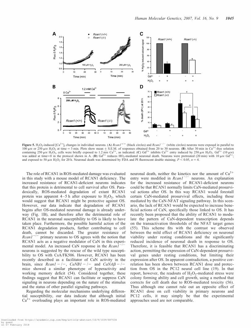
Redox status-dependent cation channels have been shown tounderlie susceptibility to OS-mediated death by inducingCa2þ overloading (34,35). We therefore determined H2O2-induced changes in Ca2þ homeostasis in Rcan1þ/þ andRcan12/2 neurons loaded with the Ca2þ indicator Fluo-3, tosee if this could account for the different sensitivities ofthese cells to H2O2 (Fig. 5). Exposure of neurons to 100or 250 mM H2O2 progressively increased the intracellularCa2þ concentration in both genotypes, with no significantdifferences detected (Fig. 5A). Mean Ca2þ responses ofRcan1þ/þ and Rcan12/2 cultures were not significantly differ-ent, whether the cells were unstimulated (fluorescenceratio ¼ 1.06 + 0.08, n ¼ 132 neurons versus 1.05 + 0.02,n ¼ 127 neurons) or stimulated with 250 mM H2O2
(2.18 + 0.06, n ¼ 170 versus 1.98 + 0.06, n ¼ 157)(P.0.05). Identical patterns were observed with higherH2O2 concentrations, although the magnitude of Ca2þ
signals was larger and the response latency shorter (data notshown). Figure 5B shows individual fluo-3 images obtainedfrom granule neurons treated with 250 mM H2O2 in aCa2þ-free solution. Under these conditions, no increases inCa2þ were observed either in Rcan1þ/þ or in Rcan12/2 cul-tures. However, both neuron genotypes showed an acute risein intracellular Ca2þ upon exposure to a Ca2þ-containing solu-tion. Thus, most of the H2O2-induced rise in Ca2þ has anextracellular origin, which suggests the presence of Ca2þ
channels sensitive to OS. The channels most likely toaccount for OS-induced Ca2þ entry belong to the TRPfamily, with the TRPM2 channel a particularly strong candi-date (35,36).
Pre-incubation with 10 mM gadolinium (Gd3þ), a non-selective blocker of TRP channels, reduced the Ca2þ increaseby 60–70% (Fig. 5C); and Gd3þ also reduced H2O2-inducedcell death (Fig. 5D), suggesting that Ca2þ influx is in partresponsible for neuronal death. Pre-incubation with the anti-oxidant agent Trolox (a vitamin-E analogue) blocked theH2O2-induced Ca2þ increase (data not shown). These dataindicate that the initial Ca2þ increase following exposure toH2O2 is dependent on extracellular Ca2þ and ROS both inwild type and in Rcan1-deficient neurons. The similar pertur-bation of calcium homeostasis by H2O2 in the two genotypesindicates that their differential susceptibility to OS is likely toinvolve a mechanism downstream of Ca2þ entry.
CaN and NFAT signaling pathways limit H2O2-induceddeath in Rcan11/1 and Rcan12/2 neurons
The principal molecular target described for RCAN1 is thephosphatase CaN. We therefore evaluated the possible
Figure 2. Rcan2/2 and Rcan1þ/þ granule neurons express similar amounts ofCaN and of RCAN-2 and RCAN-3. (A) Left: schematic diagram showing thegenomic structure of the Rcan1 wild-type (WT) allele, the targeting vector(Middle) and the homologous recombinant (KO) allele. Alternative splicingevents between exon 1 or 4 and exon 5 are indicated; these events result inRcan1.1 and Rcan1.4 transcripts, respectively. The asterisk shows the locationof the STOP codon in exon 7. Right: Southern blot of EcoRI-digested DNAfrom the wild-type parental ES cell line and three recombinant clones(clones 62, 106 and 141) hybridized with the Rcan1 3’-flanking probeshown in the diagram. (B) Rcan12/2 and Rcan1þ/þ primary neurons were cul-tured in vitro for 10 days, and analyzed by western blotting for CaN, RCAN1,RCAN2 and RCAN3. (C) Immunoflurescence with anti a-tubulin antibodyshows that Rcan12/2 neurons have no gross abnormalities in cell morphologyor neuritic complexity; scale bar 10 mM.
1042 Human Molecular Genetics, 2007, Vol. 16, No. 9
Downloaded from https://academic.oup.com/hmg/article-abstract/16/9/1039/687534by gueston 07 February 2018

involvement of CaN in OS-induced neuronal death by pre-treating Rcan1þ/þ and Rcan12/2 primary neurons with acombination of the CaN inhibitors CsA and FK506, followedby acute (15 min) exposure to 100 mM H2O2 (Fig. 6A). CsAand FK506 did not affect neuronal survival under basal con-ditions, but the inhibitors significantly enhanced neuronaldeath induced by H2O2. These data suggest that, while CaNsignaling is dispensable for cell viability under basal culture con-ditions over the time-course examined, its activity is protectivefollowing H2O2-induced stress. The extent of H2O2-mediatedcell death in Rcan12/2 neurons pretreated with CsA/FK506was similar to that detected in non-pretreated Rcan1þ/þ
neurons (Fig. 6A), which suggests that CaN is involved in thegenotype-related differential susceptibility to OS.
One of the best-characterized signaling pathways regulatedby CaN involves the activity of the nuclear factor of acti-vated T cells (NFAT) family of transcription factors. Depho-sphorylation of NFAT proteins by CaN is required for theirnuclear import and subsequent transcriptional activity(37,38). Our results showing a specific H2O2-dependentinduction of Rcan1.4 (Fig. 1A) strongly suggest that NFAT
signaling is activated after OS, which is in agreement withROS-mediated NFAT activation in other cell types (39,40).Furthermore, recent data have shown the participation ofNFAT signaling in cerebellar granule neuron survival (41).We used the VIVIT peptide to assess the possible involve-ment of NFAT signaling in OS-induced neuronal damage.VIVIT specifically inhibits NFAT-dependent transcriptionthrough its ability to block the interaction between CaNand NFAT without affecting CaN activity (42). Rcan1þ/þ
and Rcan12/2 primary neurons were transfected with GFP-VIVIT or GFP expression vectors and the proportion oftransfected cells showing nuclear PI staining was determined(Fig. 6B). Under resting conditions, VIVIT overexpressionhad no significant effect on neuronal viability. However,VIVIT-expressing neurons were appreciably more sensitiveto H2O2 (Fig. 6B), suggesting that NFAT activity has a neu-roprotective effect in the response to OS. These data supporta model in which the differential sensitivity of wild type andRCAN1-deficient neurons to OS is mediated by CaN signal-ing through a mechanism involving NFAT transcriptionalactivity.
Figure 3. Rcan12/2 neurons have increased resistance to H2O2 induced stress. (A) Dose response to H2O2-mediated damage. Neurons were cultured in vitro for7–10 days and chronically treated with the indicated concentrations of H2O2. Cell viability was determined 20 h later by MTT assay in triplicate for each con-dition and cell culture. Values are expressed as the percentage of the viability of control neurons not subjected to OS, which was assigned the value of 100%.Data are presented as the mean + SD of determinations performed in four independent paired Rcan1þ/þ and Rcan12/2 neuronal cultures. �, P, 0.05. (B) Neur-onal death induced by chronic exposure to 50 mM H2O2. The left panel shows the proportion of dead (PI-positive) cells for each genotype and condition, calcu-lated as a percentage of the total number of cells (PI-positive plus FDA-positive). For the right panel, the percentage of dead cells in Rcan12/2 cultures wassubtracted from that obtained the parallel-treated Rcan1þ/þ cultures. Data are presented as the mean + SD of determinations performed in five independentpaired Rcan1þ/þ and Rcan12/2 neuronal cultures.�, P , 0.05; ��, P, 0.001. (C) Viability of Rcan12/2 and Rcan1þ/þ neurons as a function of the exposuretime to 100 mM H2O2. Paired Rcan12/2 and Rcan1þ/þ neuronal cultures were simultaneously exposed to H2O2, and culture media were removed and replacedwith fresh conditioned media at the indicated times. Cell viability was determined by MTT assay after 20 h. The left panel shows a representative plot of fourindependent determinations. For the right panel, Rcan1þ/þ viability was subtracted from Rcan12/2 viability for each independent determination and exposuretime. The positive values obtained indicate an increased resistance of Rcan12/2 neurons to H2O2-mediated damage. Data are the mean + SD of cell viabilityvalues of Rcan12/2 minus Rcan1þ/þ �, P, 0.05.
Human Molecular Genetics, 2007, Vol. 16, No. 9 1043
Downloaded from https://academic.oup.com/hmg/article-abstract/16/9/1039/687534by gueston 07 February 2018

DISCUSSION
In this study, we have described the expression pattern of theendogenous CaN modulator RCAN1 in response to OS, andhave shown that RCAN1 is a detrimental factor in thisresponse, contributing to ROS-mediated damage.
Our analysis of the expression pattern of RCAN1 afterOS in primary granule neurons shows that H2O2-mediatedRcan1.4 induction is not associated with a concomitantincrease in RCAN1S protein levels, which is in accordancewith previous results on human non-neuronal cell lines sub-mitted to OS (27,28). These data suggest that processes thatregulate RCAN1 translation and/or stability might influencethe final expression output. Supporting this, we detectedcalpain-dependent RCAN1 degradation after exposure toH2O2. Since RCAN1 degradation is detected at 4–5 h afterOS, our results indicate that Rcan1.4 mRNA induction at 4 hafter treatment may not be relevant in maintaining RCAN1Slevels. These degradation products might not have fullRCAN1 functionality since in vitro cleavage of RCAN1 pro-duces fragments with reduced affinity for CaN (30). Ourresults further indicate that after OS CaN is able to modulateRCAN1 expression at different levels, inducing Rcan1.4
mRNA expression and limiting RCAN1 cleavage by calpain.Thus, RCAN1 expression and functionality might depend, atleast in part, on the timing and relative strengths of thesetwo processes. Our results contrast with previous reportsshowing that RCAN1L and RCAN1S become hyperpho-sphorylated shortly after exposure to H2O2, as indicated bychanges in the proportion of the RCAN1 protein bands withdifferent electrophoretic mobilities (27,28). However, thesestudies used non-neuronal cell lines, and no cell damage wasreported even though cells were treated with high H2O2 con-centrations (500 mM). The increased resistance to OS ofthese cell lines compared with primary neurons might reflectdifferences in intracellular signaling pathways, which inturn, could explain the lack of RCAN1 post-translationalmodifications in neurons. Nevertheless, OS-linked changesto the pattern of RCAN1 phosphorylation should be con-sidered in every cell type analyzed, since RCAN1 phosphory-lation status is relevant to its function as a modulator ofCaN (10,43,44). Another feature of the RCAN1 expressionpattern after OS is the shift in its cytoplasmic appearance tothe form of coarse granules. The exact nature of these struc-tures and their relevance to the neuronal response to OSremain to be elucidated. However, since OS leads to proteinaggregation (45) and recent studies have shown that RCAN1contains an aggregation-prone domain at the N-terminalregion (21), it could be hypothesized that these granulescorrespond to ROS-mediated aggregates of RCAN1.
Our data indicate that CaN is activated after exposure to H2O2,since exogenous CaN inhibitors alter not only RCAN1 mRNAexpression, but also neuronal susceptibility to ROS-mediateddamage. Similarly, H2O2-dependent activation of CaN hasbeen shown in cerebellar granule neurons (29) and in hippocam-pal slices (46). The increased OS-induced damage we detectedin the presence of exogenous CaN inhibitors suggests thatthis phosphatase protects against H2O2 toxicity in primarycerebellar neurons. Studies conducted in different cell typeshave demonstrated either pro- or antiapoptotic effectsassociated with CaN activation. For instance, there is con-siderable evidence suggesting that CaN activity contributesto neuronal apoptosis associated with glutamate excitotoxi-city and ionophore-induced Ca2þ overloading (47–50). Inother systems, such as cultured cardiac myocytes, CaN acti-vation protects against H2O2- or 2-deoxyglucose- induceddeath (51,52). In agreement with this, CaNAb-deficientmice have an elevated susceptibility to cardiac damage(53). Collectively, these accounts underscore the complexityof intracellular signaling networks within mammalian cells,with seemingly related stress stimuli able to elicit fundamen-tally different responses.
A key finding of our study is that blockade of NFATactivity increases neuronal susceptibility to ROS-induceddamage. Therefore, the protective role of CaN activity inthe response to OS might be mediated, at least in part, byNFAT transcriptional activity. On the other hand, a previousstudy has shown that oxidative damage in granule neurons ispartially due to CaN-mediated dephosphorylation of thetranscription factor CREB (29). Thus, OS-mediated CaNactivation might trigger prosurvival and detrimental signals.The strength and duration of the stimulated pathways maydetermine the overall effect on neural viability.
Figure 4. RCAN1 overexpression increases neuronal susceptibility to OS.(A) Western blot of RCAN1 protein expression. Parallel Rcan1þ/þ andRcan12/2 primary neuronal cultures were cultured for 2 days (DIV 2) andsimultaneously transduced with AdGFPLuc and AdRCAN1, MOI ¼ 2.5.RCAN1 expression was analyzed 4 days later. (B) Parallel Rcan1þ/þ andRcan12/2 primary neurons were cultured for 2 days and simultaneously trans-duced with AdRCAN1 (1 and 3) or AdGFPLuc (2 and 4) at MOI ¼ 2.5. 4 dayslater, cultures were treated with 50 mM H2O2 (3, 4 and 5), and cell viabilitywas determined by MTT assay, 6 h later in three independent wells percondition. Data are presented as the mean + SD of determinations in threeindependent experiments �, P, 0.05.
1044 Human Molecular Genetics, 2007, Vol. 16, No. 9
Downloaded from https://academic.oup.com/hmg/article-abstract/16/9/1039/687534by gueston 07 February 2018
The role of RCAN1 in ROS-mediated damage was evaluatedin this study with a mouse model of RCAN1 deficiency. Theincreased resistance of RCAN1-deficient neurons indicatesthat this protein is detrimental to cell survival after OS. Para-doxically, ROS-mediated degradation of extant RCAN1protein was apparent 4–5 h after exposure to H2O2, whichwould suggest that RCAN1 might be protective against OS.However, our data indicate that degradation of RCAN1begins after OS-mediated neuronal damage is already under-way (Fig. 1B), and therefore after the detrimental role ofRCAN1 in the neuronal susceptibility to OS is likely to havetaken place. Furthermore, the possible harmful action of theRCAN1 degradation products, further contributing to celldeath, cannot be discarded. The greater resistance ofRcan12/2 primary neurons to OS agrees with the notion thatRCAN1 acts as a negative modulator of CaN in this experi-mental model. An increased CaN response in the Rcan12/2
neurons is suggested by the rescue of the wild type suscepti-bility to OS with CsA/FK506. However, RCAN1 has beenrecently described as a facilitator of CaN activity in thebrain, since Rcan12 /2, CaNB12 /2 and CaNAb2 /2mice showed a similar phenotype of hyperactivity andworking memory deficit (54). Considered together, thesefindings suggest that RCAN1 can facilitate or suppress CaNsignaling in neurons depending on the nature of the stimulusand the status of other parallel signaling pathways.
Regarding the molecular mechanisms underlying differen-tial susceptibility, our data indicate that although initialCa2þ overloading plays an important role in ROS-mediated
neuronal death, neither the kinetics nor the amount of Ca2þ
entry were modified in Rcan12/2 neurons. An explanationfor the increased resistance of RCAN1-deficient neuronscould be that RCAN1 normally limits CaN-mediated prosurvi-val actions after OS. In this way RCAN1 would forestallcertain CaN-mediated prosurvival effects, including thosemediated by the CaN-NFAT signaling pathway. In this scen-ario, the lack of RCAN1 would be expected to increase bene-ficial actions of CaN, specifically those linked to OS. It hasrecently been proposed that the ability of RCAN1 to modu-late the pattern of CaN-dependent transcription dependson the transactivation thresholds of the NFAT target genes(55). This scheme fits with the contrast we observedbetween the mild effect of RCAN1 deficiency on neuronalviability under resting conditions and the significantlyreduced incidence of neuronal death in response to OS.Therefore, it is feasible that RCAN1 has a discriminatingaction, permitting the expression of CaN-dependent prosurvi-val genes under resting conditions, but limiting theirexpression after OS. In apparent contradiction, a positive cor-relation has been shown between RCAN1 dose and protec-tion from OS in the PC12 neural cell line (19). In thatreport, however, the readouts of H2O2-mediated stress werecolony forming ability and cell growth, using a method thatcorrects for cell death due to ROS-mediated toxicity (56).Thus although one cannot rule out an opposite effect ofRCAN1 dose on cell viability in primary neurons andPC12 cells, it may simply be that the experimentalapproaches used are not comparable.
Figure 5. H2O2-induced [Ca2þ]i changes in individual neurons. (A) Rcan1þ/þ (black circles) and Rcan12/2 (white circles) neurons were exposed in parallel to100 mM or 250 mM H2O2 at time ¼ 3 min. Plots show mean + S.E.M. of responses obtained from 20 to 30 neurons. (B) After 30 min in Ca2þ-free solutioncontaining 250 mM H2O2, cells were briefly exposed to 1.2 mM Ca2þ, as indicated. (C) Gd3þ inhibits Ca2þ entry induced by 250 mM H2O2. Gd3þ (10 mM)was added at time¼0 in the protocol shown in A. (D) Gd3þ reduces HO2-mediated neuronal death. Neurons were pretreated (20 min) with 10 mM Gd3þ;and exposed to 50 mM H2O2 for 20 h. Neuronal death was determined by FDA and PI fluorescent double staining; P, 0.05, n ¼ 4.
Human Molecular Genetics, 2007, Vol. 16, No. 9 1045
Downloaded from https://academic.oup.com/hmg/article-abstract/16/9/1039/687534by gueston 07 February 2018

Another key finding is that neuronal sensitivity to OS wasincreased by RCAN1 overexpression. Since OS has beenassociated with DS and AD, it is reasonable to hypothesizethat overexpressed RCAN1 contributes to neuronal dysfunc-tion in these neurological disorders. In summary, the resultswe have presented establish the importance of RCAN1dosage in the neuronal response to OS and further highlightthat processes modulating RCAN1 expression and dosagecould represent new therapeutic targets in the context ofneurodegenerative processes.
MATERIALS AND METHODS
Gene targeting and generation of Rcan12/2 mice
To construct the targeting vector, genomic sequences of themouse strain 129SVJ were obtained from a Lambda library
(Stratagene) and subcloned into pGem-7Zf(þ) (Promega).A 4.2 kb Rcan1 genomic fragment was then replaced by thePGKneobpA cassette. A PGKtk cassette was added at the5’-arm of the homology sequences for negative selection(Fig. 2A). The resulting construct was linearized and electro-porated into the 129OlaHsd ES cell line (E14-1) as describedpreviously (57). Clones resistant to G418 and gancyclovirwere selected for homologous recombination by Southernblotting using EcoRI-digested DNA and a 3’-flanking probe(Fig. 2A). Bona fide recombinant clones were microinjectedinto C57BL/6 blastocysts resulting in male chimeras thatwere subsequently bred with C57BL/6 females to produceheterozygous Rcan1(þ /2) offspring.
In this work, we used the progeny of wild-type Rcan1þ/þ
and knockout Rcan12/2 mice obtained after backcrossingRcan1þ/2 mice of the F1 generation with C57BL/6 mice.Animals were genotyped by Southern blotting or PCR analysisusing genomic DNA from tail biopsies. PCR oligonucleotideprimers used were the following: neo-T (5’-ATTCGCAGCGCATCGCCTTCTATCGCC-3’), Rcan1-R8 (5’-GGTGGTCCACGTGTGTGAGA-3’), and Rcan1-R15 (5’-ACGTGAACAAAGGCTGGTCCT-3’).
Animals were housed in the CRG-IMIM animal facility,and were reared and sacrificed in accordance with recommen-dations and protocols approved by the local Ethics Committee.
Neuronal cell culture and treatments
Primary cultures of mouse cerebellar granule neurons wereprepared as previously reported (58) with some modifications.In brief, cerebella from 7-day-old mouse pups were isolated,cut into small pieces and then trypsinized at 378C. Toensure comparability of results between genotypes and tominimize experimental variability, Rcan1þ/þ and Rcan12/2
neurons were established and treated in parallel. Dissociatedcells were seeded at 5 � 105 cells/cm2 into poly-lysine-coatedculture dishes, and were grown at 37ºC in a humidified 5%CO2 atmosphere in DMEM (Gibco) supplemented with 10%FCS (DMEM–FCS) containing 25 mM KCl. After 24 h,medium was replaced with a 1:1 mixture of DMEM–FCS
and NeurobasalTM
medium (Gibco) containing B-27 (Gibco)
and 25 mM KCl (NeurobasalTM
/B27) supplemented with cyto-sine arabinoside (Ara-C, 10 mM final concentration) (Sigma).Neurons were then cultured for an additional 7 to 10 days(DIV-7-DIV10) before treatments.
H2O2 solutions were freshly prepared from a 30% H2O2
stock solution (Merck). Neuronal cultures were exposed tothe indicated concentrations of H2O2 either chronically oracutely (15 min). Chronic exposure to 50 mM H2O2 andacute treatment with 100 mM H2O2 were similarly toxic.In acute treatments, medium containing H2O2 was removedafter 15 min, and replaced with conditioned medium.FK506, CsA and calpain inhibitors I and II (Calbiochem)were added 20 min before exposure to H2O2, and weremaintained throughout the experiment.
Western blot analysis
Cell homogenates were prepared from primary neurons platedin 24 well plates by rinsing with phosphate buffered saline
Figure 6. CaN and NFAT signaling modulate the neuronal response to OS bothin Rcan1þ/þ and in Rcan12/2 neurons. (A) CaN activity modulates neuronalsusceptibility to OS. Neuron cultures were pretreated with CsA (1 mM) andFK506 (0.5 mM) and exposed to 100 mM H2O2 for 15 min. H2O2 was thenwashed out and conditioned culture medium containing CsA/FK506 added.Neuronal damage was determined 20 h later, using the FDA þ PI double-staining method. The incidence of neuronal death was calculated as inFigure 3B. Notice increased sensitivity to H2O2 upon CaN inhibition. Dataare presented as the mean+ SD of parallel determinations in five independentpaired Rcan1þ/þ and Rcan12/2 neuronal cultures. �, P, 0.05. (B) NFAT sig-naling participates in the Rcan1þ/þ and Rcan12/2 neuronal response to OS.Granule neurons were transfected with VIVIT-GFP or GFP and chronicallytreated with 50 mM H2O2. The number of GFP positive cells with PI-positivenuclei was determined 16 h later and plotted as a percentage of the totalnumber of GFP positive cells. Data are presented as the mean+ SD of fourindependent pairs of transfected Rcan1þ/þ and Rcan12/2 cultures. �, P, 0.05.
1046 Human Molecular Genetics, 2007, Vol. 16, No. 9
Downloaded from https://academic.oup.com/hmg/article-abstract/16/9/1039/687534by gueston 07 February 2018

(PBS) and lysing with 80 ml/well of SDS-sample buffer.Samples were collected and immediately heated for 5 min at1008C. Proteins were resolved by 12% SDS–PAGE, and elec-troblotted onto nitrocellulose membranes (Hybond-C extra,Amersham Life Sciences). Membranes were blocked with10% skimmed milk in TBS buffer (0.1% Tween-20, 140 mM
NaCl and 10 mM Tris–HCl, pH 7.4) followed by incubationin 5% skimmed milk in TBS buffer with the primary antibodyovernight at 48C. Antibodies against the different members ofthe RCAN family were generated in our lab, using the syn-thetic peptides RPEYTPIHLS, RPGLPPSVSN or ALSERLD-CAL, which are respectively located in the carboxy-terminalregions of mouse RCAN1, RCAN2 (also named CALP2)and RCAN3 (also named CALP3). The resultant affinity-purified antibodies were highly specific and showed no cross-reactivity among the different members of the RCAN family(S. Porta, unpublished data). Anti-RCAN1, anti-RCAN2 andanti-RCAN3 were used at dilutions of 1:1000 and 1:2000and 1:100, respectively. The monoclonal antibodies to calci-neurin (Pharmigen, BD) and a-tubulin (Sigma) were usedat dilutions of 1:500 and 1:10 000, respectively. After incu-bation with primary antibody, membranes were washed inTBS buffer, and incubated for 1 h at room temperature withperoxidase-conjugated anti-mouse or anti-rabbit (Dako)secondary antibodies at a dilution of 1:2000. After washing,membranes were developed with the enhanced chemiluminis-cence system (ECL, Amersham Life Sciences).
Immunofluorescence
Primary cell cultures grown on coverslides were rinsed severaltimes with PBS and fixed for 20 min at room temperature with4% paraformaldehyde in PBS. After rinsing, cells were permea-bilized for 20 min in 0.5% Triton-X-100 in PBS. Nonspecificbinding sites were then blocked by incubating for 1 h in PBScontaining 0.2% Triton X-100 and 10% FBS. Incubation withprimary anti-RCAN1 antibody was carried out overnight at48C in PBS containing 0.2% Triton-X-100 and 1% FBS.After washing, coverslides were incubated with secondaryanti-rabbit IgG antibodies (Molecular Probes) at a dilution of1:200 for 1 h at room temperature. After washing, coverslideswere mounted in Vectashield-DAPI solution, and cells visual-ized under a Leica microscope (DMR). Images were capturedusing a digital camera (Leica DC500).
Semiquantitative RT–PCR
RNA was isolated from primary neuronal cell culturesusing the Rneasy kit (Qiagen). After treatment with DNase(Ambion), cDNA was synthesized using 1 mg of total RNA,Superscript II reverse transcriptase (Invitrogen) and randomhexamers (Roche). Rcan1 spliced transcripts were amplifiedin independent reactions using, respectively, the followingforward and reverse sets of PCR primers: Rcan1-e1(5’-ccgtagggtgactctg-3’) and Rcan1-e5a (5’-gctcttaaaatactggaaggt-3’) for Rcan1.1; and Rcan1-e4 (5’-gcgagtcgttcgttaag-3’)and Rcan1-e5b (5’-atactggaaggtggtgt-3’) for Rcan1.4.To normalize gene expression levels, the following primersspecific to ribosomal protein L7 (L7) cDNA were used:5’-gaagctcatctatgagaaggc-3’ and 5’-aagacgaaggagctgcagaac-3’.
Semi-quantitative real-time PCR was performed using FastStart DNA MasterPLUS SYBR Green I kit (Roche) anda Roche Light Cycler. To compare the abundance ofRcan1.1 and Rcan1.4 transcripts in resting cells or after H2O2
treatment, their expression was calculated relative to that ofL7 transcripts (59).
Colorimetric MTT assay to determine neuronal viability
Cells were cultured in 96-well culture dishes (Culteck) andtreated as indicated. Following treatment, mitochondrialactivity was assayed with MTT [3-(4,5-dimethylthiazol-2-yl)-2,5-diphenyltetrazolium bromide; Sigma]. Briefly, cultureswere incubated for 20 min (378C) with freshly preparedculture medium containing 0.5 mg/ml MTT. After aspirationof the medium, the dark blue crystals formed were dissolvedby adding 100 ml/well of DMSO. Absorbance readings atreference wavelengths of 570 and 630 nm were taken usinga Versa max micro plate reader.
Nuclear staining and dye-exclusion assays to determineneuronal death
Cell death was determined with the FDA/PI double stainingprocedure. Cells were incubated for 45 s at 22–258C with15 mg/ml FDA (Sigma) and 4.6 mg/ml PI (Molecular Probes,Inc., Eugene, OR, USA) in PBS. The stained cells wereimmediately examined under a Leica microscope. A blindedobserver counted the number of dead (PI stained) and living(FDA stained) neurons in three microscopic fields (40 �
magnification) for each coverslide. The values presented arethe means of counts from two coverslides per condition, total-ing �600–800 cells.
Calcium measurements
[Ca2þ]i was monitored in primary granular cells with thefluorescent Ca2þ indicator fluo 3-AM. Cells were loaded ina standard bath solution composed of 140 mM NaCl, 2.5 mM
KCl, 1.2 mM CaCl2, 0.5 mM MgCl2, 5 mM glucose and10 mM Hepes (pH 7.25 with Tris-NaOH), supplemented with5 mM fluo 3-AM (Molecular Probes, Leiden, The Netherlands)and 0.005% Pluronic F-127 (Molecular Probes). Cells werethen washed thoroughly with isotonic solution for 15 min.When indicated, Ca2þ free bathing solution composed of140 mM NaCl, 2.5 mM KCl, 1 mM EGTA, 1.5 mM MgCl2,5 mM glucose and 10 mM Hepes (pH 7.25 with Tris-NaOH)was used.
Video microscopic measurements of Ca2þ were obtained withan Olympus IX70 inverted microscope (Hamburg, Germany)fitted with a 40 � oil-immersion objective (Olympus, Hamburg,Germany). The excitation light (489 nm) was supplied by aPolychrome IV monochromator (Till Photonics, Martinsried,Germany) and directed toward the cells under study by a505DR dichromatic mirror (Omega Optical, Brattleboro,USA). Fluorescence images were first passed through a535DF emission filter (Omega Optical) and then collectedby a digital CCD camera linked to the AquaCosmos softwareprogram (Hamamatsu Photonics, Japan). Images were
Human Molecular Genetics, 2007, Vol. 16, No. 9 1047
Downloaded from https://academic.oup.com/hmg/article-abstract/16/9/1039/687534by gueston 07 February 2018

computed every 10 sec and graphs were normalized to thebasal fluorescence signal prior to the addition of H2O2.
Transfection and treatments
The GFP-VIVIT construct encodes an N-terminal fusion of thehigh affinity CaN-binding peptide (VIVIT) to GFP protein, asdescribed previously (42). At day 1 after plating, neurons weretransfected using Lipofectamine2000
TM
according to theinstructions provided. A ratio of 1 mg of DNA and 2.5 ml ofLipofectamine2000
TM
was used per well in a 24 well plate.Two days after transfection, neurons were treated with50 mM H2O2. Neuronal death was determined 20 h later bydetermining the proportion of transfected neurons (GFP posi-tive) with nuclei stained with PI. Data were obtained from atleast four independent transfected cell cultures. A total of300 neurons were counted per condition.
Adenoviral transduction
Recombinant adenoviral vectors expressing RCAN1 andEGFP (Ad-RCAN1) or Firefly Luciferase and EGFP(Ad-GFPLuc) were propagated in the HEK293 cell line andpurified by Caesium Chloride banding according to standardprocedures (60). At DIV 3–5, media were removed fromneuronal cultures. Purified virus, diluted in 50 ml ofNeurobasalTM/B27, was added to each well, and cells wereincubated for 3 h at 378C. The virus solution was removedand replaced by an equal mix of conditioned medium andfresh NeurobasalTM/B27. Four days after transduction, neur-onal cultures were subjected to OS and cell viability wasdetermined by the MTT assay, 4 to 6 h later in three indepen-dent wells per condition.
Statistical analysis
Descriptive statistical analysis was performed with SPSSsoftware (SYSTAT software, Inc, Chicago, IL, USA). TheMann–Whitney non-parametric test was used for the statisti-cal analysis (two-tailed) of the neuronal death and survivalassays in transfection experiments and transduction studies;P 4 0.05 was considered statistically significant. IC50 valueswere estimated from H2O2 dose-response curves using anon-linear model (61), and Rcan1þ/þ and Rcan12/2 werecompared with a permutation test (62); P4 0.05 wasconsidered statistically significant. In the analysis of H2O2-induced Ca2þ loading, ANOVA and Bonferroni’s tests wereused for post hoc comparison of means. The criterion for asignificant difference was P , 0.05.
ACKNOWLEDGEMENTS
We thank Dr J. Aramburu for the VIVIT-GFP expressionconstruct, Dr T. Minami for the AdRCAN1 adenovirus,Drs S. de la Luna and C. Fillat for discussion and commentson the manuscript, Dr J.R. Gonzalez for the stadistical analy-sis, and Dr S. Bartlett for linguistic help. We also thankthe staff of the IMIM animal facility and to N. Berbeland E. Ramırez for animal care. Financial Support wasreceived from the Spanish Ministry of Health ‘Fondo de
Investigaciones Sanitarias -FIS-’ (CIBER-CB06/02/0058,CIBER-CB06/07/0089, red HERACLES), the Spanish Minis-try of Education and Science (grant numbers SAF2003–1240,SAF2004–01821 and SAF2006–4793), and the Generalitat deCatalunya (SGR05–266) and the Department of Universities,Research and Information Society (2005SGR00008).S. Porta is supported by the CRG under the EU projectQLG1_CT-2002–00816; M. Huch by the Spanish Ministryof Health (BEFI predoctoral fellowship); F. Llorens by theSpanish Ministry of Education and Science (Juan de laCierva Program) and E. Martı by the CRG and the SpanishMinistry of Health (FIS).
Conflict of Interest statement. The authors wish to declare nocompeting financial interests.
REFERENCES
1. Kingsbury, T.J. and Cunningham, K.W. (2000) A conserved family ofcalcineurin regulators. Genes Dev., 14, 1595–1604.
2. Fuentes, J.J., Genesca, L., Kingsbury, T.J., Cunningham, K.W.,Perez-Riba, M., Estivill, X. and de la Luna, S. (2000) DSCR1,overexpressed in Down syndrome, is an inhibitor of calcineurin-mediatedsignaling pathways. Hum. Mol. Genet., 9, 1681–1690.
3. Rothermel, B., Vega, R.B., Yang, J., Wu, H., Bassel-Duby, R. andWilliams, R.S. (2000) A protein encoded within the Down syndromecritical region is enriched in striated muscles and inhibits calcineurinsignaling. J. Biol. Chem., 275, 8719–8725.
4. Rothermel, B.A., Vega, R.B. and Williams, R.S. (2003) The role ofmodulatory calcineurin-interacting proteins in calcineurin signaling.Trends Cardiovasc. Med., 13, 15–21.
5. Shibasaki, F., Hallin, U. and Uchino, H. (2002) Calcineurin as amultifunctional regulator. J. Biochem. (Tokyo), 131, 1–15.
6. Fuentes, J.J., Pritchard, M.A., Planas, A.M., Bosch, A., Ferrer, I. andEstivill, X. (1995) A new human gene from the Down syndrome criticalregion encodes a proline-rich protein highly expressed in fetal brain andheart. Hum. Mol. Genet., 4, 1935–1944.
7. Fuentes, J.J., Pritchard, M.A. and Estivill, X. (1997) Genomicorganization, alternative splicing, and expression patterns of the DSCR1(Down syndrome candidate region 1) gene. Genomics, 44, 358–361.
8. Casas, C., Martinez, S., Pritchard, M.A., Fuentes, J.J., Nadal, M.,Guimera, J., Arbones, M., Florez, J., Soriano, E., Estivill, X. et al. (2001)Dscr1, a novel endogenous inhibitor of calcineurin signaling, is expressedin the primitive ventricle of the heart and during neurogenesis. Mech.Dev., 101, 289–292.
9. Yang, J., Rothermel, B., Vega, R.B., Frey, N., McKinsey, T.A.,Olson, E.N., Bassel-Duby, R. and Williams, R.S. (2000) Independentsignals control expression of the calcineurin inhibitory proteins MCIP1and MCIP2 in striated muscles. Circ. Res., 87, E61–E68.
10. Genesca, L., Aubareda, A., Fuentes, J.J., Estivill, X., de La Luna, S. andPerez-Riba, M. (2003) Phosphorylation of calcipressin 1 increases itsability to inhibit calcineurin and decreases calcipressin half-life. Biochem.J., 374, 567–575.
11. U, M., Shen, L., Oshida, T., Miyauchi, J., Yamada, M. and Miyashita, T.(2004) Identification of novel direct transcriptional targets ofglucocorticoid receptor. Leukemia, 18, 1850–1856.
12. Mammucari, C., Tommasi di Vignano, A., Sharov, A.A., Neilson, J.,Havrda, M.C., Roop, D.R., Botchkarev, V.A., Crabtree, G.R. andDotto, G.P. (2005) Integration of Notch 1 and calcineurin/NFAT signalingpathways in keratinocyte growth and differentiation control. Dev. Cell, 8,665–676.
13. Strippoli, P., Petrini, M., Lenzi, L., Carinci, P. and Zannotti, M. (2000)The murine DSCR1-like (Down syndrome candidate region 1) genefamily: conserved synteny with the human orthologous genes. Gene, 257,223–232.
14. Crawford, D.R., Leahy, K.P., Abramova, N., Lan, L., Wang, Y. andDavies, K.J. (1997) Hamster adapt78 mRNA is a Down syndrome criticalregion homologue that is inducible by oxidative stress. Arch. Biochem.Biophys., 342, 6–12.
1048 Human Molecular Genetics, 2007, Vol. 16, No. 9
Downloaded from https://academic.oup.com/hmg/article-abstract/16/9/1039/687534by gueston 07 February 2018
15. Davies, K.J., Harris, C.D. and Ermak, G. (2001) The essential role ofcalcium in induction of the DSCR1 (ADAPT78) gene. Biofactors, 15,91–93.
16. Leahy, K.P., Davies, K.J., Dull, M., Kort, J.J., Lawrence, K.W. andCrawford, D.R. (1999) adapt78, a stress-inducible mRNA, is related to theglucose-regulated protein family of genes. Arch. Biochem. Biophys., 368,67–74.
17. Cook, C.N., Hejna, M.J., Magnuson, D.J. and Lee, J.M. (2005) Expressionof calcipressin1, an inhibitor of the phosphatase calcineurin, is alteredwith aging and Alzheimer’s disease. J. Alzheimers Dis., 8, 63–73.
18. Ermak, G., Morgan, T.E. and Davies, K.J. (2001) Chronic overexpressionof the calcineurin inhibitory gene DSCR1 (Adapt78) is associated withAlzheimer’s disease. J. Biol. Chem., 276, 38787–38794.
19. Ermak, G., Harris, C.D. and Davies, K.J. (2002) The DSCR1 (Adapt78)isoform 1 protein calcipressin 1 inhibits calcineurin and protects againstacute calcium-mediated stress damage, including transient oxidativestress. FASEB J., 16, 814–824.
20. Leahy, K.P. and Crawford, D.R. (2000) adapt78 protects cells againststress damage and suppresses cell growth. Arch. Biochem. Biophys., 379,221–228.
21. Ma, H., Xiong, H., Liu, T., Zhang, L., Godzik, A. and Zhang, Z. (2004)Aggregate formation and synaptic abnormality induced by DSCR1.J. Neurochem., 88, 1485–1496.
22. Chang, K.T., Shi, Y.J. and Min, K.T. (2003) The Drosophila homolog ofDown’s syndrome critical region 1 gene regulates learning: implicationsfor mental retardation. Proc. Natl. Acad. Sci. USA, 100, 15794–15799.
23. Chang, K.T. and Min, K.T. (2005) Drosophila melanogaster homolog ofDown syndrome critical region 1 is critical for mitochondrial function.Nat. Neurosci., 8, 1577–1585.
24. Cano, E., Canellada, A., Minami, T., Iglesias, T. and Redondo, J.M.(2005) Depolarization of neural cells induces transcription of the Downsyndrome critical region 1 isoform 4 via a calcineurin/nuclear factor ofactivated T cells-dependent pathway. J. Biol. Chem., 280, 29435–29443.
25. Groth, R.D. and Mermelstein, P.G. (2003) Brain-derived neurotrophicfactor activation of NFAT (nuclear factor of activated T-cells)-dependenttranscription: a role for the transcription factor NFATc4 inneurotrophin-mediated gene expression. J. Neurosci., 23, 8125–8134.
26. Graef, I.A., Wang, F., Charron, F., Chen, L., Neilson, J., Tessier-Lavigne, M. and Crabtree, G.R. (2003) Neurotrophins and netrins requirecalcineurin/NFAT signaling to stimulate outgrowth of embryonic axons.Cell, 113, 657–670.
27. Lin, H.Y., Michtalik, H.J., Zhang, S., Andersen, T.T., Van Riper, D.A.,Davies, K.K., Ermak, G., Petti, L.M., Nachod, S., Narayan, A.V. et al.(2003) Oxidative and calcium stress regulate DSCR1 (Adapt78/MCIP1)protein. Free Radic. Biol. Med., 35, 528–539.
28. Michtalik, H.J., Narayan, A.V., Bhatt, N., Lin, H.Y., Mulligan, M.T.,Zhang, S.L. and Crawford, D.R. (2004) Multiple oxidative stress-responsemembers of the Adapt78 family. Free Radic. Biol. Med., 37, 454–462.
29. See, V. and Loeffler, J.P. (2001) Oxidative stress induces neuronal deathby recruiting a protease and phosphatase-gated mechanism. J. Biol.Chem., 276, 35049–35059.
30. Cho, Y.J., Abe, M., Kim, S.Y. and Sato, Y. (2005) Raf-1 is a bindingpartner of DSCR1. Arch. Biochem. Biophys., 439, 121–128.
31. Goll, D.E., Thompson, V.F., Li, H., Wei, W. and Cong, J. (2003) Thecalpain system. Physiol. Rev., 83, 731–801.
32. Minami, T., Horiuchi, K., Miura, M., Abid, M.R., Takabe, W.,Noguchi, N., Kohro, T., Ge, X., Aburatani, H., Hamakubo, T. et al. (2004)Vascular endothelial growth factor- and thrombin-induced terminationfactor, Down syndrome critical region-1, attenuates endothelial cellproliferation and angiogenesis. J. Biol. Chem., 279, 50537–50554.
33. Alemany, R. and Curiel, D.T. (2001) CAR-binding ablation does notchange biodistribution and toxicity of adenoviral vectors. Gene Ther., 8,1347–1353.
34. Kaneko, S., Kawakami, S., Hara, Y., Wakamori, M., Itoh, E., Minami, T.,Takada, Y., Kume, T., Katsuki, H., Mori, Y. et al. (2006) A critical role ofTRPM2 in neuronal cell death by hydrogen peroxide. J. Pharmacol. Sci.,101, 66–76.
35. Hara, Y., Wakamori, M., Ishii, M., Maeno, E., Nishida, M., Yoshida, T.,Yamada, H., Shimizu, S., Mori, E., Kudoh, J. et al. (2002) LTRPC2Ca2 þ -permeable channel activated by changes in redox status conferssusceptibility to cell death. Mol. Cell, 9, 163–173.
36. Montell, C. (2005) The TRP superfamily of cation channels. Sci. STKE,2005, re3.
37. Hogan, P.G., Chen, L., Nardone, J. and Rao, A. (2003) Transcriptionalregulation by calcium, calcineurin, and NFAT. Genes Dev., 17,2205–2232.
38. Im, S.H. and Rao, A. (2004) Activation and deactivation of geneexpression by Ca2 þ /calcineurin-NFAT-mediated signaling. Mol. Cells,18, 1–9.
39. Kalivendi, S.V., Konorev, E.A., Cunningham, S., Vanamala, S.K.,Kaji, E.H., Joseph, J. and Kalyanaraman, B. (2005) Doxorubicin activatesnuclear factor of activated T-lymphocytes and Fas ligand transcription:role of mitochondrial reactive oxygen species and calcium. Biochem. J.,389, 527–539.
40. Huang, C., Li, J., Costa, M., Zhang, Z., Leonard, S.S., Castranova, V.,Vallyathan, V., Ju, G. and Shi, X. (2001) Hydrogen peroxide mediatesactivation of nuclear factor of activated T cells (NFAT) by nickelsubsulfide. Cancer Res., 61, 8051–8057.
41. Benedito, A.B., Lehtinen, M., Massol, R., Lopes, U.G., Kirchhausen, T.,Rao, A. and Bonni, A. (2005) The transcription factor NFAT3 mediatesneuronal survival. J. Biol. Chem., 280, 2818–2825.
42. Aramburu, J., Yaffe, M.B., Lopez-Rodriguez, C., Cantley, L.C.,Hogan, P.G. and Rao, A. (1999) Affinity-driven peptide selection of anNFAT inhibitor more selective than cyclosporin A. Science, 285,2129–2133.
43. Abbasi, S., Lee, J.D., Su, B., Chen, X., Alcon, J.L., Yang, J.,Kellems, R.E. and Xia, Y. (2006) Protein kinase-mediated regulation ofcalcineurin through the phosphorylation of modulatorycalcineurin-interacting protein 1. J. Biol. Chem., 281, 7717–7726.
44. Vega, R.B., Rothermel, B.A., Weinheimer, C.J., Kovacs, A.,Naseem, R.H., Bassel-Duby, R., Williams, R.S. and Olson, E.N. (2003)Dual roles of modulatory calcineurin-interacting protein 1 in cardiachypertrophy. Proc. Natl. Acad. Sci. USA, 100, 669–674.
45. Kregel, K.C. and Zhang, H.J. (2007) An integrated view of oxidativestress in aging: basic mechanisms, functional effects, and pathologicalconsiderations. Am. J. Physiol. Regul. Integr. Comp. Physiol., 292,R18–R36.
46. Kamsler, A. and Segal, M. (2003) Hydrogen peroxide modulation ofsynaptic plasticity. J. Neurosci., 23, 269–276.
47. Asai, A., Qiu, J., Narita, Y., Chi, S., Saito, N., Shinoura, N., Hamada, H.,Kuchino, Y. and Kirino, T. (1999) High level calcineurin activitypredisposes neuronal cells to apoptosis. J. Biol. Chem., 274,34450–34458.
48. Lee, B., Butcher, G.Q., Hoyt, K.R., Impey, S. and Obrietan, K. (2005)Activity-dependent neuroprotection and cAMP response element-bindingprotein (CREB): kinase coupling, stimulus intensity, and temporalregulation of CREB phosphorylation at serine 133. J. Neurosci., 25,1137–1148.
49. Wang, H.G., Pathan, N., Ethell, I.M., Krajewski, S., Yamaguchi, Y.,Shibasaki, F., McKeon, F., Bobo, T., Franke, T.F. and Reed, J.C. (1999)Ca2 þ -induced apoptosis through calcineurin dephosphorylation of BAD.Science, 284, 339–343.
50. Springer, J.E., Azbill, R.D., Nottingham, S.A. and Kennedy, S.E. (2000)Calcineurin-mediated BAD dephosphorylation activates the caspase-3apoptotic cascade in traumatic spinal cord injury. J. Neurosci., 20,7246–7251.
51. Kakita, T., Hasegawa, K., Iwai-Kanai, E., Adachi, S., Morimoto, T.,Wada, H., Kawamura, T., Yanazume, T. and Sasayama, S. (2001)Calcineurin pathway is required for endothelin-1-mediated protectionagainst oxidant stress-induced apoptosis in cardiac myocytes. Circ. Res.,88, 1239–1246.
52. De Windt, L.J., Lim, H.W., Taigen, T., Wencker, D., Condorelli, G.,Dorn, G.W., 2nd., Kitsis, R.N. and Molkentin, J.D. (2000)Calcineurin-mediated hypertrophy protects cardiomyocytes fromapoptosis in vitro and in vivo: An apoptosis-independent model of dilatedheart failure. Circ. Res., 86, 255–263.
53. Bueno, O.F., Lips, D.J., Kaiser, R.A., Wilkins, B.J., Dai, Y.S.,Glascock, B.J., Klevitsky, R., Hewett, T.E., Kimball, T.R., Aronow, B.J.et al. (2004) Calcineurin Abeta gene targeting predisposes themyocardium to acute ischemia-induced apoptosis and dysfunction. Circ.Res., 94, 91–99.
54. Sanna, B., Brandt, E.B., Kaiser, R.A., Pfluger, P., Witt, S.A.,Kimball, T.R., van Rooij, E., De Windt, L.J., Rothenberg, M.E., Tschop,M.H. et al. (2006) Modulatory calcineurin-interacting proteins 1 and 2function as calcineurin facilitators in vivo. Proc. Natl. Acad. Sci. USA,103, 7327–7332.
Human Molecular Genetics, 2007, Vol. 16, No. 9 1049
Downloaded from https://academic.oup.com/hmg/article-abstract/16/9/1039/687534by gueston 07 February 2018

55. Ryeom, S., Greenwald, R.J., Sharpe, A.H. and McKeon, F. (2003) Thethreshold pattern of calcineurin-dependent gene expression is altered byloss of the endogenous inhibitor calcipressin. Nat. Immunol., 4, 874–881.
56. Wiese, A.G., Pacifici, R.E. and Davies, K.J. (1995) Transient adaptationof oxidative stress in mammalian cells. Arch. Biochem. Biophys., 318,231–240.
57. Feliubadalo, L., Arbones, M.L., Manas, S., Chillaron, J., Visa, J.,Rodes, M., Rousaud, F., Zorzano, A., Palacin, M. and Nunes, V. (2003)Slc7a9-deficient mice develop cystinuria non-I and cystine urolithiasis.Hum. Mol. Genet., 12, 2097–2108.
58. Fujita, Y., Katagi, J., Tabuchi, A., Tsuchiya, T. and Tsuda, M. (1999)Coactivation of secretogranin-II and BDNF genes mediated by calcium
signals in mouse cerebellar granule cells. Brain Res. Mol. Brain Res., 63,316–324.
59. Pfaffl, M.W. (2001) A new mathematical model for relative quantificationin real-time RT–PCR. Nucleic Acids Res., 29, e45.
60. Becker, T.C., Noel, R.J., Coats, W.S., Gomez-Foix, A.M., Alam, T.,Gerard, R.D. and Newgard, C.B. (1994) Use of recombinant adenovirusfor metabolic engineering of mammalian cells. Methods Cell Biol., 43,161–189.
61. Venables, W.N. (2002) Modern Applied Statistics with S. Sringer-Verlag,New York.
62. Good, P.I. (1994) Permutation Tests for Testing Hypotheses.Springer-Verlag, New York.
1050 Human Molecular Genetics, 2007, Vol. 16, No. 9
Downloaded from https://academic.oup.com/hmg/article-abstract/16/9/1039/687534by gueston 07 February 2018