Transumbilical Single-Incision Laparoscopic Wedge ... · Original Article Transumbilical...
6
Original Article Transumbilical Single-Incision Laparoscopic Wedge Resection for Gastric Submucosal Tumors: Technical Challenges Encountered in Initial Experience Ji Yeon Park, Bang Wool Eom, Hongman Yoon, Keun Won Ryu, Young-Woo Kim, and Jun Ho Lee Gastric Cancer Branch, National Cancer Center, Goyang, Korea Purpose: To report the initial clinical experience with single-incision laparoscopic gastric wedge resection for submucosal tumors. Materials and Methods: The medical records of 10 patients who underwent single-incision laparoscopic gastric wedge resection be- tween July 2009 and March 2011 were reviewed retrospectively. The demographic data, clinicopathologic and surgical outcomes were assessed. Results: The mean tumor size was 2.5 cm (range, 1.2~5.0 cm), and the tumors were mostly located on the anterior wall (4/10) or along the greater curvature (4/10), of the stomach. Nine of ten procedures were performed successfully, without the use of additional trocars, or conversion to laparotomy. One patient underwent conversion to multiport laparoscopic surgery, to get simultaneous cholecys- tectomy safely. The mean operating time was 66.5 minutes (range, 24~132 minutes), and the mean postoperative hospital stay was 5 days (range, 4~7 days). No serious perioperative complications were observed. Of the 10 submucosal tumors, the final pathologic report revealed 5 gastrointestinal stromal tumors, 4 schwannomas, and 1 heterotopic pancreas. Conclusions: Single-incision laparoscopic gastric wedge resection for gastric submucosal tumors is feasible and safe, when performed by experienced laparoscopic surgeons. This technique provides favorable cosmetic results, and also short hospital stay and low morbidity, in carefully selected candidates. Key Words: Stomach neoplasms; Gastrointestinal stromal tumors; Gastrectomy; Surgical procedures, minimally invasive J Gastric Cancer 2012;12(3):173-178 http://dx.doi.org/10.5230/jgc.2012.12.3.173 Correspondence to: Jun Ho Lee Gastric Cancer Branch, National Cancer Center, 323, Ilsan-ro, Ilsandong-gu, Goyang 410-769, Korea Tel: +82-31-920-1629, Fax: +82-31-920-0069 E-mail: [email protected] Received June 3, 2012 Revised July 24, 2012 Accepted July 25, 2012 Copyrights © 2012 by The Korean Gastric Cancer Association www.jgc-online.org This is an open-access article distributed under the terms of the Creative Commons Attribution Non-Commercial License (http://creativecommons.org/ licenses/by-nc/3.0) which permits unrestricted noncommercial use, distribution, and reproduction in any medium, provided the original work is properly cited. Introduction Laparoscopic surgery is a well-established alternative to open surgery in various abdominal conditions. In general, the benefits of laparoscopy in terms of postoperative pain, recovery, and cosmetic results are widely recognized. Many surgeons have attempted to reduce the invasiveness of traditional laparoscopic surgery and to achieve better cosmetic results. Recently, two revolutionary techniques were developed: natural orifice transluminal endoscopic surgery (NOTES),(1-4) in which transabdominal incisions are completely avoided, and single- incision laparoscopic surgery (SILS),(5-9) in which laparoscopic procedures are performed through a single umbilical incision. NOTES may be the final frontier of minimally invasive surgery; however, it requires a transition from laparoscopic surgical skills to endoscopic surgical skills and further development of incomplete technology. As a bridge between NOTES and traditional laparo- scopic surgery, SILS came into the spotlight because it minimizes invasiveness by reducing the number of incisions, thereby also re- ducing the degree of postoperative pain.(10) Our institution began performing SILS for gastric submucosal tumors (SMTs) in July 2009, and we report the technical pitfalls and results of our initial experience of single-incision laparoscopic
Transcript of Transumbilical Single-Incision Laparoscopic Wedge ... · Original Article Transumbilical...

Original Article
Transumbilical Single-Incision Laparoscopic Wedge Resection for Gastric Submucosal Tumors: Technical Challenges
Encountered in Initial Experience
Ji Yeon Park, Bang Wool Eom, Hongman Yoon, Keun Won Ryu, Young-Woo Kim, and Jun Ho Lee
Gastric Cancer Branch, National Cancer Center, Goyang, Korea
Purpose: To report the initial clinical experience with single-incision laparoscopic gastric wedge resection for submucosal tumors.Materials and Methods: The medical records of 10 patients who underwent single-incision laparoscopic gastric wedge resection be-tween July 2009 and March 2011 were reviewed retrospectively. The demographic data, clinicopathologic and surgical outcomes were assessed. Results: The mean tumor size was 2.5 cm (range, 1.2~5.0 cm), and the tumors were mostly located on the anterior wall (4/10) or along the greater curvature (4/10), of the stomach. Nine of ten procedures were performed successfully, without the use of additional trocars, or conversion to laparotomy. One patient underwent conversion to multiport laparoscopic surgery, to get simultaneous cholecys-tectomy safely. The mean operating time was 66.5 minutes (range, 24~132 minutes), and the mean postoperative hospital stay was 5 days (range, 4~7 days). No serious perioperative complications were observed. Of the 10 submucosal tumors, the final pathologic report revealed 5 gastrointestinal stromal tumors, 4 schwannomas, and 1 heterotopic pancreas. Conclusions: Single-incision laparoscopic gastric wedge resection for gastric submucosal tumors is feasible and safe, when performed by experienced laparoscopic surgeons. This technique provides favorable cosmetic results, and also short hospital stay and low morbidity, in carefully selected candidates.
Key Words: Stomach neoplasms; Gastrointestinal stromal tumors; Gastrectomy; Surgical procedures, minimally invasive
J Gastric Cancer 2012;12(3):173-178 http://dx.doi.org/10.5230/jgc.2012.12.3.173
Correspondence to: Jun Ho Lee
Gastric Cancer Branch, National Cancer Center, 323, Ilsan-ro, Ilsandong-gu, Goyang 410-769, KoreaTel: +82-31-920-1629, Fax: +82-31-920-0069E-mail: [email protected] June 3, 2012Revised July 24, 2012Accepted July 25, 2012
Copyrights © 2012 by The Korean Gastric Cancer Association www.jgc-online.org
This is an open-access article distributed under the terms of the Creative Commons Attribution Non-Commercial License (http://creativecommons.org/licenses/by-nc/3.0) which permits unrestricted noncommercial use, distribution, and reproduction in any medium, provided the original work is properly cited.
Introduction
Laparoscopic surgery is a well-established alternative to open
surgery in various abdominal conditions. In general, the benefits of
laparoscopy in terms of postoperative pain, recovery, and cosmetic
results are widely recognized.
Many surgeons have attempted to reduce the invasiveness of
traditional laparoscopic surgery and to achieve better cosmetic
results. Recently, two revolutionary techniques were developed:
natural orifice transluminal endoscopic surgery (NOTES),(1-4) in
which transabdominal incisions are completely avoided, and single-
incision laparoscopic surgery (SILS),(5-9) in which laparoscopic
procedures are performed through a single umbilical incision.
NOTES may be the final frontier of minimally invasive surgery;
however, it requires a transition from laparoscopic surgical skills to
endoscopic surgical skills and further development of incomplete
technology. As a bridge between NOTES and traditional laparo-
scopic surgery, SILS came into the spotlight because it minimizes
invasiveness by reducing the number of incisions, thereby also re-
ducing the degree of postoperative pain.(10)
Our institution began performing SILS for gastric submucosal
tumors (SMTs) in July 2009, and we report the technical pitfalls
and results of our initial experience of single-incision laparoscopic
Park JY, et al.
174
gastric wedge resection for SMTs in 10 patients.
Materials and Methods
Ten patients underwent single-incision laparoscopic gastric
wedge resection for gastric SMTs between July 2009 and March
2011 at our institution. The operations were performed by two
surgeons experienced in conventional laparoscopic surgery; each
surgeon had performed as many as 200 laparoscopic gastric resec-
tions before this challenging procedure. The procedure was offered
to patients eligible for laparoscopic gastric wedge resection; those
who had tumors 2 to 5 cm in size or rapidly increasing during the
follow up period, especially if the tumor was located on the ante-
rior wall or along the greater curvature of the stomach. Since fine
needle aspiration or core needle biopsy is considered to carry a risk
of tumor dissemination, it is rarely performed before surgical re-
section in our hospital. The medical records of these patients were
reviewed retrospectively for demographic data, diagnostic modali-
ties, operative procedures, clinicopathological findings, and follow-
up. Operating time and specific tumor location in the stomach were
also recorded.
1. Surgical technique
A 3 to 4 cm-long single vertical incision was made in the um-
bilicus, and access to the peritoneal cavity was achieved via an
open technique. Then, the two surgeons used different types of
platforms to introduce laparoscopic instruments into the abdominal
cavity through a single incision. One surgeon used a homemade
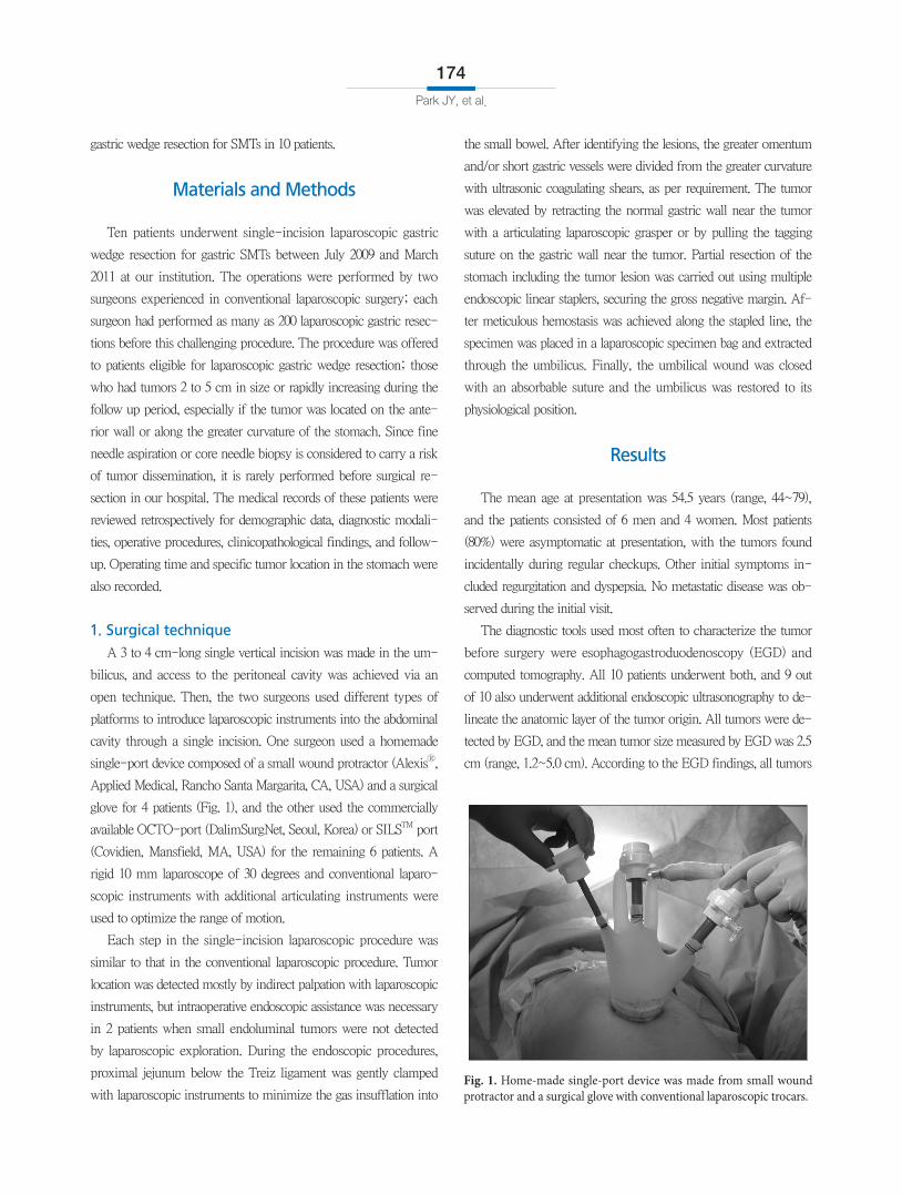
single-port device composed of a small wound protractor (AlexisⓇ,
Applied Medical, Rancho Santa Margarita, CA, USA) and a surgical
glove for 4 patients (Fig. 1), and the other used the commercially
available OCTO-port (DalimSurgNet, Seoul, Korea) or SILSTM port
(Covidien, Mansfield, MA, USA) for the remaining 6 patients. A
rigid 10 mm laparoscope of 30 degrees and conventional laparo-
scopic instruments with additional articulating instruments were
used to optimize the range of motion.
Each step in the single-incision laparoscopic procedure was
similar to that in the conventional laparoscopic procedure. Tumor
location was detected mostly by indirect palpation with laparoscopic
instruments, but intraoperative endoscopic assistance was necessary
in 2 patients when small endoluminal tumors were not detected
by laparoscopic exploration. During the endoscopic procedures,
proximal jejunum below the Treiz ligament was gently clamped
with laparoscopic instruments to minimize the gas insufflation into
the small bowel. After identifying the lesions, the greater omentum
and/or short gastric vessels were divided from the greater curvature
with ultrasonic coagulating shears, as per requirement. The tumor
was elevated by retracting the normal gastric wall near the tumor
with a articulating laparoscopic grasper or by pulling the tagging
suture on the gastric wall near the tumor. Partial resection of the
stomach including the tumor lesion was carried out using multiple
endoscopic linear staplers, securing the gross negative margin. Af-
ter meticulous hemostasis was achieved along the stapled line, the
specimen was placed in a laparoscopic specimen bag and extracted
through the umbilicus. Finally, the umbilical wound was closed
with an absorbable suture and the umbilicus was restored to its
physiological position.
Results
The mean age at presentation was 54.5 years (range, 44~79),
and the patients consisted of 6 men and 4 women. Most patients
(80%) were asymptomatic at presentation, with the tumors found
incidentally during regular checkups. Other initial symptoms in-
cluded regurgitation and dyspepsia. No metastatic disease was ob-
served during the initial visit.
The diagnostic tools used most often to characterize the tumor
before surgery were esophagogastroduodenoscopy (EGD) and
computed tomography. All 10 patients underwent both, and 9 out
of 10 also underwent additional endoscopic ultrasonography to de-
lineate the anatomic layer of the tumor origin. All tumors were de-
tected by EGD, and the mean tumor size measured by EGD was 2.5
cm (range, 1.2~5.0 cm). According to the EGD findings, all tumors
Fig. 1. Home-made single-port device was made from small wound protractor and a surgical glove with conventional laparoscopic trocars.
SILS for Gastric SMT
175
were located in the body of the stomach (4 at angle, 4 in the lower
body, and the other 2 in the mid-body). Among the 10 gastric
SMTs located in the body of stomach, 4 tumors were localized on
the anterior wall, 2 were localized on the posterior wall, and 4 were
localized along the greater curvature. Eight of 10 patients under-
went routine intraluminal endoscopic biopsy, but all failed to obtain
significant microscopic diagnosis other than chronic gastritis.
Single-incision laparoscopic gastric wedge resection was suc-
cessfully completed in 9 patients without conversion to conven-
tional laparoscopic surgery. In 1 patient, moderate hemorrhagic
event occurred during cholelcystectomy after the successful gastric
resection, and 2 additional trocars were needed to safely perform
simultaneous cholecystectomy for comorbid gall bladder stones. All
tumors were resected via an extraluminal approach. The mean op-
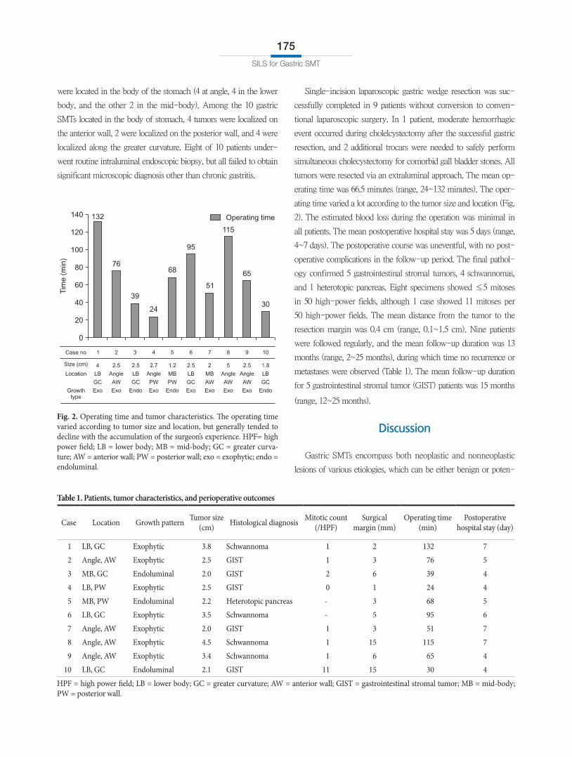
erating time was 66.5 minutes (range, 24~132 minutes). The oper-
ating time varied a lot according to the tumor size and location (Fig.
2). The estimated blood loss during the operation was minimal in
all patients. The mean postoperative hospital stay was 5 days (range,
4~7 days). The postoperative course was uneventful, with no post-
operative complications in the follow-up period. The final pathol-
ogy confirmed 5 gastrointestinal stromal tumors, 4 schwannomas,
and 1 heterotopic pancreas. Eight specimens showed ≤5 mitoses
in 50 high-power fields, although 1 case showed 11 mitoses per
50 high-power fields. The mean distance from the tumor to the
resection margin was 0.4 cm (range, 0.1~1.5 cm). Nine patients
were followed regularly, and the mean follow-up duration was 13
months (range, 2~25 months), during which time no recurrence or
metastases were observed (Table 1). The mean follow-up duration
for 5 gastrointestinal stromal tumor (GIST) patients was 15 months
(range, 12~25 months).
Discussion
Gastric SMTs encompass both neoplastic and nonneoplastic
lesions of various etiologies, which can be either benign or poten-
Fig. 2. Operating time and tumor characteristics. The operating time varied according to tumor size and location, but generally tended to decline with the accumulation of the surgeon’s experience. HPF= high power field; LB = lower body; MB = mid-body; GC = greater curva-ture; AW = anterior wall; PW = posterior wall; exo = exophytic; endo = endoluminal.
Table 1. Patients, tumor characteristics, and perioperative outcomes
Case Location Growth pattern Tumor size (cm) Histological diagnosis Mitotic count
(/HPF)Surgical
margin (mm)Operating time
(min)Postoperative
hospital stay (day)
1 LB, GC Exophytic 3.8 Schwannoma 1 2 132 7
2 Angle, AW Exophytic 2.5 GIST 1 3 76 5
3 MB, GC Endoluminal 2.0 GIST 2 6 39 4
4 LB, PW Exophytic 2.5 GIST 0 1 24 4
5 MB, PW Endoluminal 2.2 Heterotopic pancreas - 3 68 5
6 LB, GC Exophytic 3.5 Schwannoma - 5 95 6
7 Angle, AW Exophytic 2.0 GIST 1 3 51 7
8 Angle, AW Exophytic 4.5 Schwannoma 1 15 115 7
9 Angle, AW Exophytic 3.4 Schwannoma 1 6 65 4
10 LB, GC Endoluminal 2.1 GIST 11 15 30 4
HPF = high power field; LB = lower body; GC = greater curvature; AW = anterior wall; GIST = gastrointestinal stromal tumor; MB = mid-body; PW = posterior wall.
Park JY, et al.
176
tially malignant. Although endoscopy can accurately identify these
lesions, it is difficult to arrive at a preoperative histological diagnosis
through routine intraluminal endoscopic biopsy. Preoperative fine-
needle aspiration biopsy and core needle biopsy under endoscopic
ultrasonographic guidance might be useful diagnostic tools, but
are not applicable in all cases and can also lead to tumor cell dis-
semination.(11) Furthermore, even with needle biopsy specimen, it
remains difficult to predict whether the tumor poses malignant po-
tential to metastasize to distant organs.(12) Thus, without clear evi-
dence of benign features, SMTs should be considered GISTs, which
accounts for the majority of gastric SMTs, and should be resected
to arrive at a definite pathological diagnosis and ensure removal of
the lesion.
In recent years, laparoscopic wedge resection has been regarded
as safe and technically feasible with favorable oncologic outcomes
when performed by skilled surgeons.(11,13-16) Generally accepted
indications for laparoscopic management of gastric SMTs include
tumors between 2 and 5 cm in diameter, rapid increase in tumor
size during endoscopic surveillance, and presence of symptoms.
Incidentally discovered SMTs less than 2 cm in diameter are usu-
ally followed up by endoscopy or computed tomography every 6
months to 12 months. For patients with rapidly growing tumors
suspected to possess malignant potential, surgical resection is
strongly recommended regardless of tumor size.(11,14,16-18) Of
the patients for whom laparoscopic wedge resection is indicated,
those who have suitable tumor locations and characteristics can
undergo a SILS approach.
The perioperative results in our patients were thought to be
comparable to those in the previously reported laparoscopic series.
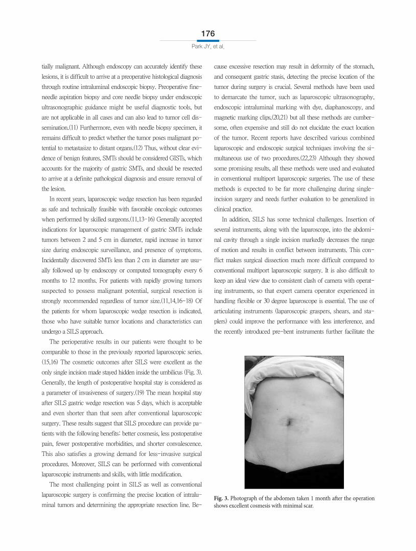
(15,16) The cosmetic outcomes after SILS were excellent as the
only single incision made stayed hidden inside the umbilicus (Fig. 3).
Generally, the length of postoperative hospital stay is considered as
a parameter of invasiveness of surgery.(19) The mean hospital stay
after SILS gastric wedge resection was 5 days, which is acceptable
and even shorter than that seen after conventional laparoscopic
surgery. These results suggest that SILS procedure can provide pa-
tients with the following benefits: better cosmesis, less postoperative
pain, fewer postoperative morbidities, and shorter convalescence.
This also satisfies a growing demand for less-invasive surgical
procedures. Moreover, SILS can be performed with conventional
laparoscopic instruments and skills, with little modification.
The most challenging point in SILS as well as conventional
laparoscopic surgery is confirming the precise location of intralu-
minal tumors and determining the appropriate resection line. Be-
cause excessive resection may result in deformity of the stomach,
and consequent gastric stasis, detecting the precise location of the
tumor during surgery is crucial. Several methods have been used
to demarcate the tumor, such as laparoscopic ultrasonography,
endoscopic intraluminal marking with dye, diaphanoscopy, and
magnetic marking clips,(20,21) but all these methods are cumber-
some, often expensive and still do not elucidate the exact location
of the tumor. Recent reports have described various combined
laparoscopic and endoscopic surgical techniques involving the si-
multaneous use of two procedures.(22,23) Although they showed
some promising results, all these methods were used and evaluated
in conventional multiport laparoscopic surgeries. The use of these
methods is expected to be far more challenging during single-
incision surgery and needs further evaluation to be generalized in
clinical practice.
In addition, SILS has some technical challenges. Insertion of
several instruments, along with the laparoscope, into the abdomi-
nal cavity through a single incision markedly decreases the range
of motion and results in conflict between instruments. This con-
flict makes surgical dissection much more difficult compared to
conventional multiport laparoscopic surgery. It is also difficult to
keep an ideal view due to consistent clash of camera with operat-
ing instruments, so that expert camera operator experienced in
handling flexible or 30 degree laparoscope is essential. The use of
articulating instruments (laparoscopic graspers, shears, and sta-
plers) could improve the performance with less interference, and
the recently introduced pre-bent instruments further facilitate the
Fig. 3. Photograph of the abdomen taken 1 month after the operation shows excellent cosmesis with minimal scar.
SILS for Gastric SMT
177
SILS procedure with lower time requirement and better maneuver-
ability.(24) Adopting right-angle light cable adaptor which makes
light cable run parallel with laparoscope and a long-shaft camera
would further reduce external collisions.(25) Nevertheless, a learn-
ing curve for these instruments is expected before their practical
use, and further development of instrumentation is required for
the widespread use of this new technique in more complicated
procedures. It should be noted that SILS can be easily converted
into conventional laparoscopic surgery by adding a few extra ports.
Some investigators introduced 2~3 mm instruments, which serves
as atraumatic retractors to secure a better surgical view and still
leaves no scar after surgery(9); this could be an alternative solution
for better surgical performance during SILS.
A small extension of the transumbilical incision can be consid-
ered for difficult-to-access tumors. Transumbilical incision with
slight extension in SILS can be used to exteriorize the lesion for
resection when the tumor is hard to access or manipulate in the
abdominal cavity; this would further facilitate SILS in many cases.
This study has several limitations. The number of patients in-
cluded in the present study was small, and the comparative data
between SILS and conventional laparoscopic approach were not
presented. Additional prospective randomized controlled trials are
needed to examine its feasibility and safety. The participating two
surgeons utilized different types of platform to perform single-
incisional wedge resection in the present study; this would have
influenced the surgical performance and serve as a limitation of the
present study.
In conclusion, gastric wedge resection using SILS for gastric
SMTs is safe and feasible when performed by experienced laparo-
scopic surgeons. This scar-free abdominal procedure is a promis-
ing alternative method for the treatment of gastric SMTs, especially
those located at the anterior wall or along the greater curvature of
the stomach. However, careful selection of patients based on tumor
location and a standardized method for tumor localization should
precede the widespread use of this new technique. Although the
immediate benefit appears to be cosmetic, potential benefits and
disadvantages require further evaluation. Prospective randomized
studies comparing SILS and conventional laparoscopic surgery for
gastric wedge resection are necessary to confirm the benefits of this
new technique.
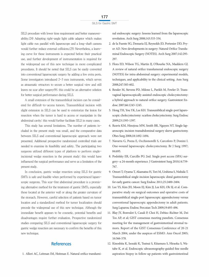
References
1. Allori AC, Leitman IM, Heitman E. Natural orifice translumi-
nal endoscopic surgery: lessons learned from the laparoscopic revolution. Arch Surg 2008;143:333-334.
2. de la Fuente SG, Demaria EJ, Reynolds JD, Portenier DD, Pry-or AD. New developments in surgery: Natural Orifice Translu-minal Endoscopic Surgery (NOTES). Arch Surg 2007;142:295-297.
3. Flora ED, Wilson TG, Martin IJ, O'Rourke NA, Maddern GJ. A review of natural orifice translumenal endoscopic surgery (NOTES) for intra-abdominal surgery: experimental models, techniques, and applicability to the clinical setting. Ann Surg 2008;247:583-602.
4. Bessler M, Stevens PD, Milone L, Parikh M, Fowler D. Trans-vaginal laparoscopically assisted endoscopic cholecystectomy: a hybrid approach to natural orifice surgery. Gastrointest En-dosc 2007;66:1243-1245.
5. Hong TH, You YK, Lee KH. Transumbilical single-port laparo-scopic cholecystectomy: scarless cholecystectomy. Surg Endosc 2009;23:1393-1397.
6. Reavis KM, Hinojosa MW, Smith BR, Nguyen NT. Single-lap-aroscopic incision transabdominal surgery sleeve gastrectomy. Obes Surg 2008;18:1492-1494.
7. Navarra G, Pozza E, Occhionorelli S, Carcoforo P, Donini I. One-wound laparoscopic cholecystectomy. Br J Surg 1997; 84:695.
8. Podolsky ER, Curcillo PG 2nd. Single port access (SPA) sur-gery--a 24-month experience. J Gastrointest Surg 2010;14:759-767.
9. Omori T, Oyama T, Akamatsu H, Tori M, Ueshima S, Nishida T. Transumbilical single-incision laparoscopic distal gastrectomy for early gastric cancer. Surg Endosc 2011;25:2400-2404.
10. Lee YS, Kim JH, Moon EJ, Kim JJ, Lee KH, Oh SJ, et al. Com-parative study on surgical outcomes and operative costs of transumbilical single-port laparoscopic appendectomy versus conventional laparoscopic appendectomy in adult patients. Surg Laparosc Endosc Percutan Tech 2009;19:493-496.
11. Blay JY, Bonvalot S, Casali P, Choi H, Debiec-Richter M, Dei Tos AP, et al; GIST consensus meeting panelists. Consensus meeting for the management of gastrointestinal stromal tu-mors. Report of the GIST Consensus Conference of 20-21 March 2004, under the auspices of ESMO. Ann Oncol 2005; 16:566-578.
12. Kinoshita K, Isozaki K, Tsutsui S, Kitamura S, Hiraoka S, Wa-tabe K, et al. Endoscopic ultrasonography-guided fine needle aspiration biopsy in follow-up patients with gastrointestinal
Park JY, et al.
178
stromal tumours. Eur J Gastroenterol Hepatol 2003;15:1189-1193.
13. DeMatteo RP, Lewis JJ, Leung D, Mudan SS, Woodruff JM, Brennan MF. Two hundred gastrointestinal stromal tumors: recurrence patterns and prognostic factors for survival. Ann Surg 2000;231:51-58.
14. Otani Y, Furukawa T, Yoshida M, Saikawa Y, Wada N, Ueda M, et al. Operative indications for relatively small (2-5 cm) gastro-intestinal stromal tumor of the stomach based on analysis of 60 operated cases. Surgery 2006;139:484-492.
15. Huguet KL, Rush RM Jr, Tessier DJ, Schlinkert RT, Hinder RA, Grinberg GG, et al. Laparoscopic gastric gastrointestinal stro-mal tumor resection: the mayo clinic experience. Arch Surg 2008;143:587-590.
16. Sasaki A, Koeda K, Obuchi T, Nakajima J, Nishizuka S, Ter-ashima M, et al. Tailored laparoscopic resection for suspected gastric gastrointestinal stromal tumors. Surgery 2010;147:516-520.
17. Ryu KJ, Jung SR, Choi JS, Jang YJ, Kim JH, Park SS, et al. Lapa-roscopic resection of small gastric submucosal tumors. Surg Endosc 2011;25:271-277.
18. Demetri GD, von Mehren M, Antonescu CR, DeMatteo RP, Ganjoo KN, Maki RG, et al. NCCN Task Force report: update on the management of patients with gastrointestinal stromal tumors. J Natl Compr Canc Netw 2010;8 Suppl 2:S1-41.
19. Kitano S, Shiraishi N, Fujii K, Yasuda K, Inomata M, Adachi Y.
A randomized controlled trial comparing open vs laparoscopy-assisted distal gastrectomy for the treatment of early gastric cancer: an interim report. Surgery 2002;131:S306-311.
20. Santambrogio R, Montorsi M, Schubert L, Pisani Cer-etti A, Costa M, Moroni E, et al. Laparoscopic ultrasound-guided resection of gastric submucosal tumors. Surg Endosc 2006;20:1305-1307.
21. Patrzyk M, Schreiber A, Heidecke CD, Glitsch A. Laser-sup-ported diaphanoscopy: an innovative technique for locating gastric stromal tumors in gastroscopic-laparoscopic rendez-vous: a case series. Endoscopy 2009;41:1090-1094.
22. Hiki N, Yamamoto Y, Fukunaga T, Yamaguchi T, Nunobe S, Tokunaga M, et al. Laparoscopic and endoscopic cooperative surgery for gastrointestinal stromal tumor dissection. Surg En-dosc 2008;22:1729-1735.
23. Tanabe K, Urabe Y, Tokumoto N, Suzuki T, Yamamoto H, Oka S, et al. A new method for intraluminal gastrointestinal stromal tumor resection using laparoscopic seromuscular dissection technique. Dig Surg 2010;27:461-465.
24. Stolzenburg JU, Kallidonis P, Oh MA, Ghulam N, Do M, Haef-ner T, et al. Comparative assessment of laparoscopic single-site surgery instruments to conventional laparoscopic in laboratory setting. J Endourol 2010;24:239-245.
25. Ragupathi M, Nieto J, Haas EM. Pearls and pitfalls in SILS col-ectomy. Surg Laparosc Endosc Percutan Tech 2012;22:183-188.