Translational genomics of sinonasal cancers · Tumor staging of sinonasal tumors is based on...
9
Contents lists available at ScienceDirect Seminars in Cancer Biology journal homepage: www.elsevier.com/locate/semcancer Review Translational genomics of sinonasal cancers Mario A. Hermsen a, ⁎ , Cristina Riobello a , Rocío García-Marín a , Virginia N. Cabal a , Laura Suárez-Fernández a , Fernando López b , José L. Llorente b a Dept. Head and Neck Oncology, Instituto de Investigación Sanitaria del Principado de Asturias (ISPA), Oviedo, Spain b Dept. Otolaryngology, Hospital Universitario Central de Asturias, Oviedo, Spain ARTICLE INFO Keywords: Sinonasal cancer Genetic analysis Diagnosis Therapeutic markers ABSTRACT The sinonasal cavities harbor a wide variety of histologically distinct cancers, the majority very aggressive with 5-year survival rates between 30–60% and local recurrence as the main cause of death. This is a complex anatomic area, close to structures such the eyes and the brain, which is of special relevance for surgery and postoperative radiotherapy. The low incidence of these rare tumors hampers accumulation of experience with diagnosis and clinical managment as well as knowledge on recurrent genetic aberrations or testing of new treatment strategies. However, recent years have seen a growing number of publications on genetic aberrations providing data that can aid or fine-tune classification and provide molecular targets for treatment with specific inhibitors. In addition, new sinonasal cancer models are created that enable preclinical testing of candidate inhibitor drugs. With more and more novel targeted therapies being developed, options for personalized treatment of sinonasal cancer patients are now opening up. 1. Introduction The sinonasal cavities comprise an anatomical area from which a large number of histologically diverse epithelial, neuroectodermal and mesenchymal neoplasms arise [1], representing approximately 3–5% of all head and neck tumors [2–5]. According to Rarecare, sinonasal tu- mors occur with an incidence of approximately 0.5–1.0 patients/ 100.000 inhabitants (www.rarecare.eu/rarecancers). The most common affected subsites are the nasal cavity, the ethmoid and the maxillary sinus, less frequent are the frontal and sphenoid sinus [2,3]. Squamous cell carcinoma (SNSCC) and intestinal-type adenocarcinoma (ITAC) make up approximately 70% of sinonasal cancers, the other 30% include malignant mucosal melanoma (MMM), olfactory neuro- blastoma (ONB), undifferentiated carcinoma (SNUC), NUT carcinoma, SMARCB1-deficient carcinoma, HPV-related multiphenotypic carci- noma (HMSC), neuroendocrine carcinoma (SNEC), non-intestinal-type adenocarcinoma (SNAC), teratocarcinosarcoma (TCS) and many other subtypes (Table 1). Mesenchymal tumors in the sinonasal cavities may not be distinct from other anatomical localizations and are therefore not included in this review. Based on their anatomical site of origin but also their distinctive etiology, epidemiology, clinical and genetic characteristics, sinonasal tumors should be considered unique entities, apart from other head and neck cancers affecting the nasopharynx, pharynx, larynx and oral cavity [1–3]. Neighboring organs as eyes and brain are a challenge for clinical management, however, recent years have seen advances in endoscopic surgical approaches and precision radiotherapy and imaging techni- ques. The low incidence of sinonasal tumors and their histological di- versity prevents accumulation of clinical experience in diagnosis, clas- sification, staging and treatment at individual hospitals. In addition, as with many rare tumors, the development of new therapeutic options is hampered by the relative scarcity of genetic data and of tumor models for preclinical testing. However, recent years have seen a growing number of genetic studies of sinonasal tumors. This article will present an up-to-date overview of genetic aberrations reported in sinonasal cancer subtypes with an emphasis on those having clinical usefulness in diagnosis, prognosis or therapy. 2. Clinical features Diagnosis of sinonasal tumors is generally late due to nonspecific symptoms similar to inflammatory diseases. The average age of onset is 50–70 years although SNUC, NUT carcinoma, SNAC and ONB also occur at younger ages [1]. Clinical examination should include a complete ear, nose and throat exploration and when malignancy is suspected also imaging tests. Computed Tomography (CT) is better for observing the bone and Magnetic Resonance Imaging (MRI) for soft tissues [6]. Especially in high-grade tumors CT may be replaced by PET- https://doi.org/10.1016/j.semcancer.2019.09.016 Received 27 August 2019; Accepted 22 September 2019 ⁎ Corresponding author. E-mail address: [email protected] (M.A. Hermsen). Seminars in Cancer Biology xxx (xxxx) xxx–xxx 1044-579X/ © 2019 Elsevier Ltd. All rights reserved. Please cite this article as: Mario A. Hermsen, et al., Seminars in Cancer Biology, https://doi.org/10.1016/j.semcancer.2019.09.016
Transcript of Translational genomics of sinonasal cancers · Tumor staging of sinonasal tumors is based on...

Contents lists available at ScienceDirect
Seminars in Cancer Biology
journal homepage: www.elsevier.com/locate/semcancer
Review
Translational genomics of sinonasal cancers
Mario A. Hermsena,⁎, Cristina Riobelloa, Rocío García-Marína, Virginia N. Cabala,Laura Suárez-Fernándeza, Fernando Lópezb, José L. Llorenteb
a Dept. Head and Neck Oncology, Instituto de Investigación Sanitaria del Principado de Asturias (ISPA), Oviedo, SpainbDept. Otolaryngology, Hospital Universitario Central de Asturias, Oviedo, Spain
A R T I C L E I N F O
Keywords:Sinonasal cancerGenetic analysisDiagnosisTherapeutic markers
A B S T R A C T
The sinonasal cavities harbor a wide variety of histologically distinct cancers, the majority very aggressive with5-year survival rates between 30–60% and local recurrence as the main cause of death. This is a complexanatomic area, close to structures such the eyes and the brain, which is of special relevance for surgery andpostoperative radiotherapy. The low incidence of these rare tumors hampers accumulation of experience withdiagnosis and clinical managment as well as knowledge on recurrent genetic aberrations or testing of newtreatment strategies. However, recent years have seen a growing number of publications on genetic aberrationsproviding data that can aid or fine-tune classification and provide molecular targets for treatment with specificinhibitors. In addition, new sinonasal cancer models are created that enable preclinical testing of candidateinhibitor drugs. With more and more novel targeted therapies being developed, options for personalizedtreatment of sinonasal cancer patients are now opening up.
1. Introduction
The sinonasal cavities comprise an anatomical area from which alarge number of histologically diverse epithelial, neuroectodermal andmesenchymal neoplasms arise [1], representing approximately 3–5% ofall head and neck tumors [2–5]. According to Rarecare, sinonasal tu-mors occur with an incidence of approximately 0.5–1.0 patients/100.000 inhabitants (www.rarecare.eu/rarecancers). The mostcommon affected subsites are the nasal cavity, the ethmoid and themaxillary sinus, less frequent are the frontal and sphenoid sinus [2,3].Squamous cell carcinoma (SNSCC) and intestinal-type adenocarcinoma(ITAC) make up approximately 70% of sinonasal cancers, the other 30%include malignant mucosal melanoma (MMM), olfactory neuro-blastoma (ONB), undifferentiated carcinoma (SNUC), NUT carcinoma,SMARCB1-deficient carcinoma, HPV-related multiphenotypic carci-noma (HMSC), neuroendocrine carcinoma (SNEC), non-intestinal-typeadenocarcinoma (SNAC), teratocarcinosarcoma (TCS) and many othersubtypes (Table 1). Mesenchymal tumors in the sinonasal cavities maynot be distinct from other anatomical localizations and are thereforenot included in this review.
Based on their anatomical site of origin but also their distinctiveetiology, epidemiology, clinical and genetic characteristics, sinonasaltumors should be considered unique entities, apart from other head andneck cancers affecting the nasopharynx, pharynx, larynx and oral cavity
[1–3]. Neighboring organs as eyes and brain are a challenge for clinicalmanagement, however, recent years have seen advances in endoscopicsurgical approaches and precision radiotherapy and imaging techni-ques. The low incidence of sinonasal tumors and their histological di-versity prevents accumulation of clinical experience in diagnosis, clas-sification, staging and treatment at individual hospitals. In addition, aswith many rare tumors, the development of new therapeutic options ishampered by the relative scarcity of genetic data and of tumor modelsfor preclinical testing. However, recent years have seen a growingnumber of genetic studies of sinonasal tumors. This article will presentan up-to-date overview of genetic aberrations reported in sinonasalcancer subtypes with an emphasis on those having clinical usefulness indiagnosis, prognosis or therapy.
2. Clinical features
Diagnosis of sinonasal tumors is generally late due to nonspecificsymptoms similar to inflammatory diseases. The average age of onset is50–70 years although SNUC, NUT carcinoma, SNAC and ONB alsooccur at younger ages [1]. Clinical examination should include acomplete ear, nose and throat exploration and when malignancy issuspected also imaging tests. Computed Tomography (CT) is better forobserving the bone and Magnetic Resonance Imaging (MRI) for softtissues [6]. Especially in high-grade tumors CT may be replaced by PET-
https://doi.org/10.1016/j.semcancer.2019.09.016Received 27 August 2019; Accepted 22 September 2019
⁎ Corresponding author.E-mail address: [email protected] (M.A. Hermsen).
Seminars in Cancer Biology xxx (xxxx) xxx–xxx
1044-579X/ © 2019 Elsevier Ltd. All rights reserved.
Please cite this article as: Mario A. Hermsen, et al., Seminars in Cancer Biology, https://doi.org/10.1016/j.semcancer.2019.09.016

CT to assess both the primary tumor and possible distant metastases(Fig. 1). Tumor staging of sinonasal tumors is based on location andextent, according to the current Union for International Cancer Control(UICC) classification system [7]. Lymph node metastasis is infrequent atdiagnosis. After treatment of the primary tumor, 10% of patients withsinonasal tumors develop distant metastasis, however, this seldom oc-curs in absence of locoregional recurrence which in fact is the maincause of sinonasal cancer mortality [8–11].
ITAC and SNSCC are etiologically related to occupational exposureto wood and leather dust, but also industrial compounds such as glues,formaldehyde, chrome, nickel, and various products used in the textileindustry. Tobacco smoking, an important etiological factor in mosthead and neck cancers, does not seem to have a key role in the devel-opment of sinonasal tumors [12–14]. Oncogenic human papillomavirusand Epstein-Barr virus have been implicated in SNSCC and HMSC andwill be discussed in the section on genetic characterization. There is amale predominance in SNSCC (2:1), ITAC (6:1) and TCS (7:1) whichmay be related to the occupational exposure but also to defects in genesresiding on the X chromosome. Chronic inflammation, a recognizedmechanism of tumorigenesis, may also play a role in sinonasal cancerdevelopment through continuous irritation by inhaled dust particlesstimulating the production of reactive oxygen species (ROS) and NFkBexpression [15]. NFkB can upregulate COX2 expression and the cano-nical WNT pathway through inhibition of GSK-3b. In turn, aberrantWNT signalling can stimulate the production of ROS and chronic in-flammation [15,16]. Preliminary evidence in sinonasal carcinomas in-deed indicated elevated NFkB, COX2 and TNFα expression [17,18] anda dominance of the G > A nucleotide transition inflammatory sig-nature in TP53 and KRAS [19–21]. Future sequencing studies should beable to shed more light on tumorigenic processes in sinonasal cancer byin-depth evaluation of mutational signatures.
Complete surgical resection with postoperative radiotherapy is themainstay of sinonasal cancer management, although treatment shouldbe adjusted individually according to tumor stage, histology, patientage, and previous treatments. Minimally invasive endoscopic ap-proaches are increasingly used as they can reduce the number ofcomplications and morbidity associated with surgery [6,22,23]. Theclose proximity of cranial nerves, eyes, internal carotid artery and brainmakes resection of sinonasal tumors with wide margins is not alwayspossible. Therefore, there is a role for systemic therapy in the man-agement of sinonasal cancer [24]. While patients with recurrent diseaseappear to respond better to salvage surgery than chemotherapy, amultimodal treatment strategy with induction chemotherapy, surgeryand radiotherapy may offer the strongest clinical benefit [25]. With theexception of low grade ONB, low grade SNAC and HMSC that appear to
Table 1Incidence and survival rates of sinonasal tumors.
Histological subtype Proportion ofsinonasal cancers
Overall survival
Squamous cell carcinoma 50% 5y: 50%Intestinal-type adenocarcinoma 13% 5y: 36–80%*Malignant mucosal melanoma 4% 5y: 31%Olfactory neuroblastoma 3% 5y: 40%**Undifferentiated carcinoma 5% 5y: 35%SMARCB1-deficient carcinoma < 1% 15 monthsNUT carcinoma < 1% 10 monthsNeuroendocrine carcinoma 3% 5y: 43%HPV-related multiphenotypic
carcinoma< 1% 5y: 100%
Non-intestinal-type adenocarcinoma < 1% 5y: 38%**Teratocarcinosarcoma < 1% 5y: 50%Other ± 20%
Legend. 5y: 5-year; *worst survival for solid and mucinous types, intermediatefor colonic and best for papillary type ITAC; **Survival rate of the high gradetumors.
Fig. 1. Imaging tests for sinonasal cancer. A) CT scan of a tumor in the nasalfossa with frontal and maxillary sinus occupation. B) MRI scan of the samepatient with better delimitation of what is tumor and and what is mucous; thetumor occupies the nasal fossa but there is no involvement of the orbit or thebrain and the frontal and maxillary sinus are occupied by retained mucous. C).ET-CT scan of the same patient showing the tumor in the nasal fossa and ab-sence of metastases in the rest of the body.
M.A. Hermsen, et al. Seminars in Cancer Biology xxx (xxxx) xxx–xxx
2

be fairly indolent [26–29], all sinonasal tumors decribed in this reviewcarry a dismal prognosis, down to a median of 10 months survival forNUT carcinoma [30], or 15 months for SMARCB1-deficient carcinoma[31]. Reported 5-year overall survival rates (Table 1) vary from 31% forMMM, 35% for SNUC, 38% for high-grade SNAC, 41% for high gradeONB, 43% for SNEC, 50% for TCS, 50% for SNSCC and 36–80% forITAC, depending on the subtype [1,32–38].
3. Histological features
All of the sinonasal cavities are lined with pseudostratified re-spiratory mucosa. Secretory glands in the submucosa produce mucousthat captures bacteria and foreign matter which ciliated cells on thesurface sweep toward the pharynx. Immunological defence mechanismsinclude antimicrobial peptides in the mucous layer and leucocytes,lymphocytes and mononuclear cells in the stroma. The roof of the nasalcavity has pseudostratified olfactory epithelium which is similar to therespiratory mucosa except for the presence of olfactory neurons whoseaxons traverse the cribiform plate and transmit signals to the olfactorybulb (Fig. 2). Normal stem cells in the basal layer of the olfactoryepithelium have been studied extensively, especially for their capacityto form new olfactory neurons after tissue damage [39]. Apart fromgenerating new neurons, these same stem cells are also precursors ofnon-neuronal, epithelial cell types. In the respiratory epithelium, stemcells have been shown capable of differentiating into cells with eitherneuroectodermal or mesodermal phenotypes [40].
Sinonasal tumors could be hypothesized to derive from stem cellscapable of undergoing differentiation into various cell types, ratherthan from differentiated, lineage-specific cells that acquire stem-cellproperties. Supporting this suggestion, many sinonasal tumors withmore than one histological appearance have been described, for ex-ample adenoid cystic carcinoma with ITAC or SNUC, or SNSCC withSNEC [41,42]. Furthermore, recurrent tumors sometimes are of a dif-ferent histological type than the original primary tumor, for instanceITAC recurring as SNUC or ONB [43]. Abnormal cell differentiationmay be an early event in malignant transformation, however, to dateonly two premalignant lesions have been described to precede sinonasaltumors: inverted papilloma in 10–25% of SNSCC [44] and 8–28% ofITAC may develop from intestinal metaplasia [45,46].
A common characteristic of all sinonasal epithelial and neu-roendocrine malignant tumors is the fact that a large proportion ofcases show very poor differentiation (Fig. 3), making their diagnosis achallenge for the pathologist, even with the aid of im-munohistochemical markers as pan-cytokeratin, synaptophysin,
chromogranin, enolase, CD56, desmin, S-100 and melan-A [47–50].Many of the tumors described in this review are known as 'small roundblue cell' tumors, including MMM, ONB, SNUC, SNSCC, NUT carcinomaand SNEC. These tumors show several overlapping histologic featureslike growth in sheets or nests, high mitotic rates and necrostic areas,and also exhibit great variation within and between cases [1,50]. Dis-tinguishing features include melanotic pigmentation in MMM, absenceof pancytokeratin and presence of neuroendocrine im-munohistochemical markers in low grade ONB, abrupt foci of squamousdifferentiation within poorly differentiated areas in NUT carcinoma,basaloid or sometimes rhabdoid features in SMARCB1-deficient carci-noma and cribriform areas resembling adenoid cystic carcinoma inHMSC. ITAC by definition displays intestinal differentiation, indeed thefour recognized subtypes, papillar, colonic, solid and mucinous (in-cluding signet ring cells) are also known in gastro-intestinal adeno-carcinomas. Also SNAC is an adenocarcinoma but shows neither sali-vary gland nor intestinal-type features. Low grade SNAC is composed ofcilindrical cells in tubular and cribriform formations while high-gradeSNAC (also named high grade non-ITAC), predominantly have a solidgrowth with occasional glandular structures. Typical for TSC is theadmixture of epithelial, mesenchymal and neuroepithelial elements(Fig. 3).
4. Genetic characteristics
'Diagnostic contamination' in published sinonasal cancer studiesoccurs and hampers the advancement of genetic and translational re-search. Increasingly, cancer types are being diagnosed and sub-classi-fied by specific molecular-genetic features [49–51]. Among sinonasaltumors, examples are NUT carcinoma defined by a chromosomaltranslocation involving the Nuclear protein in testis gene (NUT1) on15q14, SMARCB1-deficient carcinoma defined by the absence ofSMARCB1/INI1 immunohistochemical staining, both formerly diag-nosed as SNUC [30,31], and HMSC defined by the presence of high-riskHPV in the absence of the t(6;9) MYB-NFIB rearrangement, previouslyoften misclassified as high grade adenoid cystic carcinoma [26]. Also inother sinonasal cancer subtypes frequent genetic alterations have beendescribed. Their usefulness in diagnsosis, prognosis and therapy will bediscussed below.
Squamous cell carcinoma (SNSCC). Genome-wide genetic studieshave shown that SNSCC generally carry a wide variety and a largenumber of chromosomal aberrations [37,52]. Recurrent high-levelamplifications at 7p12, 11p13, 11q13, and 17q21 may indicate a rolefor oncogenes EGFR, CD44, CCND1/CTTN, and ERBB2, respectively
Fig. 2. Histological features of normal re-spiratory (A and B) and olfactory mucosa (Cand D) both containing basal, ciliated, andgoblet cells as well as occasional melanocytes.Seromucous secretory glands lie below thesurface in the lamina propria.Immunohistochemical staining with a CK17antibody shows the horizontal basal cells inboth types of mucosa (A and C). Premature andmature olfactory neurons stained with a βIII-tubulin antibody are absent in respiratory (B)and present in olfactory mucosa.
M.A. Hermsen, et al. Seminars in Cancer Biology xxx (xxxx) xxx–xxx
3

[37,52–54]. TP53 mutation has been reported in 70% of tumors [55]. Ina study on a small series of SNSCC, microsatellite instability was foundin 21% (5/21) of cases [56]. A number of recent publications havedescribed EGFR mutations, especially affecting exon 20, in 30–91% ofSNSCC associated with inverted papilloma [44,57–60]. These studiesalso indicate that EGFR-mutated cases carry a better prognosis thanwildtype tumors. Mutations in KRAS and HRAS, encoding downstreamfactors in the EGFR in the EGF signalling pathway, are almost absent inSNSCCs, while no BRAF mutations have been detected [61,62]. HPVinfection has been described in 38% of benign and 31% malignant tu-mors [57,58,63–66]. In 10–24% of inverted papilloma only non-onco-genic HPV types were found [57,58]. Possibly EGFR mutation and HPV
infection represent two different pathways of progression from invertedpapilloma to SNSCC; indeed they appear to occur in a mutually ex-clusive manner [57,58]. Finally, Doescher et al reported an incidence45% (20/44) of EBV in SNSCC associated with more frequent lymphnode or distant metastases [67].
Intestinal-type adenocarcinoma (ITAC). Microarray CGH studiesgenerally found aneuploid cases with complex chromosomal copynumber changes, with the exception of papillary-type ITAC, whichappeared predominantly diploid with few genetic aberrations [68–72].Gains at 1q22–23, 3q28–29, 6p22 and 13q31–33, and losses at4p15–16, 4q32–35 and 10q24 have been associated with worse overallsurvival [71,69–72]. Mutation of the TP53 gene with a frequency of
Fig. 3. Standard haematoxylin and eosin staining showing the histological features of various sinonasal tumor tissues. A) squamous cell carcinoma (SNSCC), B)colonic type intestinal-type adenocarcinoma (ITAC), C) malignant mucosal melanoma (MMM), D) olfactory neuroblastoma (ONB), E) undifferentiated carcinoma(SNUC), F) neuroendocrine carcinoma (SNEC), G) high grade non-intestinal-type adenocarcinoma (SNAC) and H) teratocarcinosarcoma (TCS). Magnification was10× in all images.
M.A. Hermsen, et al. Seminars in Cancer Biology xxx (xxxx) xxx–xxx
4

40–50% may be associated with exposure to wood dust[19,21,24,73–75]. Being a tumor histologically similar to intestinaladenocarcinoma, microsatellite instability and the Wnt pathway havebeen suspected and studied in ITAC. Based on mononucleotide markers,microsatellite instability was observed in 1 of 41 ITAC, suggesting thatthis mechanism of genomic instability does not play an important role,in contrast to intestinal adenocarcinoma [56]. Neither were mutationsobserved in the key Wnt components APC encoding adenomatouspolyposis coli protein or CTNNB1 encoding β‑catenin [74]. However,nuclear expression of β‑catenin has been reported in 31–53% of cases[73,76,77], indicating that activation of the Wnt pathway probablydoes contribute to the development of ITAC. Mutations in EGFR werenot found but KRAS and HRAS were mutated in approximately 15% ofITACs, and BRAF mutations were absent [54,61,74,77–81].
Malignant mucosal melanoma (MMM). Mucosal and uveal mela-nomas are more than cutaneous melanoma frequently aneuploid andcarry more copy number alterations and chromosomal rearrangements[82]. Whole chromosome arm gains of 1q, 6p and 8q occurred re-spectively in 100%, 93% and 57%, and losses of whole arms 9p and 6qin 50% and 10 in 43%. A third of MMM appeared near-diploid andcarried very few copy number changes [83]. A relatively large numberof mutation studies focused on genes known to play a role in cutaneousmelanoma, reporting the following frecuencies: 7–30% NRAS, 0–25%KIT, 8–11% TERT, 3–10% BRAF (in one study 36%), and 7% SF3B1[84–89]. Mutations affecting the TERT promoter are frequent in allmelanoma types [82]. In addition, MAPK signalling pathway activatingtranslocations involving BRAF with fusion partners ZNF767, NFIC,TMEM178B and DGKI have been described in mucosal melanoma (in-cluding head and neck) and may define a new molecular subset [90].
Olfactory neuroblastoma (ONB). In a Affymetrix microarray CGHstudy of 11 ONB, López-Hernández et al reported a markedly low fre-quency of CNAs, with gains in> 35% of cases at 7q, 14q, 18q and 20and losses at 1p, 2p, 3 and 4. Losses were more frequent than gains.Eight of 11 cases carried only whole chromosome CNAs, whereas gainsor losses of segments of chromosomes were rare (Fig. 4). Markedly,
gains at 8q, perhaps the most frequent alteration in all solid tumors,was completely absent in ONB [91]. Similar copy number data wereobtained with analyses by conventional microarray CGH on 12 ONBand methylation microarrays on 42 ONB [92,93]. Lazo de la Vega et alalso reported focal gains involving CCDN1 at 11q13 and FGFR3 at 4p16[94]. Contrary to these results, three previous works had claimed ONBto be tumors with a high level of chromosomal instability involvingdeletions of 1p, 3p/q, 9p, and 10p/q and amplifications of 17q, 17p13,20p, and 22q [95–98]. It is difficult to find a common denominator inthese data, except perhaps for the frequent finding of whole chromo-some events. The heterogeneous map of copy number alterations maybe explained by the difficulty to diagnose ONB correctly, especially thehigh grade tumors. ONB also appear to carry few mutations. Two sets ofsequencing data, both on 20 tumors using 400/560 cancer-related genepanels revealed only two cases with TP53 and two cases with DNMT3Amutation in one study and no recurrent mutations whatsoever in theother [93,94]. Gay et al studied 41 refractory or recurrent ONB sam-ples, many of which treated previously by radio/chemotherapy. Theirresults showed TP53 mutation in 17% of cases while mutations inPIK3CA, NF1, and IDH2 were noted in two cases (5%) each [99].Genome-wide methylation studies revealed a subgroup of cases withlow-grade histology defined by low level CpG methylation [93,100].
Undifferentiated carcinoma (SNUC). Similar to SNSCC and ITAC,most SNUC carry multiple gains and losses involving all chromosomes.Gains and amplifications at 3q26 and 17q23–24 are especially frequentand may indicate a role for transcription factors SOX2 and SOX9 in-volved in cellular differentiation processes in these tumors[91,101–103]. Other hotspots of amplification concern chromosomalbands 8p11 and 8q24 (Fig. 4) with possible candidate oncogenes FGFR1and cMYC [91,104]. An analysis of hotspot cancer related mutations in12 genes (AKT, BRAF, CDK4, CTNNB1, EGFR, FBXW7, JAK2, c-KIT,KRAS, PDGFR, PI3K, VEGF) in 13 tumors revealed no mutations [103].More recently, a study sequencing all coding exons of 300 cancer-re-lated genes in 11 cases showed recurrent genes mutations in IDH2,TP53, PIK3CA, ARID1A, CREBBP, KMT2D, SETD2 and TET2 [101]. A
Fig. 4. Chromosomal copy number profiling. A) Olfactory neuroblastoma exclusively showing gains and losses of whole chromosomes. B) Undifferentiated carcinomashowing a complex karyotype with copy number gains and losses affecting almost all chromosomes and a high-level amplification at chromosome band 8q24.
M.A. Hermsen, et al. Seminars in Cancer Biology xxx (xxxx) xxx–xxx
5
similar study including 10 cases reported recurrent mutations inSOX17, cKIT, SETD2 and IDH2 [102]. The most outstanding mutationconcerned IDH2, in 55% (5/11) of cases, and subsequent publicationsconfirmed the importance of this gene, presenting 49% (26/53) up to82% (14/17) of cases with IDH2 mutation [101,102,105]. It is thoughtthat the abnormal activity of mutated IDH2 causes an accumulation ofthe (R)-2-hydroxygluuotarate (2-HG) oncometabolite thus inducingglobal DNA hypermethylation and interruption of the differentiation oflineage-specific progenitor cells into terminally differentiated cells[106]. Capper et al have recently claimed ‘sinonasal IDH2 carcinoma’ asa new entity apart from IDH2 mutation characterized by high levels ofmethylation [93], however, whether or not IDH2 mutation is a char-acterizing event for a subset of SNUC remains to be seen, because theyhave also been detected in SNEC and high-grade SNAC [102,105].
SMARCB1-deficient carcinoma and NUT carcinoma. These formerSNUC variants appear to have relatively simple diploid karyotypes[30,91,107,108]. SMARCB1 inactivation can be caused by a variety ofmechanisms, including deletions, LOH and mutations, although in si-nonasal tumors no mutations have been reported yet [31]. Four sig-nalling pathways have been reported to be affected by SMARCB1: thep16INK4a-Rb-E2F pathway regulating chromosomal stability, the WNTsignaling pathway shown by WNT/β-catenin overexpression, the Shhsignaling pathway through regulation of GLI1, and repression of Poly-comb genes through elevated expression and recruitment of EZH2[109]. NUT carcinoma harbors a defining chromosomal translocationalteration involving the Nuclear protein in testis gene (NUT1 on15q14), in 70% of cases creating a fusion protein with BRD4 (19p13),and in 6% with BRD3 (9q34.2), NSD3 (8p12), ZNF532 (18q21.32),ZNF592 (15q25.3) and CIC (19q13.2) [30,110,111]. A diffuse NUT1protein expression of> 50% is also accepted for diagnosis [1].
Neuroendocrine carcinoma (SNEC). Copy number profiles withfrequent gains and losses were found in SNEC. Microarray CGH analysisof 18 cases showed hotspot gains occuring in>50% of cases at 1q, 6p,7, 8q, 12, 14, 17q, 18q and 20, and> 35% losses occurred at 5q, 16pand 22q [91]. Very little is published on gene mutations in SNEC. Theonly recurrent mutations presented so far occurred in IDH2 and TP53[102,105,112]. In a tumor showing mixed SNEC and SNSCC differ-entiation, TP53 mutation was only present in the SNEC compartment[41].
HPV-related multiphenotypic carcinoma (HMSC). Also named HPV-related sinonasal adenoid-cystic-like carcinoma, these tumors are de-fined by HPV infection in the absence of the t(6;9) MYB-NFIB re-arrangement. However, variable immunohistochemical staining ofcMYB has been reported [114]. Interestingly, not the well-known on-cogenic HPV types 16 and 18 are involved, but in two thirds of casesHPV 33 and further HPV types 35, 56, 26, and 52 [26,113–115]. To
date, there are no studies that have analyzed genetic abnormalitiesadditional to HPV infection.
Non-intestinal-type adenocarcinoma (SNAC). Recurrent transloca-tions involving ETV6 have been described in low grade SNAC.Andreasen et al first described a recurrent translocation t(12;15)(p13;q25) involving the genes ETV6 and NTRK3 in four cases and an-other translocation between ETV6 and RET [27,116]. This lattertranslocation was previously described in mammary analog of secretorycarcinoma of the salivary gland and in sinonasal secretory carcinoma,however, these are distinct tumor entities [28,117]. Yet anothertranslocation fusing SYN2 and PPARG genes was described by Soonet al [118]. The t(3;3) SYN2-PPARG fusion was previously observed inpulmonary small cell carcinoma. Villatoro et al detected CTNNB1 mu-tations in two cases of SNAC with morular metaplasia and CDX2/β-catenin nuclear expression [119]. Finally, Franchi et al reported BRAFmutations in 2 of 12 low-grade SNAC [78]. It remains to be clarifiedwhether or not all these cases with specific genetic characteristic couldbe considered unique tumor entities, distinct from the SNAC subtype.High-grade SNAC (high grade non-ITAC) have been shown to carryIDH2 mutation in approximately 25% of cases [102,105,112]. Addi-tional recurrent mutations were found in CREBBP, APC and NOTCH1[102].
Teratocarcinosarcoma (TCS). This sinonasal tumor is very rare andhardly any genetic studies are available. Trisomy chromosome 12 wasreported in two publications [120,121]. Birkeland et al found a pa-thogenic mutation in CTNNB1 and accumulation of accumulation of β-catenin protein in the nucleus, particularly in the mesenchymal com-ponent [122].
5. Actionable genetic alterations and preclinical models
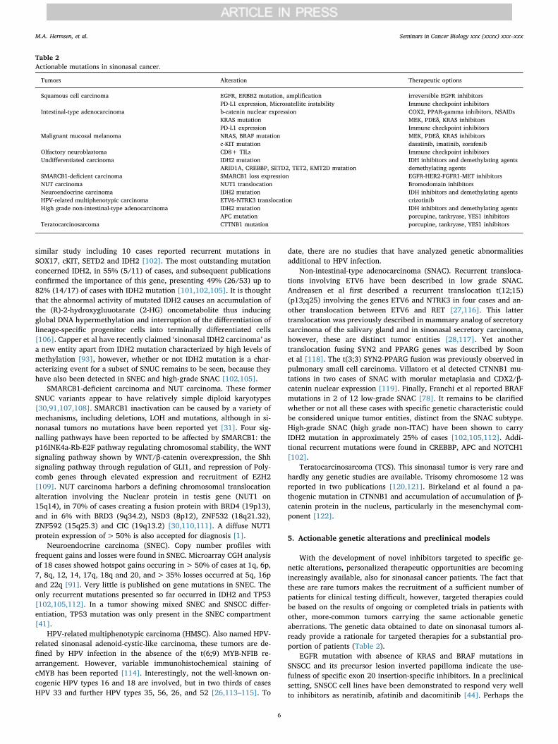
With the development of novel inhibitors targeted to specific ge-netic alterations, personalized therapeutic opportunities are becomingincreasingly available, also for sinonasal cancer patients. The fact thatthese are rare tumors makes the recruitment of a sufficient number ofpatients for clinical testing difficult, however, targeted therapies couldbe based on the results of ongoing or completed trials in patients withother, more-common tumors carrying the same actionable geneticaberrations. The genetic data obtained to date on sinonasal tumors al-ready provide a rationale for targeted therapies for a substantial pro-portion of patients (Table 2).
EGFR mutation with absence of KRAS and BRAF mutations inSNSCC and its precursor lesion inverted papilloma indicate the use-fulness of specific exon 20 insertion-specific inhibitors. In a preclinicalsetting, SNSCC cell lines have been demonstrated to respond very wellto inhibitors as neratinib, afatinib and dacomitinib [44]. Perhaps the
Table 2Actionable mutations in sinonasal cancer.
Tumors Alteration Therapeutic options
Squamous cell carcinoma EGFR, ERBB2 mutation, amplification irreversible EGFR inhibitorsPD-L1 expression, Microsatellite instability Immune checkpoint inhibitors
Intestinal-type adenocarcinoma b-catenin nuclear expression COX2, PPAR-gamma inhibitors, NSAIDsKRAS mutation MEK, PDEδ, KRAS inhibitorsPD-L1 expression Immune checkpoint inhibitors
Malignant mucosal melanoma NRAS, BRAF mutation MEK, PDEδ, KRAS inhibitorsc-KIT mutation dasatinib, imatinib, sorafenib
Olfactory neuroblastoma CD8+ TILs Immune checkpoint inhibitorsUndifferentiated carcinoma IDH2 mutation IDH inhibitors and demethylating agents
ARID1A, CREBBP, SETD2, TET2, KMT2D mutation demethylating agentsSMARCB1-deficient carcinoma SMARCB1 loss expression EGFR-HER2-FGFR1-MET inhibitorsNUT carcinoma NUT1 translocation Bromodomain inhibitorsNeuroendocrine carcinoma IDH2 mutation IDH inhibitors and demethylating agentsHPV-related multiphenotypic carcinoma ETV6-NTRK3 translocation crizotinibHigh grade non-intestinal-type adenocarcinoma IDH2 mutation IDH inhibitors and demethylating agents
APC mutation porcupine, tankryase, YES1 inhibitorsTeratocarcinosarcoma CTTNB1 mutation porcupine, tankryase, YES1 inhibitors
M.A. Hermsen, et al. Seminars in Cancer Biology xxx (xxxx) xxx–xxx
6
most promising compound being tested in an ongoing phase II clinicaltrial on non-small-cell lung cancer is poziotinib [123], which may alsobe effective against exon 20 insertion mutations in ERBB2.
IDH inhibitors may be applied in SNUC, SNEC and high grade SNAC.The FDA recently approved anti-IDH agents Enasidenib and Ivosidenibfor patients with relapsed or refractory acute myeloid leukemia [124].IDH mutations have an impact on global and gene methylation, and sodo other methylation-related genes recurrently mutated in SNUC, suchas ARID1A, CREBBP, SETD2, TET2 and KMT2D. Therefore, demethy-lation-agents such as Decitabine that inhibits DNA methyltransferase tore-activate silent genes and that is approved for treatment of myelo-dysplastic malignancies [125], may also be effective in SNUC.
Wnt pathway activation through alterations in APC or β-cateninoccur in ITAC, low grade SNAC and TCS. There are significant chal-lenges in targeting the Wnt pathway and downstream effectors as theymay not be selective. Porcupine is a Wnt pathway target that isamenable to inhibition while sparing Wnt-dependent tissues. Inhibitorsof porcupine have been shown to block Wnt signaling and tumorgrowth in vivo [126,127] and in metastatic colorectal cancer phase 1/2trials are underway [126,128]. Preclinical studies with tankyrase in-hibitors which target the β-catenin-destruction-complex are promising,but there are currently no trials. Cancers with β-catenin patway acti-vation are dependent on YES-associated protein 1 (YAP1), a transcrip-tion factor involved in stem cell differentiation. Tumor cell line andanimal model studies have shown sensitivity to the multi-kinase in-hibitor dasatinib through the inhibition of YES1 [129]. Mediated byNFkB signalling, the canonical WNT pathway is also related to chronicinflammation, a process that most likely plays a role in sinonasal cancer[17–21]. Therefore, compounds that can reduce ROS production andchronic inflammation could also decrease WNT/β-catenin signalling;candidate drugs are NSAIDs and PPAR-gamma inhibitors [16].
KRAS, NRAS and BRAF mutations have been found in ITAC, MMMand low grade SNAC and may be targets for inhibitors that targetdownstream MEK signalling. Other possibilities include interfering withbinding of mammalian PDEδ to KRAS by means of small molecules[130]. In addition, it may be possible to directly inhibit the RAS(G12C)mutant [131]. Other genetic alterations that may be targeted by specificinhibitors are c-KIT (dasatinib, sorafenib, imatinib), NUT1 (bromodo-main inhibitors), ETV6-NTRK3 fusion (crizotinib) and SMARCB1(EGFR-HER2-FGFR1-MET inhibitors) [30,132,133].
Finally, a number or recent studies on sinonasal cancer have in-dicated a role for anticancer treatment by immune checkpoint in-hibitors. PD-L1 expression on tumor cells has been detected in 32%SNSCC and 17% ITAC [134,135]. Moreover, microsatellite instability,now directly directly eligible for immunotherapy for all cancer types,was shown in 21% of SNSCC [56,77,79]. High numbers of both stromaland intra-tumoral CD8+ tumor infiltrating lymphocytes have beendemonstrated in ONB, especially in high grade tumors, and their pre-sence may serve as response markers for immunotherapeutic strategies[136].
To date, we are unaware of clinical trials involving sinonasal cancerpatients, however, candidate gene targets can first be tested in a pre-clinical setting. In recent years, a number of immortalized sinonasaltumor cell lines have been established and characterized, includingITAC, SNSCC, ONB and SNUC [53,75,137,138]. In addition, mouseorthotopic sinonasal cancer models have been created by implantingSNUC or SNSCC cells into the maxillary sinus of the mouse [138–140].These models retain the biological properties of sinonasal cancer andare important tools for functional studies on processes such as pro-liferation, differentiation, invasion, and metastasis in sinonasal tumors,and might facilitate the development and testing of new therapeuticagents.
6. Conclusion
Sinonasal cancers comprise a large number of histological subtypes
that are difficult to diagnose correctly. In addition, their low incidencehamper the accumulation of experience with surgical, radiotherapeuticand oncological treatment, reason why for best clinical care patientsshould be treated in specialized referral hospitals. The same is true formolecular-genetic research, which would benefit greatly from interna-tional collaboration. Genetic characterization can aid classification andmoreover, identify molecular targets for treatment with specific in-hibitors. With next-generation sequencing every year more accessibleand economic, knowledge on the genetic characteristics of sinonasaltumors is likely to increase considerably in the coming years, while newsinonasal cancer models are created that enable preclinical testing ofcandidate inhibitor drugs. As more and more novel targeted therapiesare being developed, new options for personalized treatment of sino-nasal cancer patients are becoming available.
Declaration of Competing Interest
The authors declare that there are no conflicts of interest.
Acknowledgements
The authors wish to thank Alessandro Franchi, University of Pisa,Italy and Blanca Vivanco, Hospital Universitario Central de Asturias,Spain for their collaboration and highly appreciated experience in thehistopathology of sinonasal cancers.
References
[1] A.K. El-Naggar, et al., WHO Classification of Tumors Pathology and Genetics ofHead and Neck Tumors, Vol, 4th ed., IARC Press, Lyon, 2017.
[2] J.H. Turner, D.D. Reh, Incidence and survival in patients with sinonasal cancer: ahistorical analysis of population-based data, Head Neck 34 (2012) 877–885.
[3] B. Ansa, et al., Paranasal sinus squamous cell carcinoma incidence and survivalbased on Surveillance, Epidemiology, and End Results data, 1973 to 2009, Cancer119 (2013) 2602–2610.
[4] D.R. Youlden, et al., International comparisons of the incidence and mortality ofsinonasal cancer, Cancer Epidemiol. 37 (2013) 770–779.
[5] S. Sanghvi, et al., Epidemiology of sinonasal squamous cell carcinoma: a com-prehensive analysis of 4,994 patients, Laryngoscope 124 (2014) 76–83.
[6] V.J. Lund, et al., European position paper on endoscopic management of tumors ofthe nose, paranasal sinuses and skull base, Rhinol. Suppl. 22 (2010) 1–143.
[7] L.H. Sobin, et al., International Union Against Cancer (UICC) TNM Classification ofMalignant Tumors, 7th ed., Wiley-Blackwell, 2009.
[8] G. Cantu, et al., Anterior craniofacial resection for malignant paranasal tumors: amonoinstitutional experience of 366 cases, Head Neck 34 (2012) 78–87.
[9] F. López, et al., The impact of histologic phenotype in the treatment of sinonasalCancer, Adv. Ther. 34 (2017) 2181–2198.
[10] S.K. Haerle, et al., Sinonasal carcinomas: epidemiology, pathology, and manage-ment, Neurosurg. Clin. N. Am. 24 (2013) 39–49.
[11] J.L. Llorente, et al., Genetic and clinical aspects of wood dust related intestinal-type sinonasal adenocarcinoma: a review, Eur. Arch. Otorhinolaryngol. 266(2009) 1–7.
[12] IARC working group on the evaluation of carcinogenic risks to humans. arsenic,metals, fibres, and dusts, IARC Monogr. Eval. Carcinog. Risks Hum. 100 (2012)11–465.
[13] C. Mensi, et al., Sinonasal cancer and occupational exposure in a population-basedregistry, Int. J. Otolaryngol. 2013 (2013) 672621.
[14] M. Bonzini, et al., Prevalence of occupational hazards in patients with differenttypes of epithelial sinonasal cancers, Rhinology 51 (2013) 31–36.
[15] K. Taniguchi, M. Karin, NF-κB, inflammation, immunity and cancer: coming ofage, Nat. Rev. Immunol. 18 (2018) 309–324.
[16] A. Vallée, Y. Lecarpentier, Crosstalk between peroxisome proliferator-activatedreceptor gamma and the canonical WNT/β-Catenin pathway in chronic in-flammation and oxidative stress during carcinogenesis, Front. Immunol. 9 (2018)745.
[17] R. Holmila, et al., COX-2 and p53 in human sinonasal cancer: COX 2 expression isassociated with adenocarcinoma histology and wood-dust exposure, Int. J. Cancer122 (2008) 2154–2159.
[18] J. Määttä, et al., Characterization of oak and birch dust-induced expression ofcytokines and chemokines in mouse macrophage RAW 264.7 cells, Toxicology 215(2005) 25–36.
[19] F. Perrone, et al., TP53, p14ARF, p16INK4a and H-ras gene molecular analysis inintestinal-type adenocarcinoma of the nasal cavity and paranasal sinuses, Int. J.Cancer 105 (2003) 196–203.
[20] R. Holmila, et al., Mutations in TP53 tumor suppressor gene in wood dust-relatedsinonasal cancer, Int. J. Cancer 127 (2010) 578–588.
[21] J. Pérez-Escuredo, et al., Wood dust-related mutational profile of TP53 in
M.A. Hermsen, et al. Seminars in Cancer Biology xxx (xxxx) xxx–xxx
7

intestinal-type sinonasal adenocarcinoma, Hum. Pathol. 43 (2012) 1894–1901.[22] S.Y. Su, et al., Endoscopic resection of sinonasal cancers, Curr. Oncol. Rep. 16
(2014) 369.[23] P. Castelnuovo, et al., Endoscopic endonasal skull base surgery: past, present and
future, Eur. Arch. Otorhinolaryngol. 267 (2010) 649–663.[24] P. Bossi, et al., TP53 status as guide for the management of ethmoid sinus in-
testinal-type adenocarcinoma, Oral Oncol. 49 (2013) 413–419.[25] E. Orlandi, et al., Locally advanced epithelial sinonasal tumors: the impact of
multimodal approach, Laryngoscope (August 1) (2019), https://doi.org/10.1002/lary.28202 [Epub ahead of print].
[26] J.A. Bishop, et al., HPV-related multiphenotypic sinonasal carcinoma: an ex-panded series of 49 cases of the tumor formerly known as HPV-related carcinomawith adenoid cystic carcinoma-like features, Am. J. Surg. Pathol. 41 (2017)1690–1701.
[27] S. Andreasen, et al., ETV6 gene rearrangements characterize a morphologicallydistinct subset of sinonasal low-grade non–intestinal-type adenocarcinoma: anovel translocation-associated carcinoma restricted to the sinonasal tract, Am. J.Surg. Pathol. 41 (2017) 1552–1560.
[28] M. Baneckova, et al., Mammary analog secretory carcinoma of the nasal cavity,Am. J. Surg. Pathol. 42 (2018) 735–743.
[29] A. Shay, et al., Survival in low-grade and high-grade sinonasal adenocarcinoma: anational cancer database analysis, Laryngoscope (May 13) (2019), https://doi.org/10.1002/lary.28052 [Epub ahead of print].
[30] C. French, NUT midline carcinoma, Nat. Rev. Cancer 14 (2014) 149–150.[31] A. Agaimy, et al., SMARCB1 (INI-1)-deficient sinonasal carcinoma: a series of 39
cases expanding the morphologic and clinicopathologic spectrum of a recentlydescribed entity, Am. J. Surg. Pathol. 41 (2017) 458–471.
[32] K.J. Chambers, et al., Incidence and survival patterns of sinonasal undifferentiatedcarcinoma in the United States, J. Neurol. Surg. B Skull Base 76 (2015) 94–100.
[33] M.K. Bhayani, et al., Sinonasal adenocarcinoma: a 16-year experience at a singleinstitution, Head Neck 36 (2014) 1490–1496.
[34] Wenig BM Undifferentiated malignant neoplasms of the sinonasal tract, Arch.Pathol. Lab. Med. 133 (2009) 699–712.
[35] M. Turri-Zanoni, et al., The clinicopathological spectrum of olfactory neuro-blastoma and sinonasal neuroendocrine neoplasms: refinements in diagnosticcriteria and impact of multimodal treatments on survival, Oral Oncol. 74 (2017)21–29.
[36] T.P. Van der Laan, et al., Meta-analysis of 701 published cases of sinonasal neu-roendocrine carcinoma: the importance of differentiation grade in determiningtreatment strategy, Oral Oncol. 63 (2016) 1–9.
[37] J.L. Llorente, et al., Sinonasal carcinoma: clinical, pathological and genetic ad-vances for new therapeutic opportunities, Nat. Rev. Clin. Oncol. 11 (2014)460–472.
[38] F. Lopez, et al., Current management of sinonasal undifferentiated carcinoma,Rhinology 53 (2015) 212–220.
[39] C.T. Leung, et al., Contribution of olfactory neural stem cells to tissue maintenanceand regeneration, Nat. Neurosci. 10 (2007) 720–726.
[40] S. Hauser, et al., Isolation of novel multipotent neural crest-derived stem cells fromadult human inferior turbinate, Stem Cells Dev. 21 (2012) 742–756.
[41] A. Franchi, et al., Primary combined neuroendocrine and squamous cell carcinomaof the maxillary sinus: report of a case with immunohistochemical and molecularcharacterization, Head Neck Pathol. 9 (2015) 107–113.
[42] S. La Rosa, et al., Mixed exocrine-neuroendocrine carcinoma of the nasal cavity:clinico-pathologic and molecular study of a case and review of the literature, HeadNeck Pathol. 7 (2013) 76–84.
[43] S.Y. Kang, et al., Sinonasal undifferentiated carcinoma and esthesioneuroblastomarecurring as nonintestinal adenocarcinoma, Laryngoscope 23 (2013) 1121–1124.
[44] A.M. Udager, et al., High frequency targetable EGFR mutations in inverted sino-nasal papilloma and associated sinonasal squamous cell carcinoma, Cancer Res. 75(2015) 2600–2606.
[45] B. Vivanco, et al., Benign lesions in mucosa adjacent to intestinal-type sinonasaladenocracinoma, Patholog. Res. Int. 2011 (2011) 230147.
[46] A. Franchi, et al., Intestinal metaplasia of the sinonasal mucosa adjacent to in-testinal-type adenocarcinoma. A morphologic, immunohistochemical, and mole-cular study, Virchows Arch. 466 (2015) 161–168.
[47] S.Y. Su, et al., Esthesioneuroblastoma, neuroendocrine carcinoma, and sinonasalundifferentiated carcinoma: differentiation in diagnosis and treatment, Int. Arch.Otorhinolaryngol. 18 (2014) S149–S156.
[48] D. Bell, et al., Neuroendocrine neoplasms of the sinonasal region, Head Neck 38(Suppl 1) (2016) E2259–E2266.
[49] J.A. Bishop, Recently described neoplasms of the sinonasal tract, Semin. Diagn.Pathol. 33 (2016) 62–70.
[50] L.D. Thompson, Small round blue cell tumors of the sinonasal tract: a differentialdiagnosis approach, Mod. Pathol. 30 (2017) S1–S26.
[51] S. Andreasen, et al., An update on head and neck cancer: new entities and theirhistopathology, molecular background, treatment, and outcome, APMIS 127(2019) 240–264.
[52] F. López, et al., Genomic profiling of sinonasal squamous cell carcinoma, HeadNeck 33 (2011) 145–153.
[53] C. García-Inclán, et al., Establishment and genetic characterization of six uniquetumor cell lines as preclinical models for sinonasal squamous cell carcinoma, Sci.Rep. 4 (2014) 4925.
[54] F. López, et al., Gene amplification and protein overexpression of EGFR andERBB2 in sinonasal squamous cell carcinoma, Cancer 118 (2012) 1818–1826.
[55] R. Holmila, et al., Mutations in TP53 tumor suppressor gene in wood dust-relatedsinonasal cancer, Int. J. Cancer 127 (2010) 578–588.
[56] J.G. Martínez, et al., Microsatellite instability analysis of sinonasal carcinomas,Otolaryngol. Head. Neck Surg. 140 (2009) 55–60.
[57] A.M. Udager, et al., Human papillomavirus (HPV) and somatic EGFR mutationsare essential, mutually exclusive oncogenic mechanisms for inverted sinonasalpapillomas and associated sinonasal squamous cell carcinomas, Ann. Oncol. 29(2018) 466–471.
[58] N. Sahnane, et al., Comprehensive analysis of HPV infection, EGFR exon 20 mu-tations and LINE1 hypomethylation as risk factors for malignant transformation ofsinonasal-inverted papilloma to squamous cell carcinoma, Int. J. Cancer 144(2019) 1313–1320.
[59] E. Sasaki, et al., Sinonasal squamous cell carcinoma and EGFR mutations: a mo-lecular footprint of a benign lesion, Histopathology 73 (2018) 953–962.
[60] Cabal V.N., et al. EGFR mutation and HPV infection in sinonasal inverted pa-pilloma and squamous cell carcinoma. Submitted 2019.
[61] J. Bornholdt, et al., K-ras mutations in sinonasal cancers in relation to wood dustexposure, BMC Cancer 8 (2008) 53.
[62] F. López, et al., KRAS and BRAF mutations in sinonasal cancer, Oral Oncol. 48(2012) 692–697.
[63] K. Syrjänen, S. Syrjänen, Detection of human papillomavirus in sinonasal pa-pillomas: systematic review and meta-analysis, Laryngoscope 123 (2013)181–192.
[64] J.A. Bishop, et al., Human papillomavirus-related carcinomas of the sinonasaltract, Am. J. Surg. Pathol. 37 (2013) 185–192.
[65] A.B. Larque, et al., High-risk human papillomavirus is transcriptionally active in asubset of sinonasal squamous cell carcinomas, Mod. Pathol. 27 (2014) 343–351.
[66] J. Laco, et al., The presence of high-risk human papillomavirus (HPV) E6/E7mRNA transcripts in a subset of sinonasal carcinomas is evidence of involvementof HPV in its etiopathogenesis, Virchows Arch. 467 (2015) 405–415.
[67] J. Doescher, et al., Epstein-Barr virus infection is strictly associated with the me-tastatic spread of sinonasal squamous-cell carcinomas, Oral Oncol. 51 (2015)929–934.
[68] M. Ariza, et al., Comparative genomic hybridization of primary sinonasal adeno-carcinomas, Cancer 100 (2004) 335–341.
[69] D. Korinth, et al., Chromosomal imbalances in wood dust-related adenocarci-nomas of the inner nose and their associations with pathological parameters, J.Pathol. 207 (2005) 207–215.
[70] M.A. Hermsen, et al., Genome-wide analysis of genetic changes in intestinal-typesinonasal adenocarcinoma, Head Neck 31 (2009) 290–297.
[71] J. Pérez-Escuredo, et al., Recurrent DNA copy number alterations in intestinal-typesinonasal adenocarcinoma, Rhinology 54 (2016) 278–286.
[72] A. López-Hernández, et al., Genomic profiling of intestinal-type sinonasal adeno-carcinoma reveals subgroups of patients with distinct clinical outcomes, HeadNeck 40 (2018) 259–273.
[73] A. Franchi, et al., Immunohistochemical investigation of tumorigenic pathways insinonasal intestinal-type adenocarcinoma. A tissue microarray analysis of 62 cases,Histopathology 59 (2011) 98–105.
[74] S.S. Yom, et al., Genetic analysis of sinonasal adenocarcinoma phenotypes: distinctalterations of histogenetic significance, Mod. Pathol. 18 (2005) 315–319.
[75] J. Pérez-Escuredo, et al., Establishment and genetic characterization of an im-mortal tumor cell line derived from intestinal-type sinonasal adenocarcinoma,Cell. Oncol. Dordr. (Dordr) 34 (2011) 23–31.
[76] J.P. Díaz-Molina, et al., Wnt-pathway activation in intestinal-type sinonasal ade-nocarcinoma, Rhinology 49 (2011) 593–599.
[77] B. Perez-Ordonez, et al., Expression of mismatch repair proteins, β‑catenin, andcadherin in intestinal-type sinonasal adenocarcinoma, J. Clin. Pathol. 57 (2004)1080–1083.
[78] A. Franchi, et al., Low prevalence of K-RAS, EGFR and BRAF mutations in sino-nasal adenocarcinomas. Implications for anti-EGFR treatments, Pathol. Oncol. Res.20 (2014) 571–579.
[79] M. Frattini, et al., Phenotype-genotype correlation: challenge of intestinal-typeadenocarcinoma of the nasal cavity and paranasal sinuses, Head Neck 28 (2006)909–915.
[80] C. García-Inclán, et al., EGFR status and KRAS/BRAF mutations in intestinal-typesinonasal adenocarcinomas, Cell. Oncol. Dordr. (Dordr) 35 (2012) 443–450.
[81] F. Projetti, et al., Epidermal growth factor receptor expression and KRAS and BRAFmutations: study of 39 sinonasal intestinal-type adenocarcinomas, Hum. Pathol.44 (2013) 2116–2125.
[82] N.K. Hayward, et al., Whole-genome landscapes of major melanoma subtypes,Nature 545 (2017) 175–180.
[83] M. Van Dijk, et al., Distinct chromosomal aberrations in sinonasal mucosal mel-anoma as detected by comparative genomic hybridization, Genes ChromosomesCancer 36 (2003) 151–158.
[84] A. Zebary, et al., KIT, NRAS and BRAF mutations in sinonasal mucosal melanoma:a study of 56 cases, Br. J. Cancer 109 (2013) 559–564.
[85] M. Amit, et al., Mutation status among patients with sinonasal mucosal melanomaand its impact on survival, Br. J. Cancer 116 (2017) 1564–1571.
[86] M. Colombino, et al., Unexpected distribution of cKIT and BRAF mutations amongsouthern Italian patients with sinonasal melanoma, Dermatology 226 (2013)279–284.
[87] M. Jangard, et al., TERT promoter mutations in sinonasal malignant melanoma: astudy of 49 cases, Melanoma Res. 25 (2014) 185–188.
[88] M. Turri-Zanoni, et al., Sinonasal mucosal melanoma: molecular profile andtherapeutic implications from a series of 32 cases, Head Neck 35 (2013)1066–1077.
[89] J.P. Wroblewska, et al., SF3B1, NRAS, KIT, and BRAF mutation; CD117 and cMYCexpression; and tumoral pigmentation in Sinonasal Melanomas: an analysis with
M.A. Hermsen, et al. Seminars in Cancer Biology xxx (xxxx) xxx–xxx
8

newly found molecular alterations and some population-based molecular differ-ences, Am. J. Surg. Pathol. 43 (2019) 168–177.
[90] H.S. Kim, et al., Oncogenic BRAF fusions in mucosal melanomas activate theMAPK pathway and are sensitive to MEK/PI3K inhibition or MEK/CDK4/6 in-hibition, Oncogene 36 (2017) 3334–3345.
[91] A. López-Hernández, et al., Genetic profiling of poorly differentiated sinonasaltumors, Sci. Rep. 8 (2018) 3998.
[92] U. Bockmuhl, et al., CGH pattern of esthesioneuroblastoma and their metastases,Brain Pathol. 14 (2004) 158–163.
[93] D. Capper, et al., DNA methylation-based reclassification of olfactory neuro-blastoma, Acta Neuropathol. 136 (2018) 255–271.
[94] L. Lazo de la Vega, et al., Comprehensive molecular profiling of olfactory neuro-blastoma identifies potentially targetable FGFR3 amplifications, Mol. Cancer Res.15 (2017) 1551–1557.
[95] H. Holland, et al., Comprehensive cytogenetic characterization of an esthesio-neuroblastoma, Cancer Genet. Cytogenet. 173 (2007) 89–96.
[96] S.H. Riazimand, et al., Analysis of cytogenetic aberrations in esthesioneuro-blastomas by comparative genomic hybridization, Cancer Genet. Cytogenet. 136(2002) 53–57.
[97] M. Guled, et al., Array comparative genomic hybridization analysis of olfactoryneuroblastoma, Mod. Pathol. 21 (2008) 770–778.
[98] R. Valli, et al., Comparative genomic hybridization on microarray (a-CGH) in ol-factory neuroblastoma: analysis of ten cases and review of the literature, GenesChromosomes Cancer 54 (2015) 771–775.
[99] L.M. Gay, et al., Comprehensive genomic profiling of esthesioneuroblastoma re-veals additional treatment options, Oncologist 22 (2017) 834–842.
[100] M. Classe, et al., Integrated multi-omic analysis of esthesioneuroblastomas iden-tifies two subgroups linked to cell ontogeny, Cell Rep. 25 (e5) (2018) 811–821.
[101] V.Y. Jo, et al., Recurrent IDH2 R172X mutations in sinonasal undifferentiatedcarcinoma, Mod. Pathol. 30 (2017) 650–659.
[102] S. Dogan, et al., Frequent IDH2 R172 mutations in undifferentiated and poorly-differentiated sinonasal carcinomas, J. Pathol. 242 (2017) 400–408.
[103] A. Gelbard, et al., Molecular profiling of sinonasal undifferentiated carcinoma,Head Neck 36 (2014) 15–21.
[104] A. Schröck, et al., Fibroblast growth factor receptor-1 as a potential therapeutictarget in sinonasal cancer, Head Neck 36 (2014) 1253-7.
[105] J.K. Mito, et al., Immunohistochemical detection and molecular characterizationof IDH-mutant sinonasal undifferentiated carcinomas, Am. J. Surg. Pathol. 42(2018) 1067–1075.
[106] C. Lu, et al., IDH mutation impairs histone demethylation and results in a block tocell differentiation, Nature 483 (2012) 474–478.
[107] E.M. Jackson, et al., Genomic analysis using high-density single nucleotide poly-morphism-based oligonucleotide arrays and multiplex ligation-dependent probeamplification provides a comprehensive analysis of INI1/SMARCB1 in malignantrhabdoid tumors, Clin. Cancer Res. 15 (2009) 1923–1930.
[108] J.K. Lee, et al., Complex chromosomal rearrangements by single catastrophic pa-thogenesis in NUT midline carcinoma, Ann. Oncol. 28 (2017) 890–897.
[109] K. Kohashi, et al., Reduced expression of SMARCB1/ INI1 protein in synovialsarcoma, Mod. Pathol. 23 (2010) 981–990.
[110] A.A. Alekseyenko, et al., Ectopic protein interactions within BRD4-chromatincomplexes drive oncogenic megadomain formation in NUT midline carcinoma,Proc. Natl. Acad. Sci. U.S.A. 114 (2017) E4184–E4192.
[111] I.M. Schaefer, et al., CIC-NUTM1 fusion: a case which expands the spectrum ofNUT-rearranged epithelioid malignancies, Genes Chromosomes Cancer 57 (2018)446–451.
[112] S. Dogan, et al., DNA methylation-based classification of sinonasal un-differentiated carcinoma, Mod. Pathol. (June 11) (2019), https://doi.org/10.1038/s41379-019-0285-x [Epub ahead of print].
[113] A.A. Shah, et al., Human papillomavirus-related multiphenotypic sinonasal car-cinoma: a case report documenting the potential for very late tumor recurrence,Head Neck Pathol. 12 (2018) 623-8.
[114] S.A. Adamane, et al., Human papillomavirus-related multiphenotypic sinonasalcarcinoma with unique HPV type 52 association: a case report with review ofliterature, Head Neck Pathol. 13 (2019) 331–338.
[115] J.F. Hang, et al., Human papillomavirus-related carcinoma with adenoid cysticlikefeatures: a series of five cases expanding the pathological spectrum,
Histopathology 71 (2017) 887–896.[116] S. Andreasen, et al., The ETV6-RET gene fusion is found in ETV6- rearranged low-
grade sinonasal adenocarcinoma without NTRK3 involvement, Am. J. Surg.Pathol. 42 (2018) 985–988.
[117] A. Skalova, et al., Molecular profiling of mammary analog secretory carcinomarevealed a subset of tumors harboring a novel ETV6-RET translocation: report of10 cases, Am. J. Surg. Pathol. 42 (2018) 234–246.
[118] G.S.T. Soon, et al., A case of nasal low-grade non-intestinal-type adenocarcinomawith aberrant CDX2 expression and a novel SYN2-PPARG gene fusion in a 13-year-old girl, Virchows Arch. 474 (2019) 619–623.
[119] T.M. Villatoro, S.K. Mardekian, Two cases of sinonasal nonintestinal-type adeno-carcinoma with squamoid morules expressing nuclear β-catenin and CDX2: acurious morphologic finding supported by molecular analysis, Case Rep. Pathol.2018 (2018) 8741017.
[120] S. Vranic, et al., Hamartomas, teratomas and teratocarcinosarcomas of the headand neck: report of 3 new cases with clinico-pathologic correlation, cytogeneticanalysis, and review of the literature, BMC Ear Nose Throat Disord. 8 (2008) 8.
[121] J. Thomas, et al., Sinonasal teratocarcinosarcoma with yolk sac elements: a neo-plasm of somatic or germ cell origin? Ann. Diagn. Pathol. 15 (2011) 135-9.
[122] A.C. Birkeland, et al., Pathogenetic analysis of sinonasal teratocarcinosarcomasreveal actionable β-catenin overexpression and a β-catenin mutation, J. Neurol.Surg. B Skull Base 78 (2017) 346–352.
[123] S. Vyse, P.H. Huang, Targeting EGFR exon 20 insertion mutations in non-small celllung cancer, Signal Transduct. Target. Ther. 4 (2019) 5.
[124] E.M. Stein, et al., Enasidenib (AG-221), a potent oral inhibitor of mutant isocitratedehydrogenase 2 (IDH2) enzyme, induces hematologic responses in patients withmyelodysplastic syndromes (MDS), Blood 128 (22) (2016).
[125] S. Turcan, et al., Efficient induction of differentiation and growth inhibition inIDH1 mutant glioma cells by the DNMT inhibitor decitabine, Oncotarget 4 (2013)1729–1736.
[126] N. Krishnamurthy, R. Kurzrock, Targeting the Wnt/beta-catenin pathway incancer: update on effectors and inhibitors, Cancer Treat. Rev. 62 (2018) 50–60.
[127] J. Liu, et al., Targeting wnt-driven cancer through the inhibition of porcupine byLGK974, Proc. Natl. Acad. Sci. U.S.A. 110 (2013) 20224-9.
[128] B.K. Koo, et al., Porcupine inhibitor suppresses paracrine wnt-driven growth ofRnf43; Znrf3-mutant neoplasia, Proc. Natl. Acad. Sci. U.S.A. 112 (2015)7548–7550.
[129] J. Rosenbluh, et al., β-Catenin-driven cancers require a YAP1 transcriptionalcomplex for survival and tumorigenesis, Cell 151 (2012) 1457–1473.
[130] G. Zimmermann, et al., Small molecule inhibition of the KRAS-PDEδ interactionimpairs oncogenic KRAS signalling, Nature 497 (2013) 638–642.
[131] J.P. O’Bryan, Pharmacological targeting of RAS: recent success with direct in-hibitors, Pharmacol. Res. 139 (2019) 503–511.
[132] Roberts, et al., Targetable kinase-activating lesions in Ph-like acute lymphoblasticleukemia, N. Engl. J. Med. 371 (2014) 1005-15.
[133] P.H. Huang, Targeting SWI/SNF mutant cancers with tyrosine kinase inhibitortherapy, Expert Rev. Anticancer Ther. 17 (2017) 1–3.
[134] C. Riobello, et al., Programmed death ligand-1 expression as immunotherapeutictarget in sinonasal cancer, Head Neck 40 (2018) 818–827.
[135] H. Quan, et al., Clinical relevance and significance of programmed death-ligand 1expression, tumor-infiltrating lymphocytes, and p16 status in sinonasal squamouscell carcinoma, Cancer Manag. Res. 11 (2019) 4335–4345.
[136] M. Classe, et al., Evaluating the prognostic potential of the Ki67 index and tumorinfiltrating lymphocytes in olfactory neuroblastoma, Histopathology (July 15)(2019), https://doi.org/10.1111/his.13954 [Epub ahead of print].
[137] P.H. Sorensen, et al., Olfactory neuroblastoma is a peripheral primitive neu-roectodermal tumor related to ewing sarcoma, Proc Natl Acad Sci U. S. A. 93(1996) 1038–1043.
[138] Y. Takahashi, et al., Establishment and characterization of novel cell lines fromsinonasal undifferentiated carcinoma, Clin. Cancer Res. 18 (2012) 6178–6187.
[139] M. Costales, et al., Orthotopic murine model of squamous cell carcinoma of thesinonasal cavities, Head Neck 37 (2015) 1769–1775.
[140] Y. Takahashi, et al., Human epidermal growth factor receptor 2/neu as a noveltherapeutic target in sinonasal undifferentiated carcinoma, Head Neck 38 (Suppl1) (2016) E1926-34.
M.A. Hermsen, et al. Seminars in Cancer Biology xxx (xxxx) xxx–xxx
9