
Transcriptional regulation of male-sterility in 7B-1...
19
RESEARCH ARTICLE Transcriptional regulation of male-sterility in 7B-1 male-sterile tomato mutant Vahid Omidvar 1 *, Irina Mohorianu 2,3 , Tamas Dalmay 3 , Yi Zheng 4 , Zhangjun Fei 4 , Anna Pucci 5 , Andrea Mazzucato 5 , Vendula Večeřova ´ 6 , Michaela Sedla ´ řova 6 , Martin Fellner 1 * 1 Laboratory of Growth Regulators, Centre of the Region Hana ´ for Biotechnological and Agricultural Research, Palacky ´ University and Institute of Experimental Botany AS CR, S ˇ lechtitelů 27, Olomouc-Holice, Czech Republic, 2 School of Computing Sciences, University of East Anglia, Norwich, United Kingdom, 3 School of Biological Sciences, University of East Anglia, Norwich, United Kingdom, 4 Boyce Thompson Institute, Cornell University, Ithaca, NY, United States of America, 5 Department of Agricultural and Forestry Sciences, University of Tuscia, Viterbo, Italy, 6 Department of Botany, Faculty of Science, Palacky ´ University in Olomouc, S ˇ lechtitelů 27, Olomouc-Holice, Czech Republic * [email protected] (VO); [email protected] (MF) Abstract The 7B-1 tomato (Solanum lycopersicum L. cv Rutgers) is a male-sterile mutant with enhanced tolerance to abiotic stress, which makes it a potential candidate for hybrid seed breeding and stress engineering. To underline the molecular mechanism regulating the male-sterility in 7B-1, transcriptomic profiles of the 7B-1 male-sterile and wild type (WT) anthers were studied using mRNA sequencing (RNA-Seq). In total, 768 differentially expressed genes (DEGs) were identified, including 132 up-regulated and 636 down-regu- lated transcripts. Gene ontology (GO) enrichment analysis of DEGs suggested a general impact of the 7B-1 mutation on metabolic processes, such as proteolysis and carbohydrate catabolic process. Sixteen candidates with key roles in regulation of anther development were subjected to further analysis using qRT-PCR and in situ hybridization. Cytological studies showed several defects associated with anther development in the 7B-1 mutant, including unsynchronized anther maturation, dysfunctional meiosis, arrested microspores, defect in callose degradation and abnormal tapetum development. TUNEL assay showed a defect in programmed cell death (PCD) of tapetal cells in 7B-1 anthers. The present study provides insights into the transcriptome of the 7B-1 mutant. We identified several genes with altered expression level in 7B-1 (including beta-1,3 glucanase, GA2oxs, cystatin, cysteine protease, pectinesterase, TA29, and actin) that could potentially regulate anther develop- mental processes, such as meiosis, tapetum development, and cell-wall formation/ degradation. Introduction In flowering plants, male-fertility is a highly regulated process, which requires proper cellular differentiation in anthers and timely regulation of microsporogenesis. Male-sterility on the PLOS ONE | DOI:10.1371/journal.pone.0170715 February 8, 2017 1 / 19 a1111111111 a1111111111 a1111111111 a1111111111 a1111111111 OPEN ACCESS Citation: Omidvar V, Mohorianu I, Dalmay T, Zheng Y, Fei Z, Pucci A, et al. (2017) Transcriptional regulation of male-sterility in 7B-1 male-sterile tomato mutant. PLoS ONE 12(2): e0170715. doi:10.1371/journal.pone.0170715 Editor: Meng-xiang Sun, Wuhan University, CHINA Received: October 12, 2016 Accepted: January 9, 2017 Published: February 8, 2017 Copyright: © 2017 Omidvar et al. This is an open access article distributed under the terms of the Creative Commons Attribution License, which permits unrestricted use, distribution, and reproduction in any medium, provided the original author and source are credited. Data availability statement: The sequencing data are available in the SRA system of the NCBI under accession number GSE85859. Funding: This work was supported by the Operational Programs Education for Competitiveness-European Social Fund, project no. CZ.1.07/2.3.00/30.0004 to MF, and by Ministry of Education, Youth and Sports, project no. LO1204. The funders had no role in study design, data collection and analysis, decision to publish, or preparation of the manuscript. Competing interests: The authors declare they have no competing interests.
Transcript of Transcriptional regulation of male-sterility in 7B-1...

RESEARCH ARTICLE
Transcriptional regulation of male-sterility in
7B-1 male-sterile tomato mutant
Vahid Omidvar1*, Irina Mohorianu2,3, Tamas Dalmay3, Yi Zheng4, Zhangjun Fei4,
Anna Pucci5, Andrea Mazzucato5, Vendula Večeřova6, Michaela Sedlařova6,
Martin Fellner1*
1 Laboratory of Growth Regulators, Centre of the Region Hana for Biotechnological and Agricultural
Research, Palacky University and Institute of Experimental Botany AS CR, Slechtitelů 27, Olomouc-Holice,
Czech Republic, 2 School of Computing Sciences, University of East Anglia, Norwich, United Kingdom,
3 School of Biological Sciences, University of East Anglia, Norwich, United Kingdom, 4 Boyce Thompson
Institute, Cornell University, Ithaca, NY, United States of America, 5 Department of Agricultural and Forestry
Sciences, University of Tuscia, Viterbo, Italy, 6 Department of Botany, Faculty of Science, Palacky University
in Olomouc, Slechtitelů 27, Olomouc-Holice, Czech Republic
* [email protected] (VO); [email protected] (MF)
Abstract
The 7B-1 tomato (Solanum lycopersicum L. cv Rutgers) is a male-sterile mutant with
enhanced tolerance to abiotic stress, which makes it a potential candidate for hybrid seed
breeding and stress engineering. To underline the molecular mechanism regulating the
male-sterility in 7B-1, transcriptomic profiles of the 7B-1 male-sterile and wild type (WT)
anthers were studied using mRNA sequencing (RNA-Seq). In total, 768 differentially
expressed genes (DEGs) were identified, including 132 up-regulated and 636 down-regu-
lated transcripts. Gene ontology (GO) enrichment analysis of DEGs suggested a general
impact of the 7B-1 mutation on metabolic processes, such as proteolysis and carbohydrate
catabolic process. Sixteen candidates with key roles in regulation of anther development
were subjected to further analysis using qRT-PCR and in situ hybridization. Cytological
studies showed several defects associated with anther development in the 7B-1 mutant,
including unsynchronized anther maturation, dysfunctional meiosis, arrested microspores,
defect in callose degradation and abnormal tapetum development. TUNEL assay showed a
defect in programmed cell death (PCD) of tapetal cells in 7B-1 anthers. The present study
provides insights into the transcriptome of the 7B-1 mutant. We identified several genes with
altered expression level in 7B-1 (including beta-1,3 glucanase, GA2oxs, cystatin, cysteine
protease, pectinesterase, TA29, and actin) that could potentially regulate anther develop-
mental processes, such as meiosis, tapetum development, and cell-wall formation/
degradation.
Introduction
In flowering plants, male-fertility is a highly regulated process, which requires proper cellular
differentiation in anthers and timely regulation of microsporogenesis. Male-sterility on the
PLOS ONE | DOI:10.1371/journal.pone.0170715 February 8, 2017 1 / 19
a1111111111
a1111111111
a1111111111
a1111111111
a1111111111
OPENACCESS
Citation: Omidvar V, Mohorianu I, Dalmay T,
Zheng Y, Fei Z, Pucci A, et al. (2017)
Transcriptional regulation of male-sterility in 7B-1
male-sterile tomato mutant. PLoS ONE 12(2):
e0170715. doi:10.1371/journal.pone.0170715
Editor: Meng-xiang Sun, Wuhan University, CHINA
Received: October 12, 2016
Accepted: January 9, 2017
Published: February 8, 2017
Copyright: © 2017 Omidvar et al. This is an open
access article distributed under the terms of the
Creative Commons Attribution License, which
permits unrestricted use, distribution, and
reproduction in any medium, provided the original
author and source are credited.
Data availability statement: The sequencing data
are available in the SRA system of the NCBI under
accession number GSE85859.
Funding: This work was supported by the
Operational Programs Education for
Competitiveness-European Social Fund, project no.
CZ.1.07/2.3.00/30.0004 to MF, and by Ministry of
Education, Youth and Sports, project no. LO1204.
The funders had no role in study design, data
collection and analysis, decision to publish, or
preparation of the manuscript.
Competing interests: The authors declare they
have no competing interests.

other hand has potential application in hybrid seed breeding and understanding its molecular
mechanism is currently an important research topic in plant science [1]. Large number of
male-sterile tomato mutants have been identified, however, in most cases the mutant gene(s)
have not been precisely identified and often mapped only to a large genomic region [1,2,3]. A
polygalacturonase gene is the only well characterized gene known so far, which is responsible
for male-sterile phenotype of ps-2 tomato mutant [1]. Male-sterile tomato mutants with
desired agricultural traits are advantageous for hybrid seed breeding. Male-sterile mutants in
tomato have been classified into functional, structural, and sporogenous classes [4]. For exam-
ple, positional sterile-2 (ps-2) tomato is a functional male-sterile mutant with defected pollen
dehiscence [1]. Stamenless-2 (sl-2) tomato is a structural mutant, which produces abnormal
stamens with aborted microspores [5]. In sporogenous mutants, microsporogenesis could
break down during meiosis, formation of tetrads or separation of microspores. In male-sterile(ms) 3 andms15 tomato mutants, pollen mother cells (PMC) collapse in pre-meiotic anthers
[6]. In ms5 andms1035 (allelic to ms10) tomato mutants, microsporogenesis beaks down at
meiosis due to aberrant regulation of tapetal cells [4,7].
Several genes with key roles in anther development have been characterized in Arabidopsis,among those, SPL/NZZ, EMS1/EXS, and TPD1 are essential for differentiation of anther wall
cells [8–11], andMS1 andMS2 are required for pollen wall formation [11,12]. In rice, GAMYB[13], MYB33/MYB65 [14], DYT1 [15], TDF1 [16], AMS [17,18], MS1 [19], PTC1 [20], TDR-2[21], UDT1 [22], TDR [23], and EAT1 [24] play key roles in tapetum development and regula-
tion of microsporogenesis. Studies in tomato and rapeseed suggest that male-sterility is, in
part, a manifestation of hormonal imbalance in flowers, particularly in stamens [25–27]. Male-
sterility is also known to be regulated by environmental factors, i.e., temperature, and photope-
riod [28,29], and it has been suggested that the effects of these external agents are mediated
through hormonal changes [26].
In most angiosperms, the anther consists of four lobes, each containing four highly special-
ized layers (from outer to inner: epidermis, endothecium, middle layer and tapetum), which
houses the reproductive cells [30]. The tapetal cells play an important physiological role as all
nutritional materials entering the sporogenous cells either passes through or originates from
the tapetum [31]. In addition, the tapetum produces callase, an enzyme which removes the cal-
lose around tetrads. Aberrant regulation of tapetum development has been often associated
with male-sterile anther phenotypes [32]. Tapetum degeneration is proposed to be triggered
by PCD processes during the late stage of pollen development, which in turn provide cellular
contents supporting pollen wall formation and maturation. Rice TDRmutant exhibits delayed
tapetal PCD and retarded degeneration, resulting in male-sterility [32].
The 7B-1 tomato mutant line (Solanum lycopersicum L. cv. Rutgers) was previously described
as a photoperiod-dependent male-sterile line [33,34]. In long days (LD), the 7B-1 flowers are
male-sterile, which produce shrunken stamens with no viable microspores, while in short days
(SD), flowers are fertile, stamens are intact and produce viable pollen. Compared to the WT, the
mutant shows reduced de-etiolation, has higher content of endogenous Abscisic acid (ABA), but
less gibberellins (GAs), indole-3-acetic acid (IAA), and cytokinins (CKs), and is hypersensitive to
exogenous ABA [35–37]. Seed germination and hypocotyl growth in 7B-1mutant are more toler-
ant to various abiotic stresses, especially under blue light [36]. Molecular studies showed defects
in blue light perception and hormonal balance in the 7B-1mutant, associated with a large num-
ber of proteins being differentially expressed between 7B-1 and WT anthers [36,38]. A recent
study by Omidvar and Fellner [39] showed distinct DNA methylation dynamics and transcrip-
tional regulation in response to different light qualities and abiotic stresses between 7B-1 and WT
seedlings. Several microRNAs (miRNAs) with key roles in regulation of anther development,
male-sterility and stress-response in 7B-1 have been identified and characterized [40,41]. With
Transcriptomic analysis of anther development
PLOS ONE | DOI:10.1371/journal.pone.0170715 February 8, 2017 2 / 19
primary effect of the 7B-1mutation yet unknown, studies indicate that modulation of the 7B-1mutation and its effect on the gene expression is coordinated through a complex interplay
between light signalling components, hormonal balance and their crosstalk with miRNAs and
DNA methylation programming, which all collectively tune the downstream gene expression
associated with anther development and male-sterility in 7B-1 anthers.
The aim of our study is to gain a deeper insight into the molecular mechanism of male-ste-
rility and transcriptional regulation of anther developmental processes in 7B-1 anthers. Using
RNA-Seq, we identified a number of genes with potential key roles in regulation of anther
development and microsporogenesis, which were differentially expressed between WT and
7B-1 anthers. Expression profiles of these candidate genes were further investigated at different
developmental stages of 7B-1 anthers using qRT-PCR and in situ hybridization. Cytological
studies showed differences between WT and 7B-1 anthers, including anther structure, callose
deposition and tapetum development.
Materials and methods
Plant materials
7B-1mutant and WT seedlings (Solanum lycopersicum L., cv. Rutgers) were grown in long
days (16/8 h light/dark) in temperature controlled growth chamber. Flower buds at different
developmental stages, including buds smaller than 4–5 mm (pre-meiotic anthers; referred to
as S1), equal to 4–5 mm (meiotic anthers; S2) and bigger than 4–5 mm (post-meiotic anthers;
S3) were collected and anthers were dissected under a stereomicroscope. Stages of flower buds
were selected according to Sheoran et al. [38] and confirmed by analysis of anther squashes.
Gibberelic acid treatment was carried out by spraying (0.1 mM GA3) directly onto the 7B-1buds at the panicle primordium stage and repeated once a week until the buds reached the
length of� 5 mm.
RNA-seq analysis
Total RNA was extracted from WT and 7B-1 anthers at different stages using the RNeasy Plant
Mini Kit (Qiagen). Samples were pooled separately in equimolar ratio and used for construc-
tion of sequencing libraries using the Truseq™ RNA Sample Prep Kit (Illumina, San Diego,
CA, USA). Sequencing was carried out on the Illumina HiSeq™ 2000 platform. Short reads and
low quality bases were trimmed using Trimmomatic [42]. The remaining reads were mapped
to the ribosome RNA database [43] using bowtie [44], allowing up to 3 mismatches and
rRNA-mapping reads were subsequently filtered out. The cleaned reads were then mapped
(allowing 2 mismatches) to the tomato reference genome ITAG v2.5 release using TopHat2
[45]. Read counts were normalized using the FPKM (fragments per kilobase per million)
approach [46]. Differential expression analysis was carried out using NOISeq [47] and pre-
sented as offset fold change (OFC), with an offset of 20 as described by Mohorianu et al. [48].
Genes with log2 (OFC)� 1.5 and probabilities > 0.95 were identified as DEGs. Gene ontolo-
gies were assigned using the Blast2go tool (http://www.blast2go.com/b2ghome). Enrichment
analysis was carried out using PANTHER [49].
Quantitative PCR
qRT-PCR experiments were carried out using the SensiFAST SYBR Lo-ROX kit (Bioline).
First-strand cDNAs were synthesized using the PrimeScript First Strand cDNA Synthesis kit
(Takara). Gene-specific primers are listed in S2 Table. Data normalization was carried out
using the CAC and α-tubulin housekeeping genes (data were shown only for CAC). PCR
Transcriptomic analysis of anther development
PLOS ONE | DOI:10.1371/journal.pone.0170715 February 8, 2017 3 / 19

thermal cycles were set for initial denaturation at 95˚C for 2 min, 40 cycles of 95˚C for 5 s, fol-
lowed by annealing/extension at 60˚C for 20 s. Differential expression values were calculated
as normalized fold changes of expression using the ΔΔCT method [50].
Light microscopy
Cryosections were prepared as described previously [40]. In brief, flower buds were embedded
in Paraplast1 PlusTM and transversal sections of 8 μM thickness were cut using a Leica Ultra-
cut R ultramicrotome (Leica Bensheim, Germany). Callose was detected by staining the tissue
sections with 0.05% (w/v) aniline blue and visualized with fluorescence microscopy (λexc =
330-385nm, λem = 480nm; Olympus BX60). In situ hybridization assay was carried out as pre-
viously described [40]. Oligo-probes (S3 Table) with sequences complementary to the candi-
date genes and murine miR122a (as a negative control) were synthesized and DIG-labelled at
5’-end by Eastport (Eastport, Czech Republic). Probe concentration and hybridization temper-
ature were experimentally optimized to 10 nM and 50˚C, respectively. In situ localization sig-
nals were detected using light microscopy in a colorimetric-based reaction using DIG-specific
antibodies coupled to alkaline phosphatase.
TUNEL assay
Anther sections were washed in PBS (160 mM NaCl, 2.7 mM KCl, 8 mM Na2HPO4, 1.5 mM
KH2PO4) for 5 min and incubated in 20 mg/mL proteinase K in proteinase K buffer (100 mM
Tris-HCl, pH 8.0, and 50 mM EDTA) for 20 min at 37˚C in a humid chamber. Sections were
washed in PBS for 5 min and fixed in 4% (w/v) paraformaldehyde in PBS for 10 min. PBS wash
was repeated twice, each for 5 min. Detection of nuclear DNA fragmentation was performed
using Terminal deoxynucleotidyl transferase (TdT) dUTP Nick-End Labeling (TUNEL) assay
(DeadEnd Fluorometric TUNEL system, Promega) according to the manufacturer’s instruc-
tions. Fluorescence signal in samples was analyzed by fluorescence microscopy (wavelength of
520 ± 20nm; Olympus BX60).
Experimental design and statistical analysis
Experiments were conducted in three biological replicates and arranged in a completely ran-
domized design. Analysis of the variance (ANOVA) and mean comparison using duncan new
multiple range test (DNMRT p = 0.05) were carried out using the SAS software version 9.2.
Results
Callose degradation is perturbed in 7B-1 anthers
We have previously showed that anther maturation in 7B-1was not synchronized and micro-
sporogenesis was impaired partially in some anthers/lobes as evidenced by arrested micro-
spores. In addition, some anthers had abnormal tapetum phenotype, where the tapetal cells
were vacuolated and failed to degenerate [41]. In this study, callose localization was examined
in WT and 7B-1 anthers during meiosis (Fig 1). At the early PMC stage, callose was detected
around the PMCs in both WT and 7B-1 anthers (Fig 1A and 1D). Callose was also detected in
WT and 7B-1meiotic anthers around the tetrads (Fig 1B and 1E). With release of microspores
from the tetrads in WT anthers, callose was completely degraded as evidenced by lack of the
signal, while it persisted around the arrested microspores in 7B-1 anthers (Fig 1C and 1F).
This result showed that callose degradation was perturbed in 7B-1 anthers at the end of meio-
sis, resulting in the arrested microspore phenotype.
Transcriptomic analysis of anther development
PLOS ONE | DOI:10.1371/journal.pone.0170715 February 8, 2017 4 / 19

Aberrant regulation of tapetum PCD in 7B-1 anthers
The PCD in tapetal cells is characterized by cleavage of the nuclear DNA. To test if 7B-1anthers are defective in PCD, we performed the TUNEL (terminal deoxynucleotidyl transfer-
ase–mediated dUTP nick-end labeling) assay (Fig 2). The assay measures nuclear DNA frag-
mentation, which can be visualized directly by fluorescence microscopy. Both WT and 7B-1
Fig 1. Callose deposition in WT (A, B, C) and 7B-1 (D, E, F) anthers. A, D: PMCs at early stage of meiosis. B,
E: tetrad stage. C, F: microspores release stage.
doi:10.1371/journal.pone.0170715.g001
Fig 2. TUNEL assay in WT and 7B-1 anthers. Panels A, B, C, D: WT anthers at PMCs, tetrads, free
binucleate microspores, and mature pollens stages, respectively. Panels E, F, G, H, I: 7B-1 anthers at
PMCs, tetrads, free binucleate microspores, arrested binucleate microspores and mature pollens stages,
respectively. Panels J, K, L, M, N: GA-treated 7B-1 anthers at the same stages as E-I.
doi:10.1371/journal.pone.0170715.g002
Transcriptomic analysis of anther development
PLOS ONE | DOI:10.1371/journal.pone.0170715 February 8, 2017 5 / 19
anthers undergoing meiosis showed TUNEL-negative signal (Fig 2A and 2E), indicating a lack
of DNA fragmentation of nuclei at the PMC stage. At the tetrad stage, the TUNEL-positive sig-
nal was marginally detectable in WT tapetal cells, but not in 7B-1, suggesting the onset of PCD
in WT tapetum (Fig 2B and 2F). At the binucleate microspore stage, strong TUNEL-positive
signal was detected in WT tapetal cells (Fig 2C), while a lack of the signal in 7B-1 tapetum indi-
cated a delay or failure of PCD in these cells (Fig 2G and 2H). At the mature pollen stage,
TUNEL-positive signal was detected in WT anthers in fully degenerated tapetal cells (Fig 2D),
while a weak signal observed in 7B-1 anthers in the vacuolated tapetal cells and collapsed
microspores (Fig 2I). These observations demonstrated that PCD in WT tapetum has com-
menced at the tetrad stage, while in 7B-1 anthers the tapetum was failed to degenerate due to
retardation or defect of PCD.
As mentioned earlier, free microspores could be marginally formed in very few of the 7B-1anthers/lobes, while in most of them, they were arrested and lysed. Strong TUNEL-positive
signal was detected in the arrested microspores, but not in the tapetal cells of either free or
arrested microspores phenotypes (Fig 2G and 2H). To test if GA3 could restore the timely
PCD in 7B-1 tapetal cells, 7B-1 buds were treated with GA3 at the panicle primordium stage.
GA3 restored the PCD of tapetal cells in anthers/lobes, which produced free microspores, but
not in those showing arrested microspores (Fig 2L and 2M). These observations confirmed
that GA is essential for triggering of PCD in 7B-1 tapetal cells.
Expression profiling revealed genes associated with male-sterile
phenotype of 7B-1 anthers
Total RNA from anthers at three developmental stages of pre-meiosis, meiosis, and post-meio-
sis (designated as S1, S2, and S3) were pooled with equimolar ratio and used for construction
of RNA-Seq libraries. Total of 14.1 and 13.9 million raw reads were sequenced for WT and 7B-1 libraries, respectively. After removal of short reads and rRNA matching reads, the clean
reads were mapped (allowing 2 mismatches) to the tomato (cv. Heinz) reference genome
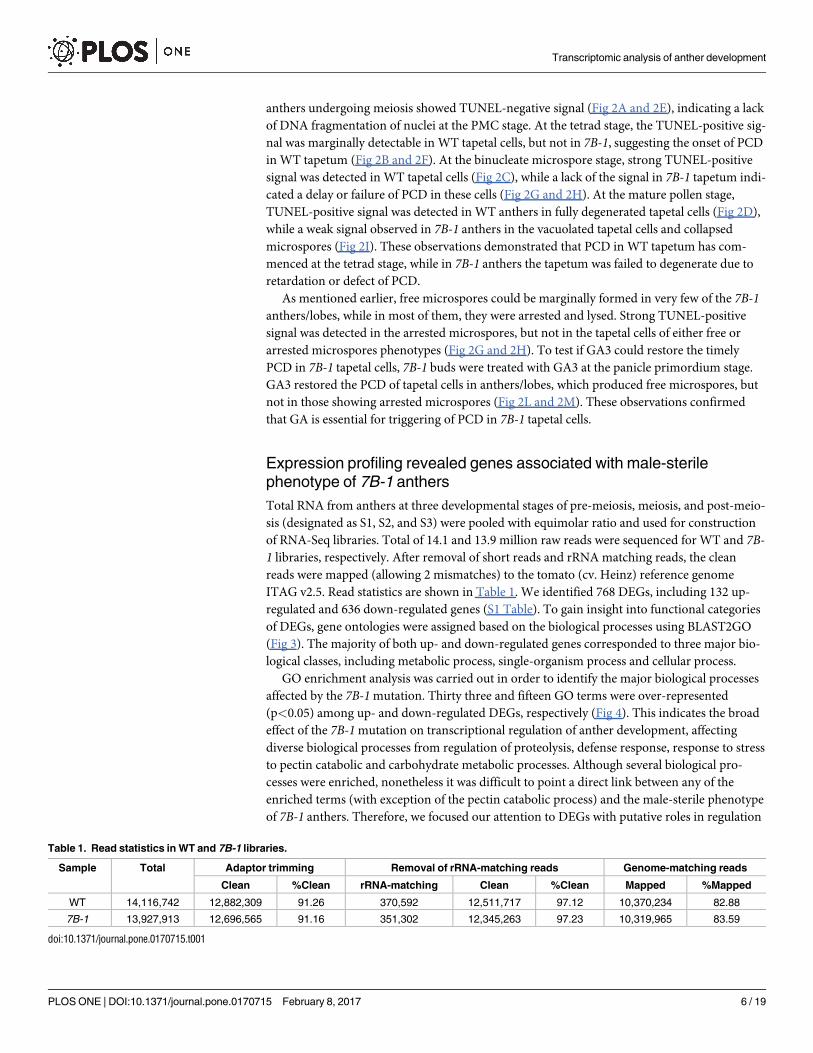
ITAG v2.5. Read statistics are shown in Table 1. We identified 768 DEGs, including 132 up-
regulated and 636 down-regulated genes (S1 Table). To gain insight into functional categories
of DEGs, gene ontologies were assigned based on the biological processes using BLAST2GO
(Fig 3). The majority of both up- and down-regulated genes corresponded to three major bio-
logical classes, including metabolic process, single-organism process and cellular process.
GO enrichment analysis was carried out in order to identify the major biological processes
affected by the 7B-1mutation. Thirty three and fifteen GO terms were over-represented
(p<0.05) among up- and down-regulated DEGs, respectively (Fig 4). This indicates the broad
effect of the 7B-1mutation on transcriptional regulation of anther development, affecting
diverse biological processes from regulation of proteolysis, defense response, response to stress
to pectin catabolic and carbohydrate metabolic processes. Although several biological pro-
cesses were enriched, nonetheless it was difficult to point a direct link between any of the
enriched terms (with exception of the pectin catabolic process) and the male-sterile phenotype
of 7B-1 anthers. Therefore, we focused our attention to DEGs with putative roles in regulation
Table 1. Read statistics in WT and 7B-1 libraries.
Sample Total Adaptor trimming Removal of rRNA-matching reads Genome-matching reads
Clean %Clean rRNA-matching Clean %Clean Mapped %Mapped
WT 14,116,742 12,882,309 91.26 370,592 12,511,717 97.12 10,370,234 82.88
7B-1 13,927,913 12,696,565 91.16 351,302 12,345,263 97.23 10,319,965 83.59
doi:10.1371/journal.pone.0170715.t001
Transcriptomic analysis of anther development
PLOS ONE | DOI:10.1371/journal.pone.0170715 February 8, 2017 6 / 19

of anther development in 7B-1mutant based on their expression, annotation and literature
search. Sixteen candidates (Table 2) with key roles in regulation of meiosis, tapetum develop-
ment, and cell-wall formation/degradation were further examined using qRT-PCR and in situhybridization.
Candidate DEGs were validated using qRT-PCR at different developmental stages of 7B-1anthers (Fig 5). Despite some quantitative differences in the expression levels, qRT-PCR results
showed the same expression pattern as RNA-seq data. Beta-1,3-glucanase was up-regulated in
Fig 3. Gene ontology of DEGs. Up-regulated (A) and down-regulated (B) genes were categorized into different biological classes and
numbers in the parenthesis indicate the frequency of members in each category.
doi:10.1371/journal.pone.0170715.g003
Transcriptomic analysis of anther development
PLOS ONE | DOI:10.1371/journal.pone.0170715 February 8, 2017 7 / 19

Fig 4. GO enrichment analysis of up- (A) and down-regulated (B) DEGs. Biological processes are listed on the Y-axis with their
enrichment folds against all tomato genes (reference) presented on the X-axis. P-values are indicated for each GO term.
doi:10.1371/journal.pone.0170715.g004
Transcriptomic analysis of anther development
PLOS ONE | DOI:10.1371/journal.pone.0170715 February 8, 2017 8 / 19

S1, S2, and more strongly in S3. NAC was up-regulated in all stages. Cystatin and gibberellin2-oxidases (GA2ox) were up-regulated with an increasing pattern during anther maturation.
Pectinesterase, myosin, polygalacturonase, pyruvate dehydrogenase kinase (PDK), beta-galactosi-dase, and zinc finger were down-regulated in S1, S3, and more strongly in S2. Glutamine syn-thetase (GS1) was slightly up-regulated in S1 and S2, but strongly down-regulated in S3. TA29and F-box were down-regulated in S1 and S2, more strongly compared to S3. Actin was down-
regulated in S1, very strongly in S2, but slightly up-regulated in S3. Cysteine protease was
down-regulated S1, S2 and more strongly in S3. MADS-boxwas down-regulated more strongly
in S2 and S3 compared to S1.
Localization profile of DEGs in 7B-1 anthers
Fig 6 shows in situ localization of beta-1,3 glucanase, GA2oxs, TA29, and pectinesterase in WT
and 7B-1 anthers. Beta-1,3 glucanase and GA2oxs were expressed in WT tapetum and binucle-
ate microspores (Fig 6A and 6C), and more strongly in 7B-1 vacuolated tapetum and arrested
microspores (Fig 6B and 6D). In WT anthers, TA29 transcripts were localized in the tapetum,
tetrads (Fig 6E), and the binucleate microspores (Fig 6F), while in 7B-1 anthers, they were
localized in the tapetum, tetrads (Fig 6G), and the arrested microspores (Fig 6H). Pectinesterasetranscripts were localized in the tapetum and the tetrads in both WT and 7B-1 anthers (Fig 6I
and 6J) as well as in the arrested binucleate microspores in 7B-1 anthers (Fig 6K). The murine
miR122a probe was used as negative control, which did not produce any hybridization signal
(Fig 6L).
Discussion
Despite the importance of male-sterility in hybrid seed breeding, the physiological mecha-
nisms, i.e. nutritional, hormonal and environmental, which regulate the male-sterility are not
Table 2. List of DEGs with potential roles in anther development in 7B-1 mutant.
GeneID Normalized reads Statistics Annotation
WT 7B-1 DE P-value
Solyc10g079860.1.1 3.73 34.55 3.21 0.99 Beta-1,3-glucanase
Solyc04g005610.2.1 11.45 54.70 2.26 0.98 NAC transcription factor
Solyc00g071180.2.1 239.56 891.96 1.90 0.97 Cystatin
Solyc01g079200.2.1 32.79 152.49 1.70 0.98 Gibberellin 2-oxidase
Solyc05g052110.2.1 60.99 9.35 -2.74 0.99 Pectinesterase
Solyc06g008530.1.1 23.17 0.87 -4.64 0.99 Myosin XI
Solyc07g044870.2.1 358.24 13.27 -4.64 1.00 Polygalacturonase
Solyc12g098930.1.1 24.02 0.79 -5.06 0.99 Pyruvate dehydrogenase kinase
Solyc05g051250.2.1 271.66 8.71 -5.06 1.00 Glutamine synthetase
Solyc02g078370.1.1 307.04 9.23 -5.06 1.00 Anther-specific protein TA29
Solyc10g086460.1.1 291.50 9.83 -5.06 1.00 Actin
Solyc01g111540.2.1 172.06 5.29 -5.06 1.00 Beta-galactosidase
Solyc07g053460.2.1 75.02 1.37 -5.64 1.00 Cysteine proteinase
Solyc06g005180.1.1 33.48 0.63 -5.64 0.99 Zinc finger transcription factor
Solyc06g059970.2.1 204.97 2.67 -6.64 1.00 MADS-box transcription factor
Solyc06g059820.1.1 29.45 0.35 -6.64 0.99 F-box transcription factor
DE is differential expression values, which were calculated as log2-fold changes of the expression. Positive and negative values mean up- and down-
regulation of expression in 7B-1, respectively.
doi:10.1371/journal.pone.0170715.t002
Transcriptomic analysis of anther development
PLOS ONE | DOI:10.1371/journal.pone.0170715 February 8, 2017 9 / 19

Fig 5. qRT-PCR analysis of DEGs in 7B-1 anthers. Expression changes are presented as normalized fold changes (FC) between
7B-1 and WT reference tissue. Positive and negative values indicate up- and down-regulation of the expression, respectively. Two-
fold threshold was considered as a cutoff value for significant changes in the expression. Error bars represent standard errors of three
biological replicates.
doi:10.1371/journal.pone.0170715.g005
Transcriptomic analysis of anther development
PLOS ONE | DOI:10.1371/journal.pone.0170715 February 8, 2017 10 / 19

yet fully understood. Until now, only a small number of genes have been identified that are
specifically involved in this developmental process and the molecular mechanism of genetic
male-sterility is still largely unknown. The transcriptomic profiling in our study showed differ-
ential expression of a large number of genes between WT and 7B-1 anthers. Majority of DEGs
Fig 6. In situ localization of beta-1,3-glucanase, GA2oxs, TA29 and pectinesterase. A and B:
localization of beta-1, 3-glucanase in WT and 7B-1 anthers respectively at binucleate microspores stage. C
and D: GA2ox in WT and 7B-1 anthers at binucleate microspores stage, respectively. E and F: TA29 in WT
anthers at tetrads and binucleate microspores stages, respectively. G and H: TA29 in 7B-1 anthers at tetrads
and arrested binucleate microspores stages, respectively. I, J, K: pectinesterase in WT anthers at tetrads, in
7B-1 anthers at tetrads, and in 7B-1 anthers at arrested binucleate microspores stages, respectively. L:
negative control, where a murine miR122a-specific probe was used to ensure that the experimental staining is
not an artifact.
doi:10.1371/journal.pone.0170715.g006
Transcriptomic analysis of anther development
PLOS ONE | DOI:10.1371/journal.pone.0170715 February 8, 2017 11 / 19

belonged to three major biological classes, including metabolic process, single-organism pro-
cess and cellular process. This indicates that diverse gene regulation pathways are affected by
or involved in the regulation of male-sterility in 7B-1 anthers. Further examination of GO
terms showed enrichment of several biological processes, including those of special interest
related to protein and carbohydrate metabolic processes. Several pectinesterase and pectatelyase-related genes were enriched within down-regulated DEGs, which were further char-
acterized. Enrichment analysis suggested a broad impact of 7B-1mutation primarily on the
metabolism. Sixteen candidates were identified with potential roles in regulation of anther
development and male-sterility in 7B-1 anthers and further characterized in different develop-
mental stages between WT and 7B-1 anthers. These DEGs and their roles are discussed below.
During meiosis, tapetal cells undergo PCD and release beta-1,3-glucanase, which hydroly-
ses the callose from tetrads [51]. Persistent callose or delay in its dissolution could result in col-
lapse of the developing microspores [52]. While callose was no longer detectable in the early
microspore stage in WT anthers, it persisted around the tetrads and newly formed microspores
in 7B-1 anthers, resulting in an arrested-microspore phenotype. A similar phenotype was
observed in male-sterile anthers of Brassica napus, where callose was persistent around the tet-
rads [53]. qRT-PCR analysis showed up-regulation of beta-1,3-glucanase in 7B-1 anthers and
in situ hybridization showed the prominent expression of this enzyme in 7B-1 tapetum at late
stage of meiosis, where tapetal cells were vacuolated but not degenerated. Delay of tapetum
degeneration in 7B-1 anthers could have led to beta-1,3-glucanase build-up level in these cells
as detected by qRT-PCR and in situ hybridization signal, while callose around the newly
formed microspores was not degraded, probably due to lack of the acting enzyme.
Several pectinesterase and pectate lyase-related genes were enriched within down-regulated
DEGs. In addition to pectinesterase, several other cell wall modifying enzymes, including beta-galactosidase, a cellulose-modifying enzyme, and polygalacturonase which is a pectin-modify-
ing enzyme [54,55] were strongly down-regulated in 7B-1meiotic anthers. In qrt1 and qrt2mutants of Arabidopsis thaliana, microspores were arrested as pectin was not degraded in pri-
mary cell walls around tetrads [56]. Pectinesterase transcripts were localized in tapetum, tetrads
and arrested binucleate microspores in 7B-1 anthers. Suppression of the pectin-modifying
enzymes in 7B-1 anthers were more pronounced during meiosis (stage S2), which could have
impaired enzymatic degradation of cell wall pectin around tetrads, resulting in an arrested-
microspores phenotype, similar to those observed in qrtmutants.
Previously, we found that cystatin and cysteine protease were up- and down-regulated in 7B-1 anthers, respectively with a pattern correlated to tapetum degeneration during anther devel-
opment [41]. Similar results were observed using mRNA-seq and qRT-PCR in the present
study. TUNEL assay showed a delay of PCD in 7B-1 tapetal cells. There results strongly suggest
that suppression of cysteine protease could have caused a delay or defect of PCD in tapetal cells.
GA plays an important role in floral organ growth, especially anther development. Tapetum is
an important source of bioactive gibberellins in anthers [57], and alteration of GA level is
often associated with abnormalities in anther development and male-sterility. GA-deficient
mutants of tomato, rice and Arabidopsis exhibited common defects in PCD of tapetal cells,
resulting in a post-meiotic arrest in male-sterile stamens [13,58,59]. In sl-2 tomato mutant
GA3 could restore the male-fertility [5,60]. Application of GA3 also partially restored the
male-fertility in 7B-1 anthers (Omidvar et al., unpublished data). GA2oxs regulates the GA
level through inactivation of endogenous bioactive GAs [61]. 7B-1 seedlings have a lower GA
level compared to WT. Up-regulation of GA2oxs in 7B-1 anthers could have decreased the GA
level in 7B-1 anthers, resulting in a defect in PCD of tapetal cells. Using TUNEL assay, we
showed that application of GA3 restored the PCD of tapetal cells in 7B-1 anthers similar to
those of WT, which suggests that GA3 is likely to regulate the initiation of PCD in tapetal cells.
Transcriptomic analysis of anther development
PLOS ONE | DOI:10.1371/journal.pone.0170715 February 8, 2017 12 / 19

Another gene which has been differentially expressed between WT and 7B-1 anthers was
TA29. It is a tapetal-specific gene in tobacco, and its promoter region has been used for engi-
neering of male-sterility in tobacco as well as other crops [62–65]. Although TA29 is not func-
tionally characterized with respect to regulation of male-sterility, silencing of this gene in
tobacco has resulted in male-sterile transgenic plants, where tapetum was prematurely degen-
erated [65]. In our study TA29 was strongly down-regulated in 7B-1meiotic anthers, where
the TA29 transcripts were predominantly localized in the tapetal cells and tetrads and arrested
binucleate microspores. Down-regulation of TA29 in 7B-1 anthers did not result in premature
degeneration of tapetum, but it could be associated with the defect of PCD in tapetal cells as it
was strongly down-regulated and localized in undegenerated tapetal cells in late meiotic 7B-1anthers.
Aberrant regulation of actin-, tubulin-, and myosin-related genes could disrupt the organi-
zation of actin and microtubules in meiotic cytoskeleton, thus leading to defective cytokinesis
in developing pollens and male-sterility in crops [66,67]. In our study actin andmyosin were
down-regulated in 7B-1 anthers. In addition, actin depolymerizing factors 3/10, and beta-tubu-lin were also down-regulated in 7B-1 anthers (not validated by qRT-PCR). These observations
indicate that the actin cytoskeleton balance may be disturbed in 7B-1 anthers, which could have
directly affected the meiosis and pollen cell wall development. A case study showed that suppres-
sion of pyruvate dehydrogenase kinase in transgenic tobacco has led to tapetum perturbation
and male-sterility [68]. The importance of glutamine synthetase in pollen reproduction has been
shown in rice [69], maize [70], and tobacco [71]. Down-regulation of these two enzymes in 7B-1anthers could also be associated with tapetum perturbation and meiosis break-down. In addition
to the above mentioned genes, several transcription factors, including F-box,MADS-box and zincfinger genes were down-regulated, whileNAC was up-regulated in 7B-1 anthers. Overexpression
of RMF (reduced male fertility) gene, encoding a F-box protein in Arabidopsis caused the delay in
tapetum degeneration and male-sterility [72]. Li et al. [73] showed that suppression of a F-box
protein-encoding gene,OsADF (anther development F-box), perturbed tapetum degeneration
and resulted in male-sterility in rice.MADS-box transcription factors play important roles in floral
organ development, anther dehiscence and pollen maturation [74,75]. Arabidopsis MS1 gene en-
codes PHD-type zinc finger protein, which is redundantly expressed in tapetum and regulates
timely PCD in tapetal cells [11,76]. SeveralNAC transcription factors were differentially expressed
between wild type and male-sterile flower buds of Brassica rapa [77].NACs are key regulators of
secondary wall thickening in anther tissue [78]. Although differential expression of these tran-
scription factors in our study could be associated with the 7B-1mutation and male-sterility phe-
notype, understating the exact function of these genes require further functional analysis.
A number of genes and transcription factors have been identified that control the tape-
tum formation and development [16,17,79–82]. However, little is known about the genetic
basis regulating the PCD of tapetum during pollen development. In Arabidopsis ms1 and
rice tdr male-sterile mutants, tapetum aberrations were associated with failure or delay of
PCD [32,76]. TUNEL assay in our study showed a delay of PCD in 7B-1 tapetal cells, where
presence of large autophagic vacuolated tapetal cells at this stage suggested the necrotic-
based breakdown of cells rather than the normal regulated PCD process. TUNEL-positive
signal in arrested 7B-1microspores was indicative of a PCD-based breakdown, likely as a
result of the tapetum aberration. Treatment of GA-deficient male-sterile anthers of rice
with GA3, restored the PCD of tapetal cells [13]. GA3 restored the PCD in 7B-1 anthers
similar to those in WT, which suggest that GA3 is likely to regulate the PCD onset in 7B-1anthers.
Transcriptomic analysis of anther development
PLOS ONE | DOI:10.1371/journal.pone.0170715 February 8, 2017 13 / 19

Conclusions
Overall in our study, we found that anther development and microsporogenesis in 7B-1anthers was perturbed as evidenced by unsynchronized anther growth, dysfunctional meiosis,
arrested microspores, defects in callose degradation, retarded PCD and abnormal tapetum
profile. In situ localization signals for beta-1,3 glucanase, GA2oxs, TA29, and pectinesterasewere coincided with qRT-PCR data, which confirmed the temporal gene expression results,
suggesting that these genes could be closely related to tapetum development and regulation of
meiosis in 7B-1 anthers. Our findings provide the first insights into the gene regulatory net-
works underlying the 7B-1mutation and transcriptome dynamic between WT and 7B-1anthers (Fig 7). It showed that 7B-1mutation has predominantly affected genes regulating
metabolic processes, and pointed out the distinct gene expression dynamic between 7B-1 and
WT anthers. However, there is often a complex interplay of genes, transcription factors, hor-
monal balance, and environmental stimuli, which collaboratively regulate the male-sterility
phenotypes and has to be taken into consideration.
Supporting information
S1 Table. List of differentially expressed genes.
(DOCX)
Fig 7. Schematic diagram of transcriptional regulation of male-sterility in 7B-1 anthers.
doi:10.1371/journal.pone.0170715.g007
Transcriptomic analysis of anther development
PLOS ONE | DOI:10.1371/journal.pone.0170715 February 8, 2017 14 / 19

S2 Table. List of the primers used for qRT-PCR analysis.
(DOCX)
S3 Table. List of the DIG-labeled oligo-probes used for in situ hybridization.
(XLSX)
Acknowledgments
We thank Renata Plotzova and Vera Chytilova for their excellent technical assistance. We
thank Vipen K. Sawhney (University of Saskatchewan, Canada) for providing the seeds of 7B-1mutant. We thank the bioinformatics team at ScienceVision Sdn Bhd (Malaysia) for their tech-
nical advises. We thank J. Naus (Department of Biophysics, Palacky University in Olomouc,
Czech Republic) for measurements of the PFD of the lights.
Author contributions
Conceptualization: VO MF.
Data curation: VO.
Formal analysis: VO.
Funding acquisition: MF.
Investigation: VO MF.
Methodology: VO VV MS IM TD YZ ZF.
Project administration: MF.
Software: VO IM TD YZ ZF.
Supervision: MF.
Validation: VO AP AM.
Visualization: VO.
Writing – original draft: VO.
Writing – review & editing: VO.
References
1. Gorguet B, Schipper D, van Lammeren A, Visser RG, van Heusden AW. ps-2, the gene responsible for
functional sterility in tomato, due to non-dehiscent anthers, is the result of a mutation in a novel polyga-
lacturonase gene. Theor Appl Genet. 2009; 118:1199–1209 doi: 10.1007/s00122-009-0974-9 PMID:
19219598
2. Emmanuel E, Levy AA. Tomato mutants as tools for functional genomics. Curr Opin Plant Biol. 2002;
5:112–117 PMID: 11856605
3. Lu Q, Li XH, Guo D, Xu CG, Zhang Q. Localization of pms3, a gene for photoperiod-sensitive genic
male sterility, to a 284-kb DNA fragment. Mol Genet Genomics. 2005; 273:507–511 doi: 10.1007/
s00438-005-1155-4 PMID: 15912317
4. Gorman SW, McCormick S. Male sterility in tomato. Crit Rev Plant Sci. 1997; 16:31–53
5. Sawhney VK, Bhadula SK. Microsporogenesis in the normal and male-sterile stamenless-2 mutant of
tomato. Can J Bot. 1988; 66:2013–2021
6. Rick CM. Genetics and development of nine male-sterile tomato mutants. Hilgardia. 1948; 18:599–633
7. Jeong HJ, Kang JH, Zhao M, Kwon JK, Choi HS, Hwan J, et al. Tomato Male sterile 1035 is essential
for pollen development and meiosis in anthers. J Exp Bot. 2014;
Transcriptomic analysis of anther development
PLOS ONE | DOI:10.1371/journal.pone.0170715 February 8, 2017 15 / 19

8. Canales C, Bhatt AM, Scott R, Dickinson H. EXS, a putative LRR receptor kinase, regulates male germ-
line cell number and tapetal identity and promotes seed development in Arabidopsis. Curr Biol. 2002;
12:1718–1727 PMID: 12401166
9. Zhao DZ, Wang GF, Speal B, Ma H. The excess microsporocytes1 gene encodes a putative leucine-
rich repeat receptor protein kinase that controls somatic and reproductive cell fates in the Arabidopsis
anther. Genes Dev. 2002; 16:2021–2031 doi: 10.1101/gad.997902 PMID: 12154130
10. Ito T, Nagata N, Yoshiba Y, Ohme-Takagi M, Ma H, Shinozaki K. Arabidopsis MALE STERILITY1
encodes a PHD-Type transcription factor and regulates pollen and tapetum development. Plant Cell.
2007; 19:3549–3562 doi: 10.1105/tpc.107.054536 PMID: 18032630
11. Yang C, Vizcay-Barrena G, Conner K, Wilson ZA. MALE STERILITY1 is required for tapetal develop-
ment and pollen wall biosynthesis. Plant Cell. 2007; 19:3530–3548 doi: 10.1105/tpc.107.054981 PMID:
18032629
12. Aarts MG, Hodge R, Kalantidis K, Florack D, Wilson ZA, Mulligan BJ, et al. The Arabidopsis male steril-
ity 2 protein shares similarity with reductases in elongation/condensation complexes. Plant J. 1997;
12:615–623 PMID: 9351246
13. Aya K, Ueguchi-Tanaka M, Kondo M, Hamada K, Yano K, Nishimura M, Matsuoka M. Gibberellin modu-
lates anther development in rice via the transcriptional regulation of GAMYB. Plant Cell. 2009;
21:1453–1472 doi: 10.1105/tpc.108.062935 PMID: 19454733
14. Millar AA, Gubler F. The Arabidopsis GAMYB-like genes, MYB33 and MYB65, are microRNA-regulated
genes that redundantly facilitate anther development. Plant Cell. 2005; 17:705–721 doi: 10.1105/tpc.
104.027920 PMID: 15722475
15. Zhang W, Sun Y, Timofejeva L, Chen C, Grossniklaus U, Ma H. Regulation of Arabidopsis tapetum
development and function by DYSFUNCTIONAL TAPETUM1 (DYT1) encoding a putative bHLH tran-
scription factor. Development. 2006; 133: 3085–3095 doi: 10.1242/dev.02463 PMID: 16831835
16. Zhu J, Chen H, Li H, Gao JF, Jiang H, Wang C, et al. Defective in Tapetal development and function 1 is
essential for anther development and tapetal function for microspore maturation in Arabidopsis. Plant J.
2008; 55:266–277 doi: 10.1111/j.1365-313X.2008.03500.x PMID: 18397379
17. Xu J, Yang C, Yuan Z, Zhang D, Gondwe MY, Ding Z, et al. The ABORTED MICROSPORES regulatory
network is required for postmeiotic male reproductive development in Arabidopsis thaliana. Plant Cell.
2010; 22:91–107 doi: 10.1105/tpc.109.071803 PMID: 20118226
18. Yang X, Wu D, Shi J, He Y, Pinot F, Grausem B, et al. Rice CYP703A3, a cytochrome P450 hydroxy-
lase, is essential for development of anther cuticle and pollen exine. J Integr Plant Biol. 2014;
19. Wilson ZA, Morroll SM, Dawson J, Swarup R, Tighe PJ. The Arabidopsis MALE STERILITY1 (MS1)
gene is a transcriptional regulator of male gametogenesis, with homology to the PHD-finger family of
transcription factors. Plant J. 2001; 28:27–39 PMID: 11696184
20. Li H, Yuan Z, Vizcay-Barrena G, Yang C, Liang W, Zong, et al. PERSISTENT TAPETAL CELL1
encodes a PHD-finger protein that is required for tapetal cell death and pollen development in rice.
Plant Physiol. 2011; 156(2):615–630 doi: 10.1104/pp.111.175760 PMID: 21515697
21. Fu Z, Yu J, Cheng X, Zong X, Xu J, Chen M, et al. The rice basic helix-loop-helix transcription factor
TDR INTERACTING PROTEIN2 is a central switch in early anther development. Plant Cell 2014;
26:1512–1524 doi: 10.1105/tpc.114.123745 PMID: 24755456
22. Jung KH, Han MJ, Lee YS, Kim YW, Hwang I, Kim MJ, et al. Rice Undeveloped Tapetum1 is a major
regulator of early tapetum development. Plant Cell. 2005; 17:2705–2722 doi: 10.1105/tpc.105.034090
PMID: 16141453
23. Zhang DS, Liang WQ, Yuan Z, Li N, Shi J, Wang J, et al. Tapetum degeneration retardation is critical for
aliphatic metabolism and gene regulation during rice pollen development. Mol Plant. 2008; 1:599–610
doi: 10.1093/mp/ssn028 PMID: 19825565
24. Niu N, Liang W, Yang X, Jin W, Wilson ZA, Hu J, et al. EAT1 promotes tapetal cell death by regulating
aspartic proteases during male reproductive development in rice. Nat Commun. 2013; 4:1445 doi: 10.
1038/ncomms2396 PMID: 23385589
25. Singh S, Sawhney VK. Cytokinins in a normal and the ogura (ogu) cytoplasmic male-sterile line of rape-
seed (Brassica napus). Plant Sci. 1992; 86:147–154
26. Singh S, Sawhney VK, Pearce DW. Temperature effects on endogenous indole-3-acetic acid levels in
leaves and stamens of the normal and male sterile ‘stamenless 2’ mutant of tomato. Plant Cell Environ.
1992; 15:373–377
27. Shukla A, Sawhney VK. Abscisic acid: one of the factors affecting male sterility in Brassica napus. Phy-
siol Plantarum. 1994; 91:522–528
28. Smith MB, Horner HT, Palmer RG. Temperature and photoperiod effects on sterility in a cytoplasmic-
male-sterile soybean. Crop Sci. 2001; 41:702–704
Transcriptomic analysis of anther development
PLOS ONE | DOI:10.1371/journal.pone.0170715 February 8, 2017 16 / 19

29. Guo RX, Sun DF, Tan ZB, Rong DF, Li CD. Two recessive genes controlling thermophotoperiod-sensi-
tive male sterility in wheat. Theor Appl Genet. 2006; 112:1271–1276 doi: 10.1007/s00122-006-0228-z
PMID: 16465548
30. Goldberg RB, Beals TP, Sanders PM. Anther development: basic principles and practical applications.
Plant Cell. 1993; 5:1217–1229 doi: 10.1105/tpc.5.10.1217 PMID: 8281038
31. Piffanelli P, Ross JHE, Murphy DJ. Biogenesis and function of the lipidic structures of pollen grains. Sex
Plant Reprod. 1998; 11:65–80
32. Li N, Zhang DS, Liu HS, Yin CS, Li XX, Liang WQ, et al. The rice tapetum degeneration retardation
gene is required for tapetum degradation and anther development. Plant Cell. 2006; 18: 2999–3014 doi:
10.1105/tpc.106.044107 PMID: 17138695
33. Sawhney VK. Genic male sterility In, Shivanna KR Sawhney VK editors Pollen biotechnology for crop
production and improvement. Cambridge, Cambridge University Press pp. 1997;183–198
34. Sawhney VK. Photoperiod-sensitive male-sterile mutant in tomato and its potential use in hybrid seed
production. J Hortic Sci Biotech. 2004; 79:138–141
35. Fellner M, Zhang R, Pharis RP, Sawhney VK. Reduced de-etiolation of hypocotyl growth in a tomato
mutant is associated with hypersensitivity to and high endogenous levels of abscisic acid. J Exp Bot.
2001; 52:725–738 PMID: 11413209
36. Fellner M, Sawhney VK. The 7B-1 mutant in tomato shows blue-light-specific resistance to osmotic
stress and abscisic acid. Planta. 2002; 214:675–682 doi: 10.1007/s004250100671 PMID: 11882935
37. Bergougnoux V, Zalabak D, Jandova M, Novak O, Wiese-Klinkenberg A, Fellner M. Effect of blue light
on endogenous isopentenyladenine and endoreduplication during photomorphogenesis and de-etiola-
tion of tomato Solanum lycopersicum L seedlings. PLoS One. 2012; 7:e45255 doi: 10.1371/journal.
pone.0045255 PMID: 23049779
38. Sheoran IS, Rossb A, Olsonb D, Sawhney VK. Differential expression of proteins in the wild type and
7B-1 male-sterile mutant anthers of tomato Solanum lycopersicum), A proteomic analysis. J Proteo-
mics. 2009; 71:624–636 doi: 10.1016/j.jprot.2008.10.006 PMID: 19032992
39. Omidvar V, Fellner M. DNA methylation and transcriptomic changes in response to different lights and
stresses in 7B-1 male-sterile tomato. PLoS ONE. 2015; 10: e0121864 doi: 10.1371/journal.pone.
0121864 PMID: 25849771
40. Omidvar V, Mohorianu I, Dalmay T, Fellner M. Identification of miRNAs with potential roles in regulation
of anther development and male-sterility in 7B-1 male-sterile tomato mutant. BMC Genomics. 2015a;
16:878 doi: 10.1186/s12864-015-2077-0 PMID: 26511108
41. Omidvar V, Mohorianu I, Dalmay T, Fellner M. MicroRNA regulation of abiotic stress response in 7B-1
male-sterile tomato mutant. Plant Genome. 2015b;
42. Bolger AM, Lohse M, Usadel B. Trimmomatic: a flexible trimmer for Illumina sequence data. Bioinfor-
matics. 2014; 30:2114–2120 doi: 10.1093/bioinformatics/btu170 PMID: 24695404
43. Quast C, Pruesse E, Yilmaz P, Gerken J, Schweer T, Yarza P, et al. The SILVA ribosomal RNA gene
database project: improved data processing and web-based tools. Nucleic Acids Res. 2013; 41: D590–
6 doi: 10.1093/nar/gks1219 PMID: 23193283
44. Langmead B, Trapnell C, Pop M, Salzberg SL. Ultrafast and memory-efficient alignment of short DNA
sequences to the human genome. Genome Biol. 2009; 10: R25 doi: 10.1186/gb-2009-10-3-r25 PMID:
19261174
45. Kim D, Pertea G, Trapnell C, Pimentel H, Kelley R, Salzberg SL. TopHat2: accurate alignment of tran-
scriptomes in the presence of insertions, deletions and gene fusions. Genome Biol. 2013; 14: R36 doi:
10.1186/gb-2013-14-4-r36 PMID: 23618408
46. Robinson MD, Oshlack A. A scaling normalization method for differential expression analysis of RNA-
seq data. Genome Biol. 2010; 11:R25 doi: 10.1186/gb-2010-11-3-r25 PMID: 20196867
47. Tarazona S, Garcıa-Alcalde F, Dopazo J, Ferrer A, Conesa A. Differential expression in RNA-seq: a
matter of depth. Genome Res. 2011; 21: 2213–2223 doi: 10.1101/gr.124321.111 PMID: 21903743
48. Mohorianu I, Schwach F, Jing R, Lopez-Gomollon S, Moxon S, Szittya G, et al. Profiling of short RNAs
during fleshy fruit development reveals stage-specific sRNAome expression patterns. Plant J. 2011;
67:232–246 doi: 10.1111/j.1365-313X.2011.04586.x PMID: 21443685
49. Mi H, Muruganujan A, Casagrande JT, Thomas PD. Large-scale gene function analysis with the PAN-
THER classification system. Nat Protocol. 2013; 8:1551–1566
50. Livak KJ, Schmittgen TD. Analysis of relative gene expression data using real-time quantitative PCR
and the 2(-Delta Delta C(T)). Methods. 2001; 25:402–408 doi: 10.1006/meth.2001.1262 PMID:
11846609
Transcriptomic analysis of anther development
PLOS ONE | DOI:10.1371/journal.pone.0170715 February 8, 2017 17 / 19

51. Scott RJ, Spielman M, Dickinson HG. Stamen structure and function. Plant Cell. 2004; 16(Suppl): S46–
S60
52. Lu P, Maofeng C, Jiange Y, Gang N, Guoliang W, Hong M. The Arabidopsis CALLOSE DEFECTIVE
MICROSPORE1 gene is required for male fertility through regulating callose metabolism during micro-
sporogenesis. Plant Physiol. 2014; 164:1893–1904 doi: 10.1104/pp.113.233387 PMID: 24567187
53. Zhu Y, Xiaoling D, Zhengfu Z, Shengqian X, Bin Y, Wen J, et al. Separation defect of tapetum cells and
microspore mother cells results in male sterility in Brassica napus: the role of abscisic acid in early
anther development. Plant Mol Biol. 2010; 72:111–123 doi: 10.1007/s11103-009-9556-0 PMID:
19862484
54. Nakamura A, Maeda H, Mizuno M, Koshi Y, Nagamatsu Y. beta-Galactosidase and its significance in
ripening of "Saijyo" Japanese Persimmon fruit. Biosci Biotechnol Biochem. 2003; 67:68–76 doi: 10.
1271/bbb.67.68 PMID: 12619675
55. Lazan H, Ng SY, Goh LY, Ali ZM. Papaya beta-galactosidase/galactanase isoforms in differential cell
wall hydrolysis and fruit softening during ripening. Plant Physiol Biochem. 2004; 42(11):847–53 doi: 10.
1016/j.plaphy.2004.10.007 PMID: 15694277
56. Rhee SY, Somerville CR. Tetrad pollen formation in quartet mutants of Arabidopsis thaliana is associ-
ated with persistence of pectic polysaccharides of the pollen mother cell wall. Plant J. 1998; 15:79–88
PMID: 9744097
57. Kaneko M, Itoh H, Inukai Y, Sakamoto T, Ueguchi-Tanaka M, Ashikari M, et al. Where do gibberellin
biosynthesis and gibberellin signaling occur in rice plants?. Plant J. 2003; 35:104–115 PMID: 12834406
58. Jacobsen SE, Olszewski NE. Characterization of the arrest in anther development associated with gib-
berellin deficiency of the gib-1 mutant of tomato. Plant Physiol. 1991; 97:409–414 PMID: 16668400
59. Plackett ARG, Powers SJ, Fernandez-Garcia N, Urbanova T, Takebayashi Y, Seo M, et al. Analysis of
the developmental roles of the Arabidopsis gibberellin 20-oxidases demonstrates that GA20ox1, -2,
and -3 are the dominant paralogs. Plant Cell. 2012; 24:941–960 doi: 10.1105/tpc.111.095109 PMID:
22427334
60. Rastogi R, Sawhney VK. Flower Culture of a Male Sterile Stamenless-2 Mutant of Tomato (Lycopersi-
con esculentum). Amer J Bot. 1988; 75:513–518.
61. Ross JJ, Reid JB, Swain SM, Hasan O, Poole AT, Hedden P, et al. Genetic regulation of gibberellin
deactivation in Pisum. Plant J. 1995; 7: 513–523
62. Koltunow AM, Truettner J, Cox KH, Wallroth M, Goldberg RB. Different temporal and spatial gene
expression patterns occur during anther development. Plant Cell. 1990; 2:1201–1224 doi: 10.1105/tpc.
2.12.1201 PMID: 12354953
63. Mariani C, De Beuckeleer M, Truettner J, Leemans J, Goldberg RB. lnduction of male sterility in plants
by a chimaeric ribonuclease gene. Nature. 1990; 347:737–741
64. Rong Z, Yu-le L, Feng Z, Sheng-guo L, Liang-yi K, Peng L. Induction of male sterility in oilseed rape by
TA29-barnase gene. Acta Botanica Sinica. 1996; 38:582–585
65. Nawaz-ul-Rehman MS, Mansoor S, Khan AA, Zafar Y, Briddon RW. RNAi-mediated male sterility of
tobacco by silencing TA29. Mol Biotechnol. 2007; 36(2):159–165 PMID: 17914195
66. Zhang J, Zhang C, Cheng Y, Qi L, Wang S, Hou X. Microtubule and male sterility in a gene-cytoplasmic
male sterile line of non-heading Chinese cabbage. J Sci Food Agric. 2012; 92:3046–3054 doi: 10.1002/
jsfa.5722 PMID: 22581783
67. Xu C, Liu Z, Zhang L, Zhao C, Yuan S, Zhang F. Organization of actin cytoskeleton during meiosis I in a
wheat thermo-sensitive genic male sterile line Protoplasma. 2013; 250:415–422 doi: 10.1007/s00709-
012-0386-6 PMID: 22350736
68. Yui R, Iketani S, Mikami T, Kubo T. Antisense inhibition of mitochondrial pyruvate dehydrogenase sub-
unit in anther tapetum causes male sterility. Plant J. 2003; 34:57–66 PMID: 12662309
69. Tabuchi M, Sugiyama K, Ishiyama K, Inoue E, Sato T, Takahashi H, et al. Severe reduction in growth
rate and grain filling of rice mutants lacking OsGS1;1, a cytosolic glutamine synthetase1;1. Plant J.
2005; 42: 641–651 doi: 10.1111/j.1365-313X.2005.02406.x PMID: 15918879
70. Martin A, Lee J, Kichey T, Gerentes D, Zivy M, Tatout C, et al. Two cytosolic glutamine synthetase iso-
forms of maize are specifically involved in the control of grain production. Plant Cell. 2006; 18:3252–
3274 doi: 10.1105/tpc.106.042689 PMID: 17138698
71. Mamun AN. Reversible male sterility in transgenic tobacco carrying a dominant-negative mutated gluta-
mine synthetase gene under the control of microspore-specific promoter. Indian J Exp Biol. 2007;
45:1022–1030 PMID: 18254207
72. Kim OK, Jung JH, Park CM. An Arabidopsis F-box protein regulates tapetum degeneration and pollen
maturation during anther development. Planta. 2010; 232:353–366 doi: 10.1007/s00425-010-1178-x
PMID: 20458496
Transcriptomic analysis of anther development
PLOS ONE | DOI:10.1371/journal.pone.0170715 February 8, 2017 18 / 19

73. Li L, Li Y, Song S, Deng H, Li N, Fu X, et al. An anther development F-box (ADF) protein regulated by
tapetum degeneration retardation (TDR) controls rice anther development. Planta. 2015; 241:157–166
doi: 10.1007/s00425-014-2160-9 PMID: 25236969
74. Schreiber DN, Bantin J, Dresselhaus T. The MADS box transcription factor ZmMADS2 is required for
anther and pollen maturation in maize and accumulates in apoptotic bodies during anther dehiscence.
Plant Physiol. 2004; 134:1069–1079 doi: 10.1104/pp.103.030577 PMID: 15001699
75. Huang F, Xu G, Chi Y, Liu H, Xue Q, Zhao T, et al. A soybean MADS-box protein modulates floral organ
numbers, petal identity and sterility. BMC Plant Biol. 2014; 14:89 doi: 10.1186/1471-2229-14-89 PMID:
24693922
76. Vizcay-Barrena G, Wilson ZA. Altered tapetal PCD and pollen wall development in the Arabidopsis ms1
mutant. J Exp Bot. 2006; 57:2709–2717 doi: 10.1093/jxb/erl032 PMID: 16908508
77. Dong X, Feng H, Xu M, Lee J, Kim YK, Lim YP, et al. Comprehensive analysis of genic male sterility-
related genes in Brassica rapa using a newly developed Br300K Oligomeric Chip. PLoS ONE. 2013; 8
(9): e72178 doi: 10.1371/journal.pone.0072178 PMID: 24039743
78. Distelfeld A, Pearce SP, Avni R, Scherer B, Uauy C, Piston F, et al. Divergent functions of orthologous
NAC transcription factors in wheat and rice. Plant Mol Biol. 2012; 78:515–524 doi: 10.1007/s11103-
012-9881-6 PMID: 22278768
79. Sun YJ, Hord CLH, Chen CB, Ma H. Regulation of Arabidopsis early anther development by putative
cell-cell signaling molecules and transcriptional regulators J Integr Plant Biol. 2007; 49:60–68
80. Liu Z, Bao E, Liang W, Yin J, Zhang D. Identification of gamyb-4 and analysis of the regulatory role of
GAMYB in rice anther development. J Integr Plant Biol. 2010; 52:670–678 doi: 10.1111/j.1744-7909.
2010.00959.x PMID: 20590996
81. Zhu J, Lou Y, Xu X, Yang ZN. A genetic pathway for tapetum development and function in Arabidopsis.
J Integr Plant Biol. 2011; 53:892–900 doi: 10.1111/j.1744-7909.2011.01078.x PMID: 21957980
82. Gu JN, Zhu J, Yu Y, Teng XD, Lou Y, Xu XF. DYT1 directly regulates the expression of TDF1 for tape-
tum development and pollen wall formation in Arabidopsis. Plant J. 2014; 80:1005–1013 doi: 10.1111/
tpj.12694 PMID: 25284309
Transcriptomic analysis of anther development
PLOS ONE | DOI:10.1371/journal.pone.0170715 February 8, 2017 19 / 19