Strumentazione Biomedica 2 Tomografia computerizzata a raggi X - 2.

Tomografia Computerizzata
(TC)
IngleseEnglish
FranceseFrançais
Arabo
Cinese中文
Russo Русский
Urdu
PunjabiHindi
Dipartimento interaziendale diagnostica per immaginiDirettore dr. Pierpaolo Pattacini
Azienda Unità Sanitaria Locale di Reggio EmiliaAzienda Ospedaliera di Reggio EmiliaArcispedale S. Maria Nuova

La Tomografia Computerizzata (TC) è un esame diagnostico basato sull’uso di radiazioni ionizzanti e combina i tradizionali raggi X con programmi computerizzati. Con la TC si ottiene l’immagine radiologica di una sezione trasversale del corpo umano.È utile per lo studio della patologia traumatica, malformativa, infiammatoria e neoplastica (tumorale) di tutte le parti del corpo umano. Rispetto alla radiografia tradizionale, la TC evidenzia anche minime differenze di densità tra i diversi tessuti di un organo, permettendo di visualizzare strutture localizzate anche in profondità.
Durante l’esame, il paziente è sdraiato sopra ad un lettino che scorre all’interno di un grande anello, dove è collocato il tubo radiogeno. Questo tubo, che ruota, emette un fascio di radiazioni X sulla parte da esaminare. Le informazioni originate dalle radiazioni vengono registrate ed elaborate da un sistema elettronico, per poi essere ricostruite in immagini.
La TC si può eseguire con e senza somministrazione di mezzi di contrasto: l’esame con contrasto richiede la somministrazione endovenosa di una sostanza iodata, che favorisce la visualizzazione delle strutture più vascolarizzate (cioè più irrorate dal sangue).In molte circostanze si richiede al paziente di trattenere il respiro per pochi secondi. L’esame non supera generalmente i 30 minuti.

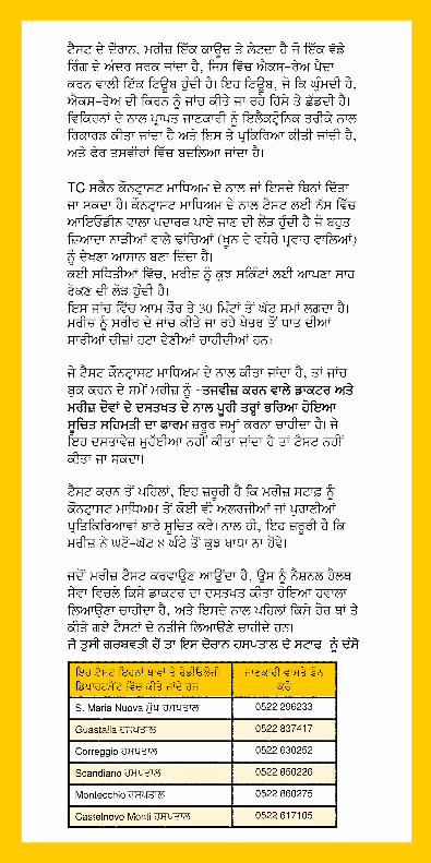
Questi esami vengono eseguiti presso la Radiologia di
Per informazioni tel
Arcispedale S. Maria Nuova 0522 296233 Ospedale di Guastalla 0522 837417Ospedale di Correggio 0522 630252Ospedale di Scandiano 0522 850226Ospedale di Montecchio 0522 860275Ospedale di Castelnovo Monti 0522 617105
Il paziente deve togliere dalla parte corporea interessata all’indagine gli oggetti metallici.
In caso di esame con l’utilizzo del mezzo di contrasto, all’atto della prenotazione il paziente deve essere in possesso del consenso informato compilato e firmato dal medico che prescrive l’esame e dal paziente. In assenza di tale documento l’esame non verrà eseguito.
È importante che il paziente segnali, prima dell’esecuzione dell’esame, l’esistenza di eventuali allergie o di precedenti reazioni al mezzo di contrasto e rispetti inoltre il digiuno da almeno 8 ore.
Il paziente deve presentarsi munito della richiesta motivata e firmata da un medico del Servizio Sanitario Nazionale e portare eventuali indagini precedenti eseguite in altra sede.
È importante segnalare al personale l’eventuale stato di gravidanza.

IngleseEnglish
Computed Tomography (TC)Computed Tomography (TC) is a diagnostic exam that uses ionising radiations. It is a combination of traditional X-rays and computerised programs. With TC, a radiological image of a transverse section of the body is obtained. It is useful for examining pathology due to trauma, malformation, inflammation and tumours in all parts of the body. As compared with traditional radiography, TC shows even minimal differences in density between the various tissues in an organ, thus enabling even deeply located structures to be seen.
During the test, the patient lies on a coach that slides inside a large ring containing the tube which generates the X-rays. This tube, which rotates, directs a beam of X-rays onto the part being examined.The information obtained with the radiations is recorded and processed electronically, and is then converted into images.
A TC scan can be given with or without a contrast medium. The test with a contrast medium requires intravenous administration of a substance containing iodine, which makes highly vascular structures (those with greater blood flow) easier to see. In many situations, the patient must hold his or her breath for a few seconds. The exam generally takes less than 30 minutes. The patient must remove all metal objects from the area of the body being examined.
If the test is run with a contrast medium, the patient must submit - when the exam is booked - a duly filled-up informed consent form signed by both the prescribing doctor and the patient. The test cannot be run unless this document is provided.Before the test is performed, it is essential for the patient to notify the staff of any allergies or previous reactions to the contrast medium. Also, the patient must have been fasting for at least 8 hours.
When the patient shows up for the test, he or she must bring a referral signed by a physician in the National Health service, along with the results of any tests that were previously performed elsewhere. Please inform the staff if you think that you may be pregnant.
These tests are performed at the Radiology department
For informationphone
S. Maria Nuova Main Hospital 0522 296233 Guastalla Hospital 0522 837417Correggio Hospital 0522 630252Scandiano Hospital 0522 850226Montecchio Hospital 0522 860275Castelnovo Monti Hospital 0522 617105

FranceseFrançais
Tomographie assistée par ordinateur (TC)La tomographie assistée par ordinateur (TC) est un examen diagnostique basé sur l’utilisation de radiations ionisantes, qui combine les rayons X traditionnels à des programmes informatisés. La tomographie assistée par ordinateur permet d’obtenir l’image radiologique d’une section transversale du corps humain. Elle est utile dans l’étude de la pathologie traumatique, des malformations, inflammatoire et néoplasique (tumorale) de l’ensemble du corps humain. Par rapport à la radiographie conventionnelle, la TC fait apparaître des différences de densité même minimes entre les différents tissus d’un organe, et permet d’observer des structures localisées en profondeur.
Pendant l’examen, le patient est allongé sur une couchette qui coulisse à l’intérieur d’un grand anneau équipé du tube radiogène. Ce tube rotatif émet un faisceau de rayons X sur la partie à examiner. Les informations mises en évidence par les radiations sont enregistrées et traitées par un système électronique, puis reconstruites en images.
La TC peut être réalisée avec ou sans produit de contraste : l’examen avec produit de contraste requiert l’administration endoveineuse d’une substance iodée qui favorise la visualisation des structures les plus vascularisées (les plus irriguées par le sang).Il est souvent demandé au patient de retenir sa respiration pendant quelques secondes. Généralement, l’examen ne dépasse pas 30 minutes. Le patient doit ôter les objets métalliques présents sur la partie du corps concernée par l’examen.En cas d’examen avec un produit de contraste, le patient doit au moment de la prise de rendez-vous être en possession du formulaire de consentement éclairé rempli et signé par le patient et par le médecin prescrivant l’examen. En l’absence de ce document, l’examen ne sera pas effectué.
Il est important que le patient signale, avant l’exécution de l’examen, l’existence d’allergies éventuelles ou de précédentes réactions au produit de contraste, et qu’il soit à jeun d’au moins 8 heures.Le patient doit se présenter avec une demande motivée et signée par un médecin du service sanitaire national et apporter avec lui les analyses précédentes éventuellement effectuées auprès d’autres établissements. Si vous pensez d’être enciente veuillez en informer le personnel.
Ces examens sont effectués dans le service de radiologie de
Informations Tél.
Arcispedale S. Maria Nuova 0522 296233Hôpital de Guastalla 0522 837417Hôpital de Correggio 0522 630252Hôpital de Scandiano 0522 850226Hôpital de Montecchio 0522 860275Hôpital de Castelnovo Monti 0522 617105
Arabo

Cinese中文
计算机 X 线断层扫描 (TC)计算机 X 线断层扫描 (TC) 是一种利用电离辐射进行医疗诊断的方法。它是传统 X 射线和计算机程序的结合体。利用 TC 可获取人体横截面的放射影像。TC 可用于检测因创伤、畸形、炎症和肿瘤引起的人体任何部分的病变。与传统的放射线扫描相比,TC 可检测到器官内各组织间密度的细微差别,确保能够观察到深层结构。
检测期间,患者需躺在长榻上,然后被推入装备有显像管(可产生 X 射线)的大型圆环中。该旋转式显像管引导 X 射线照射在需要检测的部位。放射物所获取的信息将被记录下来并通过电子处理,最终转换成图像。
TC 在有无造影剂的情况下均可进行。使用造影剂的检测需要静脉注射含碘物质,确保高度血管结构(血流量较大的血管)易于观察。通常情况下,患者须憋气几秒钟。该检测一般需要不到 30 分钟的时间。患者必须将所有金属物质从需要检测的部位移除。
如果在使用造影剂的情况下进行检测,患者须在预约成功后提交经处方医生和患者双方签字的正式填写的知情同意书。提供该文件后才能接受该项检测。
如有任何过敏症或对造影剂的过往不良反应,患者应在检测前告知相关人员。患者还必须在接受检测至少 8 小时前停止进食。
接受检查时,患者须携带由英国国民健康服务的医生签字的转诊书和任何过往检测结果。 如果你认为自己怀孕了,请告诉医务人员.
检测地点位于如下医院的放射科 咨询 电话
S. Maria Nuova 总医院 0522 296233
Guastalla 医院 0522 837417
Correggio 医院 0522 630252
Scandiano 医院 0522 850226
Montecchio 医院 0522 860275
Castelnovo Monti 医院 0522 617105
RussoРусский
Компьютерная томография (TC)Компьютерная томография (TC) – это диагностическое исследование, основанное на использовании ионизирующих излучений, которое сочетает традиционные рентгеновские лучи с компьютерными программами. С помощью компьютерной томографии получают рентгеновские изображения поперечного сечения тела человека.Компьютерная томография полезна для исследования травматических, воспалительных патологий, патологий новообразований (опухолей), в том числе злокачественных образований любых частей человеческого тела. По сравнению с традиционной рентгенографией, компьютерная томография выявляет даже минимальные различия плотности между различными тканями какого-либо органа, позволяя визуализировать структуры, находящиеся даже в глубине.
Во время проведения исследования пациент ложится на кушетку, которая задвигается вовнутрь большого кольца, где размещена рентгеновская трубка. Эта трубка, вращаясь, испускает пучок рентгеновских лучей на обследуемую часть тела. Данные, получаемые в результате облучения, регистрируются и обрабатываются электронной системой для последующего воссоздания в виде изображений.
Компьютерную томографию можно выполнять без и с контрастным веществом: процедура с использованием контрастного вещества требует внутривенной инъекции парамагнитного вещества, которое способствует визуализации структур с наибольшим скоплением кровяных сосудов (то есть в большей мере орошаемых кровью).Во многих случаях от пациента требуется задержать дыхание в течение несколько секунд. Процедура обычно длиться не более 30 минут. Пациент должен снять с обследуемой части тела все металлические предметы.
В случае исследования с использованием контрастного вещества, во время записи пациент должен иметь при себе информированное согласие, заполненное и подписанное врачом, который выписал направление на исследование, и самим пациентом. В случае отсутствия такого документа, исследование не будет выполнено.
Важно, чтобы пациент сообщил до начала проведения исследования о возможном существовании аллергий или предыдущих реакциях на контрастное вещество, а также чтобы он не ел ничего в течение не менее 8 часов до начала проведения исследования. Пациент должен явиться с мотивированным направлением, подписанным врачом.Сообщите, пожалуйста, персоналу, если полагаете, что Вы беременны.
Эти исследования проводятся в отделении рентгенологии
Для получения информации тел.
Главная больница S. Maria Nuova 0522 296233Больница Guastalla 0522 837417
Больница Correggio 0522 630252Больница Scandiano 0522 850226Больница Montecchio 0522 860275Больница Castelnovo Monti 0522 617105

Urdu

Hindi

Punjabi