TMJ & Muscles of mastication D.Rania Gabr D.Sama. D.Elsherbiny.
13
TMJ & Muscles of mastication D.Rania Gabr D.Sama. D.Elsherbiny
-
Upload
darlene-scott -
Category
Documents
-
view
228 -
download
1
Transcript of TMJ & Muscles of mastication D.Rania Gabr D.Sama. D.Elsherbiny.
TMJ & Muscles of mastication
D.Rania GabrD.Sama.D.Elsherbiny
Objectives• Identify the parts of the mandible.• Know the type and formation of temporomandibular
joint.• Understand the attachment of the capsule and
ligaments of the temporomandibular joint.• Explain the mechanism of movements taking place at
temporomandibular joint.• Define the stability factors of the joint.• Explain the muscles of mastication with their origin,
insertion, nerve supply and actions.• Discuss maxillary artery.

Action1. All elevate the mandible except lateral pterygoid “depress”2. All protrude the mandible except temporalis “retract”3. 2 muscles (medial & lateral pterygoid) produce side to side
movement.
Nerve supply: Mandibular nerve (trigeminal nerve).


5
TEMPORAL FASCIA
Attachment:
Above: Superior temporal line
Below: It splits into 2 layers and insert into respective borders of zygomatic bone
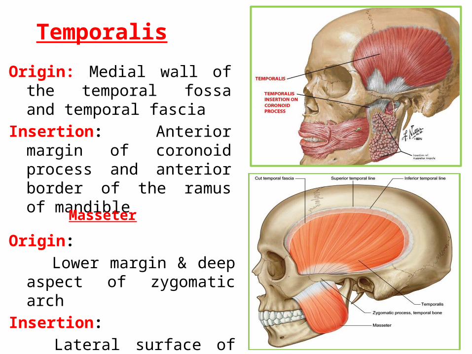
TemporalisOrigin: Medial wall of the
temporal fossa and temporal fascia
Insertion: Anterior margin of coronoid process and anterior border of the ramus of mandible
6
Masseter
Origin:
Lower margin & deep aspect of zygomatic arch
Insertion:
Lateral surface of ramus of mandible

Lateral pterygoid Origin:
1. Upper head: Infratemporal surface of greater wing of sphenoid
2. Lower head: Lateral surface of the lateral pterygoid plate
Insertion: Pterygoid fovea & capsule of TMJ.
7
Medial pterygoidOrigin:
1. Deep head: from the medial surface of lateral pterygoid plate
2. Superficial head: Tuberosity of maxilla
Insertion: inner surface of angle of mandible.
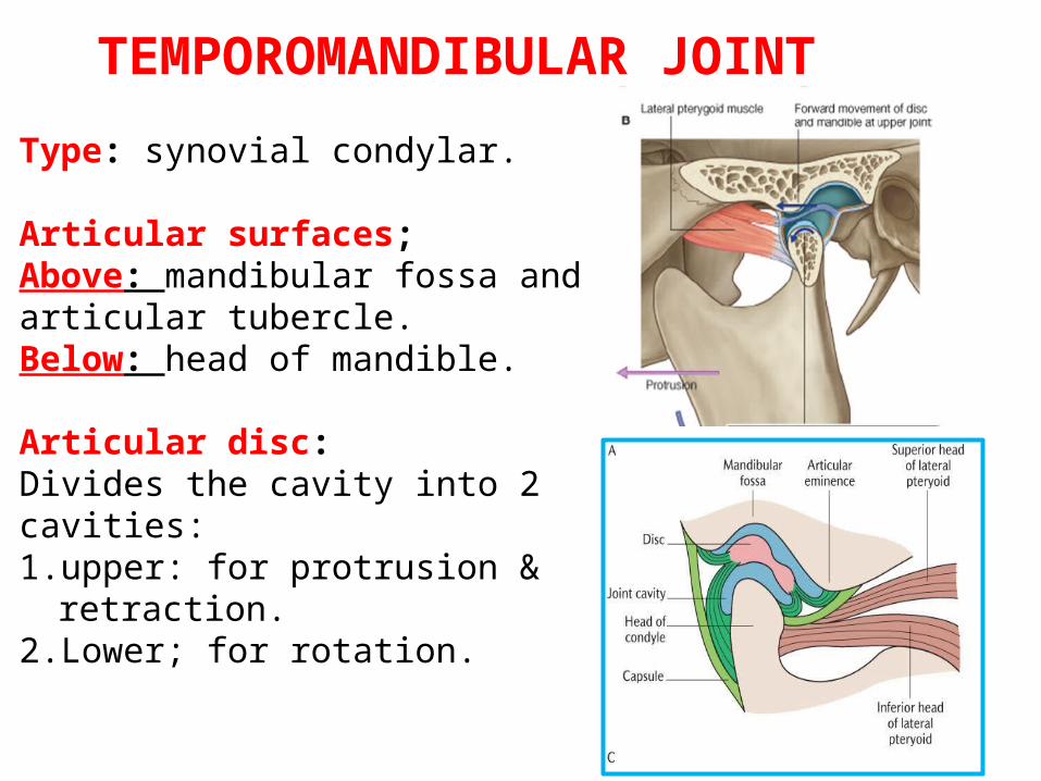
TEMPOROMANDIBULAR JOINT
Type: synovial condylar.
Articular surfaces;Above: mandibular fossa and articular tubercle.Below: head of mandible.
Articular disc: Divides the cavity into 2 cavities:1. upper: for protrusion & retraction.2. Lower; for rotation.

Ligaments:1. lateral temporomandibular
ligament.2. Sphenomandibular
ligament.3. Stylomandibular ligament.
Arterial supply: Maxillary artery and superficial temporal artery.
Nerve supply: Auriculotemporal nerve and nerve to massster
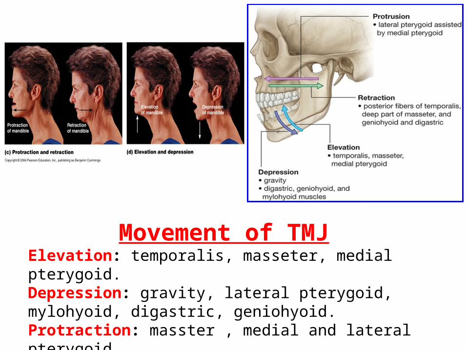
Movement of TMJElevation: temporalis, masseter, medial pterygoid.Depression: gravity, lateral pterygoid, mylohyoid, digastric, geniohyoid.Protraction: masster , medial and lateral pterygoid.Retraction: temporalis and digastric
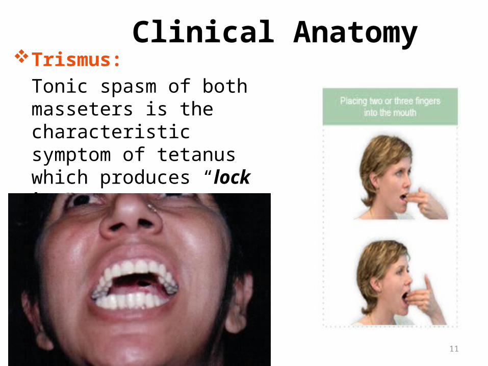
Clinical AnatomyTrismus:
Tonic spasm of both masseters is the characteristic symptom of tetanus which produces “lock jaw”
11
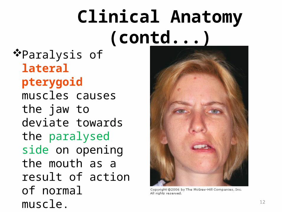
Clinical Anatomy (contd...)Paralysis of lateral
pterygoid muscles causes the jaw to deviate towards the paralysed side on opening the mouth as a result of action of normal muscle.
12