TITRATION OF AVANCED PAP THERAPIES : WHAT … OF AVANCED PAP THERAPIES : WHAT TO USE, WHEN, WHY AND...
81
TITRATION OF AVANCED PAP THERAPIES : WHAT TO USE, WHEN, WHY AND HOW Kala Bingham, CRT RCP RPSGT Sleep Care Manager The Sleep Wellness Institute
-
Upload
truongdieu -
Category
Documents
-
view
214 -
download
0
Transcript of TITRATION OF AVANCED PAP THERAPIES : WHAT … OF AVANCED PAP THERAPIES : WHAT TO USE, WHEN, WHY AND...

TITRATION OF AVANCED PAP
THERAPIES : WHAT TO USE, WHEN,
WHY AND HOW
Kala Bingham, CRT RCP RPSGT
Sleep Care Manager
The Sleep Wellness Institute

General Titration Goals
THE GOALS SHOULD BE INDIVIDUALIZED TO MEET THE PATIENT
NEED
AIRWAY MANAGEMENT-KEEP THE AIRWAY OPEN
STABILIZE BREATHING PATTERNS BY MONITORING THE PATIENTS RESPONSE
TO THERAPY
SET PARAMETERS FOR OPTIMAL THERAPY
ENSURE MASK FIT
Sleep disordered breathing
OSA Central Hypoventilation
CPAP
BiPAP
Noninvasive ventilation:
BiPAP BiPAP
ASV AVAPS

BiLevel: indications for
use
OSA patients with: intolerance of CPAP pressures
hypoxemia despite resp event control
elevated CO2 levels despite resp event control
Hypoventilation syndrome
Complex or Central Sleep Apnea

Provides two independently set pressures
to maintain airway stability and support
ventilation requirements while the patient
sleeps
IPAP
EPAP
What is BiLevel

BiLevel terms
Rise Time=the time it takes for BiPAP to
change from EPAP to IPAP. You can
adjust for patient comfort
Tidal Volume -Vt

Titration BiLevel for control of
apnea:
Increase expiratory pressure (EPAP) in a stepwise
fashion to control obstructive apnea
Increase Inspiratory pressure (IPAP) in a stepwise
fashion (maintaining at least 4 cm difference from
EPAP) to control hypopneas and snoring.
IPAP 4-6 cm > EPAP and snoring or hypopneas
persist, trial of increasing EPAP.
Central apneas back up rate

Titration of BiLevel for persistent
hypoxemia in OSA
IPAP/EPAP control of apnea, hypopnea and snoring.
Hypoxemia persists increase IPAP in 2 cm increments.
IPAP > 4 cm above level for control of OSA without benefit to sats or increases not tolerated reduce to lowest effective level + add supplemental O2 to keep sats > 89-90%
NOT ALL HYPOXEMIA IS HYPOVENTILATION

Effects of sleep on normal
ventilation
Sleep
Decrease muscle
tone
Increased airway
resistance
Decrease drive
to breath
↑ PaCO2 2 – 8 mm Hg
↓ PaO2 3 – 10 mm Hg
Nl wake PaCO2 38-43 mm
Nl sleep PaCO2 37-50 mm

Hypoventilation-Respiratory
Insufficiency
The state in which a reduced amount of air
enters the alveoli of the lungs resulting in: Pa02 falls
PaC02 rises
Occurs due to:
1. decrease tidal volume (Vt)
2. increased dead space (Vd)
3. decreased respiratory rate (RR)

Development of respiratory insufficiency
HYPOVENTILATION
Thoracic
disease
Decrease muscle
tone &
tidal volume
Increased airway
resistance
Decrease drive
to breath
Hypoventilation↑CO2
↓ O2 Respiratory
insufficiency
Night N+Day
Abnormal PaCO2 in
sleep > 50 or change >
7 mm Hg from wake to
sleep
Decreased RR
Decreased CO2
response

Common causes of thoracic
disorder
1. Respiratory muscle weakness –decreased Vt, increased dead space, increased RR
Amyotrophic Lateral Sclerosis
Muscular Dystrophy
Spinal Muscular Atrophy
Post-Polio Syndrome

Common causes of thoracic
disorder
2. Restrictive thoracic –decrease in the lung’s ability to expand due to an external restriction of the chest wall or stiffness of the lung tissue.
Kyphoscoliosis
Sarcoidosis

Common causes of thoracic
disorders
3. Obstructive lung disease –Increased airway resistance, partial air-flow obstruction, increased dead space, air trapping.
COPD
Emphysema
Severe Asthma
Overlap syndrome
Common causes of thoracic
disorders
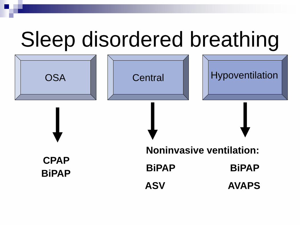
4. Obesity hypoventilation syndrome –
decreased Vt, increased RR

Titration options for patients
with hypoventilation or
respiratory insufficiency
1. Bi-Level PAP
2. Average Volume Assured
Pressure Support (AVAPS)

Titration of BiLevel for
persistent hypoxemia in OSA
Titrate BiLevel to pressures appropriate for
control of apnea, hypopnea and snoring
If hypoxemia persists, increase IPAP in
2cm increments in attempt to improve 02
saturation

Titration of BiLevel for
persistent hypoxemia in OSA
If increasing IPAP >4 cm above level
appropriate for control of OSA without
benefit to sats or increases not tolerated,
add supplemental 02 as needed to
maintain sats > 89-90%
NOT ALL HYPOXEMIA IS
HYPOVENTILATION

BiLevel S (spontaneous mode)
Used with patients who are able to
maintain a constant respiratory rate, but
require an IPAP:EPAP pressure difference
to augment tidal volume while you sleep.

BiLevel S (spontaneous mode)
Can be used with the following patients:
Obesity hypoventilation
Neuromuscular weakness disorders
Restrictive thoracic disease
Obstructive lung disease

BiLevel S/T (timed back up
rate)
This mode is used with patients that
require:
Time rate from the device to support their
inconsistent respiratory pattern (more
common in NM disease)

BiLevel S/T (timed back up
rate)
Pressure support to augment their tidal
volume when the device provides a breath to
the patient
Patient has the ability to spontaneously
initiate breaths or tolerate timed back up
breaths from the device

How can we affect ventilation?
To increase ventilation:
1. Insure patent airway.
2. Increase Vt.
I:E differential
Vt setting with AVAPS.
3. Increase respiratory rate
4. Body position
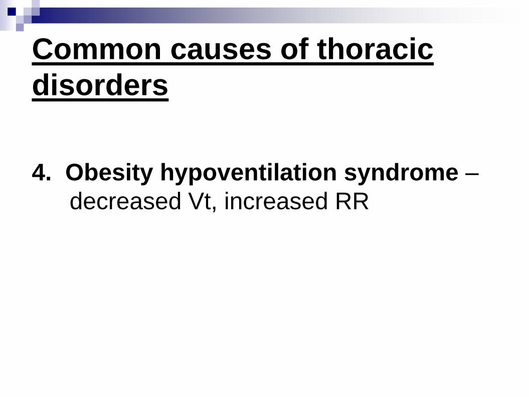
Titration of BiLevel for control of
hypoventilation
1. Transcutaneous (TCCO2) or End-Tidal CO2 (ETCO2) monitoring.
2. Excessive leakage must be prevented.
3. Initiate BiLevel at IPAP/EPAP 10/4 cm or EPAP at pressure previously demonstrated as effective to control obstructive apnea. InitiateIPAP at (EPAP +6 cm).
4. Increase EPAP only until obstructive events are controlled.
Increase EPAP increase IPAP same
***Want lowest possible EPAP

Titration of BiLevel for control of
hypoventilation (cont’d)
5. Increase IPAP (as tolerated) until the
following parameters are achieved:
a. TCCO2 or ETCO2 < 50 mm (or RR 2-4
BPM < baseline wake RR )
b. Minimal hypopneas
c. Improvement in O2 sat if > 89%

Titration of BiLevel for control
of hypoventilation (cont’d)
5. Central apneas or inconsistent efforts
back up rate = match RR during relaxed
wake.
6. Increase RR in increments of 2 BPM if CO2
remains > 50 mm despite use of maximally
tolerated IPAP.

Bi-Level with (AVAPS)
• Fixed EPAP
• Vt selected – based on IBW (8-10
ml/kg)
• Adjusts pressure support (IPAP-EPAP)
to maintain a consistent tidal volume
• Able to provide a constant tidal volume
as patient ventilation changes.
• Allows for compensation of Intra-night and
inter-night changes in breathing status
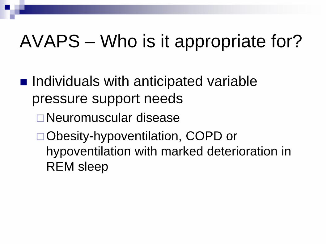
AVAPS – Who is it appropriate for?
Individuals with anticipated variable
pressure support needs
Neuromuscular disease
Obesity-hypoventilation, COPD or
hypoventilation with marked deterioration in
REM sleep

AVAPS auto-titration algorithm
IPAPmax
IPAPmin
EPAP
Vte = Vt Patient
1 cm H2O/min
Target Vt
Automatically adjusts the IPAP within a preset range to maintain a
consistent tidal volume IPAP will automatically increase or decrease

AVAPS is NOT recommended for
patients with periodic breathing
Treatment of periodic breathing requires a rapid and variable breath by breath response system so the patients PaCO2 stabilizes quickly
AVAPS does not have a quick variable response to changes in tidal volume.

31
• Set mode to S/T with AVAPS on
• Establish initial settings as indicated below
• Ensure proper mask fit to allow algorithm to work effectively
• Have patient breathe on bi-level device at basic settings below
• May Adjust IPAP, I-Time and Rate to patient comfort
Titration protocol with AVAPS for respiratory
insufficiencyGoal: Adjust user parameters for efficacy and adherence
EPAP
IPAPmin
IPAPmax
Rise Time
4 cm H2O
10 cm H2O
25 cm H2O
2 or 3
I -Time
Rate
1.2 sec.
8-10 BPM or
2 below wake rateAcclim
atio
n Z
on
e
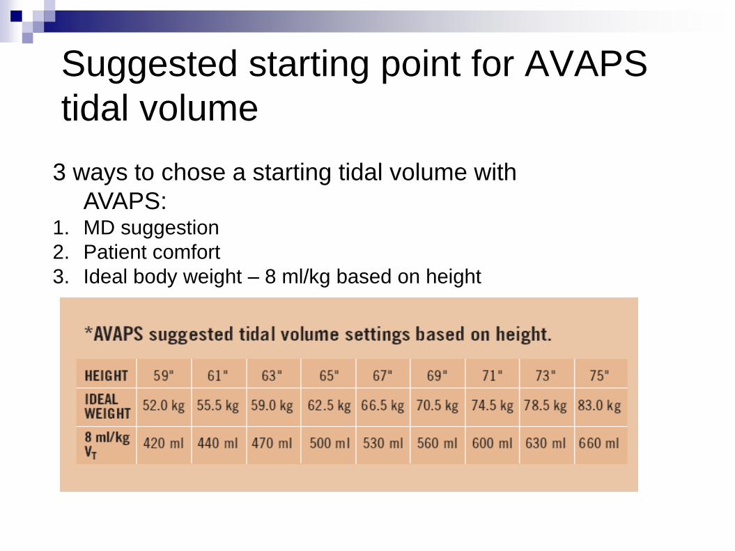
Suggested starting point for AVAPS
tidal volume
3 ways to chose a starting tidal volume with
AVAPS: 1. MD suggestion
2. Patient comfort
3. Ideal body weight – 8 ml/kg based on height

At lights out
observe for
patient’s inability
to maintain sleep
due to
obstructive
apneas
For patient comfort
and to allow sleep
onset increase
EPAP to open the
airway
At lights out
observe for
indications of
therapy
intolerance
For patient comfort and
to allow sleep onset
increase IPAPmin by 2
cm H2O
Titra
tio
n Z
on
e
if yesif yes
Monitoring patient response at lights out

Unable to
maintain sleep
due to
obstructive
apneas
Observe for
inadequate back-
up rate
Unable to
maintain SaO2
>90% for 5
continuous
minutes
Increase rate by 2
bpm. Assess I-Time
and Rise Time for
comfort
if yes
Increase EPAP and
IPAPmin by same
amount to open the
airway
Unable to
maintain tidal
volume with
sleep
Increase IPAPmin
setting to increase
tidal volume .
If IPAP reaching max
and Vt too low,
increase IPAP max.
CHECK LEAK
Titra
tio
n Z
on
e
Return to * Return to *Return to *Return to *
Add supplemental
oxygen
if yesif yesif yes
if no if noand if no
* Monitor patient PSG
Wait… Observe… Think
Patience is the key to successful titration

Complex and Central Sleep Apnea
Definition
Treatment approaches

3 main forms of Central Sleep Apnea
Idiopathic Central Sleep ApneaBrain issue with control of respiration
Narcotics
Periodic breathing Heart failure
Chemoreceptor issue/CO2 issue
Narcotics
Complex Sleep Apnea “CPAP Emergent central events”
Chemoreceptor issue

Idiopathic central sleep apnea –
PSG view• No output from
respiratory
center of the
brain causing
lack of
movement of the
thorax.
• No movement
of thorax &
abdomen
causes apnea

Idiopathic central sleep apneaCause of Idiopathic Central Apnea:
The respiratory center of the brain does not fire during sleep causing periodic apnea (see below)
Seen during the diagnostic night and titration night
Generally seen in non REM sleep clears during REM sleep
Generally seen in younger populations
May appear as part of a neurological disease process or injury
Relationship between chronic opioid therapy and central sleep apnea1
Impacts very small population of people
Apnea Apnea
1 Webster,et al. American Academy of Pain Medicine 2007

Treatment recommendations for idiopathic
central sleep apnea
Oxygen therapy**
Must have desaturation ≤88% for 5 minutes or longer to qualify for oxygen therapy (CMS guidelines) OR ≤88% for 5 minutes with history of either CHF, Pulm. HTN, Cor Pulmonale or increased RBC count
Medications: Theophylline 1, 3
Acetazolamide 2, 3
Gradual reduction of opioid medications may improve narcotic-induced CSA3
BiPAP S/T or ASV
1 Orth, et al. Resp. Med. 2005;99:471-476
2 Javahari, S. AJRCCM: 2006:173(2) 234-237
3 Eckert, et al. Chest. 2007; 131:595-607
Remember:
<2% of SDB

Periodic breathingCharacteristics: waxing and waning breathing
pattern
Length of cycle is based on disease process causing the breathing pattern Longer events for patients in heart failure1
50-70 second events of CSR then followed by normal respiration (waxing and waning of respiration) in patients with heart failure1
Shorter events in those at altitude/neurological disorder/renal failure1
(picture B)
20 – 40 seconds on length1
40
20-40 sec
1 Thomas, et. al. Curr. Opin Pulm Med. 2005
A B
50-70 sec
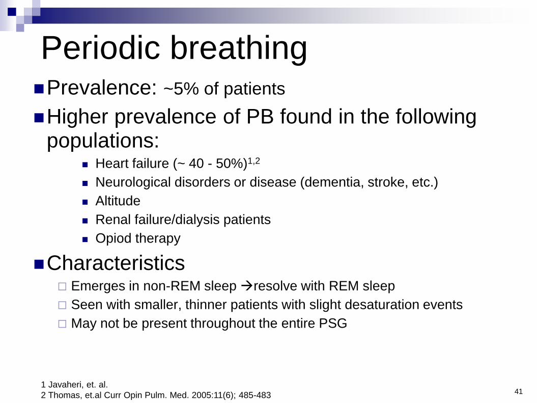
Periodic breathingPrevalence: ~5% of patients
Higher prevalence of PB found in the following populations:
Heart failure (~ 40 - 50%)1,2
Neurological disorders or disease (dementia, stroke, etc.)
Altitude
Renal failure/dialysis patients
Opiod therapy
Characteristics Emerges in non-REM sleep resolve with REM sleep
Seen with smaller, thinner patients with slight desaturation events
May not be present throughout the entire PSG
411 Javaheri, et. al.
2 Thomas, et.al Curr Opin Pulm. Med. 2005:11(6); 485-483

Periodic breathing sample
42

Treatment of periodic breathing (PB)
Medical management of underlying disease
Medical Management of Heart Failure is KEY in treatment
of PB1
Mainly PB, (PB > 50%), CSA > 5, AHI or RDI > 5
CPAP Therapy1
Auto Servo Ventilation3
Bi-Level Therapy with back up rate2
Mainly OSA (< 50% PB), CPAP or BiPAP S should be
prescribed and patient followed for signs of emerging or non-
resolving PB
43
1 Javaheri, et. al. Curr Treatment Option in CV Med: 2005:7:295-306
2 Kasi, et. al. Circ. J.; 200569:913-921
3 Teschler et al, AJRCCM, 164:614-419, 2001

44
Complex sleep apnea
OSA which converts to central apnea with CPAP application
Typically emerges during titration
Not obvious during diagnostic PSG
Often occurs at ~ 30 second intervals vs. 60-90 second intervals with CSR
Minimal data available
Estimated prevalence 1/7 or ~15% of the SDB population
1 Morgenthaler, et. al. Sleep 2006; 29 (9):1203-1209

45
Central apnea
emerges on
patient with
OSA and
CPAP therapy
on 7 cm H2O
(seen with
highlighted
area)
Complex Sleep Apnea

Complex sleep apnea
Due to a combination of upper airway resistance and
abnormal respiratory drive1,2
OSA eliminated with CPAP allows for normal RR.
The change of the RR changes CO2
Brain reads the change in CO2 as “hyperventilation” central apneas
during the CPAP titration
Central apnea CO2 rises re-establishes drive to breathe
Chemoreceptor issues are unmasked when OSA is eliminated
Often an temporary abnormality of ventilatory control
46
1 Interview with Dr. Younes & Dr. Sanders
2 Morgenthaler, et.al. Sleep 2006
Complex
~35
sec
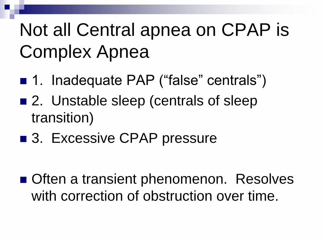
Not all Central apnea on CPAP is
Complex Apnea
1. Inadequate PAP (“false” centrals”)
2. Unstable sleep (centrals of sleep
transition)
3. Excessive CPAP pressure
Often a transient phenomenon. Resolves
with correction of obstruction over time.

Treatment options for complex sleep
apnea
CPAP + Time on Therapy to reset
chemoreceptors1
30-day trial on CPAP follow up patient re: EDS and
compliance data AHI, if improved keep on CPAP
No improvement in daytime sleepiness after
30 days, try alternatives Auto Servo Ventilation
Bi-Level therapy with backup rate
1 Dernaika T et.al; Chest 2006 s;130(4)129
2 Adult Sleep Apnea Task Force, AASM, ; Journal of Clinical Sleep Medicine 2009; 5(3)48

BiPAP autoSV
ADVANCED
Overview

5050
A AA
H
S S
A
SH A
Looks like Auto CPAP! EPAP only changes every 2 minAUTO EPAP
SV works ‘on top’ of Auto EPAPEPAPmin
EPAPmin
EPAPmax
EPAPmax
Pressure max
Pressure max

51
30cm H2O
15cm H2O
4cm H2O
20cm H2O
15cm
H2O
0cm
H2O
Default Settings
Auto-EPAP Looks like Auto CPAP! EPAP only changes every 2 min
SV works ‘on top’ of Auto EPAP

52
Servo Ventilation Algorithm
4 Minutes
On a breath by breath basis peak flow is captured
Peak flow is monitored over a moving 4 minute window
As 1 breath is added, the initial breath falls off
At every point within this 4 minute period an Average Peak Flow is
calculated
The Peak flow target is established around that average and is based on
the patient’s needs

53
IF: Peak flow is at target
THEN: autoSV delivers CPAP pressure
Servo Ventilation Algorithm – Normal Breathing

54
IF: Peak flow falls below target
THEN: autoSV increases pressure support
Servo Ventilation Algorithm – Decreased Flow

BiPAP autoSV Titration Protocol
55

56
• Establish initial settings as indicated below
• Ensure proper mask fit to allow algorithm to work effectively
• Have patient breathe on autoSV Advanced at basic settings below
• Adjust EPAPmin, Bi-Flex and PSmin settings to patient comfort
Titration protocol for BiPAP autoSV Advanced
for periodic and complex breathingGoal: Adjust user parameters for efficacy and adherence
*If pt has known CPAP pressure of <10 set EPAPmin at 4 cm H2O or patient comfort
*If pt has known CPAP pressure of >10 set EPAPmin at 6-8 cm H2O or patient comfort
EPAPmin
EPAPmax
PSmin
PSmax
4 cm H2O*
15 cm H2O
0 cm H2O
15 cm H2O
Max pressure
Rate
Bi-Flex
30 cm H2O
Auto
2 or 3Acclim
atio
n Z
on
e
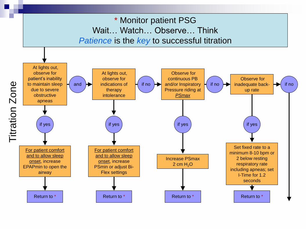
At lights out,
observe for
patient’s inability
to maintain sleep
due to severe
obstructive
apneas
Observe for
inadequate back-
up rate
Observe for
continuous PB
and/or Inspiratory
Pressure riding at
PSmax
Set fixed rate to a
minimum 8-10 bpm or
2 below resting
respiratory rate
including apneas; set
I-Time for 1.2
seconds
if yes
For patient comfort
and to allow sleep
onset, increase
EPAPmin to open the
airway
At lights out,
observe for
indications of
therapy
intolerance
For patient comfort
and to allow sleep
onset, increase
PSmin or adjust Bi-
Flex settings
Titra
tio
n Z
on
e
Return to * Return to *Return to *Return to *
Increase PSmax
2 cm H2O
if yesif yesif yes
if no if noand if no
* Monitor patient PSG
Wait… Watch… Observe… Think
Patience is the key to successful titration

Complex Sleep Apnea
CSA patients may challenge even the
most experienced, skilled sleep
technologist
Helpful hints for CSA titrations
If changes are needed-Watch, Wait and
Observe

OSA
CSA
Complex ~85% - 90% of patients
have OSA
Treatment includes:
CPAP or BiPAP S
~5% have CSA or CSR
Treatment includes:
Medication mgmt.
Oxygen therapy
Bi-level therapy
ASV therapy
~10% have
Complex SDB
Treatment
includes:
CPAP therapy
CPAP + Oxygen
Bi-level therapy
ASV
Summary of treatment strategies for
SDB patients
Eckert, et al. Chest. 2007; 131:595-607

Medicare RAD policy requirements for
central or complex sleep apnea
Medicare Definition of Complex Sleep
Apnea
Persistence or emergence of central events upon exposure to
CPAP/BiPAP when obstructive events have disappeared
Mainly obstructive or mixed apneas on diagnostic sleep
study, > 5 events / hour, OA>CA
On CPAP/BiPAP patterns of central apnea that meet the
definition of Central Sleep Apnea

Medicare definition of central sleep
apnea
Central sleep apnea
Apnea index > 5
Central apnea >50% of the total apneas
Central apneas > 5 times per hour

Respiratory Assist Device
(RAD)
Coverage-The treating physician must fully
document in the patients medical record
symptoms characteristic of sleep-
associated hypoventilation.
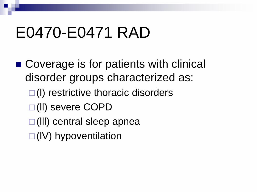
E0470-E0471 RAD
Coverage is for patients with clinical
disorder groups characterized as:
(l) restrictive thoracic disorders
(ll) severe COPD
(lll) central sleep apnea
(lV) hypoventilation
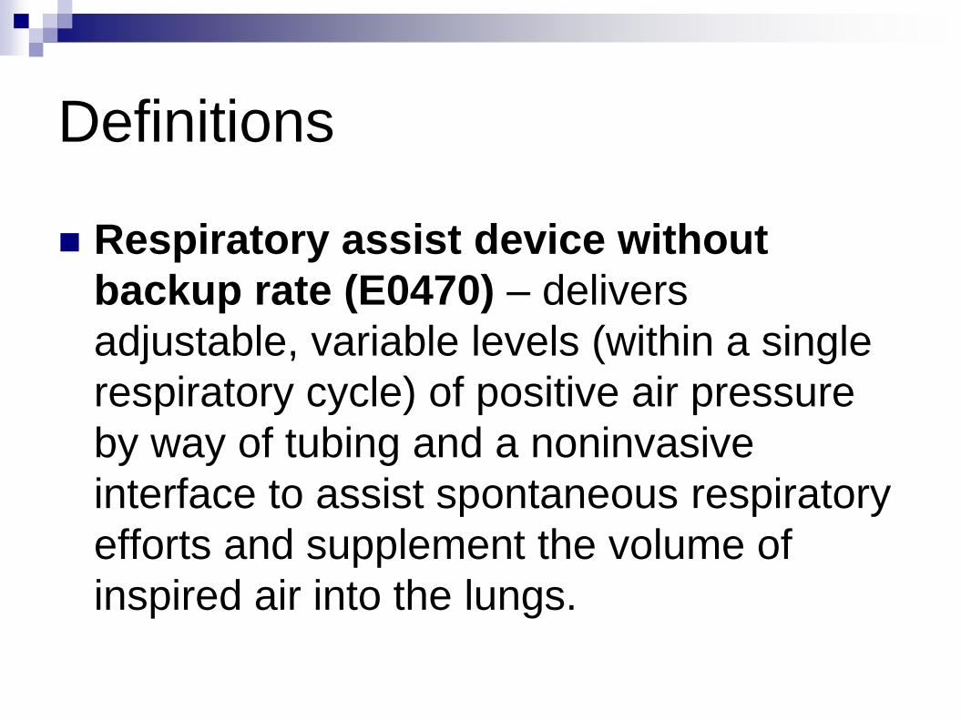
Definitions
Respiratory assist device without
backup rate (E0470) – delivers
adjustable, variable levels (within a single
respiratory cycle) of positive air pressure
by way of tubing and a noninvasive
interface to assist spontaneous respiratory
efforts and supplement the volume of
inspired air into the lungs.

Definitions
Respiratory assist device with backup
rate (E0471) – delivers adjustable,
variable levels (within a single respiratory
cycle) of positive air pressure by way of
tubing and a noninvasive interface to
assist spontaneous respiratory efforts and
supplement the volume of air into the
lungs. Back up rate

Definitions
FlO2 – the fractional concentration of
oxygen delivered to the patient for
inspiration. A patient’s prescribed FlO2
refers to the oxygen concentration the
patient normally breathes when not
undergoing testing to qualify for a RAD.

Definitions
FEV1 – the forced expired volume in 1
second
FVC – the forced vital capacity
FRC - forced residual volume
ABG’s – Arterial Blood Gas

Restrictive Thoracic Disorders
Documentation of neuromuscular disease
or severe thoracic cage abnormality in the
patient’s medical record

Restrictive Thoracic Disorders
Perform one of the following
ABG’s(done while awake and on prescribed
FiO2 ) PaCO2 ≥ 45 mm Hg OR
Sleep oximetry-O2 saturation ≤ 88% for ≥ 5
minutes, minimum 2 hours of recording time
OR
For neuromuscular-Either FVC < 50% of
predicted or MIP < 60 cm H2O

COPD
ABG’s done while awake on prescribed
Fi02 with a PaCO2 > 52 mmHg
Sleep oximetry-02 sats < 88% for > for 5
continuous minutes
Qualify for E0470 (no back up rate)

COPD-Situation 1
After initial use of E0470
ABG’s-shows PaCO2 worsens >7 mm Hg
compared to original ABG
Facility-based PSG-demonstrates oxygen
saturation ≤ 88% for ≥ a cumulative 5
minutes, minimum 2 hours nocturnal
recording time

COPD-Situation 2
61 days after initial issue of E0470
ABG-done while awake and on prescribed
FiO2) shows PaCO2 ≥ 52 mm Hg;
Sleep Oximetry-demonstrates oxygen
saturation ≤ 88% for ≥ a cumulative 5
minutes, minimum 2 hours nocturnal
recording time

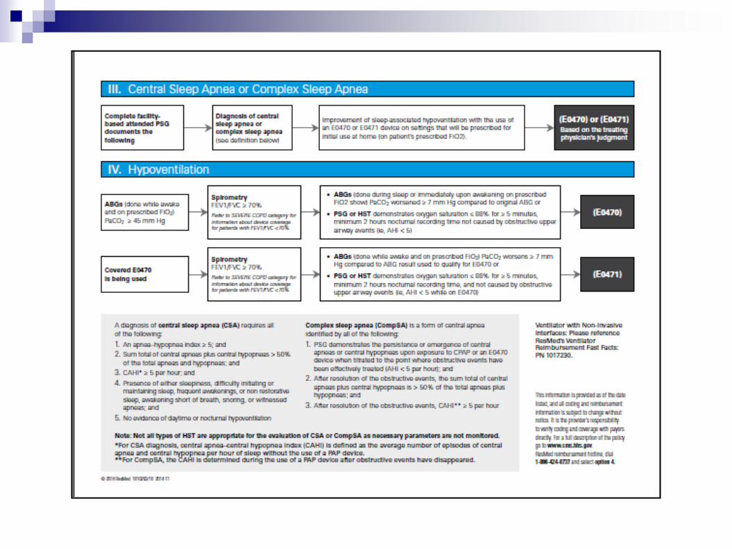
Central Sleep Apnea/Complex
Sleep Apnea
Completed facility-based attended PSG
documents the following
Diagnosis of CSA/Comp SA
Improvement of sleep-association
hypoventilation with the use of E0470 or
E0471 on settings that will be prescribed
for initial home use
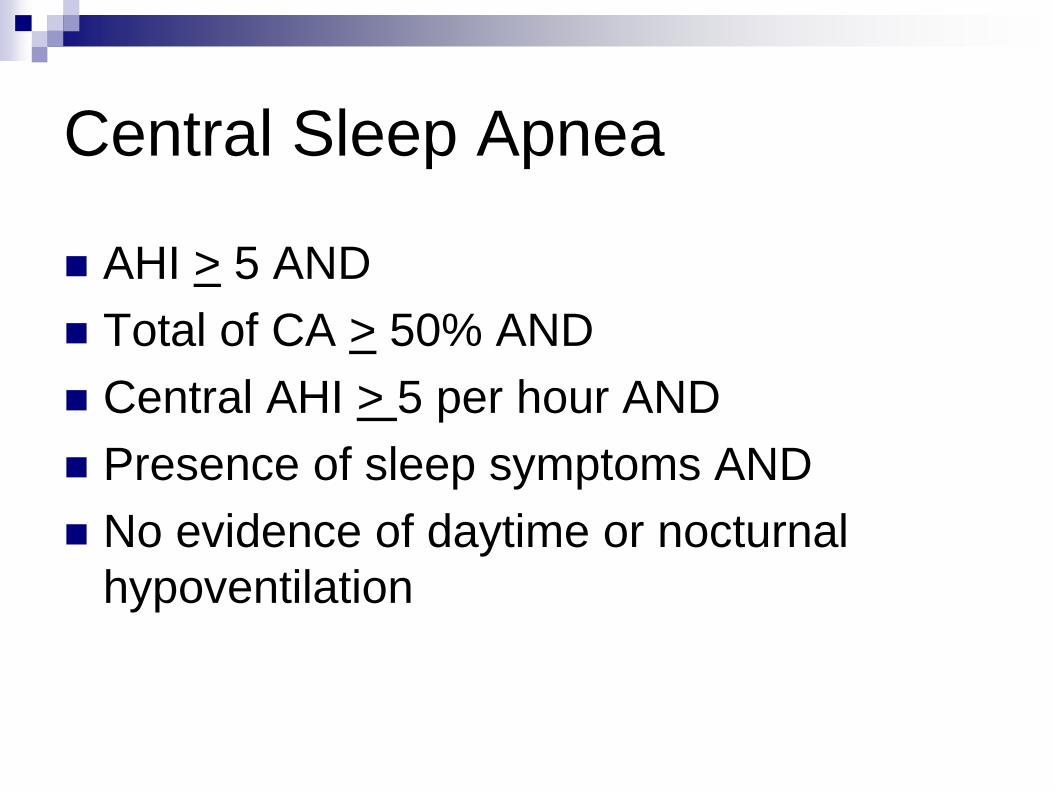
Central Sleep Apnea
AHI > 5 AND
Total of CA > 50% AND
Central AHI > 5 per hour AND
Presence of sleep symptoms AND
No evidence of daytime or nocturnal
hypoventilation
Complex Sleep Apnea
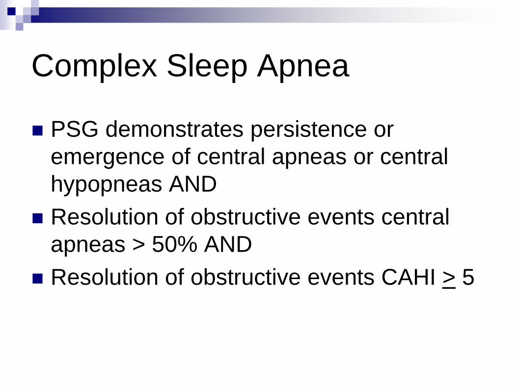
PSG demonstrates persistence or
emergence of central apneas or central
hypopneas AND
Resolution of obstructive events central
apneas > 50% AND
Resolution of obstructive events CAHI > 5

Hypoventilation (E04070)
ABG’s done while awake and on
prescribed Fi02 with the PaC02 > 45
mmHg AND
Spirometry FEV1/FVC 70% AND
ABG’s during sleep or immediate
awakening worsen PaC02 OR
PSG/HST

Hypoventilation (E0471)
Covered E0470 is being used AND
Spirometry FEV1/FVC > 70% AND
ABG’s done while awake and on
prescribed Fi02 worsens > 7mm Hg
compared to ABG result used for E0470
OR
PSG or HST

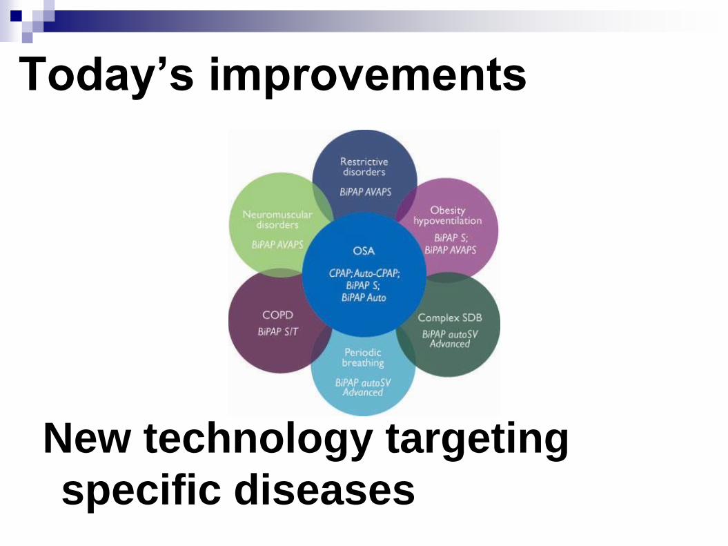
Today’s improvements
New technology targeting
specific diseases

Thank you