Title: Patterns in palaeontology: How and why did the ... · The phylum Arthropoda encompasses...
10
Title: Patterns in palaeontology: How and why did the arthropod shed its skin? Moulting in living and fossil arthropods Author(s): Harriet B. Drage *1 Volume: 6 Article: 7 Page(s): 110. Published Date: 01/07/2016 PermaLink: http://www.palaeontologyonline.com/articles/2016/arthropodmoulting/ IMPORTANT Your use of the Palaeontology [online] archive indicates your acceptance of Palaeontology [online]'s Terms and Conditions of Use, available at http://www.palaeontologyonline.com/siteinformation/termsandconditions/ . COPYRIGHT Palaeontology [online] (www.palaeontologyonline.com) publishes all work, unless otherwise stated, under the Creative Commons Attribution 3.0 Unported (CC BY 3.0) license. This license lets others distribute, remix, tweak, and build upon the published work, even commercially, as long as they credit Palaeontology[online] for the original creation. This is the most accommodating of licenses offered by Creative Commons and is recommended for maximum dissemination of published material. Further details are available at http://www.palaeontologyonline.com/siteinformation/copyright/ . CITATION OF ARTICLE Please cite the following published work as: Drage, Harriet B. 2016. Patterns in palaeontology: How and why did the arthropod shed its skin? Moulting in living and fossil arthropods, Palaeontology Online, Volume 6, Article 7, 110.
Transcript of Title: Patterns in palaeontology: How and why did the ... · The phylum Arthropoda encompasses...

Title: Patterns in palaeontology: How and why did the arthropod shed its skin?
Moulting in living and fossil arthropods Author(s): Harriet B. Drage *1 Volume: 6 Article: 7 Page(s): 110. Published Date: 01/07/2016 PermaLink: http://www.palaeontologyonline.com/articles/2016/arthropodmoulting/
IMPORTANT
Your use of the Palaeontology [online] archive indicates your acceptance of Palaeontology [online]'s Terms and
Conditions of Use, available at http://www.palaeontologyonline.com/siteinformation/termsandconditions/ .
COPYRIGHT Palaeontology [online] (www.palaeontologyonline.com) publishes all work, unless otherwise stated, under the Creative Commons Attribution 3.0 Unported (CC BY 3.0) license.
This license lets others distribute, remix, tweak, and build upon the published work, even commercially, as long as they credit Palaeontology[online] for the original creation. This is the most accommodating of licenses offered by Creative Commons and is recommended for maximum dissemination of published material.
Further details are available at http://www.palaeontologyonline.com/siteinformation/copyright/ .
CITATION OF ARTICLE Please cite the following published work as:
Drage, Harriet B. 2016. Patterns in palaeontology: How and why did the arthropod shed its skin? Moulting in living and
fossil arthropods, Palaeontology Online, Volume 6, Article 7, 110 .
Patterns in palaeontology: How and why did the arthropod shed its skin? Moulting in living and fossil arthropods
by Harriet B. Drage *1 Introduction:
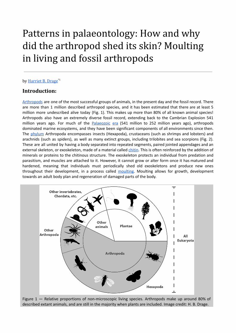
Arthropods are one of the most successful groups of animals, in the present day and the fossil record. There are more than 1 million described arthropod species, and it has been estimated that there are at least 5 million more undescribed alive today (Fig. 1). This makes up more than 80% of all known animal species! Arthropods also have an extremely diverse fossil record, extending back to the Cambrian Explosion 541 million years ago. For much of the Palaeozoic era (541 million to 252 million years ago), arthropods dominated marine ecosystems, and they have been significant components of all environments since then. The phylum Arthropoda encompasses insects (Hexapoda), crustaceans (such as shrimps and lobsters) and arachnids (such as spiders), as well as many extinct groups, including trilobites and sea scorpions (Fig. 2). These are all united by having a body separated into repeated segments, paired jointed appendages and an external skeleton, or exoskeleton, made of a material called chitin . This is often reinforced by the addition of minerals or proteins to the chitinous structure. The exoskeleton protects an individual from predation and parasitism, and muscles are attached to it. However, it cannot grow or alter form once it has matured and hardened, meaning that individuals must periodically shed old exoskeletons and produce new ones throughout their development, in a process called moulting . Moulting allows for growth, development towards an adult body plan and regeneration of damaged parts of the body.
Figure 1 — Relative proportions of nonmicroscopic living species. Arthropods make up around 80% of described extant animals, and are still in the majority when plants are included. Image credit: H. B. Drage.
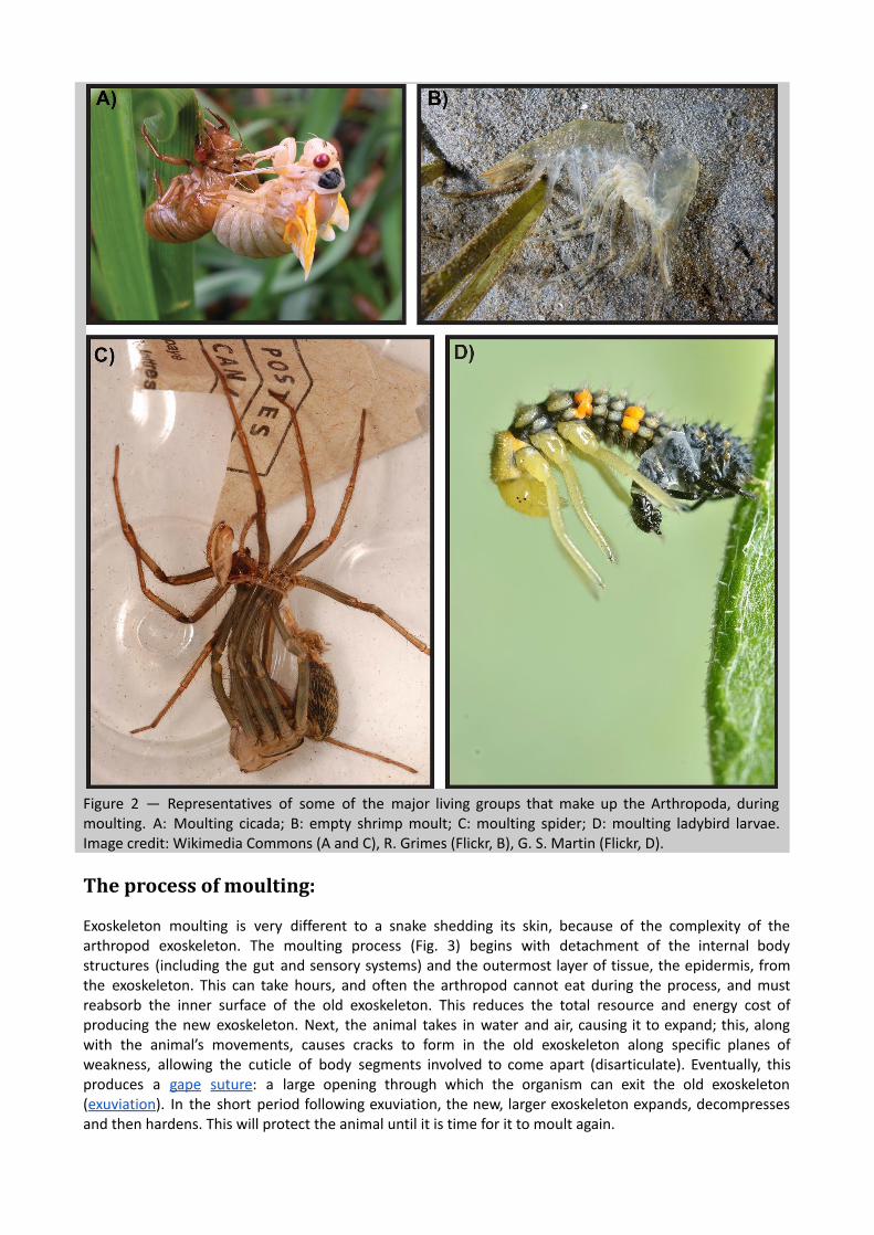
Figure 2 — Representatives of some of the major living groups that make up the Arthropoda, during moulting. A: Moulting cicada; B: empty shrimp moult; C: moulting spider; D: moulting ladybird larvae. Image credit: Wikimedia Commons (A and C), R. Grimes (Flickr, B), G. S. Martin (Flickr, D).
The process of moulting:
Exoskeleton moulting is very different to a snake shedding its skin, because of the complexity of the arthropod exoskeleton. The moulting process (Fig. 3) begins with detachment of the internal body structures (including the gut and sensory systems) and the outermost layer of tissue, the epidermis, from the exoskeleton. This can take hours, and often the arthropod cannot eat during the process, and must reabsorb the inner surface of the old exoskeleton. This reduces the total resource and energy cost of producing the new exoskeleton. Next, the animal takes in water and air, causing it to expand; this, along with the animal’s movements, causes cracks to form in the old exoskeleton along specific planes of weakness, allowing the cuticle of body segments involved to come apart (disarticulate). Eventually, this produces a gape suture : a large opening through which the organism can exit the old exoskeleton ( exuviation ). In the short period following exuviation, the new, larger exoskeleton expands, decompresses and then hardens. This will protect the animal until it is time for it to moult again.

Figure 3 — A generalized process of exoskeleton moulting. Source .
How do we identify moults in the fossil record?
Because the presence of an exoskeleton is a defining characteristic of Arthropoda, a number of fossilized arthropods should show evidence of moulting (Fig. 4). But how can we distinguish moulted exoskeletons from the carcasses of deceased individuals in the fossil record? Firstly, palaeontologists studying arthropod moulting in the fossil record need to examine and compare a number of potential fossil moults and carcasses. The identification of suspected gape sutures at the same location in the preserved exoskeletons of multiple fossils suggests a way of moulting for that group. For example, a repeatedly observable disarticulation between the cephalon (headshield) and thorax (body) in a species, with the fossil otherwise fully intact, indicates a gape suture used for moulting at this location.
Figure 4 — Examples of arthropod moult assemblages preserved in the fossil record. A: Meyeria magna decapod crustacean, OUMNH K.755; B: Asaphiscus wheeleri , KUMIP 153922; C: Ogygopsis klotzi, OUMNH AT.205; D: adelopthalmid eurypterid (sea scorpion), OUMNH D.2184. Scale bars: 5 mm in A and B, 10 mm in C and D.

Specimens are usually considered to be carcasses if they are completely articulated, or are broken or entirely disarticulated. It may also be possible to identify other features that have been linked to moulting, such as crushing of particular elements, compression and overlapping of body segments or a systematic orientation of the limbs. However, the circumstances of fossil preservation should also be considered. Environmental conditions (such as fastflowing river currents) or decay processes at the time of fossilization may result in disarticulation of a complete carcass that looks like moulting. Subsequent deformation can also produce repeated crushing and overlapping features, making carcasses look like moults.
Figure 5 — Moulting in chelicerate animals. A: empty moult of a tarantula, showing the carapace flipped forward. Image credit: Frosted Peppercorn (Flickr). B: Horseshoe crab carcass (right) having just emerged from the old exoskeleton on the left. Image credit: Wikimedia Commons. C. Counting from the top left, 1 and 5 are moults from living scorpions that show extension of the chelicerae and pedipalps respectively, and 2 and 6 show the same in fossil scorpion moults. 3 and 7 are carcasses of living scorpions that show retraction of the chelicerae and pedipalps respectively, while 4 and 8 show the same in fossilised scorpion carcasses. From figures 710 and 1518 of McCoy & Brandt (2009).

For some arthropod groups, it is particularly difficult to study moulting (Fig. 5). Horseshoe crabs (Xiphosura) and scorpions (Scorpiones, both part of Chelicerata) moult using a horizontal gape suture at the front of the animal, without requiring disarticulation of any body segments (Fig. 5B). The suture closes after exuviation, often leaving a completely intact moulted exoskeleton that may be mistaken for a carcass. In a scenario such as this, it is important to carefully compare numerous fossil specimens, and to identify any repeated limb orientations or segment preservation styles. For example, splaying and extension of the limbs in modern moults has allowed researchers to describe the moulting behaviour of extinct scorpions from museum collections (Fig. 5C).
What can we learn from modern and fossil moults?
The successful identification of moults in the fossil record has allowed for the reconstruction of moulting behaviours in many arthropod groups. Some of these, such as trilobites or eurypterids (sea scorpions), have no living relatives, and the moult fossil record therefore provides the only available source of behavioural and developmental data. For other groups, studies of moulting have shown that highly specific anatomylinked behaviours were ancestral and evolved long ago. For example, crabs that lived around 70 million years ago moulted like modern crabs. Studying fossilized moults enables us to trace the origins and prevalence of different moulting strategies, and the effects that these had on the longterm evolution of morphology (body shape), behaviour, development and ecology in arthropods. The evolutionary histories of moulting in a few key arthropod groups are discussed below.
Moulting in Decapoda
Decapoda, a group of crustaceans, includes the shrimp, lobsters and crabs. At least two moulting behaviours can be observed for living decapods, and these extend far back into their early fossil record. Shrimp and lobsters, which have a long body formed of a carapace (shell) and segmented abdomen, moult through a gape suture formed between these two parts. The animal then withdraws its limbs from in front of the body, and emerges forwards and upwards from this gape (Fig. 6). Crabs, which have a rounder dorsal (back) carapace, also break along the back edge of the shell, disarticulating it from the abdomen at the rear end. This often involves the carapace rotating away or flipping forwards towards the head. To exit the old exoskeleton, the crab moves backwards, withdrawing its legs last from in front of the body (Fig. 7). Understanding moulting in crabs has had an impact on the food industry. Edible ‘softshell crabs’ are not a single species, but actually crabs that have been cooked immediately after moulting, before the new exoskeleton can harden. We can also use these descriptions of moulting in living representatives to identify fossilized moulted exoskeletons, when we see similar gape sutures. For all these crustaceans, this style of moulting can be seen throughout their history.
Figure 6 — Video of a moulting juvenile rock lobster. Creative Commons licensed.
Figure 7 — Video of a moulting spider crab. Creative Commons licensed.
Moulting in Chelicerata
The group Chelicerata includes the spiders and scorpions (Arachnida), and the horseshoe crabs and extinct sea scorpions (together, the Merostomata). Spiders, potentially including fossils from the Devonian (419 million to 359 million years ago) and Permian (299 million to 252 million years ago) periods, moult using a similar behaviour to crabs. Their similar anatomy means that they use the same backwards movement out of an opening at the back and side of the old exoskeleton, leaving behind moults with disrupted carapaces and clear stepwise bodysize increases. This can be seen in images of moulted pet tarantula that are reminiscent of a horror movie (Figs 2C, 5A).
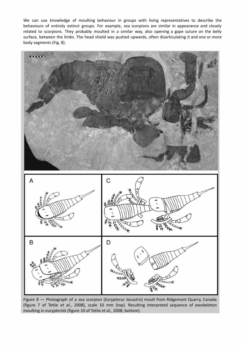
We can use knowledge of moulting behaviour in groups with living representatives to describe the behaviours of entirely extinct groups. For example, sea scorpions are similar in appearance and closely related to scorpions. They probably moulted in a similar way, also opening a gape suture on the belly surface, between the limbs. The head shield was pushed upwards, often disarticulating it and one or more body segments (Fig. 8).
Figure 8 — Photograph of a sea scorpion ( Eurypterus lacustris ) moult from Ridgemont Quarry, Canada (figure 7 of Tetlie et al., 2008), scale 10 mm (top). Resulting interpreted sequence of exoskeleton moulting in eurypterids (figure 10 of Tetlie et al., 2008; bottom).

Moulting in Trilobita
Trilobites were one of the most successful groups of Palaeozoic arthropods. They were an extremely anatomically, geographically and ecologically diverse group (Fig. 9), represented by more than 20,000 species and found on every continent. Their ultimate demise in the mass extinction at the end of the Permian period followed a series of declines in diversity. This means that we can only reconstruct their moulting behaviour through comparison to extant groups such as those described above, and through the preservation of fossilized moults. For trilobites, moulting is one of the few direct behaviours that we can reconstruct, and it is important for helping us to understand their ecology, evolutionary relationships and morphological evolution.
Figure 9 — The diversity of trilobites at the level of orders in the Linnean system is extremely high. Image credit: photographs Wikimedia Commons and Creative Commons licensed, drawings by H. B. Drage.
Trilobite moulting is particularly interesting to look at because it separates these animals from the rest of Arthropoda. Trilobites as a whole show a diverse range of moulting behaviours. Even more unusually, individual species could use more than one method of moulting, depending on their environment or body dimensions. Most species had sutures on the back surface between lateral sections of the head called free

cheeks (or librigenae). During moulting, opening of these sutures was used to create a gape suture (Fig. 10A, D, E). In some lineages, these facial sutures have become fused, which is occasionally associated with reduction of size of the eyes. These species usually moulted through movements that disarticulated the entire cephalon (Fig. 10B). However, trilobite species tend to show great fluidity around this general pattern. Many species that usually moulted by opening the facial sutures have moult assemblages preserved with disarticulation of the whole cephalon, or the remainder of the cephalon detached following removal of the free cheeks (Fig. 10F). And in rare events, probably during difficult moults, other parts of the exoskeleton would become disarticulated, for example between body segments (Fig. 10C, D, F), or the tail shield (pygidium). This behavioural variability within single species may have been integral to the evolution of trilobites. Preliminary work has suggested that it is not isolated to certain groups, and may be linked to their patterns of diversity through the Palaeozoic era (541 million to 252 million years ago). Perhaps their flexibility during moulting helped them to become one of the most diverse and ecologically important groups during the first half of the Palaeozoic?
Figure 10 — Trilobite moult assemblages showing varying moulting behaviours. A: Acidaspis coronata , OUMNH C.17494; B: Ogygiocarella debuchii , OUMNH B.263; C: Acadoparadoxides sp., PMU 25636; D: Acadoparadoxides sp., PMU 25995; E: Eccaparadoxides sp., PMY 25636; F: Accadoparadoxides sp., PMU25690. Scale bars: 5 mm in A, C, E, F; 10 mm in B, D. Image credit: figure 2 in Daley & Drage (2016).
Why is moulting important to study?
Moulting is arguably the most significant recurring event in the life of an arthropod. It has been suggested that up to 90% of arthropod deaths take place during moulting, but this includes potentially difficult moults late in life, when the individual has become relatively large. This is because the act of moulting is very energy intensive and can be fatal if it goes on for too long, and it leaves the animal exceptionally vulnerable

to predation. Some groups, such as mud lobsters and potentially some phacopid trilobites, burrowed into the seafloor sediment as protection during moulting events. How arthropods moult, how long this takes, how they protect themselves during moulting and their moultingadapted characteristics are key to our understanding of their evolution. In particular, these may have shaped their total morphology, life cycles and behaviour, and exploring this allows us to better understand the macroevolution of the Arthropoda.
Suggestions for further reading:
Brandt, D. S. Ecdysial efficiency and evolutionary efficacy among marine arthropods: implications for trilobite survivorship. Alcheringa 26, 399–421 (2002). DOI: 10.1080/03115510208619264 Daley, A. C. & Drage, H. B. 2016. The fossil record of ecdysis, and trends in the moulting behaviour of trilobites. Arthropod Structure & Development 45, 71–96 (2016). DOI: 10.1016/j.asd.2015.09.004 Ewer, J. How the ecdysozoan changed its coat. PloS Biology 3, e349 (2005). DOI: 10.1371/journal.pbio.0030349 Gon, S. III. A Guide to the Orders of Trilobites (2016). [ View ] Henningsmoen, G. Moulting in trilobites. Fossils and Strata 4, 179–200 (1975). McCoy, V. E. & Brandt, D. S. Scorpion taphonomy: criteria for distinguishing fossil scorpion molts and carcasses. Journal of Arachnology 37, 312–320 (2009). [ View ] Whittington, H. B. Articulation and exuviation in Cambrian trilobites. Philosophical Transactions of the Royal Society B 329B, 27–46 (1990). DOI: 10.1098/rstb.1990.0147
1 Department of Zoology, University of Oxford, The Tinbergen Building, South Parks Road, Oxford OX1 3PS