Title Anterior approach to cervical spine Author(s) Cheung...
28
Title Anterior approach to cervical spine Author(s) Cheung, KMC; Mak, KC; Luk, KDK Citation Spine, 2012, v. 37 n. 5, p. E297-E302 Issued Date 2012 URL http://hdl.handle.net/10722/170192 Rights This is a non-final version of an article published in final form in Spine, 2012, v. 37 n. 5, p. E297-E302; This work is licensed under a Creative Commons Attribution-NonCommercial-NoDerivatives 4.0 International License.
Transcript of Title Anterior approach to cervical spine Author(s) Cheung...

Title Anterior approach to cervical spine
Author(s) Cheung, KMC; Mak, KC; Luk, KDK
Citation Spine, 2012, v. 37 n. 5, p. E297-E302
Issued Date 2012
URL http://hdl.handle.net/10722/170192
Rights
This is a non-final version of an article published in final form inSpine, 2012, v. 37 n. 5, p. E297-E302; This work is licensed undera Creative Commons Attribution-NonCommercial-NoDerivatives4.0 International License.

Editorial Manager(tm) for Spine Journal Manuscript Draft
Manuscript Number: SPINE F11111 Title: Anterior approach to cervical spine Article Type: Focus Paper Keywords:
Cervical Spine; surgery Corresponding Author: Kenneth MC. Cheung, FRCS, FHKCOS, FHKAM(Orth) Corresponding
Author's Institution: The University of Hong Kong Medical Centre First Author: Kenneth MC. Cheung, FRCS, FHKCOS,
FHKAM(Orth) Order of Authors: Kenneth MC. Cheung, FRCS, FHKCOS, FHKAM(Orth);K.C. Mak, FRCS(Ed),
FHKAM(Orth),;Keith D.K. Luk, MCh(Ortho), FRCSE, FRCSG, FRACS, FHKAM (Ortho)

Title Page
Anterior approach to cervical spine

Running Head: Anterior approach to cervical spine Order of Authors: Kenneth M C Cheung, MD, FRCS, FHKAM(Orth), K C Mak, FRCS(Ed), FHKAM(Orth), Keith D K Luk, MCh(Ortho), FRCSE, FRCSG, FRACS, FHKAM (Ortho) Keywords: anterior cervical decompression; transoral approach; anterolateral approach; manubriotomy
Correspondence: Kenneth M.C. Cheung, MBBS, MD, FRCS, FHKCOS, FHKAM(Orth) Department of Orthopaedics & Traumatology Queen Mary Hospital 102 Pokfulam Road Hong Kong, SAR, China Tel: +852 2255 4254 Fax: +852 2817 4392 Email: [email protected] Affiliations: Department of Orthopaedics and Traumatology, The University of Hong Kong, Pokfulam, Hong Kong SAR, China Disclosure: The authors have no financial or competing interests to disclose.

*Structured Abstract (300 words)
Abstract
Study Design: Review of surgical technique
Objective: To provide accounts of the authors’ preferred methods for performing anterior
cervical surgery with personal tips and pearls.
Summary of Background Data: Many have described the various anterior surgical approaches to the cervical
spine, and in this review we hope to describe our preferences, highlighted with some tips and pearls.
Methods: Various accounts of the transoral, the anterolateral (Smith-Robinson) and the split manubrium
approaches were reviewed and used as the basis of the review. Additional notes with
regards to the authors’ preference were noted to provide further guidance. The description were
delineated from the most cephalad to the most caudal.
Results: The transoral, the anterolateral (Smith-Robinson) approach and the manubriotomy approach were
described. Each account starts with the basic pre-operative considerations, then describes the incision and the
main anatomical landmarks, and finally concludes with closure and main complications to monitor for. A brief
description of the main pathologies that each approach may address is also provided.
Conclusion: The 3 anterior approaches to the cervical spine are direct and elegant solutions to pathologies arising
from the anterior column. They supplement the more commonly used posterior approaches, which provide
stronger multilevel fixation, and thus is an essential tool in the armamentarium of a spine surgeon.

*Key Points (3-5 main points of the article)
Key Points
1 The transoral approach provides access to C0 to C2 with prerequisites including adequate interdental distance, neck hyperextension, and specialized retractors.
2 Once the anterior tubercle of C1 is identified for the incision of the transoral approach, the main caution is not to go wider than 15mm from the midline least the vertebral artery is injured.
3 The anterolateral (Smith-Robinson) approach is the most commonly employed of the three and allows access from C3 to cervicothoracic junction.
4 We prefer to approach from the right side for C3 to C6, and from the left for cervicothoracic junction to avoid need to protect the recurrent laryngeal nerve.
5 The manubriotomy approach can either be 1-or 2-sided thereby affording customized access as far down as T4, without the morbidity of a full sternal split or clavicular osteotomies.

*Mini Abstract (50 words)
Mini-abstract
The classic transoral and anterolateral (Smith-Robinson) approaches which allow direct anterior
access to the cervical spine is described with the authors’ preferences highlighted. The 1-or 2sided
manubriotomy provides an approach to the cervicothoracic junction without the morbidity of the
full sternotomy.


*References (cited in order of appearance)
References
1. Fang HSY, Ong GB. Direct Anterior Approach to the Upper Cervical Spine. Journal of Bone and Joint Surgery 1962;44:1588-604.
2. Smith GW, Robinson RA. The treatment of certain cervical-spine disorders by anterior removal of the intervertebral disc and interbody fusion. J Bone Joint Surg Am 1958;40-A:607-24.
3. Southwick WO, Robinson RA. Surgical approaches to the vertebral bodies in the cervical and lumbar regions. J Bone Joint Surg Am 1957;39-A:631-44.
4. Luk KD, Cheung KM, Leong JC. Anterior approach to the cervicothoracic junction by unilateral or bilateral manubriotomy. A report of five cases. J Bone Joint Surg Am 2002;84A:1013-7.
5. Shaha AR, Johnson R, Miller J, et al. Transoral-transpharyngeal approach to the upper cervical vertebrae. Am J Surg 1993;166:336-40.
6. Biyani A, An H. Anterior upper cervical spine approaches. In Herkowitz HN ed. The cervical spine surgery atlas. 2nd ed. Philadelphia: Lippincott Williams & Wilkins, 2003:69
89. 1. Henn JS, Lee MC, Rhoton ALJ. Transoral approach to craniocervical junction and upper cervical spine.
In Kim DH, Henn JS, Vaccaro AR, et al. eds. Surgical anatomy & techniques to the spine. Philadelphia: Saunders Elsevier, 2006:3-32.
2. Winter R, Lonstein J, Dennis F, et al. Anterior upper cervical procedures. Atlas of spine surgery. Philadelphia: W.B. Saunders, 1995:1-17.
3. Crockard H. Midline ventral approaches to the craniocervical junction and upper cervical spine. In Sherk H ed. The cervical spine: an atlas of cervical procedures. Philadelphia: JB Lippincott Co., 1994:93-112.
4. Mendoza N, H.A. C. Anterior transoral procedures. In An HS, Riley LHI eds. An atlas of surgery of the spine. London: Martin Dunitz, 1998:55-69.
5. Robinson RA, Smith G. Anterolateral cervical disk removal and interbody fusion for cervical disk syndrome. Bull Johns Hopkins Hosp 1955;96:223-4.
6. German J, Benzel E, Alexander J. Anatomy and surgical approaches and exposure of the vertebral column, the cervical spine. In Benzel E ed. Spine surgery: technique, complication avoidance and management. New York: Churchill Livingstone, 1999:145-56.
7. Silber J, Albert T. Anterior and anterolateral, mid and lower cervical spine approaches: transverse and longitudinal (C3 to C7). In Herkowitz HN ed. The cervical spine surgery atlas. Philadelphia: Lippincott Williams & Wilkins, 2003:91-8.
8. Hillard VH, Apfelbaum RI. Surgical management of cervical myelopathy: indications and techniques for multilevel cervical discectomy. Spine J 2006;6:242S-51S.
9. Chang U, Lee MC, Kim DH. Anterior approach to the midcervical spine. In Kim DH, Henn JS, Vaccaro A, et al. eds. Surgical anatomy & techniques to the spine. Philadelphia: Saunders Elsevier, 2006:45-56.
10. Curylo LJ, Mason HC, Bohlman HH, et al. Tortuous course of the vertebral artery and anterior cervical decompression: a cadaveric and clinical case study. Spine (Phila Pa 1976) 2000;25:2860-4.
11. Birch R, Bonney G, Marshall RW. A surgical approach to the cervicothoracic spine. J Bone Joint Surg Br 1990;72:904-7.

12. Fang HS, Ong GB, Hodgson AR. Anterior spinal fusion: The operative approaches. Clin Orthop Relat Res 1964;35:16-33.
13. Darling GE, McBroom R, Perrin R. Modified anterior approach to the cervicothoracic junction. Spine (Phila Pa 1976) 1995;20:1519-21.
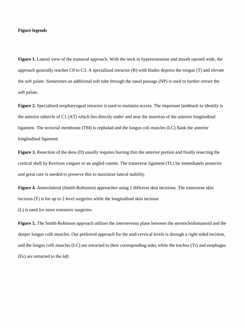
Figure legends
Figure 1. Lateral view of the transoral approach. With the neck in hyperextension and mouth opened wide, the
approach generally reaches C0 to C3. A specialized retractor (R) with blades depress the tongue (T) and elevate
the soft palate. Sometimes an additional soft tube through the nasal passage (NP) is used to further retract the
soft palate.
Figure 2. Specialized oropharyngeal retractor is used to maintain access. The important landmark to identify is
the anterior tubercle of C1 (AT) which lies directly under and near the insertion of the anterior longitudinal
ligament. The tectorial membrane (TM) is cephalad and the longus coli muscles (LC) flank the anterior
longitudinal ligament.
Figure 3. Resection of the dens (D) usually requires burring thin the anterior portion and finally resecting the
cortical shell by Kerrison ronguer or an angled curette. The transverse ligament (TL) lie immediately posterior
and great care is needed to preserve this to maximize lateral stability.
Figure 4. Anterolateral (Smith-Robinson) approaches using 2 different skin incisions. The transverse skin
incision (T) is for up to 2 level surgeries while the longitudinal skin incision
(L) is used for more extensive surgeries.
Figure 5. The Smith-Robinson approach utilizes the internervous plane between the sternocleidomastoid and the
deeper longus colli muscles. Our preferred approach for the mid-cervical levels is through a right sided incision,
and the longus colli muscles (LC) are retracted to their corresponding sides while the trachea (Tr) and esophagus
(Es) are retracted to the left.

Figure 6. The manubriotomy skin incision is a longitudinal incision that extends from the manubrial notch (MN)
to 3 cm caudad to the sternal angle. The incision may be extended cephalad anterior the the sternocleidomastoid
(SCM) if more proximal levels require operating. The medial end of the right clavicle (C) is shown.
Figure 7. The longitudinal osteotomy is made over the midline of the manubrium with the
transverse limb cutting across the 2nd
intercostal space. Therefore an L-shaped or inverted T-shaped cut is created
depending on the width of the approach required.
Figure 8. This schematic diagram illustrates the positions of the main vessels relative to the 3rd thoracic vertebra
(T3). On the right the 3thoracic vertebra is flanked by the right brachiocephalic vein (RBv) and artery (A). The
left brachiocephalic vein (LBv) crosses caudal and anterior to the 3rd
thoracic vertebra while the left common
carotid (LCC) flanks the left. The esophagus and trachea are not shown here as it is retracted to the left under the
retractor (TE).

Figures ( TIFF, EPS, or PPT only)
Figure 1. Lateral view of the
transoral approach. With the
neck in hyperextension and
mouth opened wide, the
approach generally reaches C0
to C3. A specialized retractor
(R) with blades depress the
tongue (T) and elevate the soft
palate. Sometimes an additional
soft tube through the nasal
passage (NP) is used to further
retract the soft palate.

Figure 2. Specialized oropharyngeal retractor is used to maintain access. The important landmark to identify is
the anterior tubercle of C1 (AT) which lies directly under and near the insertion of the anterior longitudinal
ligament. The tectorial membrane (TM) is cephalad and the longus coli muscles (LC) flank the anterior
longitudinal ligament.
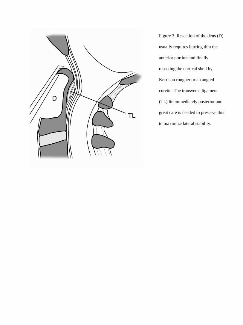
Figure 3. Resection of the dens (D)
usually requires burring thin the
anterior portion and finally
resecting the cortical shell by
Kerrison ronguer or an angled
curette. The transverse ligament
(TL) lie immediately posterior and
great care is needed to preserve this
to maximize lateral stability.

Figure 4.
Anterolateral
(Smith-Robinson)
approaches using 2
different skin
incisions. The
transverse skin
incision (T) is for
up to 2 level
surgeries while the
longitudinal skin
incision
(L) is used for more extensive surgeries.

Figure 5. The Smith-Robinson approach utilizes the internervous plane between the sternocleidomastoid and the
deeper longus colli muscles. Our preferred approach for the mid-cervical levels is through a right sided incision,
and the longus colli muscles (LC) are retracted to their corresponding sides while the trachea (Tr) and esophagus
(Es) are retracted to the left.

Figure 6. The manubriotomy skin incision is
a longitudinal incision that extends from the
manubrial notch (MN) to 3 cm caudad to the
sternal angle. The incision may be extended
cephalad anterior the the sternocleidomastoid
(SCM) if more proximal levels require
operating. The medial end of the right
clavicle (C) is shown.
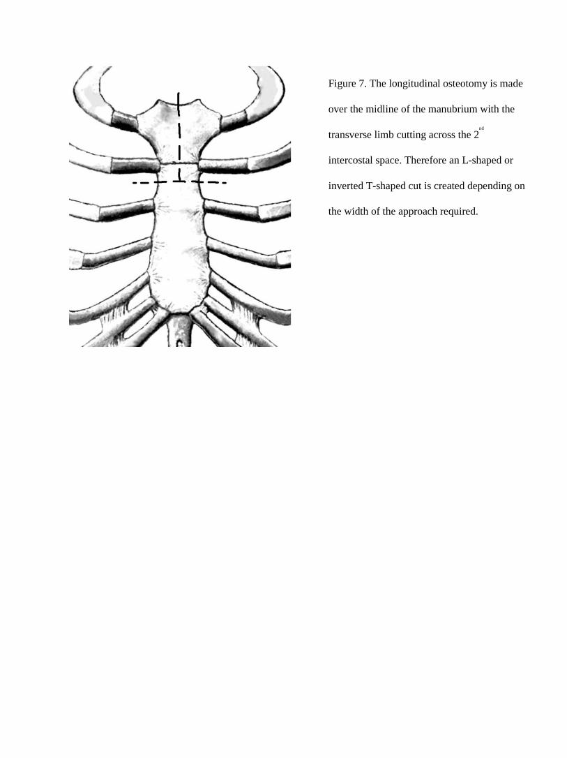
Figure 7. The longitudinal osteotomy is made
over the midline of the manubrium with the
transverse limb cutting across the 2nd
intercostal space. Therefore an L-shaped or
inverted T-shaped cut is created depending on
the width of the approach required.

Figure 8. This schematic diagram illustrates the positions of the main vessels relative to the
rd rd
3thoracic vertebra (T3). On the right the
3thoracic vertebra is flanked by the right
brachiocephalic vein (RBv) and artery (A).
The left brachiocephalic vein (LBv) crosses
caudal and anterior to the 3rd
thoracic vertebra
while the left common carotid (LCC) flanks
the left. The esophagus and trachea are not
shown here as it is retracted to the left under
the retractor (TE).