Thrombospondin-1 and CD47 regulate blood pressure and...
10
Thrombospondin-1 and CD47 regulate blood pressure and cardiac responses to vasoactive stress Jeff S. Isenberg a, 1 ,2 , Yan Qin b, 1 , Justin B. Maxhimer a , John M. Sipes a , Daryl Despres c , Jurgen Schnermann b , William A. Frazier d , David D. Roberts a, a Laboratory of Pathology, Center for Cancer Research, National Cancer Institute, Bethesda, Maryland 20892, United States b Kidney Diseases Branch, National Institute of Diabetes and Digestive and Kidney Diseases, Bethesda, Maryland 20892, United States c Mouse Imaging Facility, National Institutes of Health, Bethesda, Maryland 20892, United States d Department of Biochemistry and Molecular Biophysics, Washington University School of Medicine, St. Louis, Missouri 63110, United States abstract article info Article history: Received 2 September 2008 Received in revised form 17 December 2008 Accepted 5 January 2009 Keywords: Thrombospondin-1 CD47 Nitric oxide Blood pressure Cardiac output Nitric oxide (NO) locally regulates vascular resistance and blood pressure by modulating blood vessel tone. Thrombospondin-1 signaling via its receptor CD47 locally limits the ability of NO to relax vascular smooth muscle cells and increase regional blood ow in ischemic tissues. To determine whether thrombospondin-1 plays a broader role in central cardiovascular physiology, we examined vasoactive stress responses in mice lacking thrombospondin-1 or CD47. Mice lacking thrombospondin-1 exhibit activity-associated increases in heart rate, central diastolic and mean arterial blood pressure and a constant decrease in pulse pressure. CD47-decient mice have normal central pulse pressure but elevated resting peripheral blood pressure. Both null mice show exaggerated decreases in peripheral blood pressure and increased cardiac output and ejection fraction in response to NO. Autonomic blockade also induces exaggerated hypotensive responses in awake thrombospondin-1 null and CD47 null mice. Both null mice exhibit a greater hypotensive response to isourane, and autonomic blockage under isourane anesthesia leads to premature death of thrombos- pondin-1 null mice. Conversely, the hypertensive response to epinephrine is attenuated in thrombospondin-1 null mice. Thus, the matricellular protein thrombospondin-1 and its receptor CD47 serve as acute physiological regulators of blood pressure and exert a vasopressor activity to maintain global hemodynamics under stress. Published by Elsevier B.V. 1. Introduction Cardiovascular homeostasis requires constant regulation of tissue perfusion and blood ow through coordinated interactions of the autonomic nervous system, heart, lungs and blood vessels. Meeting regional metabolic demands requires rapid and efcient redistribu- tion of blood ow. Nitric oxide (NO) is a major physiological regulator of blood vessel diameter and blood ow (Arnal et al., 1999; Ignarro, 2002). In response to specic stressors, arterial endothelium increases its production of NO, which diffuses into the adjacent vascular smooth muscle cells (VSMC) and causes cGMP-mediated relaxation by activating soluble guanylate cyclase (sGC). This results in vessel dilation and increased blood ow. Direct cGMP-dependent and indirect activation of cGMP phosphodiesterases (PDE) provides negative feedback to limit NO/cGMP signaling (Mullershausen et al., 2003). The matricellular protein thrombospondin-1 (TSP1), which is produced by vascular cells and circulates at 100–200 pM levels in plasma (Bergseth et al., 2000), controls a second pathway that limits NO signaling by preventing activation of sGC (Isenberg et al., 2005b, 2006b). TSP1 also inhibits signaling downstream of cGMP, and in platelets cGMP-dependent protein kinase (cGK) is a second target of TSP1 (Isenberg et al., 2008b). Physiological levels of TSP1 potently inhibit NO-driven relaxation of contracting VSMC and thereby limit the ability of NO to increase tissue blood ow at rest and under stress (Isenberg et al., 2007a). The ability of TSP1 to block NO/cGMP signaling in vascular cells requires its cell surface receptor CD47 (Isenberg et al., 2006a). Targeting of either TSP1 or CD47 relieves the inhibition of sGC and signicantly enhances tissue survival and blood ow after local ischemic challenges (Isenberg et al., 2007b,c). NO also has systemic cardiovascular activities. Some NOS knockout mice exhibit hypertensive phenotypes (Ortiz and Garvin, 2003). Systemic administration of NO donors or nitrovasodilators alters blood pressure and cardiac function, leading to extensive use of these agents for treating chronic and acute cardiovascular diseases ( Hermann et al., 2006). Based on their ability to limit NO signaling, we hypothesized that TSP1 and CD47 could also regulate systemic cardiovascular responses. We show here that TSP1 and CD47-null mice exhibit alterations in resting blood pressure and Matrix Biology 28 (2009) 110–119 Corresponding author. NIH, Building 10 Room 2A33, Bethesda, MD 20892-1500, United States. Tel.: +1 301 496 6264. E-mail address: [email protected] (D.D. Roberts). 1 These authors contributed equally to the manuscript. 2 Present address: Hemostasis and Vascular Biology Research Institute and the Department of Medicine, University of Pittsburgh, Pittsburgh, PA 15260. 0945-053X/$ – see front matter. Published by Elsevier B.V. doi:10.1016/j.matbio.2009.01.002 Contents lists available at ScienceDirect Matrix Biology journal homepage: www.elsevier.com/locate/matbio
Transcript of Thrombospondin-1 and CD47 regulate blood pressure and...

Thrombospondin-1 and CD47 regulate blood pressure and cardiac responses tovasoactive stress
Jeff S. Isenberg a,1,2, Yan Qin b,1, Justin B. Maxhimer a, John M. Sipes a, Daryl Despres c, Jurgen Schnermann b,William A. Frazier d, David D. Roberts a,!a Laboratory of Pathology, Center for Cancer Research, National Cancer Institute, Bethesda, Maryland 20892, United Statesb Kidney Diseases Branch, National Institute of Diabetes and Digestive and Kidney Diseases, Bethesda, Maryland 20892, United Statesc Mouse Imaging Facility, National Institutes of Health, Bethesda, Maryland 20892, United Statesd Department of Biochemistry and Molecular Biophysics, Washington University School of Medicine, St. Louis, Missouri 63110, United States
a b s t r a c ta r t i c l e i n f o
Article history:Received 2 September 2008Received in revised form 17 December 2008Accepted 5 January 2009
Keywords:Thrombospondin-1CD47Nitric oxideBlood pressureCardiac output
Nitric oxide (NO) locally regulates vascular resistance and blood pressure by modulating blood vessel tone.Thrombospondin-1 signaling via its receptor CD47 locally limits the ability of NO to relax vascular smoothmuscle cells and increase regional blood !ow in ischemic tissues. To determine whether thrombospondin-1plays a broader role in central cardiovascular physiology, we examined vasoactive stress responses in micelacking thrombospondin-1 or CD47. Mice lacking thrombospondin-1 exhibit activity-associated increases inheart rate, central diastolic and mean arterial blood pressure and a constant decrease in pulse pressure.CD47-de"cient mice have normal central pulse pressure but elevated resting peripheral blood pressure. Bothnull mice show exaggerated decreases in peripheral blood pressure and increased cardiac output andejection fraction in response to NO. Autonomic blockade also induces exaggerated hypotensive responses inawake thrombospondin-1 null and CD47 null mice. Both null mice exhibit a greater hypotensive response toiso!urane, and autonomic blockage under iso!urane anesthesia leads to premature death of thrombos-pondin-1 null mice. Conversely, the hypertensive response to epinephrine is attenuated in thrombospondin-1null mice. Thus, the matricellular protein thrombospondin-1 and its receptor CD47 serve as acute physiologicalregulators of blood pressure and exert a vasopressor activity to maintain global hemodynamics under stress.
Published by Elsevier B.V.
1. Introduction
Cardiovascular homeostasis requires constant regulation of tissueperfusion and blood !ow through coordinated interactions of theautonomic nervous system, heart, lungs and blood vessels. Meetingregional metabolic demands requires rapid and ef"cient redistribu-tion of blood !ow. Nitric oxide (NO) is a major physiological regulatorof blood vessel diameter and blood !ow (Arnal et al., 1999; Ignarro,2002). In response to speci"c stressors, arterial endothelium increasesits production of NO, which diffuses into the adjacent vascular smoothmuscle cells (VSMC) and causes cGMP-mediated relaxation byactivating soluble guanylate cyclase (sGC). This results in vesseldilation and increased blood !ow. Direct cGMP-dependent andindirect activation of cGMP phosphodiesterases (PDE) providesnegative feedback to limit NO/cGMP signaling (Mullershausen et al.,
2003). The matricellular protein thrombospondin-1 (TSP1), which isproduced by vascular cells and circulates at 100–200 pM levels inplasma (Bergseth et al., 2000), controls a second pathway that limitsNO signaling by preventing activation of sGC (Isenberg et al., 2005b,2006b). TSP1 also inhibits signaling downstream of cGMP, and inplatelets cGMP-dependent protein kinase (cGK) is a second target ofTSP1 (Isenberg et al., 2008b). Physiological levels of TSP1 potentlyinhibit NO-driven relaxation of contracting VSMC and thereby limitthe ability of NO to increase tissue blood !ow at rest and under stress(Isenberg et al., 2007a). The ability of TSP1 to block NO/cGMPsignaling in vascular cells requires its cell surface receptor CD47(Isenberg et al., 2006a). Targeting of either TSP1 or CD47 relieves theinhibition of sGC and signi"cantly enhances tissue survival and blood!ow after local ischemic challenges (Isenberg et al., 2007b,c).
NO also has systemic cardiovascular activities. Some NOS knockoutmice exhibit hypertensivephenotypes (Ortiz andGarvin, 2003). Systemicadministration of NO donors or nitrovasodilators alters blood pressureand cardiac function, leading to extensive use of these agents for treatingchronic and acute cardiovascular diseases (Hermann et al., 2006). Basedon their ability to limitNOsignaling,wehypothesized that TSP1andCD47could also regulate systemic cardiovascular responses.Weshowhere thatTSP1 andCD47-nullmice exhibit alterations in resting bloodpressure and
Matrix Biology 28 (2009) 110–119
! Corresponding author. NIH, Building 10 Room 2A33, Bethesda, MD 20892-1500,United States. Tel.: +1 301 496 6264.
E-mail address: [email protected] (D.D. Roberts).1 These authors contributed equally to the manuscript.2 Present address: Hemostasis and Vascular Biology Research Institute and the
Department of Medicine, University of Pittsburgh, Pittsburgh, PA 15260.
0945-053X/$ – see front matter. Published by Elsevier B.V.doi:10.1016/j.matbio.2009.01.002
Contents lists available at ScienceDirect
Matrix Biology
j ourna l homepage: www.e lsev ie r.com/ locate /matb io
hyperdynamic responses to vasoactive challenges. These results demon-strate for the "rst time that a matricellular protein can acutely regulateblood pressure and cardiovascular responses to stress.
2. Results
2.1. TSP1-null mice are hypertensive with activity
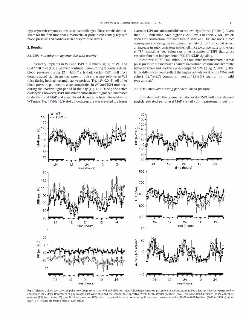
Telemetry implants in WT and TSP1 null mice (Fig. 1) or WT andCD47nullmice (Fig. 2) allowedcontinuousmonitoringof central arterialblood pressure during 12 h light/12 h dark cycles. TSP1 null micedemonstrated signi"cant decreases in pulse pressure relative to WTmice during both active and inactive periods (Fig. 1, Pb0.045). All otherblood pressure parameters were comparable in WT and TSP1 null miceduring the inactive light period of the day (Fig. 1A). During the activedark cycles, however, TSP1 null mice demonstrated signi"cant increasesin diastolic and MAP and a signi"cant decrease in hear rate relative toWTmice (Fig.1, Table 1). Systolic blood pressurewas elevated to a lesser
extent inTSP1nullmice anddidnot achieve signi"cance (Table1).Giventhat TSP1 null mice have higher cGMP levels in their VSMC, whichdecreases contraction, the increases in MAP and DBP are not a directconsequence of losing the vasopressor activity of TSP1 but could re!ectan increase in autonomic tone in thenullmice to compensate for the lossof TSP1 signaling (see below) or other activities of TSP1 that affectvascular function independent of CD47/cGMP signaling.
In contrast to TSP1 null mice, CD47 null mice demonstrated normalpulse pressure but increased changes in diastolic pressure and heart ratebetween active and inactive cycles compared toWT (Fig. 2, Table 1). Thelatter differences could re!ect the higher activity level of the CD47 nullcohort (23.7±2.72 counts/min versus 11.7±3.8 counts/min in wildtype animals).
2.2. CD47 modulates resting peripheral blood pressure
Consistent with the telemetry data, awake TSP1 null mice showedslightly elevated peripheral MAP via tail cuff measurement, but this
Fig. 1. Telemetry blood pressure and pulse recordings in consciousWTand TSP1 null mice. Following transmitter placement in age and sexmatched mice, the mice were permitted toequilibrate for 7 days. Recordings of physiologic data were obtained the second post-operative week. Mean arterial pressure (MAP), diastolic blood pressure (DBP), and pulsepressure (PP), heart rate (HR), systolic blood pressure (SBP), and activity level data are presented ±SE for three consecutive wake (18:00 to 6:00 h)–sleep (6:00 to 1800 h) cyclesover 72 h. Results are from 4 mice of each strain.
111J.S. Isenberg et al. / Matrix Biology 28 (2009) 110–119
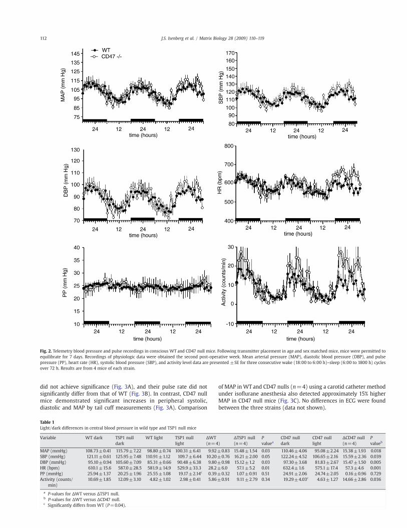
did not achieve signi"cance (Fig. 3A), and their pulse rate did notsigni"cantly differ from that of WT (Fig. 3B). In contrast, CD47 nullmice demonstrated signi"cant increases in peripheral systolic,diastolic and MAP by tail cuff measurements (Fig. 3A). Comparison
of MAP in WT and CD47 nulls (n=4) using a carotid catheter methodunder iso!urane anesthesia also detected approximately 15% higherMAP in CD47 null mice (Fig. 3C). No differences in ECG were foundbetween the three strains (data not shown).
Fig. 2. Telemetry blood pressure and pulse recordings in conscious WT and CD47 null mice. Following transmitter placement in age and sex matched mice, mice were permitted toequilibrate for 7 days. Recordings of physiologic data were obtained the second post-operative week. Mean arterial pressure (MAP), diastolic blood pressure (DBP), and pulsepressure (PP), heart rate (HR), systolic blood pressure (SBP), and activity level data are presented ±SE for three consecutive wake (18:00 to 6:00 h)–sleep (6:00 to 1800 h) cyclesover 72 h. Results are from 4 mice of each strain.
Table 1Light/dark differences in central blood pressure in wild type and TSP1 null mice
Variable WT dark TSP1 nulldark
WT light TSP1 nulllight
!WT(n=4)
!TSP1 null(n=4)
Pvaluea
CD47 nulldark
CD47 nulllight
!CD47 null(n=4)
Pvalueb
MAP (mmHg) 108.73±0.41 115.79±7.22 98.80±0.74 100.31±6.41 9.92±0.83 15.48±1.54 0.03 110.46±4.06 95.08±2.24 15.38±1.93 0.018SBP (mmHg) 121.11±0.61 125.95±7.48 110.91±1.12 109.7±6.44 10.20±0.76 16.21±2.00 0.05 122.24±4.52 106.65±2.16 15.59±2.36 0.039DBP (mmHg) 95.10±0.94 105.60±7.09 85.31±0.66 90.48±6.38 9.80±0.98 15.12±1.2 0.03 97.30±3.68 81.83±2.67 15.47±1.50 0.005HR (bpm) 610.1±15.6 587.0±28.5 581.9±14.9 529.9±33.3 28.2±6.0 57.1±5.2 0.01 632.4±1.6 575.1±17.4 57.3±4.6 0.001PP (mmHg) 25.94±1.37 20.25±1.96 25.55±1.08 19.17±2.14c 0.39±0.32 1.07±0.91 0.51 24.91±2.06 24.74±2.05 0.16±0.96 0.729Activity (counts/
min)10.69±1.85 12.09±3.10 4.82±1.02 2.98±0.41 5.86±0.91 9.11±2.79 0.34 19.29±4.03c 4.63±1.27 14.66±2.86 0.036
a P-values for !WT versus !TSP1 null.b P-values for !WT versus !CD47 null.c Signi"cantly differs from WT (P=0.04).
112 J.S. Isenberg et al. / Matrix Biology 28 (2009) 110–119

2.3. TSP1 and CD47 limit blood pressure responses to NO
The above moderate differences in basal blood pressure para-meters suggest that endogenous TSP1 and CD47 play subtle roles inresting blood pressure regulation. In addition to their commonregulation of NO/cGMP signaling, the differences between TSP1-and CD47-null mice could re!ect anatomical effects of these genedeletions or the ability of TSP1 to act through additional TSP1receptors. Furthermore, several homeostatic pathways could com-pensate for the expected hypertensive activity of TSP1/CD47 signal-
ing. Because our primary goal was de"ne the roles of TSP1 and CD47 inacute cardiovascular regulation, we next examined acute responses ofthe mice to speci"c vasoactive challenges. Because TSP1/CD47signaling limits NO responses in VSMC in vitro by preventing sGCactivation (Isenberg et al., 2006a,b) and tissue blood !ow in responseto exogenous NO in vivo (Isenberg et al., 2007a), we proposed that thenull mice should exhibit a greater acute hypotensive responsefollowing a systemic NO challenge. Age and sex matched WT andTSP1-null mice were challenged via i.p. injection using 1 µl/g of100 mM DEA/NO, and awake resting peripheral blood pressure and
Fig. 3. TSP1 and CD47 limit blood pressure changes in response to NO. Age and sex matched awakeWT, TSP1 and CD47 null mice underwent analysis of blood pressure (A) and pulse(B) via tail cuff. Blood pressure was assessed via a carotid arterial catheter in 5 WT and 4 CD47 !/! mice under anesthesia (C). Awake WT and TSP1 null were treated with a rapidreleasing NO-donor (1 µl/g i.p. of 100 mM DEA/NO) and blood pressure measured by tail cuff (D). Awake WT, TSP1 and CD47 null mice underwent treatment with an intermediatereleasing NO-donor (1 µl/g i.p of 100mM PAPA/NO) and blood pressure (E) and pulse (F) measurements obtained via tail cuff. Results are of the mean±SD of 8mice each ofWTandTSP1 null and 4 CD47-null. Awake WT and TSP1 null mice underwent telemetric analysis of physiologic data before and after treatment with PAPA/NO. Pulse pressure (PP±SE, G) ispresented from 4 mice of each strain. Experiments were repeated a minimum of 3 times. Asterisk (!) indicates pressure values following treatment that signi"cantly differ frombaseline pressure values (Pb0.05).
113J.S. Isenberg et al. / Matrix Biology 28 (2009) 110–119

pulse were determined by tail cuff. NO treatment signi"cantlydecreased systolic, diastolic, and MAP in WT mice, but signi"cantlygreater decreases in blood pressuremeasurementswere noted inmicelacking TSP1 (Fig. 3D). Mean pulse values tended to increase modestlyin all mice following DEA/NO challenge (data not shown). Timecourse data demonstrated delayed recovery of baseline pressure levelsin TSP1 null versus WT mice (data not shown).
Similar differences were found when mice were challenged usingan NO donor with slower release kinetics (PAPA/NO, t1/2=15 min,Thomas et al., 2002, Fig. 3E), with the drop in peripheral MAP alwaysgreater in the absence of TSP1. Interestingly, CD47 null mice treatedwith PAPA/NO showed the greatest drop in peripheral MAP (Fig. 3E).Mean pulse was only modestly increased above resting values in micetreated with PAPA/NO (Fig. 3F). Central pulse pressure measurementsusing telemetry con"rmed an enhanced hypotensive response toPAPA/NO challenge in the TSP1 nulls (Fig. 3G). These results areconsistent with the increased responsiveness of sGC to exogenous NOin VSMC from both null mice in vitro (Isenberg et al., 2006a).
2.4. TSP1 and CD47 limit cardiac responses following NO challenge
In addition to its effects on arterial tone, NO increases cardiacfunction in both normal (Prendergast et al., 1997) and failing hearts.(Inglessis et al., 2004) To investigate the potential role of its antagonist
TSP1 in systemic cardiac physiology, age and sex matched WT, TSP1and CD47 null mice underwent cardiac Doppler analysis (Fig. 4).Under 1.5% iso!urane anesthesia and at constant core temperature(35.5 °C), TSP1 and CD47 null mice demonstrated elevated heart rates(Fig. 4A). These differences were maintained following i.p. injectionwith a rapidly releasing NO donor (0.5 µl/g body weight of 100 mMDEA/NO, t1/2=2–4 min, Thomas et al., 2002), which increased heartrate in all mice. Both null strains demonstrated differences in ejectionfractions compared to WT mice after induction of iso!urane generalanesthesia (data not shown).
More profound alterations in cardiac ejection fraction and cardiacoutput were observed after exogenous NO challenge. WT micedemonstrated mild increases in ejection fraction and cardiac output(12.2±5.3 and 13.2±15.3% increases respectively) which rapidlyreturned to pre-treatment baseline (Fig. 4B, C). In contrast, TSP1 nullmice demonstrated dramatic increases in ejection fraction and cardiacoutput (44±2.6 and 48±1.4% respectively). CD47 null mice showedsimilarly enhanced cardiac output and ejection fraction when treatedwith the NO donor (43.3±10.6 and 54.5±8.6 respectively). In bothTSP1 and CD47 null mice, the increase in cardiac function alwayspersisted longer than in WT mice.
The above cardiac responses may be secondary to direct effects ofTSP1/CD47 on arterial tone. However, TSP1/CD47 signaling is knownto limit tissue cGMP in skeletal muscle (Isenberg et al., 2007b),
Fig. 4. TSP1 and CD47 modulate cardiac responses to vasoactive challenge. Age and sex matched WT (n=4), TSP1 null (n=4), and CD47 null (n=4) underwent echocardiography.After baseline recordings,micewere challengedwithNO(0.5"l/g bodyweight of 100mMDEA/NO i.p.) anddata gathered at 2, 5 and15min. Heart rate (A) is presented as themeanvalues±SD, whereas ejection fraction (B) and cardiac output (C) as presented as percent control. Asterisk (!) indicates curves are of statistically signi"cant difference when compared withcorresponding WT mice (A, B) or individual points of curves are statistically signi"cant compared to WT (C) (Pb0.05). Age and sex matched WT, TSP1 and CD47 null mice underwenteuthanasia via cervical dislocation. Hearts were excised, pulverized in liquid nitrogen and tissue cGMP (D) or cAMP (E) levels determined. Results are expressed as themean±SD for 6 ofeach strain (cGMP) and 4 of each strain (cAMP). Asterisk (!) indicates statistically signi"cant difference between TSP1 and CD47 nullwhen comparedwith correspondingWTsamples (D,E) (Pb0.05).
114 J.S. Isenberg et al. / Matrix Biology 28 (2009) 110–119

suggesting that some cardiac changes in the nulls may represent localeffects of TSP1 in the heart. Consistent with this hypothesis, analysis oftissue cGMP levels in left ventricular samples demonstrated asigni"cant elevation of cardiac muscle cGMP in TSP1 and CD47 nullmice compared to WT (Fig. 4D). Cardiac function also depends oncAMP levels, which in turn are regulated by several PDEs that aremodulated by cGMP (Zaccolo and Movsesian, 2007). Whole tissuecAMP levels were markedly increased in hearts from TSP1 and CD47null mice (Fig. 4E).
2.5. TSP1 limits cardiovascular collapse following autonomic blockade
Input from the autonomic nervous system is a critical homeostaticmechanism to maintain MAP and to minimize alterations in bloodpressure due to vascular stress. Conversely, autonomic blockade usinga centrally active agent such as hexamethonium chloride removessympathetic tone and enhances cardiovascular responses to altera-tions in NO levels (Scrogin et al., 1998; Shibao et al., 2007), Becauseautonomic tone and TSP1 signaling limit the vasodilation response toNO via different mechanisms, we proposed that autonomic blockademight further enhance responses of TSP1 null mice to endogenous NO.Telemetric blood pressure analysis demonstrated dramatically greaterdecreases in central MAP (Pb0.05) after central autonomic blockadeusing a limited dose of hexamethonium chloride in awake TSP1 andCD47 null mice compared to WT (Fig. 5A). The hypotensive responsein TSP1 and CD47 null mice following autonomic blockade was alsosigni"cantly prolonged compared to WT mice.
We further compared responses to autonomic blockade in micemaintained under general anesthesia using 1.5% iso!urane bychallenging with a higher dose of hexamethonium than used inFig. 5A. Cutaneous perfusion was measured every 2.5 min via Doppler(Fig. 5B). Both WT and TSP1 null mice demonstrated decreasedcutaneous perfusion and eventual cardiovascular collapse and death.However, loss of perfusion and death was signi"cantly faster in theabsence of TSP1 and diverged from that of WT mice after 5 min. WTmice sustained cutaneous perfusion for an additional 10 min,indicating an important role for endogenous TSP1 in maintainingperfusion under this combined stress.
2.6. TSP1 augments acute blood pressure responses to epinephrine
Autonomic stimulation of sympathetic nerves leads to norepi-nephrine-stimulated vasoconstriction of arteries (Lee et al., 2003).This can be mimicked by treatment with epinephrine (0.05 µg/animalvia i.p. injection), which produced the expected increase in peripheralMAP in WT mice (Fig. 5C). However, TSP1 null mice did not show asigni"cant increase in MAP, presumably because of the greateropposing NO signaling in these mice. Mean pulse values weremoderately increased in both WT and TSP null mice (Fig. 5D).Remarkably, the same dose of epinephrine proved fatal to CD47 nullmice, precluding further measurements (data not shown).
2.7. TSP1 limits blood pressure response to iso!urane
Central anesthetic agents are known to have strong effects uponblood pressure and cardiac performance (Becker and Haas, 2007;Reich et al., 2005; Torri et al., 2000). To assess whether differentialresponses to anesthesia contributed to the greater sensitivity of TSP1null mice in Fig. 5B, we compared peripheral blood pressure responsesto iso!urane. Induction of iso!urane inhalation anesthesia resulted ina greater decrease in tail cuff blood pressure in TSP1 null mice than inWT (Fig. 5E).
To determine whether the differential response to iso!urane wasassociated with altered circulating TSP1 levels following exposure toiso!urane we determined plasma TSP1 levels in groups of 5 WT micebefore and after anesthesia. Plasma TSP1 levels tended to decrease
following 30 min under inhalation iso!urane (34±18 versus 13±10 ng/ml), but the difference did not achieve signi"cance (P=0.066by two-tailed t-test). Although this could suggest that iso!uranelowers blood pressure inWTmice by decreasing TSP1, we caution thatcirculating TSP1 should not acutely increase blood pressure because itcannot cross the subendothelial basementmembrane to engage VSMCCD47 and inhibit their relaxation by NO.
3. Discussion
Previous studies have shown that disrupting some components ofelastic matrix in blood vessels can affect blood pressure. Partial lossof vascular elastin in mice results in elevated pulmonary circulatorypressures, altered vascular compliance, and cardiac hypertrophy(Shifren et al., 2008; Wagenseil et al., 2007, 2005). Loss of theassociated protein "bulin-5 also causes a decrease in vascularcompliance (Kelleher et al., 2004). Finally, polymorphisms in"brillin-1 in humans are associated with elevated carotid pulsepressure (Medley et al., 2002). These phenotypes can all be attributedto the effects of these structural matrix proteins on the elasticproperties of resistance vessels. In contrast, TSP1 is not a structuralcomponent of blood vessels. However, its presence in the ECM at verylow levels is suf"cient to limit VSMC responses to the potentphysiological vasodilator NO (Isenberg et al., 2007a, 2006b).
The present results demonstrate a broader role for TSP1 incontrolling peripheral and central blood pressure and cardiovascularresponses to several vasoactive challenges. TSP1 null mice have alower central pulse pressure than WT mice and exhibit severalactivity-dependent alterations in central and peripheral bloodpressure parameters. These differences are not consistent with asimple hypotensive response due to elevated vascular cGMP levels inthe TSP1 null but may re!ect homeostatic efforts to compensate forloss of the hypertensive activity of TSP1. However, the enhancedhypotensive responses of TSP and CD47 null mice to NO challengeare consistent with and extend our previous "nding that TSP1 nullmice at rest demonstrate a greater regional increase in skeletalmuscle blood !ow in response to NO (Isenberg et al., 2007a).Hydrolysis of cGMP by PDEs has been considered the major negativeregulator of NO signaling in vascular physiology (Mullershausenet al., 2003; Rybalkin et al., 2003). However, TSP1 also regulatesNO signaling by limiting sGC and cGK activation (Isenberg et al.,2005b). Elevated basal cGMP levels in tissue and vascular cells fromTSP1 or CD47 null mice show that cGMP signaling is continuouslymodulated by physiological concentrations of TSP1 independentof PDE activity (Isenberg et al., 2006a). Thus, TSP1 in the ECMsurrounding VSMC constantly signals via CD47 to enhance bloodpressure and temper both regional and systemic vasodilator activitiesof endogenous NO.
The null mice also exhibit exaggerated responses to anesthesia orloss of autonomic regulation and a more limited response, in the caseof TSP1 null mice, to epinephrine. These results indicate aninterdependence of TSP1/CD47 and autonomic regulation. Becauseautonomic tone also differs between asleep and awake animals, aslight sympathetic overcompensation may account for the increasedMAP in TSP1 null mice during their active cycles. This could be acompensation for the decreased pulse pressure in the TSP1 nulls,which is consistent with TSP1 being a vasopressor. However,additional studies will be required to identify why CD47 null micelack a decreased pulse pressure. These differences between the CD47null and TSP1 null mice may re!ect some compensation for loss ofTSP1 by other thrombospondins expressed in vascular cells such asTSP2 and TSP4 (Lamy et al., 2007; Lopes et al., 2003; Stenina et al.,2003a). Our recent data indicates that TSP4 has some activity toinhibit NO/cGMP signaling in endothelial cells via CD47, but TSP2 ismuch less active, and both are weaker than TSP1 (Isenberg et al.,2009). Furthermore, the absence of TSP2 in vascular cells does not
115J.S. Isenberg et al. / Matrix Biology 28 (2009) 110–119

result in elevated cGMP levels. Alternatively, other CD47 ligands suchas SIRP# (Lee et al., 2007) may in!uence cardiovascular signalingthrough CD47 in a TSP1-independent manner.
Cardiac function is regulated by cGMP and cAMP. As previouslyshown for cGMP in skeletal muscle (Isenberg et al., 2007b), cGMPlevels are increased in cardiac muscle of TSP1 and CD47 nulls.Although cGMP can in!uence cAMP levels by modulating PDEs, thisfeedback is unlikely to explain the greater increase in cAMP in nullheart tissue. CD47 ligation in VSMC and T cells decreases cAMP levelsby inhibiting adenylate cyclase in a heterotrimeric Gi-dependentmanner (Manna and Frazier, 2003; Wang et al., 1999), suggesting thatTSP1/CD47 signaling may regulate cardiac cAMP levels via similarmechanism. However, the enhanced cardiac responses to NO in TSP1and CD47 null mice probably also re!ect compensation for differencesin peripheral vasodilation in these mice.
Because homeostatic compensation mechanisms limit changes inresting cardiovascular parameters, the roles of TSP1 and CD47 inmaintaining systemic cardiovascular physiology becomemore evidentfollowing vasoactive challenge. In WT mice, moderate doses of NOenhance cardiac function and heart rate (Kojda and Kottenberg, 1999;Mohan et al., 1996; Shah and MacCarthy, 2000), but these responsesare modest and rapidly dampened by autonomic compensation. Incontrast, a similar NO challenge in TSP1 or CD47 null mice inducesgreater increases in heart rate, cardiac output and ejection fractionwith delayed return to baseline.
Lack of TSP1 is also associated with less elevation of blood pressureto epinephrine. Conversely, chemical sympathectomy with hexam-ethonium, which effectively ablates the vasoconstrictor effects ofperipheral epinephrine on vascular tone, enhances hypotension inTSP1 and CD47 null mice. Absent sympathetic tone, TSP1 is requiredfor maintenance of blood pressure and cardiac output. Under suchconditions, lack of TSP1 leads to accelerated loss of blood pressure,tissue perfusion and death compared to WT.
Because loss of TSP1/CD47 signaling can alter blood pressure andhemodynamic responses to stress, the potential for altered vascularmatrix TSP1 levels to increase blood pressure or decrease compliancein response to physiological NO signaling should be considered. TSP1expression is elevated in the blood vessel wall or surrounding matrixin atherosclerosis, restenosis, tissue ischemia, and diabetes (Chenet al., 1999; Riessen et al., 1998; Roth et al., 1998; Stenina et al., 2003b).In these diseases, accumulation of TSP1 in the matrix surroundingVSMC could chronically limit NO-mediated vasodilation. Somecancers are associated with elevated circulating plasma TSP1 levels(Yamashita et al., 1998). However, it remains unclear whetherelevated circulating TSP1 could be hypertensive since it would nothave access to CD47 on VSMC.
Given the association between a coding polymorphism in TSP1 andearly coronary artery disease (Topol et al., 2001; Zwicker et al., 2006),our results raise the intriguing possibility that altering the antagonismbetween the TSP/CD47 and NO/cGMP pathways could lead tocardiovascular pathology in people. This polymorphism alters theconformation of signature domain of TSP1, which contains the CD47binding site (Isenberg et al., 2009), but it is not known whether thepolymorphism directly affects CD47 binding. Furthermore, theseresults suggest that inhibitors of TSP1/CD47 interactions could havetherapeutic application to regulate blood pressure, cardiac dynamics,and regional blood !ow.
4. Experimental procedures
4.1. Animals
Wild type (WT), TSP1 and CD47 null mouse colonies in a pureC57BL/6 background were housed under pathogen-free conditionsand had ad libitum access to "ltered water and standard chow. Bothnull strains were rederived with back crossing before use, and theCD47 null strain was back crossed against the wild type C57BL/6colony within one year of initiating this study. Handling and care ofanimals were in compliance with guidelines of the Animal Care andUse Committee of the National Cancer Institute.
4.2. Reagents
The NO donor diethylamine NONOate (DEA/NO) was provided byDr. Larry Keefer (NCI, Frederick, Maryland). The NO donor (Z)-1-[N-(3-ammoniopropyl)-N-(n-propyl)amino]-NONOate (PAPA/NO) wasfrom Cayman Chemical Company (Ann Arbor, MI). Iso!urane (For-ane®, USP) was from Baxter Healthcare Corp. (Deer"eld, IL).Epinephrine hydrochloride and hexamethonium chloride were fromSigma-Aldrich (St. Louis, MO).
4.3. Tail cuff blood pressure and pulse measurements
Peripheral systolic, diastolic andmean arterial pressure (MAP) andpulse were measured using a computerized tail cuff system (HatterasSystems, MC4000 Blood Pressure Analysis System, Cary, NC). Eachanimal underwent a cycle of 10 preliminary and 50 experimentalpressures measurements for data acquisition and calculation. Micewere acclimated to the measuring system for 4 days with studiesperformed on the 5th day. Recordings were obtained at the same timeeach day.
4.4. Internal blood pressure measurement via telemetry
Mice were anesthetized with ketamine/xylazine (90 and 10 mg/kg, respectively). The telemeter catheter was inserted into the leftcarotid artery at the aortic arch, and the telemeter body (modelTA11PA-C20, Data Sciences International, St. Paul, MN) placed in asubcutaneous pocket. The signal was processed using a RPC-1receiver, a 20-channel data-exchange matrix, APR-1 ambient pres-sure monitor, and a Dataquest ART 2.3 acquisition system (DataSciences International). Data was acquired for 10 s every 2 min and100-min averages calculated.
4.5. ECG measurements
ECG measurements were obtained using the ECGenieTm ECGScreening System (Mouse Speci"cs, Inc., Boston, MA). Mice wereplaced on the recording stage and allowed to acclimate to the system,and measurements were obtained passively.
4.6. In vivo echocardiography
Analysis of murine hearts in WT, TSP1 and CD47 null age and sexmatched mice was performed using a Vevo 770 High-Resolution In
Fig. 5. TSP1 protects against cardiovascular collapse following autonomic blockade and regulates response to epinephrine and general anesthesia. Telemetric transmitters wereplaced in age and sex matchedWT, TSP1 and CD47 null mice (A). On post-operative day 7 mice were treated with hexamethonium chloride (25 µg/g in 100 µl of sterile PBS i.p.) andMAP±SE determined. Experiments were repeated a minimum of three times in 4 mice of each strain. (B) WT and TSP1 null mice underwent laser Doppler analysis of cutaneousperfusion at 2.5 min intervals±hexamethonium (30 µg/g) i.p. Results are the mean±SD for 4 mice of each strain. Asterisk indicates values are of statistically signi"cant differencewhen compared with correspondingWTmice (Pb0.05). Age and sex matched mice underwent blood pressure (C) and pulse (D) analysis via tail cuff before and after treatment withepinephrine (0.05 µg/animal i.p.). Asterisk (!) indicates arterial pressure following treatment signi"cantly differs comparedwith baseline (C) (Pb0.05). Age and sexmatchedmice ata core temperature of 35.5 °C underwent blood pressure analysis before and after receiving 1.5% inhalation iso!urane (E). Asterisk (!) indicates signi"cant difference in change inpressure between TSP1 compared to wild type following treatment (Pb0.05).
116 J.S. Isenberg et al. / Matrix Biology 28 (2009) 110–119

117J.S. Isenberg et al. / Matrix Biology 28 (2009) 110–119

Vivo Imaging System (VisualSonics, Toronto, ON, Canada) with a RMV707 “high frame” scan head for high frame rate and real-time smallanimal imaging with a center frequency of 30 MHz and a frequencyband 15–45 MHz. Mice were maintained on 1.5% iso!urane and coretemperature of 35.5 °C. Long-axis imaging was taken to visualize leftventricle, right ventricle, ascending aorta, and right ventricularout!ow tract. The short-axis imaging was taken to view the LV andRV movement during diastole and systole stages. Anatomical M-mode(AM-Mode) provided the ability to obtain anatomically correct LVmeasurements. All data and images were analyzed by AdvancedCardiovascular Package Software (VS-11560, VisualSonics, Toronto,ON, Canada). Measurements from three continuous cardiac cycleswere averaged to obtain a value for each data point.
4.7. Skin blood !ow
Skin blood !ow was measured on the dorsum of age and sexmatched mice using laser Doppler imaging (MoorLD1-2$, MoorInstruments, Devon, England) (Isenberg et al., 2007b). Anesthesiawas provided by 1.5% inhalation iso!urane. Core temperature wasmonitored by rectal probe. After equilibration to the experimentalset-up, analysis of blood !ow was performed. The followinginstrument settings were used: override distance 21 cm; scan time4 ms/pixel. Results are expressed as a percent of the control baselinefor the ROI.
4.8. Plasma TSP1 analysis
Plasma samples were collected using EDTA as anti-coagulant fromgroups of 5WTmicewith or without prior anesthesia for 30min using1.5% inhalation iso!urane. TSP1 levels were quanti"ed using aheparin-BSA capture enzyme-linked immunoassay with detectionusing a polyclonal rabbit anti-TSP1 antibody as previously described(Isenberg et al., 2005a).
4.9. Cardiac cGMP and cAMP
cGMP and cAMP from cardiac muscle tissue were determined byimmunoassay as previously described (Isenberg et al., 2007b).
4.10. Statistics
Results are presented as the mean±SD or mean±SE withsigni"cance calculated by the Student's t test or one-way ANOVA withTukey post hoc test and signi"cance taken at P valuesb0.05. Datarepresents results of studies performed in a total of 117mice distributedas follows: WT, n=46; TSP1 null, n=46; CD47 null, n=25.
Acknowledgments
We thank Dr. Larry Keefer for providing reagents and KevinKaltenbronn and Dr. Ken Blumer for performing the carotid pressuremeasurements. This work was supported by the Intramural ResearchProgram of the NIH, National Cancer Institute, Center for CancerResearch (D.D.R.), National Institute of Diabetes and Digestive andKidney Diseases (J.S.) and NIH grants HL54390 and GM57573 (W.A.F.).
References
Arnal, J.F., Dinh-Xuan, A.T., Pueyo, M., Darblade, B., Rami, J., 1999. Endothelium-derivednitric oxide and vascular physiology and pathology. Cell. Mol. Life Sci. 55,1078–1087.
Becker, D.E., Haas, D.A., 2007. Management of complications during moderate and deepsedation: respiratory and cardiovascular considerations. Anesth. Prog. 54, 59–68quiz 69.
Bergseth, G., Lappegard, K.T., Videm, V., Mollnes, T.E., 2000. A novel enzymeimmunoassay for plasma thrombospondin. Comparison with beta-thromboglobu-lin as platelet activation marker in vitro and in vivo. Thromb. Res. 99, 41–50.
Chen, D., Asahara, T., Krasinski, K., Witzenbichler, B., Yang, J., Magner, M., Kearney, M.,Frazier, W.A., Isner, J.M., Andres, V., 1999. Antibody blockade of thrombospondinaccelerates reendothelialization and reduces neointima formation in balloon-injured rat carotid artery. Circulation 100, 849–854.
Hermann, M., Flammer, A., Luscher, T.F., 2006. Nitric oxide in hypertension. J. Clin.Hypertens. (Greenwich) 8, 17–29.
Ignarro, L.J., 2002. Nitric oxide as a unique signaling molecule in the vascular system: ahistorical overview. J. Physiol. Pharmacol. 53, 503–514.
Inglessis, I., Shin, J.T., Lepore, J.J., Palacios, I.F., Zapol, W.M., Bloch, K.D., Semigran, M.J.,2004. Hemodynamic effects of inhaled nitric oxide in right ventricular myocardialinfarction and cardiogenic shock. J. Am. Coll. Cardiol. 44, 793–798.
Isenberg, J.S., Calzada,M.J., Zhou, L.,Guo,N., Lawler, J.,Wang,X.Q., Frazier,W.A.,Roberts,D.D.,2005a. Endogenous thrombospondin-1 is not necessary for proliferation but ispermissive for vascular smooth muscle cell responses to platelet-derived growthfactor. Matrix Biol. 24, 110–123.
Isenberg, J.S., Ridnour, L.A., Perruccio, E.M., Espey, M.G., Wink, D.A., Roberts, D.D., 2005b.Thrombospondin-1 inhibits endothelial cell responses to nitric oxide in a cGMP-dependent manner. Proc. Natl. Acad. Sci. U. S. A. 102, 13141–13146.
Isenberg, J.S., Ridnour, L.A., Dimitry, J., Frazier, W.A., Wink, D.A., Roberts, D.D., 2006a.CD47 is necessary for inhibition of nitric oxide-stimulated vascular cell responsesby thrombospondin-1. J. Biol. Chem. 281, 26069–26080.
Isenberg, J.S., Wink, D.A., Roberts, D.D., 2006b. Thrombospondin-1 antagonizes nitricoxide-stimulatedvascular smoothmuscle cell responses. Cardiovasc. Res. 71, 785–793.
Isenberg, J.S., Hyodo, F., Matsumoto, K., Romeo, M.J., Abu-Asab, M., Tsokos, M.,Kuppusamy, P., Wink, D.A., Krishna, M.C., Roberts, D.D., 2007a. Thrombospondin-1limits ischemic tissue survival by inhibiting nitric oxide-mediated vascular smoothmuscle relaxation. Blood 109, 1945–1952.
Isenberg, J.S., Hyodo, F., Pappan, L.K., Abu-Asab, M., Tsokos, M., Krishna, M.C., Frazier, W.A.,Roberts, D.D., 2007b. Blocking thrombospondin-1/CD47 signaling alleviates deleter-ious effects of aging on tissue responses to ischemia. Arterioscler. Thromb. Vasc. Biol.27, 2582–2588.
Isenberg, J.S., Romeo, M.J., Abu-Asab, M., Tsokos, M., Oldenborg, A., Pappan, L., Wink, D.A.,Frazier, W.A., Roberts, D.D., 2007c. Increasing survival of ischemic tissue by targetingCD47. Circ. Res. 100, 712–720.
Isenberg, J.S., Romeo, M.J., Yu, C., Yu, C.K., Nghiem, K., Monsale, J., Rick, M.E., Wink, D.A.,Frazier, W.A., Roberts, D.D., 2008b. Thrombospondin-1 stimulates platelet aggrega-tion by blocking the antithrombotic activity of nitric oxide/cGMP signaling. Blood111, 613–623.
Isenberg, J.S., Annis, D.S., Pendrak,M.L., Ptaszynska, M., Frazier, W.A., Mosher, D.F., Roberts,D.D., 2009. Differential interactions of thrombospondins-1, -2 and -4 with CD47 andeffects on cGMP signaling and Ischemic injury responses. J Biol Chem. 284,1116–1125.
Kelleher, C.M., McLean, S.E., Mecham, R.P., 2004. Vascular extracellular matrix and aorticdevelopment. Curr. Top. Dev. Biol. 62, 153–188.
Kojda, G., Kottenberg, K., 1999. Regulation of basal myocardial function by NO.Cardiovasc. Res. 41, 514–523.
Lamy, L., Foussat, A., Brown, E.J., Bornstein, P., Ticchioni, M., Bernard, A., 2007.Interactions between CD47 and thrombospondin reduce in!ammation. J. Immunol.178, 5930–5939.
Lee, C.R.,Watkins,M.L., Patterson, J.H.,Gattis,W.,O'Connor, C., Gheorghiade,M.,Adams Jr., K.F.,2003. Vasopressin: a new target for the treatment of heart failure. Am.Heart J.146, 9–18.
Lee, W.Y., Weber, D.A., Laur, O., Severson, E.A., McCall, I., Jen, R.P., Chin, A.C., Wu, T.,Gernert, K.M., Parkos, C.A., 2007. Novel structural determinants on SIRP alpha thatmediate binding to CD47. J. Immunol. 179, 7741–7750.
Lopes, N., Gregg, D., Vasudevan, S., Hassanain, H., Goldschmidt-Clermont, P., Kovacic, H.,2003. Thrombospondin 2 regulates cell proliferation induced by Rac1 redox-dependent signaling. Mol. Cell. Biol. 23, 5401–5408.
Manna, P.P., Frazier, W.A., 2003. The mechanism of CD47-dependent killing of T cells:heterotrimeric Gi-dependent inhibition of protein kinase A. J. Immunol. 170,3544–3553.
Medley, T.L., Cole, T.J., Gatzka, C.D., Wang, W.Y., Dart, A.M., Kingwell, B.A., 2002. Fibrillin-1genotype is associated with aortic stiffness and disease severity in patients withcoronary artery disease. Circulation 105, 810–815.
Mohan, P., Brutsaert, D.L., Paulus,W.J., Sys, S.U., 1996. Myocardial contractile response tonitric oxide and cGMP. Circulation 93, 1223–1229.
Mullershausen, F., Friebe, A., Feil, R., Thompson, W.J., Hofmann, F., Koesling, D., 2003.Direct activation of PDE5 by cGMP: long-term effects within NO/cGMP signaling.J. Cell Biol. 160, 719–727.
Ortiz, P.A., Garvin, J.L., 2003. Cardiovascular and renal control in NOS-de"cient mousemodels. Am. J. Physiol., Regul. Integr. Comp. Physiol. 284, R628–R638.
Prendergast, B.D., Sagach, V.F., Shah, A.M., 1997. Basal release of nitric oxide augmentsthe Frank-Starling response in the isolated heart. Circulation 96, 1320–1329.
Reich, D.L., Hossain, S., Krol, M., Baez, B., Patel, P., Bernstein, A., Bodian, C.A., 2005.Predictors of hypotension after induction of general anesthesia. Anesth. Analg. 101,622–628 table of contents.
Riessen, R., Kearney, M., Lawler, J., Isner, J.M., 1998. Immunolocalization of thrombospon-din-1 in human atherosclerotic and restenotic arteries. Am. Heart J. 135, 357–364.
Roth, J.J., Gahtan, V., Brown, J.L., Gerhard, C., Swami, V.K., Rothman, V.L., Tulenko, T.N.,Tuszynski, G.P., 1998. Thrombospondin-1 is elevated with both intimal hyperplasiaand hypercholesterolemia. J. Surg. Res. 74, 11–16.
Rybalkin, S.D., Yan, C., Bornfeldt, K.E., Beavo, J.A., 2003. Cyclic GMP phosphodiesterasesand regulation of smooth muscle function. Circ. Res. 93, 280–291.
Scrogin,K.E.,Hatton,D.C.,Chi,Y., Luft, F.C.,1998. Chronicnitricoxide inhibitionwith L-NAME:effects on autonomic control of the cardiovascular system. Am. J. Physiol. 274,R367–R374.
Shah, A.M., MacCarthy, P.A., 2000. Paracrine and autocrine effects of nitric oxide onmyocardial function. Pharmacol. Ther. 86, 49–86.
118 J.S. Isenberg et al. / Matrix Biology 28 (2009) 110–119

Shibao, C., Raj, S.R., Gamboa, A., Diedrich, A., Choi, L., Black, B.K., Robertson, D., Biaggioni, I.,2007. Norepinephrine transporter blockade with atomoxetine induces hypertensionin patients with impaired autonomic function. Hypertension 50, 47–53.
Shifren, A., Durmowicz, A.G., Knutsen, R.H., Faury, G., Mecham, R.P., 2008. Elastininsuf"ciency predisposes to elevated pulmonary circulatory pressures throughchanges in elastic artery structure. J. Appl. Physiol. 105, 1610–1619.
Stenina, O.I., Desai, S.Y., Krukovets, I., Kight, K., Janigro, D., Topol, E.J., Plow, E.F., 2003a.Thrombospondin-4 and its variants: expression and differential effects on endothelialcells. Circulation 108, 1514–1519.
Stenina, O.I., Krukovets, I., Wang, K., Zhou, Z., Forudi, F., Penn, M.S., Topol, E.J., Plow, E.F.,2003b. Increased expression of thrombospondin-1 in vessel wall of diabetic Zuckerrat. Circulation 107, 3209–3215.
Thomas, D.D., Miranda, K.M., Espey,M.G., Citrin, D., Jourd'heuil, D., Paolocci, N., Hewett, S.J.,Colton, C.A., Grisham, M.B., Feelisch, M., Wink, D.A., 2002. Guide for the use of nitricoxide (NO) donors as probes of the chemistry of NO and related redox species inbiological systems. Methods Enzymol. 359, 84–105.
Topol, E.J., McCarthy, J., Gabriel, S., Moliterno, D.J., Rogers, W.J., Newby, L.K., Freedman, M.,Metivier, J., Cannata, R., O'Donnell, C.J., Kottke-Marchant, K., Murugesan, G., Plow, E.F.,Stenina, O., Daley, G.Q., 2001. Single nucleotide polymorphisms in multiple novelthrombospondin genes may be associated with familial premature myocardialinfarction. Circulation 104, 2641–2644.
Torri, G., Casati, A., On behalf of the Italian Research Group on sevo!urane, 2000.Cardiovascular homeostasis during inhalational general anesthesia: a clinicalcomparison between sevo!urane and iso!urane. J. Clin. Anesth. 12, 117–122.
Wagenseil, J.E., Nerurkar, N.L., Knutsen, R.H., Okamoto, R.J., Li, D.Y., Mecham, R.P., 2005.Effects of elastin haploinsuf"ciency on the mechanical behavior of mouse arteries.Am. J. Physiol., Heart Circ. Physiol. 289, H1209–H1217.
Wagenseil, J.E., Knutsen, R.H., Li, D.Y., Mecham, R.P., 2007. Elastin-insuf"cient mice shownormal cardiovascular remodeling in 2K1C hypertension despite higher baselinepressure and unique cardiovascular architecture. Am. J. Physiol., Heart Circ. Physiol.293, H574–H582.
Wang, X.Q., Lindberg, F.P., Frazier, W.A., 1999. Integrin-associated protein stimulatesalpha2beta1-dependent chemotaxis via Gi-mediated inhibition of adenylatecyclase and extracellular-regulated kinases. J. Cell Biol. 147, 389–400.
Yamashita, Y., Kurohiji, T., Tuszynski, G.P., Sakai, T., Shirakusa, T., 1998. Plasma thrombos-pondin levels in patients with colorectal carcinoma. Cancer 82, 632–638.
Zaccolo, M., Movsesian, M.A., 2007. cAMP and cGMP signaling cross-talk: role ofphosphodiesterases and implications for cardiac pathophysiology. Circ. Res. 100,1569–1578.
Zwicker, J.I., Peyvandi, F., Palla, R., Lombardi, R., Canciani, M.T., Cairo, A., Ardissino, D.,Bernardinelli, L., Bauer, K.A., Lawler, J., Mannucci, P., 2006. The thrombospondin-1N700S polymorphism is associated with early myocardial infarction withoutaltering von Willebrand factor multimer size. Blood 108, 1280–1283.
119J.S. Isenberg et al. / Matrix Biology 28 (2009) 110–119