CD47 Is Upregulated on Circulating Hematopoietic Stem Cells and ...
15
CD47 Is Upregulated on Circulating Hematopoietic Stem Cells and Leukemia Cells to Avoid Phagocytosis Siddhartha Jaiswal, 1, * Catriona H.M. Jamieson, 2 Wendy W. Pang, 1 Christopher Y. Park, 1 Mark P. Chao, 1 Ravindra Majeti, 1 David Traver, 3 Nico van Rooijen, 4 and Irving L. Weissman 1, * 1 Ludwig Center at Stanford, Stanford Cancer Center, Department of Pathology, and Institute for Stem Cell Biology and Regenerative Medicine, Stanford University School of Medicine, Stanford, CA 94305, USA 2 Stem Cell Research Program, Moores UCSD Cancer Center 3 Section of Cell and Developmental Biology University of California San Diego, La Jolla, CA, 92093, USA 4 Department of Molecular Cell Biology, Vrije Universiteit, VUMC, Amsterdam, The Netherlands *Correspondence: [email protected] (S.J.), [email protected] (I.L.W.) DOI 10.1016/j.cell.2009.05.046 SUMMARY Macrophages clear pathogens and damaged or aged cells from the blood stream via phagocytosis. Cell- surface CD47 interacts with its receptor on macro- phages, SIRPa, to inhibit phagocytosis of normal, healthy cells. We find that mobilizing cytokines and inflammatory stimuli cause CD47 to be transiently upregulated on mouse hematopoietic stem cells (HSCs) and progenitors just prior to and during their migratory phase, and that the level of CD47 on these cells determines the probability that they are engulfed in vivo. CD47 is also constitutively upregu- lated on mouse and human myeloid leukemias, and overexpression of CD47 on a myeloid leukemia line increases its pathogenicity by allowing it to evade phagocytosis. We conclude that CD47 upregulation is an important mechanism that provides protection to normal HSCs during inflammation-mediated mobi- lization, and that leukemic progenitors co-opt this ability in order to evade macrophage killing. INTRODUCTION Hematopoietic stem cells (HSCs) have the ability to migrate to ectopic niches in fetal and adult life via the blood stream (Chris- tensen et al., 2004; Cumano and Godin, 2007; Morrison et al., 1995; Wright et al., 2001b). Once in the blood, HSCs home to VCAM-1 + endothelia using integrin a4b1(Wagers et al., 2002), then navigate the vascular beds of the marrow, spleen, and liver before returning to potential niches that secrete SDF-1, for which HSCs have responsive CXCR4 chemokine receptors (Peled et al., 1999; Wright et al., 2002). Macrophages lining vascular sinusoids at these sites function to remove damaged cells and foreign parti- cles from the blood stream, sensing altered surface molecules. During inflammatory responses, macrophages become more phagocytically active in order to more effectively clear offending pathogens (Wright et al., 1989). At these times, host cells are at increased risk of clearance and newly arriving stem cells might require additional protection against phagocytosis. Like normal HSCs, leukemic stem and progenitor cells also have an intrinsic ability to migrate via circulation to ectopic marrow sites. By analogy with the normal HSCs, the leukemic stem cells (LSCs) must also be able to navigate through macrophage-lined vasculature of spleen, liver, and marrow as they travel to ectopic niches. CD47 is an immunoglobulin-like protein that is known to interact functionally with integrins (Brown and Frazier, 2001) and thromb- spondin-1 (Gao et al., 1996; Liu et al., 2001). It has been implicated in functions as diverse as neutrophil migration (Lindberg et al., 1996), axon extension (Miyashita et al., 2004), and T cell costimu- lation (Reinhold et al., 1997). In addition, CD47 is capable of inter- acting with its receptor SIRPa (Jiang et al., 1999) on macrophages to negatively regulate phagocytosis (Brown and Frazier, 2001). Lack of autonomous CD47 expression results in phagocytosis of red blood cells (Oldenborg et al., 2000), as well as T cells and whole bone marrow cells in a transplant setting (Blazar et al., 2001). Thus, CD47 functions as a ‘‘don’t eat me’’ signal to ensure that autologous cells are not inappropriately phagocytosed. While the absence of CD47 is known to result in phagocytosis, the effect of differential CD47 expression is unclear. IAP +/ plate- lets and erythrocytes may be more prone to phagocytosis than their wild-type counterparts (Olsson et al., 2005, 2007), which suggests that CD47 downregulation might lead to clearance of these cells as they age. However, no formal exploration of the role of CD47 expression on normal or leukemic hematopoietic stem and progenitor cells, which are also physiologically migrating cells, has been undertaken. Hence, we performed experiments to under- stand the effect of differential CD47 expression on these cells. RESULTS CD47 Expression Is Increased in HSCs after Mobilization or Induced Inflammation Mobilization of marrow stem and progenitor cells involves several steps in which they come into contact with macrophages. We Cell 138, 271–285, July 24, 2009 ª2009 Elsevier Inc. 271
-
Upload
truongliem -
Category
Documents
-
view
217 -
download
1
Transcript of CD47 Is Upregulated on Circulating Hematopoietic Stem Cells and ...

CD47 Is Upregulated on CirculatingHematopoietic Stem Cells andLeukemia Cells to Avoid PhagocytosisSiddhartha Jaiswal,1,* Catriona H.M. Jamieson,2 Wendy W. Pang,1 Christopher Y. Park,1 Mark P. Chao,1 Ravindra Majeti,1
David Traver,3 Nico van Rooijen,4 and Irving L. Weissman1,*1Ludwig Center at Stanford, Stanford Cancer Center, Department of Pathology, and Institute for Stem Cell Biology and RegenerativeMedicine, Stanford University School of Medicine, Stanford, CA 94305, USA2Stem Cell Research Program, Moores UCSD Cancer Center3Section of Cell and Developmental BiologyUniversity of California San Diego, La Jolla, CA, 92093, USA4Department of Molecular Cell Biology, Vrije Universiteit, VUMC, Amsterdam, The Netherlands*Correspondence: [email protected] (S.J.), [email protected] (I.L.W.)DOI 10.1016/j.cell.2009.05.046
SUMMARY
Macrophages clear pathogens and damaged or agedcells from the blood stream via phagocytosis. Cell-surface CD47 interacts with its receptor on macro-phages, SIRPa, to inhibit phagocytosis of normal,healthy cells. We find that mobilizing cytokines andinflammatory stimuli cause CD47 to be transientlyupregulated on mouse hematopoietic stem cells(HSCs) and progenitors just prior to and duringtheir migratory phase, and that the level of CD47 onthese cells determines the probability that they areengulfed in vivo. CD47 is also constitutively upregu-lated on mouse and human myeloid leukemias, andoverexpression of CD47 on a myeloid leukemia lineincreases its pathogenicity by allowing it to evadephagocytosis. We conclude that CD47 upregulationis an important mechanism that provides protectionto normal HSCs during inflammation-mediated mobi-lization, and that leukemic progenitors co-opt thisability in order to evade macrophage killing.
INTRODUCTION
Hematopoietic stem cells (HSCs) have the ability to migrate toectopic niches in fetal and adult life via the blood stream (Chris-tensen et al., 2004; Cumano and Godin, 2007; Morrison et al.,1995; Wright et al., 2001b). Once in the blood, HSCs home toVCAM-1+ endothelia using integrin a4b1 (Wagers et al., 2002),then navigate the vascular beds of the marrow, spleen, and liverbefore returning to potential niches that secrete SDF-1, for whichHSCs have responsive CXCR4 chemokine receptors (Peled et al.,1999; Wright et al., 2002). Macrophages lining vascular sinusoidsat these sites function to remove damaged cells and foreign parti-cles from the blood stream, sensing altered surface molecules.During inflammatory responses, macrophages become morephagocytically active in order to more effectively clear offending
pathogens (Wright et al., 1989). At these times, host cells are atincreased risk of clearance and newly arriving stem cells mightrequire additional protection against phagocytosis.
Like normal HSCs, leukemic stem and progenitor cells alsohave an intrinsic ability tomigrateviacirculation toectopicmarrowsites. By analogy with the normal HSCs, the leukemic stem cells(LSCs) must also be able to navigate through macrophage-linedvasculature of spleen, liver, and marrow as they travel to ectopicniches.
CD47 is an immunoglobulin-like protein that is known to interactfunctionally with integrins (Brown and Frazier, 2001) and thromb-spondin-1 (Gao et al., 1996; Liu et al., 2001). It has been implicatedin functions as diverse as neutrophil migration (Lindberg et al.,1996), axon extension (Miyashita et al., 2004), and T cell costimu-lation (Reinhold et al., 1997). In addition, CD47 is capable of inter-acting with its receptor SIRPa (Jiang et al., 1999) on macrophagesto negatively regulate phagocytosis (Brown and Frazier, 2001).Lack of autonomous CD47 expression results in phagocytosisof red blood cells (Oldenborg et al., 2000), as well as T cells andwhole bone marrow cells in a transplant setting (Blazar et al.,2001). Thus, CD47 functions as a ‘‘don’t eat me’’ signal to ensurethat autologous cells are not inappropriately phagocytosed.
While the absence of CD47 is known to result in phagocytosis,the effect of differential CD47 expression is unclear. IAP+/! plate-lets and erythrocytes may be more prone to phagocytosis thantheir wild-type counterparts (Olsson et al., 2005, 2007), whichsuggests that CD47 downregulation might lead to clearance ofthese cells as they age. However, no formal exploration of the roleof CD47 expression on normal or leukemic hematopoietic stemand progenitor cells, which are also physiologically migrating cells,hasbeenundertaken. Hence,weperformedexperiments tounder-stand the effect of differential CD47 expression on these cells.
RESULTS
CD47 Expression Is Increased in HSCs after Mobilizationor Induced InflammationMobilization of marrow stem and progenitor cells involves severalsteps in which they come into contact with macrophages. We
Cell 138, 271–285, July 24, 2009 ª2009 Elsevier Inc. 271

A
C
F
B
E
D
Figure 1. Mobilization or Inflammation Induces CD47 Upregulation in Hematopoietic Stem and Progenitor Cells(A) Expression level of CD47 on cKit+ cells is shown for day 2 Cy/G-mobilized BM.
(B) Myeloid progenitor and stem cell gates are shown for day 2 mobilized BM. Histograms on right show level of CD47 expression in marrow LT-HSCs and GMPs
for steady state (light gray shaded histogram), day 2 mobilized (black line), and day 5 mobilized (dark gray shaded histogram).
(C) Relative MFI of CD47 for GMPs on days 0–5 of Cy/G mobilization. Results were normalized so that steady-state GMPs had MFI 100.
272 Cell 138, 271–285, July 24, 2009 ª2009 Elsevier Inc.

thus utilized the cyclophosphamide/G-CSF (Cy/G) protocol(Morrison et al., 1997) to experimentally induce mobilization inmice. We found that there was a notable increase of CD47 oncKit+ bone marrow cells on day 2 (Figure 1A), when bone marrowHSCs reach their maximum level, with CD47 increasing approx-imately 4-fold by this time point (Figure 1B). The increase wasseen at all levels of the myeloid progenitor hierarchy, as long-term hematopoietic stem cells (LT-HSCs) as well as granulo-cyte-macrophage progenitors (GMPs) displayed this increasein CD47 expression (Figure 1B). By day 5, when egress fromthe marrow to distant marrow/spleen/liver sites had nearlystopped, the levels of CD47 on the marrow HSC/progenitorshad returned to nearly normal levels. In Figure 1C, the mean fluo-rescence intensity of CD47 expression on GMPs is shown ondays 0 to 5 of mobilization.
These results predicted that mobilized human HSCs in theblood would also have increased levels of CD47 on their cellsurfaces. We examined the CD47 expression level on humanbone marrow, cord blood (CB), and mobilized peripheral blood(MPB) HSCs. The results show that human HSCs in circulationhave significantly higher levels of CD47 expression than sessileHSCs (Figure 1E).
Endotoxins such as lipopolysaccharide (LPS) are also thoughtto contribute to bone marrow mobilization (Cline and Golde,1977) by causing activation of macrophages and a proinflamma-tory response (Fenton and Golenbock, 1998), as well asincreasing the phagocytic capacity of macrophages (Wrightet al., 1989). We tested whether LPS administration in micewould affect CD47 expression in stem and progenitor cells. Mir-roring the pattern seen in Cy/G-induced mobilization, LPScaused expansion of stem and progenitor cells by 2 days post-treatment, followed by migration to the spleen and liver (Fig-ure 1D). On day 2 after LPS administration, stem and progenitorcells in the marrow had upregulated CD47 to a similar degree asin Cy/G mobilization. By day 5, when the inflammatory responsehad resolved, the levels of the protein had dropped to steady-state levels (Figure 1D).
CD47 Upregulation during Mobilization Is Unlikelyto Be Necessary for MigrationSince CD47 was consistently upregulated in the mobilizationresponse, we tested the ability of stem and progenitor cells tomobilize following Cy/G. The CD47 knockout mouse has defectsin migration of neutrophils to sites of inflammation (Lindberg et al.,1996) and of dendritic cells to secondary lymphoid organs (Vanet al., 2006). The exact role of CD47 in migration of these cellsis unknown, but it may relate to poor integrin binding in the circu-lation or lack of interaction with SIRPa on endothelial cells. Hencewe reasoned that if CD47 was involved in the migration capacityof these cells during mobilization, then IAP!/!mice would displayreduced numbers of cells in the peripheral organs after Cy/G.
We thus administered Cy/G to both wild-type and knockoutmice and sacrificed mice on days 2–5. For each mouse, we
analyzed the number of stem and progenitor cells in peripheralblood, marrow, spleen, and liver. We used the KLS (lineage-negative, cKit-positive, Sca1-positive) population as a surrogatefor HSCs because the number of CD34! cells drops consider-ably in proliferative states, making accurate calculation of LT-HSC numbers difficult.
We found that there was little difference in numbers of mobi-lized KLS or GMPs between wild-type and IAP!/! mice (Fig-ure 1F). There was a modest decrease in the ability of IAP!/!
mice to move progenitors to the spleen by day 3 (about 10-foldless), but by days 4 and 5 they had restored normal numbers ofcells to the periphery. The marrow and liver compartments inIAP!/! had no difference in HSCs or GMPs from wild-typemice. Additionally, there was no difference in HSC numbers inperipheral blood by day 4 (data not shown). Hence, IAP!/!
mice do not have a significant mobilization defect. While thisresult does not support a role for CD47 in the migration phaseof mobilization, it does not rule out a role for protection againstphagocytosis since macrophages from IAP!/! mice also fail toproperly phagocytose CD47-deficient cells, likely due to aberrantmacrophage education (Wang et al., 2007).
CD47-Deficient Mice Have No Gross HematopoieticDefect, but IAP!/! HSCs Fail to Engraft Wild-Type MiceWe examined stem and progenitor frequencies in IAP+/! andIAP!/! mice. The relative frequency of cells in the stem andmyeloid progenitor compartment did not differ between thesemice and wild-type mice (Figure 2A). We then tested stem cellsfrom these mice for their ability to form colonies in an in vitroassay. We examined colony formation at day 7 and found thatthere was no major difference between wild-type and IAP!/!
stem cells in the number and type of colonies formed (Figure 2B).We also tested whether bone marrow cells from IAP!/! mice
could rescue recipient mice from the effects of lethal irradiation.Typically, a dose of 2 3 105 bone marrow cells will rescue 100%of recipient mice in this assay. In agreement with previous results(Blazar et al., 2001), we found that IAP!/! bone marrow could notrescue these recipients (Figure 2C). However, administration ofthese cells did prolong life span; normally, mice die betweenday 12 and 15 after irradiation, but mice that received IAP!/!
bone marrow lived about 7–10 days longer (Figure 2C). We do notyet know the reason for the prolongation of life span in this case.
Next, we sorted Flk2! CD34! KLS stem cells from wild-typeand IAP!/! cells and transplanted them into lethally irradiatedwild-type recipients along with 2 3 105 competitor cells. Noneof the mice that received IAP!/! HSCs had any engraftment ofdonor cells, indicating that CD47 was indeed required to beexpressed intrinsically for the HSCs to transplant (Figures 2Dand 2E). We speculated that this was due to phagocytosis ofCD47 null cells, as has been shown for erythrocytes andT cells. To test this, we enriched cKit+ cells from the bone marrowof wild-type and IAP!/! mice and coincubated them with bonemarrow-derived macrophages. IAP!/! stem and progenitor cells
(D) Myeloid progenitor and stem cell gates are shown for day 2 BM post-LPS treatment. Histograms show level of CD47 expression on day 2 post-LPS (black line),
day 5 post-LPS (dark gray shaded histogram), steady state (light gray shaded histogram), and IAP!/! (black shaded histogram).
(E) MFI for human CD47 on HSCs from human bone marrow (NBM), cord blood (CB), or mobilized peripheral blood (MPB).
(F) Evaluation of KLS cells in the hematopoetic organs of IAP+/+ and IAP!/! mice mobilized on days 2 through 5. Two mice are analyzed per genotype per day.
Cell 138, 271–285, July 24, 2009 ª2009 Elsevier Inc. 273

were readily phagocytosed in this assay, whereas wild-type cellswere only minimally phagocytosed (Figures 2F and 2G). Theseresults were not due to increased apoptosis of the IAP!/! cellsin these culture conditions, as there was no difference in AnnexinV positivity between the groups (Figure S4A available online).
We also tested HSC migration using the parabiosis model inwhich two mice are joined surgically to allow their circulatorysystems to form anastamoses and a shared blood system
(Wright et al., 2001b). This model allows the examination ofmigration in a more physiological setting because stem cellsare continuously seeded into the blood stream from the marrowover time. We joined an IAP!/!mouse with a congenic wild-typeGFP+ mouse. For both pairs of mice that were parabiosed, bonemarrow HSC chimerism was seen in the IAP!/! mouse but notthe wild-type mouse (Figure S1). Thus, IAP!/! HSCs are clearedeven during physiological migration.
A B C
D E
F G
Figure 2. CD47-Deficient HSCs Are Efficiently Phagocytosed by Macrophages but Otherwise Exhibit Normal Hematopoietic DevelopmentalPotential(A) Stem cells (left column) are gated on Lin! cKit+ Sca1+ cells. Myeloid progenitors (right column) are gated on Lin! cKit+ Sca1+ cells. Frequency in whole bone
marrow is shown adjacent to each gated population.
(B) Colony output on day 7 of individually sorted LT-HSCs. G: granulocyte; M: macrophage; GM: granulocyte and macrophage; GEMM: granulocyte, macro-
phage, erythroid, and megakaryocyte; Meg: megakaryocyte.
(C) Survival curve of recipient mice given lethal radiation and transplanted with the cells shown, n = 5 for each group.
(D) Examples of chimerism plots at 4 weeks post-transplant for IAP+/+ or IAP!/! donors.
(E) Summary of chimerism analysis of mice transplanted with either 50 or 500 IAP+/+ or IAP!/! cells.
(F) Results of phagocytosis assays using IAP+/+ or IAP!/! cKit-enriched bone marrow. n = 3, error bars represent 1 standard deviation (SD).
(G) Photomicrographs of phagocytosis assays taken after 2 hr.
274 Cell 138, 271–285, July 24, 2009 ª2009 Elsevier Inc.
CD47 Heterozygous HSCs Have Reduced FitnessRelative to Wild-Type HSCs due to MacrophageClearanceOur observation that CD47 expression increases in states ofstress and mobilization led us to hypothesize that hematopoieticstem and progenitor cells (HSPCs) that were genetically hemizy-gous for CD47 might be more prone to phagocytosis and clear-ance by macrophages over time, as has been seen for platelets
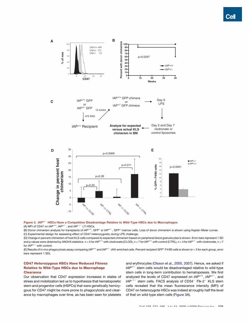
and erythrocytes (Olsson et al., 2005, 2007). Hence, we asked ifIAP+/! stem cells would be disadvantaged relative to wild-typestem cells in long-term contribution to hematopoiesis. We firstanalyzed the levels of CD47 expressed on IAP+/+, IAP+/!, andIAP!/! stem cells. FACS analysis of CD34! Flk-2! KLS stemcells revealed that the mean fluorescence intensity (MFI) ofCD47 on heterozygote HSCs was indeed at roughly half the levelof that on wild-type stem cells (Figure 3A).
A B
C
D E
Figure 3. IAP+/! HSCs Have a Competitive Disadvantage Relative to Wild-Type HSCs due to Macrophages(A) MFI of CD47 on IAP+/+, IAP+/!, and IAP!/! LT-HSCs.
(B) Donor chimerism analysis for transplants of IAP+/+, GFP+ or IAP+/!, GFP+ marrow cells. Loss of donor chimerism is shown using Kaplan-Meier curves.
(C) Experimental design for assessing effect of CD47 heterozygosity during LPS challenge.
(D) Change in percent chimerism of host KLS cells compared to expected chimerism based on peripheral blood granulocytes is shown. Error bars represent 1 SD
and p values were obtained by ANOVA statistics. n = 5 for IAP+/+ with clodronate (CLOD), n = 7 for IAP+/+ with control (CTRL), n = 4 for IAP+/!with clodronate, n = 7
for IAP+/! with control.
(E) Results of in vivo phagocytosis assay comparing IAP+/+ and IAP+/! cKit-enriched cells. Percent recipient GFP+ F4/80 cells is shown (n = 3 for each group, error
bars represent 1 SD).
Cell 138, 271–285, July 24, 2009 ª2009 Elsevier Inc. 275
We then transplanted one cohort of sublethally irradiatedrecipients with 2 3 106 wild-type whole bone marrow cells andanother with the same dose of IAP+/! bone marrow cells. Sucha dose would be expected to contain roughly 50–100 HSCs.Since granulocyte chimerism in the peripheral blood is a goodsurrogate marker of stem cell fitness (Bhattacharya et al.,2006), we analyzed cells from the blood of these recipients atperiodic intervals. When wild-type marrow was transplantedinto wild-type recipients, granulocyte chimerism was maintainedfor at least 40 weeks. However, when IAP+/! cells were trans-planted, 4 out of 10 mice lost donor chimerism over time, despitehaving a successful engraftment initially (p = 0.0297, Figures 3Band S2A). It was possible that there were fewer LT-HSCs per unitof marrow in IAP+/! or IAP!/! mice compared to wild-type. Totest if IAP!/! mice had fewer functional HSCs, we transplantedeither IAP+/+ or IAP!/! marrow cells into IAP!/! donors. Wealso cotransplanted IAP+/!marrow into wild-type mice togetherwith wild-type marrow. We did not find any statistical differencein donor granulocyte chimerism for up to 12 weeks in either case(Figures S2B and S2C), indicating that there was not a demon-strable deficit in stem cell numbers in IAP+/! or IAP!/! mice.
Interestingly, the IAP+/! chimeras that did not lose donorengraftment over time did not appear to have a quantitativedeficit in chimerism; rather they seemed to have levels of donorcells similar to those in the wild-type chimeras (Figure S2A). Wethus hypothesized that physiological insults leading to chronic oracute inflammatory states that occurred in some mice, but notothers, led to their losing IAP+/! donor chimerism over time,but at steady state the donor IAP+/! cells were not affected.
To test this, we created chimeras by transplanting whole bonemarrow from either IAP+/+ GFP+ or IAP+/! GFP+ mice into suble-thally irradiated wild-type syngeneic recipients. Since IAP!/!
mice have no known in vivo (Lindberg et al., 1996) or in vitro(Figure 2B) defect in granulopoiesis, blood granulocyte chime-rism can be used in this setting to estimate donor HSC engraft-ment. After 12 weeks, contribution of donor HSCs was assessed.At this early time point, IAP+/! mice actually had a higher bloodgranulocyte contribution, but there was considerable variationwithin each group. The mice were challenged with a sublethaldose of LPS (day 0), then given clodronate or control lipsomeson day 2 and day 7. Clodronate liposomes have been shownto specifically but transiently deplete marrow, spleen, and livermacrophages when injected intravenously (Van Rooijen andSanders, 1994). At day 14, the mice were euthanized andactual marrow KLS chimerism was assessed and compared toexpected chimerism based on blood granulocyte chimerism(Figure 3D). Raw values are shown in Table S1. The results indi-cate that there were more host (wild-type) KLS cells in the bonemarrow of the IAP+/! chimeras than expected. When macro-phages were depleted, the chimerism was at the level that wouldbe expected based on blood granulocyte chimerism. ANOVAshowed a significant interaction among IAP genotype (IAP+/+
versus IAP+/!) and treatment with and without clodronate(CLOD versus CTRL) (p = 0.0069). When the group includingthe IAP+/! chimeras treated with control liposomes (IAP+/!
CTRL) was excluded, there was no statistical difference betweenthe groups (p = 0.28). Analysis also showed that differencesbetween the treatment with and without clodronate were not
significant in IAP+/+ mice (p = 0.20) but were significant in IAP+/!
mice (p = 0.011).It was possible that these results were due to increased
apoptosis of IAP+/! cells relative to IAP+/+. To test this, wemeasured the percent of Annexin V+ 7-AAD! KLS cells in IAP+/+
and IAP+/! mice treated with LPS in the presence of control orclodronate liposomes. There was no difference between thegroups (Figure S4B).
To establish that macrophages were indeed responsible forclearing the cells with reduced CD47 expression, we challengeddonor IAP+/+, GFP+ and IAP+/!, GFP+ mice with LPS. After 3 days,we harvested bone marrow from these mice and transplantedcKit-enriched cells intrasplenically into GFP-negative wild-typerecipient mice similarly challenged with LPS 3 days prior. After2 hr the recipient spleens were harvested and analyzed byFACS. There were significantly more F4/80+ macrophages thatwere GFP+ (representing macrophages containing phagocy-tosed GFP+ cells) in mice injected with IAP+/! cells comparedto IAP+/+ (p < 0.0001, Figures 3E and S3).
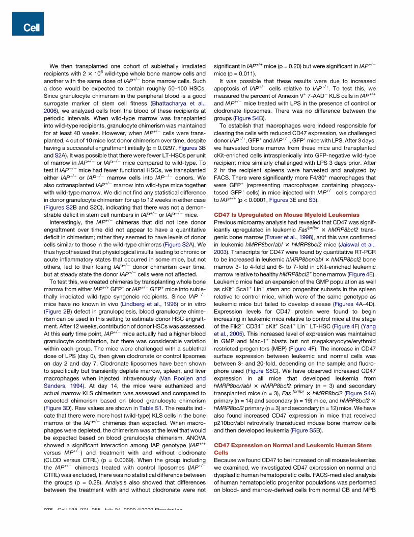
CD47 Is Upregulated on Mouse Myeloid LeukemiasPrevious microarray analysis had revealed that CD47 was signif-icantly upregulated in leukemic Faslpr/lpr 3 hMRP8bcl2 trans-genic bone marrow (Traver et al., 1998), and this was confirmedin leukemic hMRP8bcr/abl 3 hMRP8bcl2 mice (Jaiswal et al.,2003). Transcripts for CD47 were found by quantitative RT-PCRto be increased in leukemic hMRP8bcr/abl 3 hMRP8bcl2 bonemarrow 3- to 4-fold and 6- to 7-fold in cKit-enriched leukemicmarrow relative to healthy hMRP8bcl2+ bone marrow (Figure 4E).Leukemic mice had an expansion of the GMP population as wellas cKit+ Sca1+ Lin! stem and progenitor subsets in the spleenrelative to control mice, which were of the same genotype asleukemic mice but failed to develop disease (Figures 4A–4D).Expression levels for CD47 protein were found to beginincreasing in leukemic mice relative to control mice at the stageof the Flk2! CD34! cKit+ Sca1+ Lin! LT-HSC (Figure 4F) (Yanget al., 2005). This increased level of expression was maintainedin GMP and Mac-1+ blasts but not megakaryocyte/erythroidrestricted progenitors (MEP) (Figure 4F). The increase in CD47surface expression between leukemic and normal cells wasbetween 3- and 20-fold, depending on the sample and fluoro-phore used (Figure S5C). We have observed increased CD47expression in all mice that developed leukemia fromhMRP8bcr/abl 3 hMRP8bcl2 primary (n = 3) and secondarytransplanted mice (n = 3), Fas lpr/lpr 3 hMRP8bcl2 (Figure S4A)primary (n = 14) and secondary (n = 19) mice, and hMRP8bcl2 3hMRP8bcl2 primary (n = 3) and secondary (n = 12) mice. We havealso found increased CD47 expression in mice that receivedp210bcr/abl retrovirally transduced mouse bone marrow cellsand then developed leukemia (Figure S5B).
CD47 Expression on Normal and Leukemic Human StemCellsBecause we found CD47 to be increased on all mouse leukemiaswe examined, we investigated CD47 expression on normal anddysplastic human hematopoietic cells. FACS-mediated analysisof human hematopoietic progenitor populations was performedon blood- and marrow-derived cells from normal CB and MPB
276 Cell 138, 271–285, July 24, 2009 ª2009 Elsevier Inc.

C
E
A B
F
D
GH
Figure 4. CD47 Is Upregulated in Murine and Human Myeloid LeukemiaTypical stem and progenitor plots are shown for leukemic hMRP8bcrabl 3 hMRP8bcl2 cells compared to control nonleukemic animals. Lin! cKit+ Sca1+ gated
cells from control bone marrow (A) and leukemic spleen (B) and Lin! cKit+ Sca1! gated cells from control bone marrow (C) and leukemic spleen (D). Frequency is
shown as a percentage of entire marrow or spleen mononuclear fraction.
(E) Quantitative RT-PCR for CD47. Data are shown from three sets of mice transplanted with either leukemic or control hRMP8bcrabl 3 hMRP8bcl2 BM cells and
then sacrificed 2–6 weeks later. Results were normalized to beta-actin and 18S rRNA expression. Fold change relative to control transplanted whole Bcl-2+ BM
cells was determined. Error bars represent 1 SD.
(F) Histograms show expression of CD47 on gated populations for leukemic (gray) and control (black) mice.
(G) Comparative FACS histograms of human CD47 expression by normal (red; n = 6) and acute myeloid leukemic (AML, blue; n = 6) hematopoietic stem cells
(HSCs; CD34+CD38!CD90+Lin!) and progenitors (CD34+CD38+Lin!).
(H) Comparative FACS histograms of CD47 expression by normal (red) and chronic myeloid leukemia (blue) hematopoietic stem cells (HSCs;
CD34+CD38!CD90+Lin), committed progenitors (CD34+CD38+Lin!), and Lin+ cells.
Cell 138, 271–285, July 24, 2009 ª2009 Elsevier Inc. 277

(n = 16) and myeloproliferative disorders (MPDs) including poly-cythemia vera (PV; n = 16), myelofibrosis (MF; n = 5), essentialthrombocythemia (ET; n = 7), chronic myelomonocytic leukemia(CMML; n = 11), and atypical chronic myeloid leukemia (aCML;n = 1), as well as myeloid blast crisis (BC) phase chronic myeloidleukemia (CML; n = 19), chronic phase CML (n = 7), and acutemyeloid leukemia (AML; n = 13). This analysis demonstratedthat the GMP population was expanded in MPDs with myeloidskewed differentiation potential including atypical CML, prolifer-ative phase CMML, and acute leukemia including blast crisisCML and AML (Figures S6A and S6C). AML HSCs and progeni-tors uniformly exhibited higher levels of CD47 expressioncompared with normal controls (Figure 4G); every sample fromCML-BC (n = 19) and AML (n = 13) had elevated levels ofCD47. Moreover, progression from chronic phase CML to blastcrisis was associated with a significant increase in CD47 expres-sion (Figure 4H). Using the methods described in this study, wehave found that human CD47 protein expression in CML-BCincreased 2.2-fold in CD90+ CD34+ CD38! Lin! cells relative tonormal CB and MPB (p = 6.3 3 10!5), 2.3-fold in CD90!
CD34+ CD38!Lin! cells relative to normal (p = 4.3 3 10!5),and 2.4-fold in CD34+ CD38+ Lin! cells (p = 7.6 3 10!6) (Figures4G, 4H, and S6B). Since CB and MPB HSCs have almost doublethe amount of CD47 as normal marrow (Figure 1E), these figureslikely underestimate the true increase in CD47 compared tonormal marrow.
We also examined CD47 expression in other myeloprolifera-tive disorders, such as polycythemia vera, post-polycythemicmyeloid metaplasia with myelofibrosis, essential thrombocythe-mia, agnogenic myeloid metaplasia, and chronic myelomono-cytic leuekmia. None of the samples we examined from thesedisorders displayed increased CD47 (data not shown). It is likelythat the fixed, high expression of CD47 on AML cells andCML-BC cells represents a late event in malignant progression,whether by genetic or epigenetic heritably increased expression.A summary of all the human samples analyzed is shown inTable S2.
CD47 Expression Can Rescue the In Vivo Growth Defectof the AML Cell Line MOLM-13 via a Mechanism OtherThan MigrationIn order to experimentally test the significance of elevated CD47expression on myeloid leukemias, we decided to examinewhether the level of CD47 expressed on myeloid leukemia couldaffect its pathogenicity in vivo. We first screened a panel ofhuman AML cell lines for human CD47 expression. On the basisof this analysis, we found significant variation in the amount ofcell surface CD47 (adjusted for cell size) in the varying lines(Figures S7A and S7B). We then tested whether there was vari-ation in the ability of some of these cell lines to engraft in immu-nocompromised mice. Human HL-60 and Kasumi-1 cells, whichexpress moderate to high levels of human CD47, were able toengraft C57Bl/6 or Balb/c recombination-activating gene 2,common gamma chain-deficient (RAG2!/!, gc!/!) mice (FiguresS6C and S6D), which are reported to have little or no interactionbetween their SIRPa and human CD47 (Takenaka et al., 2007).MOLM-13 cells, which are derived from a patient with AML 5a,have low endogenous CD47 expression and were unable to
engraft in either of these strains. Even when transplanted intononobese diabetic severe combined immunodeficiency, inter-leukin 2 receptor common gamma chain!/! (NOG) mice thatare reported to have more effective interaction between theirmouse SIRPa and human CD47, and thus should be permissivefor human donor cells (Takenaka et al., 2007), MOLM-13 cellsfailed to engraft (Figure S7C).
Since MOLM-13 cells failed to engraft in any of the mousemodels tested, we decided to use them for further analysis ofthe role of CD47 in leukemogenicity. MOLM-13 cells were trans-duced with either control (Tet) or CD47-expressing (Tet-CD47)lentiviruses (Figure 5A), and stable integrants were propagatedon the basis of GFP expression. We first tested if ectopic expres-sion of the murine CD47 (muCD47) protein could rescue thegrowth defect of these cells. The cells were transplanted intrave-nously in a competitive setting with untransduced MOLM-13cells into T-, B-, and NK-deficient RAG2!/!, gc!/! mice. Onlycells transduced with Tet-CD47 were able to give rise to tumorsin these mice, efficiently engrafting bone marrow, spleen, liver,and peripheral blood (Figures 5B and 5C).
Since CD47 has been reported to be important for the migra-tion of hematopoietic cells, one possibility for the lack of growthof Tet MOLM-13 cells in mice was their inability to migrate toniches. To test this, Tet MOLM-13 or Tet-CD47 MOLM-13 cellswere directly injected into a single femoral cavity of immunode-ficient Balb/c mice. Tet-CD47 MOLM-13 cells were able tospread to all bones and other hematopoietic tissues of recipientmice and lead to death, whereas Tet MOLM-13 cells hadminimal, if any, engraftment and only at the site of injection,without morbidity by 75 days after transplant (Figures 5D–5F).These results suggested a function other than or in addition tomigration for CD47 in MOLM-13 engraftment.
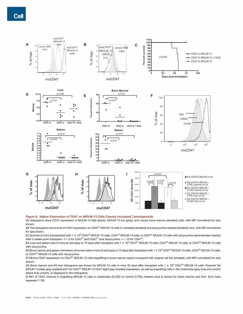
The Level of CD47 Expressed on MOLM-13 CellsDetermines Tumorigenic PotentialOur results up to this point had indicated that ectopicallyexpressing muCD47 on a nonengrafting human cell line couldrescue its growth defect. These experiments did not, however,answer whether the level of CD47 expressed on a leukemiccell could affect its tumor-forming potential. To model the tumor-igenic effect of having high versus low CD47 expression, wesorted clones of muCD47 expressing MOLM-13 cells into highand low expressors. When adjusted for cell size, CD47 densityon the CD47lo MOLM-13 cells was approximately equal tomouse bone marrow cells, whereas CD47hi MOLM-13 cellshad 9-fold higher expression, an increase commensurate withthe change seen in CD47 expression on primary leukemic cellscompared to their normal counterparts (Figures 6A and 6B).When high- or low-expressing cells were transplanted into recip-ients, only mice transplanted with high-expressing cells suc-cumbed to disease by 75 days of age (Figure 6C). Furthermore,organomegaly was more pronounced in these mice (Figure 6D).As expected, the infiltration of MOLM-13 cells in bone marrowand spleen of recipient mice was also much higher for micetransplanted with CD47hi MOLM-13 cells as well (Figure 6E).We also examined the level of CD47 expression in two micethat received CD47lo MOLM-13 cells but had significant marrowengraftment. In both cases, the persisting cells after 75 days had
278 Cell 138, 271–285, July 24, 2009 ª2009 Elsevier Inc.

much higher levels of CD47 than the original line (Figure 6F), indi-cating that a strong selection pressure exists in vivo for highlevels of CD47 expression on MOLM-13 leukemia cells.
We were also able to modulate CD47 expression with doxycy-cline because we utilized the Tet-responsive (Tet-OFF) promoterelement to control expression of CD47 in MOLM-13 cells. Begin-ning 2 weeks after transplantation with CD47hi MOLM-13 cells,a cohort of mice was given doxycycline to extinguish CD47expression and followed for up to 75 days post-transplant.
During this time, none of the mice given doxycycline succumbedto disease or had large infiltration of MOLM-13 cells in hemato-poietic organs (Figures 6D and 6E) other than 1–2 large extrapar-enchymal masses seen in the livers. At the dose of doxycyclineused in this experiment, muCD47 expression in MOLM-13 cellswas reduced to levels below that of normal mouse bone marrowbut notably was not completely absent (Figure 6B). Thus, a sus-tained high level of CD47 expression is required for robustMOLM-13 growth in hematopoietic organs.
A B C
D E
F
Figure 5. Overexpression of Murine CD47 Rescues the Growth Defect of MOLM-13 Myeloid Leukemia Cells(A) GFP and human CD45 chimerism for mice transplanted with untransduced MOLM-13 cells (5 3 105) and either 5 3 105 Tet (n = 6) or Tet-CD47 MOLM-13 (n = 8)
cells.
(B) MOLM-13 chimerism in hematopoietic tissues was determined by human CD45 chimerism and measurement of tumor lesion size.
(C) Hematoxylin and eosin sections of Tet-CD47 MOLM-13 transplanted liver (2003) (top panel). Periportal (arrow) and sinusoidal (arrowhead) tumor infiltration is
evident. Examples of liver tumor formation and hepatomegaly in Tet-CD47 MOLM-13 transplanted mice versus control transplanted mice (middle panel). GFP
fluorescence demonstrates tumor nodule formation as well diffuse infiltration (bottom panel).
(D) 1 3 106 Tet (n = 5) or Tet-CD47 MOLM-13 (n = 4) cells were injected into the right femur of RAG2!/!, Gc!/!mice and the tissues were analyzed 50–75 days later
for chimerism of MOLM-13 cells in BM.
(E) Survival curve of mice transplanted intrafemorally with Tet or Tet-CD47 MOLM-13 cells.
(F) Representative FACS plots from mice transplanted intrafemorally with Tet or Tet-CD47 MOLM-13 cells. R, right; L, left.
Cell 138, 271–285, July 24, 2009 ª2009 Elsevier Inc. 279
A B
D E F
C
HG I
Figure 6. Higher Expression of CD47 on MOLM-13 Cells Causes Increased Tumorigenicity(A) Histograms show CD47 expression in MOLM-13 high (black), MOLM-13 low (gray), and mouse bone marrow (shaded) cells, with MFI normalized for size
shown.
(B) The histograms show level of CD47 expression on CD47hi MOLM-13 cells in untreated (shaded) and doxycycline-treated (shaded) mice, with MFI normalized
for size shown.
(C) Survival of mice transplanted with 1 3 106 CD47hi MOLM-13 cells, CD47lo MOLM-13 cells, or CD47hi MOLM-13 cells with doxycycline administration started
after 2 weeks post-transplant. n = 5 for CD47hi and CD47hi plus doxycycline. n = 10 for CD47lo.
(D) Liver and spleen size of mice at necropsy or 75 days after transplant with 1 3 106 CD47hi MOLM-13 cells, CD47lo MOLM-13 cells, or CD47hi MOLM-13 cells
with doxycycline.
(E) Bone marrow and spleen chimerism of human cells in mice at necropsy or 75 days after transplant with 1 3 106 CD47hi MOLM-13 cells, CD47lo MOLM-13 cells,
or CD47hi MOLM-13 cells with doxycycline.
(F) Murine CD47 expression on CD47lo MOLM-13 cells engrafting in bone marrow (open) compared with original cell line (shaded), with MFI normalized for size
shown.
(G) Bone marrow and (H) liver histograms are shown for MOLM-13 cells in mice 35 days after transplant with 1 3 106 CD47lo MOLM-13 cells. Parental Tet
MOLM-13 (dark gray shaded) and Tet CD47lo MOLM-13 CD47 (light gray shaded) expression, as well as engrafting cells in the clodronate (gray line) and control
(black line) cohorts, is displayed in the histograms.
(I) MFI of CD47 channel in engrafting MOLM-13 cells in clodronate (CLOD) or control (CTRL) treated mice is shown for bone marrow and liver. Error bars
represent 1 SD.
280 Cell 138, 271–285, July 24, 2009 ª2009 Elsevier Inc.

Macrophages Mediate Selective Pressure for CD47hi
Variants of MOLM-13 CellsBecause we had observed that mice transplanted with CD47lo
MOLM-13 cells had selection over time for higher-expressingclones in vivo, we wondered if this selection pressure was medi-ated by macrophages. To test this, we used clodronate lipo-somes to deplete macrophages prior to and after transplant ofCD47lo MOLM-13 cells and sacrificed mice after 5 weeks.When control liposomes were used, the level of CD47 increasedsignificantly on the engrafted human cells. However, when clodr-onate-containing liposomes were used, the expression of CD47on MOLM-13 cells was maintained at the level of the parental line(Figures 6G–6I). Thus, macrophages mediate selective pressurefor high-expressing CD47 clones in vivo.
Interestingly, when clodronate was used to deplete macro-phages in the immunodeficient mice, the level of engraftmentwas comparable between the control Tet MOLM-13 cells andthe CD47 MOLM-13 cells (Figure S8), suggesting that depletionof macrophages could compensate for lack of CD47 expressionon MOLM-13 cells as well.
Ability to Evade Macrophage Phagocytosis Correlateswith CD47 Expression LevelWe incubated Tet (vector only), bulk (unsorted) Tet-CD47, Tet-CD47hi, or Tet-CD47lo MOLM-13 cells with bone marrow-derivedmacrophages (BMDM) for 2–24 hr and assessed phagocytosis bycounting the number of ingested GFP+ cells under a microscope(Figures 7A and 7B) or by evaluating the frequency of GFP+
macrophages by flow cytometry (Figure S9A). There was a strongcorrelation between level of CD47 expressed and the likelihoodof phagocytosis (Figure 7A). When CD47lo RFP and CD47hi GFPMOLM-13 cells were coincubated with macrophages in the samewells, the low-expressing cells were far more likely to be phago-cytosed (Figures 7C and 7D). Thus, in a mixed population of cellswith varying levels of CD47 expression, the low expressiors weremore likely to be cleared by macrophages over time. Theseresults were not due to differences in apoptosis betweenCD47-expressing and control MOLM-13 cells, as both groupshad similar levels of Annexin V positivity in vitro (Figure S4C).
We also injected MOLM-13 cells into mice and analyzedhematopoietic organs 2 hr later for evidence of macrophage
A B
C D
Figure 7. Increasing CD47 Expression on MOLM-13 Cells Confers Increasing Ability to Evade Macrophage Phagocytosis(A) Tet, Tet-CD47lo, Tet-CD47 bulk, or Tet CD47hi MOLM-13 cells were incubated with bone marrow-derived macrophages (BMDM) for 2 hr and phagocytic index
was determined. Error bars represent 1 SD (n = 6 for each time point).
(B) Photomicrographs of BMDMs incubated with Tet or Tet-CD47 MOLM-13 cells at 2 and 24 hr (4003).
(C) CD47hi GFP and CD47lo MOLM-13 RFP cells were coincubated with BMDMs for 2 hr. Phagocytic index is shown for three separate samples for CD47hi GFP
(green) and CD47lo MOLM-13 RFP (red) cells.
(D) Photomicrographs show brightfield (top left), RFP (top right), GFP (bottom left), and merged (bottom right) images of CD47hi GFP and CD47lo MOLM-13 RFP
cells that were coincubated with BMDMs for 24 hr.
Cell 138, 271–285, July 24, 2009 ª2009 Elsevier Inc. 281
phagocytosis. Macrophages in bone marrow, spleen, and liverall had a higher GFP+ fraction when injected with Tet MOLM-13cells as compared to muCD47-expressing cells, indicating thatthe muCD47! cells were more likely to be phagocytosed in vivoas well (Figure S9B).
Finally, we asked if inhibiting macrophage SIRPa using ablocking antibody could abrogate the protection against phago-cytosis that CD47 provided MOLM-13 cells. Indeed, when thisreceptor was blocked, CD47-expressing MOLM-13 cells werephagocytosed at the same rate as control MOLM-13 cells(Figure S10).
DISCUSSION
Physiological Significance of CD47 UpregulationWe demonstrate here that HSPCs upregulate CD47 in responseto an insult that induces mobilization. This then leads to thesurprising finding that HSPCs themselves are regulated byphagocytosis in vivo. Hence, our results indicate that protectionfrom phagocytosis is essential for HSC survival during migrationto the periphery after a strong mobilizing or inflammatory stim-ulus. A recent report indicates that HSPCs can circulate throughthe lymphatic system as well (Massberg et al., 2007). It will beinteresting to see if these HSPCs upregulate CD47 to evadethe numerous sinusoidal macrophages in lymph nodes.
Since leukemias upregulate CD47 as well, it appears that theyco-opt this normal physiological response to stress-inducedmobilization as a means to protect themselves at the expenseof normal, non-neoplastic progenitors. In this manner, leukemicstem cells could quickly gain a survival advantage over normalstem cells, leading to their eventual take-over of the hematopoi-etic environment.
The regulatory mechanisms for CD47 expression in mobiliza-tion and leukemia remain unknown. The fact that CD47 is upre-gulated and downregulated in such a temporally precise mannerduring mobilization suggests that there is tight control over itsexpression in normal physiology. Leukemias, however, seemto have constitutively high CD47 expression. The only publishedregulator of CD47 expression is the transcription factor nuclearrespiratory factor 1 (Nrf1) (Chang and Huang, 2004). It remainsto be determined if Nrf1 or other regulatory elements are involvedin the progression of human myeloid leukemia, and if there isa common regulatory pathway that operates in mobilizationas well.
Macrophages Are Important Mediators of TumorImmunosurveillanceMany examples of tumor clearance by T, B, and NK cells havebeen described in the literature, indicating that a healthy immunesystem is essential for regulating nascent tumor growth.However, to date, few examples indicating that macrophage-mediated phagocytosis can check tumor development havebeen produced. Collectively, our studies reveal that ectopicoverexpression of CD47 can enable otherwise immunogenictumor cells to grow rapidly in a T, B, and NK cell-deficienthost. Furthermore, this is likely to reflect a mechanism used byprimary human myeloid leukemias to evade the host immunesystem since CD47 is upregulated in nearly all sufficiently
advanced murine and human myeloid leukemias examinedthus far.
This form of immune evasion is particularly important sincethese cancers occupy sites of high macrophage infiltration.The leukemias studied here are clonal yet found throughout themarrow (and in mouse, spleen, and liver); these are organswith abundant intra-tissue macrophages as well as macro-phage-lined sinusoids at the entry site of circulating blood cells.CD47 was first cloned as an ovarian tumor cell marker, indicatingthat it may play a role in preventing phagocytosis of other tissuecancers as well (Campbell et al., 1992). Furthermore, solidtumors often metastasize to macrophage-rich tissues such asliver, lung, bone, and lymph nodes, indicating that they mustbe able to escape macrophage-mediated killing in those tissues.Preventing CD47-SIRPa interaction could be doubly effectivebecause antigens from phagocytosed tumor cells may bepresented by professional antigen-presenting cells such asmacrophages or dendritic cells to activate an adaptive immuneresponse, leading to further tumor destruction.
CD47 May Be Upregulated because Some CancersAre More Immunogenic Than Normal TissueBut why would leukemic progenitor cells require additionalprotection against phagocytosis? Like aged neutrophils or eryth-rocytes, tumor cells can display signs of cellular ‘‘damage’’ and/or glycosidic variations on their cell surface. There are well-estab-lished data that tumors can be recognized by NK cells via ligandsthat mark ‘‘stressed self’’ (Gasser et al., 2005). We propose thatleukemic cells likewise express stress ligands that mark themfor phagocytosis; by upregulating CD47, this innate immunitycheck on tumor growth can be circumvented. One of thesepro-phagocytic ligands, calreticulin, is known to be upregulatedin aged cells. It has been shown that its effects can be counter-acted by expression of CD47 (Gardai et al., 2005). It remains tobe seen if this or other stress ligands are expressed on nascentleukemic cells to mark them for destruction.
Therapeutics Targeting CD47 May Be Clinically Usefulfor Treating CancerOthers have shown that antibodies that crosslink CD47 on thesurface of chronic lymphocytic leukemia cells can induce a cas-pase-independent cell death (Mateo et al., 1999; Uno et al.,2007). This phenomenon, while promising, appears to functionprimarily by inducing apoptosis, not necessarily by promotingphagocytosis. In fact, we show in a companion paper (Majetiet al., 2009 [this issue of Cell]), that anti-CD47 blocked CD47hi
human AML cells are phagocytosed by macrophages, withoutfirst becoming apoptotic.
In addition, our recent results in human AML indicate thatincreasing CD47 transcript levels are associated with shortenedsurvival (Majeti et al., 2009), allowing the level of expression of thisgene to potentially be used as an independent prognostic vari-able. Combined with the data presented here, where we showthat CD47 expression levels affect the fitness of both normalHSCs and leukemic cells, we propose that CD47 is a compellingtarget for therapeutic strategies that reduce its functional interac-tion with macrophage SIRPa and thus render these cancerssusceptible to innate and adaptive immune system clearance.
282 Cell 138, 271–285, July 24, 2009 ª2009 Elsevier Inc.

EXPERIMENTAL PROCEDURES
MiceCD47-deficient IAP!/!mice on a C57Bl/6 background were obtained from Eric
Brown and bred onto our wild-type colony. IAP!/!mice were crossed to beta-
actin GFP (Wright et al., 2001a) or CD45.1 C57Bl/6 mice from our colony to
obtain GFP+ or congenic strains. hMRP8bcrabl, hMRP8bcl2, and Faslpr/lpr
transgenic mice were obtained as previously described (Traver et al., 1998;
Jaiswal et al., 2003). C57Bl/6 mice from our colony were used as a source
of wild-type cells. C57Bl/6 or Balb/c RAG2!/! common gamma chain (Gc)!/!
mice were bred in our laboratory. NOD.Cg-PrkdcscidIl2rgtm1Wjl/SzJ (NOG)
mice were obtained from The Jackson Laboratory and bred in a pathogen-
free environment at our facility. For transplant experiments, cells were trans-
planted into either immunodeficient mice given a radiation dose of 4 Gy using
gamma rays from a cesium irradiator (Phillips), or CD45.2 C57Bl6/Ka mice
given a radiation dose of 9.5 Gy. Sublethal radiation was given at a dose of
4.75 Gy. Mice were euthanized when moribund.
Cells were injected via tail vein in sterile PBS using a 27-gauge needle. Donor
and recipient mice were 6–12 weeks of age. Parabiosis was performed as
previously described (Wright et al., 2001b). Mice were mobilized with cyclo-
phosphamide (Sigma) (200 mg/kg) and G-CSF (Neupogen) (250 mg/kg) as
previously described (Morrison et al., 1997). Bacterial LPS from E. coli
055:B5 (Sigma) was administered at a dose of 40 mg/kg into the peritoneal
cavity.
Cell LinesMOLM-13 cells were obtained from DSMZ. HL-60, Jurkat, U937, K562, and
Kasumi-1 cells were obtained from ATCC. Cells were maintained in Iscove’s
modified Dulbecco’s media (IMDM) plus 10% fetal bovine serum (FBS)
(Hyclone).
Quantitative RT-PCR AnalysisBone marrow was obtained from leukemic hMRP8bcr/abl 3 hMRP8bcl2 mice
or hMRP8bcl2 control mice. Cells were cKit enriched using cKit microbeads
and an autoMACS column (Miltenyi) and cDNA was created from RNA using
standard methods (Invitrogen, SuperScript II). cDNA corresponding to approx-
imately 1000 cells was used per PCR reaction. Quantitative PCR was per-
formed with a SYBR green kit on an ABI Prism 7000 PCR (Applied Biosystems)
machine.
Cell Staining and Flow CytometryMouse tissues were prepared and stained for stem, progenitor, and peripheral
blood cells as previously described (Akashi et al., 2000; Yang et al., 2005;
Bhattacharya et al., 2006). Mouse CD47 antibody (clone mIAP301, ATCC)
was assessed using biotinylated antibody produced in our lab followed by
staining with streptavidin-conjugated Quantum Dot 605 (Chemicon). Samples
were analyzed using a FACSAria (Beckton Dickinson).
Normal human bone marrow and blood samples were obtained with
informed consent from 20- to 25-year-old paid donors who were hepatitis A,
B, C, and HIV negative by serology (All Cells). Leukemic blood and marrow
cells were obtained with informed consent from previously untreated patients
at Stanford University Medical Center according to IRB approved methods.
HSCs and progenitors were stained and identified as previously described
(Manz et al., 2002). Anti-human CD47 FITC (clone B6H12, PharMingen) was
used to assess CD47 expression in all human samples. Following staining,
cells were analyzed using a modified FACS Vantage (Becton Dickinson) or a
FACSAria.
Engraftment of MOLM-13 cells was assessed by using anti-human CD45
PE-Cy7 (PharMingen), anti-mouse CD45.2 APC (clone AL1-4A2 produced in
our lab), and anti-mouse CD47 Alexa-680 (mIAP301, produced in our lab).
All samples were resuspended in propidium iodide containing buffer before
analysis to exclude dead cells. FACS data was analyzed using FloJo software
(Treestar).
Lentiviral Preparation and TransductionpRRL.sin-18.PPT.Tet07.IRES.GFP.pre, CMV, VSV, and tet trans-activator
(tTA) plasmids were obtained from Luigi Naldini (Vigna et al., 2002). The full-
length murine cDNA for CD47 form 2 was provided by Eric Brown (Genentech).
Plasmid DNA was transfected into 293T cells and supernatant harvested and
concentrated by standard protocols. Cells were transduced with lentivirus for
48 hr and GFP+ cells were sorted to purity and grown for several generations to
ensure stability of the transgenes.
In Vitro Phagocytosis AssaysBone marrow-derived macrophages were prepared and harvested by incuba-
tion in trypsin/EDTA (GIBCO) for 5 min and gentle scraping. Macrophages
were plated at 5 3 104 cells per well in a 24-well tissue culture plate (Falcon)
for 24 hr. Fresh media were added 2 hr before 2.5 3 105 target cells were
added to the macrophage-containing wells and incubated at 37"C for the indi-
cated times. After coincubation, wells were washed thoroughly with IMDM
three times and examined under an Eclipse T5100 (Nikon) using an enhanced
green fluorescent protein (GFP) or Texas Red filter set (Nikon). Phagocytic
index was calculated using the following formula: phagocytic index = number
of ingested cells/(number of macrophages/100). At least 200 macrophages
were counted per well.
Macrophage DepletionClodronate was a gift of Roche Diagnostics GmbH. It was encapsulated in
liposomes as previously described (Van Rooijen and Sanders, 1994). Macro-
phages were depleted by injecting intravenously (i.v.) 200 ml of the final
liposome preparation 48 hr prior to cell transplant. Mice were injected with
100 ml every week thereafter to maintain depletion until they were euthanized.
StatisticsStatistics were calculated using Prism 4.0 software (GraphPad Software, Inc.)
The Welch corrected student’s t test was used to calculate all column statis-
tics. Two-tailed p values are shown in figures. For survival curves, p values
were obtained using a logrank test. For comparison of groups, one-way
ANOVA and tests of contrast were used where indicated.
Additional Experimental Procedures are available in Supplemental Data.
SUPPLEMENTAL DATA
Supplemental Data include two tables, ten figures, and Supplemental Experi-
mental Procedures and can be found with this article online at http://www.cell.
com/supplemental/S0092-8674(09)00651-5.
ACKNOWLEDGMENTS
The work in this study was supported in part by National Institutes of Health
Grants 5R01CA086017-08, 5R01HL058770-08, and 5P01DK053074-08
awarded to I.L.W., the de Villier award of the Leukemia Society, the Ludwig
Institute, and a gift from the Smith Family Fund. The authors would also like
to thank Christina Muscat for antibody production, Libuse Jerabek for capable
laboratory management, and Eric Brown and Mette Johanssen for providing
valuable advice and reagents.
Author contributions: S.J., C.H.M.J., W.W.P., M.P.C., R.M., and I.L.W.
designed experiments. S.J., C.H.M.J., W.W.P., C.Y.P., M.P.C., and D.T. per-
formed experiments and analyzed data. N.v.R. provided reagents. S.J.,
C.H.M.J., and I.L.W. wrote the manuscript. All authors endorse the full content
of this work.
Received: June 30, 2008
Revised: March 4, 2009
Accepted: May 21, 2009
Published: July 23, 2009
REFERENCES
Akashi, K., Traver, D., Miyamoto, T., and Weissman, I.L. (2000). A clonogenic
common myeloid progenitor that gives rise to all myeloid lineages. Nature 404,
193–197.
Cell 138, 271–285, July 24, 2009 ª2009 Elsevier Inc. 283

Bhattacharya, D., Rossi, D.J., Bryder, D., and Weissman, I.L. (2006). Purified
hematopoietic stem cell engraftment of rare niches corrects severe lymphoid
deficiencies without host conditioning. J. Exp. Med. 203, 73–85.
Blazar, B.R., Lindberg, F.P., Ingulli, E., Panoskaltsis-Mortari, A., Oldenborg,
P.A., Iizuka, K., Yokoyama, W.M., and Taylor, P.A. (2001). CD47 (integrin-
associated protein) engagement of dendritic cell and macrophage counterre-
ceptors is required to prevent the clearance of donor lymphohematopoietic
cells. J. Exp. Med. 194, 541–549.
Brown, E.J., and Frazier, W.A. (2001). Integrin-associated protein (CD47) and
its ligands. Trends Cell Biol. 11, 130–135.
Campbell, I.G., Freemont, P.S., Foulkes, W., and Trowsdale, J. (1992). An
ovarian tumor marker with homology to vaccinia virus contains an IgV-like
region and multiple transmembrane domains. Cancer Res. 52, 5416–5420.
Chang, W.T., and Huang, A.M. (2004). Alpha-Pal/NRF-1 regulates the
promoter of the human integrin-associated protein/CD47 gene. J. Biol.
Chem. 279, 14542–14550.
Christensen, J.L., Wright, D.E., Wagers, A.J., and Weissman, I.L. (2004). Circu-
lation and chemotaxis of fetal hematopoietic stem cells. PLoS Biol. 2, E75. 10.
1371/journal.pbio.0020075.
Cline, M.J., and Golde, D.W. (1977). Mobilization of hematopoietic stem
cells (CFU-C) into the peripheral blood of man by endotoxin. Exp. Hematol.
5, 186–190.
Cumano, A., and Godin, I. (2007). Ontogeny of the hematopoietic system.
Annu. Rev. Immunol. 25, 745–785.
Fenton, M.J., and Golenbock, D.T. (1998). LPS-binding proteins and recep-
tors. J. Leukoc. Biol. 64, 25–32.
Gao, A.G., Lindberg, F.P., Finn, M.B., Blystone, S.D., Brown, E.J., and Frazier,
W.A. (1996). Integrin-associated protein is a receptor for the C-terminal
domain of thrombospondin. J. Biol. Chem. 271, 21–24.
Gardai, S.J., McPhillips, K.A., Frasch, S.C., Janssen, W.J., Starefeldt, A.,
Murphy-Ullrich, J.E., Bratton, D.L., Oldenborg, P.A., Michalak, M., and
Henson, P.M. (2005). Cell-surface calreticulin initiates clearance of viable or
apoptotic cells through trans-activation of LRP on the phagocyte. Cell 123,
321–334.
Gasser, S., Orsulic, S., Brown, E.J., and Raulet, D.H. (2005). The DNA damage
pathway regulates innate immune system ligands of the NKG2D receptor.
Nature 436, 1186–1190.
Jaiswal, S., Traver, D., Miyamoto, T., Akashi, K., Lagasse, E., and Weissman,
I.L. (2003). Expression of BCR/ABL and BCL-2 in myeloid progenitors leads to
myeloid leukemias. Proc. Natl. Acad. Sci. USA 100, 10002–10007.
Jiang, P., Lagenaur, C.F., and Narayanan, V. (1999). Integrin-associated
protein is a ligand for the P84 neural adhesion molecule. J. Biol. Chem. 274,
559–562.
Lindberg, F.P., Bullard, D.C., Caver, T.E., Gresham, H.D., Beaudet, A.L., and
Brown, E.J. (1996). Decreased resistance to bacterial infection and granulo-
cyte defects in IAP-deficient mice. Science 274, 795–798.
Liu, Y., Merlin, D., Burst, S.L., Pochet, M., Madara, J.L., and Parkos, C.A.
(2001). The role of CD47 in neutrophil transmigration. Increased rate of migra-
tion correlates with increased cell surface expression of CD47. J. Biol. Chem.
276, 40156–40166.
Majeti, R., Chao, M.P., Alizadeh, A.A., Pang, W.W., Jaiswal, S., Gibbs, K.D.,
Jr., van Rooijen, N., and Weissman, I.L. (2009). CD47 is an adverse prognostic
factor and therapeutic antibody target on human acute myeloid leukemia stem
cells. Cell 138, this issue, 286–299.
Manz, M.G., Miyamoto, T., Akashi, K., and Weissman, I.L. (2002). Prospective
isolation of human clonogenic common myeloid progenitors. Proc. Natl. Acad.
Sci. USA 99, 11872–11877.
Massberg, S., Schaerli, P., Knezevic-Maramica, I., Kollnberger, M., Tubo, N.,
Moseman, E.A., Huff, I.V., Junt, T., Wagers, A.J., Mazo, I.B., et al. (2007).
Immunosurveillance by hematopoietic progenitor cells trafficking through
blood, lymph, and peripheral tissues. Cell 131, 994–1008.
Mateo, V., Lagneaux, L., Bron, D., Biron, G., Armant, M., Delespesse, G., and
Sarfati, M. (1999). CD47 ligation induces caspase-independent cell death in
chronic lymphocytic leukemia. Nat. Med. 5, 1277–1284.
Miyashita, M., Ohnishi, H., Okazawa, H., Tomonaga, H., Hayashi, A., Fujimoto,
T.T., Furuya, N., and Matozaki, T. (2004). Promotion of neurite and filopodium
formation by CD47: roles of integrins, Rac, and Cdc42. Mol. Biol. Cell 15,
3950–3963.
Morrison, S.J., Hemmati, H.D., Wandycz, A.M., and Weissman, I.L. (1995). The
purification and characterization of fetal liver hematopoietic stem cells. Proc.
Natl. Acad. Sci. USA 92, 10302–10306.
Morrison, S.J., Wright, D.E., and Weissman, I.L. (1997). Cyclophosphamide/
granulocyte colony-stimulating factor induces hematopoietic stem cells to
proliferate prior to mobilization. Proc. Natl. Acad. Sci. USA 94, 1908–1913.
Oldenborg, P.A., Zheleznyak, A., Fang, Y.F., Lagenaur, C.F., Gresham, H.D.,
and Lindberg, F.P. (2000). Role of CD47 as a marker of self on red blood cells.
Science 288, 2051–2054.
Olsson, M., Bruhns, P., Frazier, W.A., Ravetch, J.V., and Oldenborg, P.A.
(2005). Platelet homeostasis is regulated by platelet expression of CD47 under
normal conditions and in passive immune thrombocytopenia. Blood 105,
3577–3582.
Olsson, M., Nilsson, A., and Oldenborg, P.A. (2007). Dose-dependent
inhibitory effect of CD47 in macrophage uptake of IgG-opsonized murine
erythrocytes. Biochem. Biophys. Res. Commun. 352, 193–197.
Peled, A., Petit, I., Kollet, O., Magid, M., Ponomaryov, T., Byk, T., Nagler, A.,
Ben-Hur, H., Many, A., Shultz, L., et al. (1999). Dependence of human stem
cell engraftment and repopulation of NOD/SCID mice on CXCR4. Science
283, 845–848.
Reinhold, M.I., Lindberg, F.P., Kersh, G.J., Allen, P.M., and Brown, E.J. (1997).
Costimulation of T cell activation by integrin-associated protein (CD47) is an
adhesion-dependent, CD28-independent signaling pathway. J. Exp. Med.
185, 1–11.
Takenaka, K., Prasolava, T.K., Wang, J.C., Mortin-Toth, S.M., Khalouei, S.,
Gan, O.I., Dick, J.E., and Danska, J.S. (2007). Polymorphism in Sirpa modu-
lates engraftment of human hematopoietic stem cells. Nat. Immunol. 8,
1313–1323.
Traver, D., Akashi, K., Weissman, I.L., and Lagasse, E. (1998). Mice defective
in two apoptosis pathways in the myeloid lineage develop acute myeloblastic
leukemia. Immunity 9, 47–57.
Uno, S., Kinoshita, Y., Azuma, Y., Tsunenari, T., Yoshimura, Y., Iida, S.,
Kikuchi, Y., Yamada-Okabe, H., and Fukushima, N. (2007). Antitumor activity
of a monoclonal antibody against CD47 in xenograft models of human
leukemia. Oncol. Rep. 17, 1189–1194.
Van Rooijen, N., and Sanders, A. (1994). Liposome mediated depletion of
macrophages: mechanism of action, preparation of liposomes and applica-
tions. J. Immunol. Methods 174, 83–93.
Van, V.Q., Lesage, S., Bouguermouh, S., Gautier, P., Rubio, M., Levesque, M.,
Nguyen, S., Galibert, L., and Sarfati, M. (2006). Expression of the self-marker
CD47 on dendritic cells governs their trafficking to secondary lymphoid
organs. EMBO J. 25, 5560–5568.
Vigna, E., Cavalieri, S., Ailles, L., Geuna, M., Loew, R., Bujard, H., and Naldini,
L. (2002). Robust and efficient regulation of transgene expression in vivo by
improved tetracycline-dependent lentiviral vectors. Mol. Ther. 5, 252–261.
Wagers, A.J., Allsopp, R.C., and Weissman, I.L. (2002). Changes in integrin
expression are associated with altered homing properties of Lin(-/lo)Thy1.1
(lo)Sca-1(+)c-kit(+) hematopoietic stem cells following mobilization by
cyclophosphamide/granulocyte colony-stimulating factor. Exp. Hematol. 30,
176–185.
Wang, H., Madariaga, M.L., Wang, S., Van Rooijen, N., Oldenborg, P.A., and
Yang, Y.G. (2007). Lack of CD47 on nonhematopoietic cells induces split
macrophage tolerance to CD47null cells. Proc. Natl. Acad. Sci. USA 104,
13744–13749.
Wright, D.E., Cheshier, S.H., Wagers, A.J., Randall, T.D., Christensen, J.L., and
Weissman, I.L. (2001a). Cyclophosphamide/granulocyte colony-stimulating
284 Cell 138, 271–285, July 24, 2009 ª2009 Elsevier Inc.

factor causes selective mobilization of bone marrow hematopoietic stem cells
into the blood after M phase of the cell cycle. Blood 97, 2278–2285.
Wright, D.E., Wagers, A.J., Gulati, A.P., Johnson, F.L., and Weissman, I.L.
(2001b). Physiological migration of hematopoietic stem and progenitor cells.
Science 294, 1933–1936.
Wright, D.E., Bowman, E.P., Wagers, A.J., Butcher, E.C., and Weissman, I.L.
(2002). Hematopoietic stem cells are uniquely selective in their migratory
response to chemokines. J. Exp. Med. 195, 1145–1154.
Wright, S.D., Tobias, P.S., Ulevitch, R.J., and Ramos, R.A. (1989). Lipopoly-
saccharide (LPS) binding protein opsonizes LPS-bearing particles for
recognition by a novel receptor on macrophages. J. Exp. Med. 170, 1231–
1241.
Yang, L., Bryder, D., Adolfsson, J., Nygren, J., Mansson, R., Sigvardsson, M.,
and Jacobsen, S.E. (2005). Identification of Lin(-)Sca1(+)kit(+)CD34(+)Flt3-
short-term hematopoietic stem cells capable of rapidly reconstituting and
rescuing myeloablated transplant recipients. Blood 105, 2717–2723.
Cell 138, 271–285, July 24, 2009 ª2009 Elsevier Inc. 285