Three-Dimensional Structures of Pathogenic and Saprophytic ...
8
Three-Dimensional Structures of Pathogenic and Saprophytic Leptospira Species Revealed by Cryo-Electron Tomography Gianmarco Raddi, a Dustin R. Morado, a Jie Yan, c David A. Haake, d,e X. Frank Yang, b and Jun Liu a Department of Pathology and Laboratory Medicine, University of Texas Medical School at Houston, Houston, Texas, USA a ; Department of Microbiology and Immunology, Indiana University School of Medicine, Indianapolis, Indiana, USA b ; Department of Medical Microbiology and Parasitology, College of Medicine, Zhejiang University, Hangzhou, People’s Republic of China c ; Division of Infectious Diseases, Veterans Affairs Greater Los Angeles Healthcare System, Los Angeles, California, USA d ; and Departments of Medicine, Urology, and Microbiology, Immunology, and Molecular Genetics, University of California, Los Angeles, Los Angeles, California, USA e Leptospira interrogans is the primary causative agent of the most widespread zoonotic disease, leptospirosis. An in-depth struc- tural characterization of L. interrogans is needed to understand its biology and pathogenesis. In this study, cryo-electron tomog- raphy (cryo-ET) was used to compare pathogenic and saprophytic species and examine the unique morphological features of this group of bacteria. Specifically, our study revealed a structural difference between the cell envelopes of L. interrogans and Lepto- spira biflexa involving variations in the lipopolysaccharide (LPS) layer. Through cryo-ET and subvolume averaging, we deter- mined the first three-dimensional (3-D) structure of the flagellar motor of leptospira, with novel features in the flagellar C ring, export apparatus, and stator. Together with direct visualization of chemoreceptor arrays, DNA packing, periplasmic filaments, spherical cytoplasmic bodies, and a unique “cap” at the cell end, this report provides structural insights into these fascinating Leptospira species. T he genus Leptospira belongs to the phylum Spirochetes and includes both pathogenic and saprophytic species (37). L. in- terrogans is the predominant pathogenic species causing leptospi- rosis, a global reemerging zoonotic disease (for reviews, see refer- ences 17 and 20). In nature, L. interrogans colonizes the renal tubules of reservoir hosts (e.g., wild rodents). Humans are in- fected via contact with infected animals or contaminated soil or water. Outbreaks occur frequently in tropical regions (2, 20), with more than 500,000 reported severe cases of leptospirosis annually with a 10% mortality rate (29). In contrast, Leptospira biflexa is a free-living saprophytic organism that is unable to infect the mam- malian host despite its extensive genetic and structural similarities with L. interrogans. Recent comparative genomic analyses of L. interrogans and L. biflexa have identified many unique genes in L. interrogans that may be potentially associated with virulence and pathogenesis (6, 35, 40, 42, 49). Leptospira spp. share a number of general features with other spirochetes, yet they differ in many ways from other spirochetal pathogens, such as Treponema pallidum and Borrelia burgdorferi (42). As with other spirochetes, Leptospiral motility is driven by the rotation of periplasmic flagella (PF) that are located between the inner membrane (IM) and outer membrane (OM). PF of Lep- tospira species extend a relatively short distance from each pole, resulting in the characteristic hook-shaped ends of the cells (11). Leptospira also has an abundance of lipopolysaccharide (LPS) that is absent from both T. pallidum and B. burgdorferi (42). Cryo-electron tomography (cryo-ET) is a powerful three- dimensional (3-D) imaging technique that is capable of visualiz- ing cellular components in living organisms at the molecular level (23, 26, 30). This technique involves collecting a tilt series of im- ages from different angles at high magnification and computa- tionally reconstructing a 3-D density map of the intact organism. Recently, cryo-ET has been extensively utilized to study many spirochetes (1, 7, 14, 15, 19, 28, 32, 33), particularly, in our hands, to study the intact flagellar motor structure of B. burgdorferi (25) as well as the cell envelope architecture of T. pallidum (24). In this work, we employed cryo-ET for a systematic compari- son of cellular structures from pathogenic L. interrogans and sap- rophytic L. biflexa. Our study revealed cellular features unique to Leptospira spp., as well as structural differences between patho- genic L. interrogans and nonpathogenic L. biflexa. Possible impli- cations of our findings in leptospiral motility, physiology, and pathogenesis are discussed. MATERIALS AND METHODS Leptospira culture preparation. L. interrogans serovar Lai strain Lai 56601 and L. biflexa serovar Patoc strain Patoc I (Paris) were kindly pro- vided by James Matsunaga at the VA Medical Center in Los Angeles, CA. Leptospira spp. were cultured in 50 ml Ellinghausen-McCullough- Johnson-Harris (EMJH) medium at 30°C under aerobic conditions for 100 h to reach exponential growth phase. Leptospiral pellets were har- vested by centrifugation at 4,000 g at 22°C for 20 min and resuspended in EMJH medium. Cryo-ET data collection and 3-D reconstruction. Viable bacterial cultures were centrifuged to increase the concentration to 2 10 9 cells/ ml. four-microliter samples were deposited onto freshly glow-discharged holey carbon grids for 1 min. The grids were blotted with filter paper and rapidly frozen in liquid ethane using a gravity-driven plunger apparatus as previously described (25). The resulting frozen-hydrated specimens were imaged at 170°C using a Polara G2 electron microscope (FEI Company, Hillsboro, OR) equipped with a field emission gun and a 4,000 (4K) 4K charge-coupled-device (CCD) (16-megapixel) camera (TVIPS; GMBH, Germany). The microscope was operated at 300 kV with a magnification of 31,000, resulting in an effective pixel size of 5.6 Å after 2 2 binning. Received 7 November 2011 Accepted 28 December 2011 Published ahead of print 6 January 2012 Address correspondence to Frank Yang, [email protected], or Jun Liu, [email protected]. Supplemental material for this article may be found at http://jb.asm.org/. Copyright © 2012, American Society for Microbiology. All Rights Reserved. doi:10.1128/JB.06474-11 0021-9193/12/$12.00 Journal of Bacteriology p. 1299 –1306 jb.asm.org 1299 on March 21, 2018 by guest http://jb.asm.org/ Downloaded from
Transcript of Three-Dimensional Structures of Pathogenic and Saprophytic ...
Three-Dimensional Structures of Pathogenic and SaprophyticLeptospira Species Revealed by Cryo-Electron Tomography
Gianmarco Raddi,a Dustin R. Morado,a Jie Yan,c David A. Haake,d,e X. Frank Yang,b and Jun Liua
Department of Pathology and Laboratory Medicine, University of Texas Medical School at Houston, Houston, Texas, USAa; Department of Microbiology and Immunology,Indiana University School of Medicine, Indianapolis, Indiana, USAb; Department of Medical Microbiology and Parasitology, College of Medicine, Zhejiang University,Hangzhou, People’s Republic of Chinac; Division of Infectious Diseases, Veterans Affairs Greater Los Angeles Healthcare System, Los Angeles, California, USAd; andDepartments of Medicine, Urology, and Microbiology, Immunology, and Molecular Genetics, University of California, Los Angeles, Los Angeles, California, USAe
Leptospira interrogans is the primary causative agent of the most widespread zoonotic disease, leptospirosis. An in-depth struc-tural characterization of L. interrogans is needed to understand its biology and pathogenesis. In this study, cryo-electron tomog-raphy (cryo-ET) was used to compare pathogenic and saprophytic species and examine the unique morphological features of thisgroup of bacteria. Specifically, our study revealed a structural difference between the cell envelopes of L. interrogans and Lepto-spira biflexa involving variations in the lipopolysaccharide (LPS) layer. Through cryo-ET and subvolume averaging, we deter-mined the first three-dimensional (3-D) structure of the flagellar motor of leptospira, with novel features in the flagellar C ring,export apparatus, and stator. Together with direct visualization of chemoreceptor arrays, DNA packing, periplasmic filaments,spherical cytoplasmic bodies, and a unique “cap” at the cell end, this report provides structural insights into these fascinatingLeptospira species.
The genus Leptospira belongs to the phylum Spirochetes andincludes both pathogenic and saprophytic species (37). L. in-
terrogans is the predominant pathogenic species causing leptospi-rosis, a global reemerging zoonotic disease (for reviews, see refer-ences 17 and 20). In nature, L. interrogans colonizes the renaltubules of reservoir hosts (e.g., wild rodents). Humans are in-fected via contact with infected animals or contaminated soil orwater. Outbreaks occur frequently in tropical regions (2, 20), withmore than 500,000 reported severe cases of leptospirosis annuallywith a 10% mortality rate (29). In contrast, Leptospira biflexa is afree-living saprophytic organism that is unable to infect the mam-malian host despite its extensive genetic and structural similaritieswith L. interrogans. Recent comparative genomic analyses of L.interrogans and L. biflexa have identified many unique genes in L.interrogans that may be potentially associated with virulence andpathogenesis (6, 35, 40, 42, 49).
Leptospira spp. share a number of general features with otherspirochetes, yet they differ in many ways from other spirochetalpathogens, such as Treponema pallidum and Borrelia burgdorferi(42). As with other spirochetes, Leptospiral motility is driven bythe rotation of periplasmic flagella (PF) that are located betweenthe inner membrane (IM) and outer membrane (OM). PF of Lep-tospira species extend a relatively short distance from each pole,resulting in the characteristic hook-shaped ends of the cells (11).Leptospira also has an abundance of lipopolysaccharide (LPS) thatis absent from both T. pallidum and B. burgdorferi (42).
Cryo-electron tomography (cryo-ET) is a powerful three-dimensional (3-D) imaging technique that is capable of visualiz-ing cellular components in living organisms at the molecular level(23, 26, 30). This technique involves collecting a tilt series of im-ages from different angles at high magnification and computa-tionally reconstructing a 3-D density map of the intact organism.Recently, cryo-ET has been extensively utilized to study manyspirochetes (1, 7, 14, 15, 19, 28, 32, 33), particularly, in our hands,to study the intact flagellar motor structure of B. burgdorferi (25)as well as the cell envelope architecture of T. pallidum (24).
In this work, we employed cryo-ET for a systematic compari-son of cellular structures from pathogenic L. interrogans and sap-rophytic L. biflexa. Our study revealed cellular features unique toLeptospira spp., as well as structural differences between patho-genic L. interrogans and nonpathogenic L. biflexa. Possible impli-cations of our findings in leptospiral motility, physiology, andpathogenesis are discussed.
MATERIALS AND METHODSLeptospira culture preparation. L. interrogans serovar Lai strain Lai56601 and L. biflexa serovar Patoc strain Patoc I (Paris) were kindly pro-vided by James Matsunaga at the VA Medical Center in Los Angeles, CA.Leptospira spp. were cultured in 50 ml Ellinghausen-McCullough-Johnson-Harris (EMJH) medium at 30°C under aerobic conditions for100 h to reach exponential growth phase. Leptospiral pellets were har-vested by centrifugation at 4,000 � g at 22°C for 20 min and resuspendedin EMJH medium.
Cryo-ET data collection and 3-D reconstruction. Viable bacterialcultures were centrifuged to increase the concentration to �2 � 109 cells/ml. four-microliter samples were deposited onto freshly glow-dischargedholey carbon grids for 1 min. The grids were blotted with filter paper andrapidly frozen in liquid ethane using a gravity-driven plunger apparatus aspreviously described (25). The resulting frozen-hydrated specimens wereimaged at �170°C using a Polara G2 electron microscope (FEI Company,Hillsboro, OR) equipped with a field emission gun and a 4,000 (4K) � 4Kcharge-coupled-device (CCD) (16-megapixel) camera (TVIPS; GMBH,Germany). The microscope was operated at 300 kV with a magnificationof �31,000, resulting in an effective pixel size of 5.6 Å after 2 � 2 binning.
Received 7 November 2011 Accepted 28 December 2011
Published ahead of print 6 January 2012
Address correspondence to Frank Yang, [email protected], or Jun Liu,[email protected].
Supplemental material for this article may be found at http://jb.asm.org/.
Copyright © 2012, American Society for Microbiology. All Rights Reserved.
doi:10.1128/JB.06474-11
0021-9193/12/$12.00 Journal of Bacteriology p. 1299–1306 jb.asm.org 1299
on March 21, 2018 by guest
http://jb.asm.org/
Dow
nloaded from

Using the FEI “batch tomography” program, low-dose single-axis tilt se-ries were collected from each bacterium at �4- to �6-�m defocus with acumulative dose of �100 e�/Å2 distributed over 65 images, covering anangular range from �64° to �64°, with an angular increment of 2°. Tiltedimages were aligned and reconstructed using the software packageProtomo (48). In total, 127 and 420 reconstructions were generated fromL. interrogans and L. biflexa, respectively.
Subvolume averaging of flagellar motor. A total of 496 subvolumes(320 � 320 � 320 voxels) containing entire flagellar motors and theirsurrounding membranes were extracted from the original 547 tomo-grams. The initial orientation of each flagellar motor in each organism wasdetermined manually. Subvolume analysis of flagellar motors was carriedout as described before (24, 25).
Visualization of 3-D reconstruction. Tomographic reconstructionswere visualized using IMOD software (18), and surface rendering of fla-gellar structures was carried out with the software package UCSF Chimera(38).
Reconstructive microscopy. L. biflexa and L. interrogans at a concen-tration of �1 � 109 cells/ml were mounted with Hanks ’ buffered saltsolution (HBSS) on pads in preparation for imaging. 4=,6-Diamidino-2-phenylindole (DAPI) and FM 1-43FX (Invitrogen) were added to the cellsat concentrations of 2.5 �g/ml and 5 �g/ml, respectively, and simultane-ously incubated for 15 min. Specimens were scanned with an AppliedPrecision DeltaVision (Issaquah, WA) system fitted with an Olympus IX
70 inverted microscope employing a 100-W mercury arc lamp for illumi-nation (Olympus America, Melville, NY) and excitation/emission filtersets (Chroma Technology Corp, Brattleboro, VT) specific for each of thefluorescent dyes. The filter set combination for DAPI (blue membrane-permeating DNA-specific dye) was a 340-nm excitation filter with a bandpass of 20 nm and an emission filter of 390 nm with a band pass of 20 nm.FM 1-43FX (cell envelope stain for cell localization) was visualized with anexcitation filter of 555 nm, band pass of 28 nm, and an emission filter of617 nm, band pass of 73 nm. Image scans for each dye were acquired inseries of Z sections at a step size of 0.02 �m with a Sony Interline CCDcamera—16 total images for the combined reconstruction of the two dyesused. Objective magnification was �100. Deconvolution and imageanalysis were performed on a Linux/RedHat workstation employingthe SoftWoRx software program (Applied Precision).
RESULTS AND DISCUSSION
Three-dimensional (3-D) reconstructions of 547 different organ-isms were generated using cryo-ET to examine the cellular char-acteristics of pathogenic and saprophytic Leptospira (Fig. 1). Thedata presented are organized from the exterior to the center of theorganisms as follows: cell envelope, cell end “cap,” periplasmicfilaments, flagellar motor, chemoreceptor receptors, sphericalbodies, and DNA.
FIG 1 Ultrastructure from 3-D reconstruction of intact L. interrogans (A) or L. biflexa (B). The outer membrane (OM), inner membrane (IM), peptidoglycanlayer (PG), and periplasmic flagellum (PF), the “cap” at the cell end, and a spherical body (SB) can be discerned from the picture. Zoom-in views reveal thestructural details of the cell envelope of L. interrogans (C) or L. biflexa (D), respectively. LPS extends from the outer leaflet to a strong continuous density layer.Density profiles across the membranes of L. interrogans and L. biflexa (dashed line) are shown in panels E and F, respectively. The LPS layer was considerablythicker in L. interrogans than that in L. biflexa.
Raddi et al.
1300 jb.asm.org Journal of Bacteriology
on March 21, 2018 by guest
http://jb.asm.org/
Dow
nloaded from

Ultrastructure of leptospiral cell envelope. Leptospira has adual-membrane architecture similar to that of Gram-negativebacteria. Leptospiral LPS and outer membrane proteins are majorcomponents of the outer membrane and play important roles inpathogenesis during infection (12, 34, 47). Cryo-ET of intact or-ganisms provided a detailed view of the leptospiral cell envelope(Fig. 1). The envelope is composed of an IM, OM, and peptidogly-can (PG) layer. The IM contains two density layers correspondingto the phospholipid bilayer with a 4.0-nm spacing (Fig. 1E and F).There are three distinct density layers associated with the Lepto-spira OM, which is considerably thicker than the OMs of B. burg-dorferi and T. pallidum (24, 25). The first two density layers likelycorrespond to the inner leaflet and the outer leaflet of the OM,with a spacing of 5.3 nm and 5.8 nm in L. interrogans and L. biflexa,respectively. A major structural difference between L. interrogansand L. biflexa was the third layer of the OM, which appears morepronounced in L. interrogans than in L. biflexa (Fig. 1C, D, E, andF). This third layer was 9.2 nm away from the outer leaflet of theOM in L. interrogans, compared to 6.0 nm in L. biflexa. ProteinaseK treatment was ineffective in eliminating any of the OM layers(see Fig. S1 in the supplemental material), suggesting that none ofthe three density layers is formed solely by outer membrane pro-teins (which is unlike what has been observed in B. burgdorferi[25]). In addition, the outermost density layer is absent in T. pal-lidum, a spirochete that lacks both LPS and major outer surfaceproteins (24), suggesting that LPS likely extends from the outerleaflet of the OM to the third density layer on the envelope ofLeptospira. LPS appears more abundant and longer (9.2 nm) in L.interrogans than that in L. biflexa (6.0 nm) (Fig. 1E and F). Thisfinding supports the model that LPS is highly variable on cellsurfaces of pathogenic and saprophytic species and plays an essen-tial role in leptospiral virulence (34, 47).
Cryo-ET revealed the most detailed images of Leptospira spe-cies to date (Fig. 1C and D). Multiple density layers were observedin the periplasmic space, in an arrangement similar to that ob-served in T. pallidum (24). Therefore, we hypothesize that spiro-chetes share a similar envelope architecture (24), with a thin PGlayer lying between two putative lipoprotein layers that are asso-ciated with either the IM or the OM. In addition, we observed thatmembrane vesicles are commonly formed by the OM withoutobvious involvement of the PG/IM layer (see Fig. S2 in the sup-plemental material), suggesting that despite the presence of LPS,the OM is fluid and loosely connected to the cell body.
Cryo-ET also revealed a “cap”-like structure at the polar endsin both L. biflexa (Fig. 2A to D) and L. interrogans (Fig. 2E to H).Multiple density layers are located between the tips of OM and IM.The “cap” structure is distinct from the cone-shaped structuresreported at the cell ends of Treponema primitia (33) and T. palli-dum (15, 24). The leptospiral cell end is known for its attachmentto the host cells (17, 22, 35), suggesting that this “cap” structuremight play a role in mediating such interactions. The nature of thepolar “cap,” as well as its physiological functions, remains to beexplored.
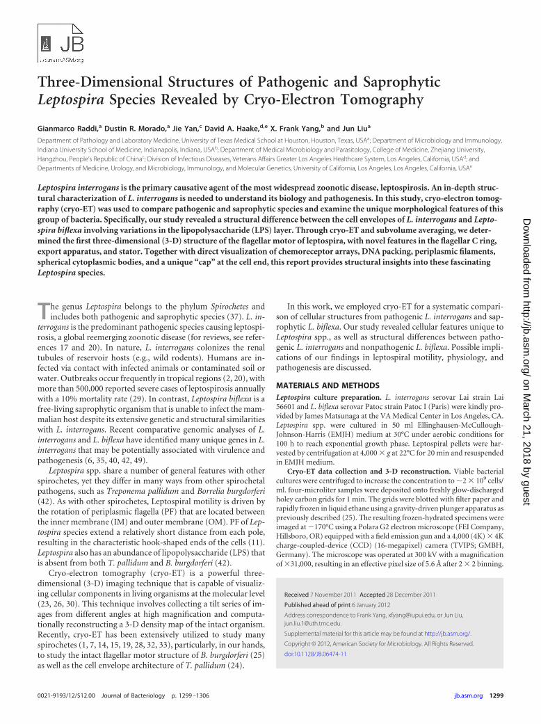
Novel filaments in the periplasm. Cryo-ET revealed two typesof filaments in the periplasmic space: flagella and novel periplas-mic filaments (Fig. 3; see also Fig. S3 in the supplemental mate-rial). They differ in their diameters, locations, and possible func-tions. Flagella of Leptospira are located near the cell termini. Theyare responsible for bacterial motility and for the unique hookshape of the cell end(s) (5), but they do not determine the spiralshape of the cell (39). In this respect, Leptospira spp. are verydifferent from B. burgdorferi, in which the flagella define the flatwave shape of the cell, since inactivation of flagellar genes resultsin long, rod-shaped bacteria (31). Additional filaments werefound in the periplasmic space of both Leptospira species. Thediameter of these filaments is considerably smaller than that offlagellar filaments (8 nm versus 22 nm). They wrap around the cellbody in a right-handed fashion, particularly in the middle of theelongated organisms (Fig. 3), where PF is absent. Given that PF ofLeptospira does not play a major role in defining the spiral shape ofthe cell body (39), it has been postulated that the cell morphologyof Leptospira is determined by the cytoskeleton, the PG layer, oranother undefined structure (17). The presence of these addi-tional filaments in the periplasmic space is intriguing, since theyrepresent novel candidates for shape determination in Leptospira.Further studies are required to provide direct evidence for thishypothesis.
Molecular architecture of leptospiral flagellar motor. Flagellaplay important roles in spirochetal motility and host tissue inva-sion (16, 43). Unlike many other spirochetes (e.g., T. pallidum andB. burgdorferi) that have multiple flagellar motors, Leptospira spe-cies have a single flagellar motor at each end, and yet the organismis highly motile. To better understand this fascinating nanoma-chine, a total of 496 motors were extracted from 3-D reconstruc-tions of intact organisms, and the molecular architecture of theintact flagellar motor was determined by subvolume averaging
FIG 2 A gallery of the cell end reconstructions reveals a “cap”-like feature of L. biflexa (A to D) and L. interrogans (E to H).
Cryo-Electron Tomography of Leptospira
March 2012 Volume 194 Number 6 jb.asm.org 1301
on March 21, 2018 by guest
http://jb.asm.org/
Dow
nloaded from
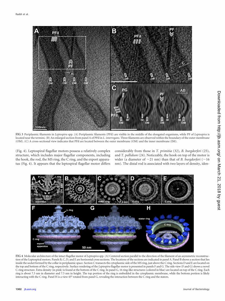
(Fig. 4). Leptospiral flagellar motors possess a relatively complexstructure, which includes major flagellar components, includingthe hook, the rod, the MS ring, the C ring, and the export appara-tus (Fig. 4). It appears that the leptospiral flagellar motor differs
considerably from those in T. primitia (32), B. burgdorferi (25),and T. pallidum (24). Noticeably, the hook on top of the motor iswider (a diameter of �21 nm) than that of B. burgdorferi (�16nm). The distal rod is associated with two layers of density, iden-
FIG 3 Periplasmic filaments in Leptospira spp. (A) Periplasmic filaments (PFil) are visible in the middle of the elongated organisms, while PF of Leptospira islocated near the termini. (B) An enlarged section from panel A of PFil in L. interrogans. Three filaments are observed within the boundary of the outer membrane(OM). (C) A cross-sectional view indicates that PFil are located between the outer membrane (OM) and the inner membrane (IM).
FIG 4 Molecular architecture of the intact flagellar motor of Leptospira spp. (A) Centered section parallel to the direction of the filament of an asymmetric reconstruc-tion of the Leptospiral motors. Panels B, C, D, and E are horizontal cross sections. The locations of the sections are indicated in panel A. Panel B shows a section that liesinside the socket formed by the collar in periplasmic space. Section C transects the cytoplasmic side of the MS ring, just above the C ring. Sections D and E are located onthe top and bottom of the C ring, respectively. Surface rendering of the Leptospira flagellar motor is presented in panels F and G. The side view (F and G) shows a novelC-ring structure. Extra density (in pink) is found at the bottom of the C ring. In panel G, 16 ring-like structures (colored in blue) are located on top of the C ring. Eachring is about 7.3 nm in diameter and 7.5 nm in height. The top portion of the ring is embedded in the cytoplasmic membrane, while the bottom portion is likelyinteracting with the C ring. Panel H is a view 45° rotated from panel G, revealing the interaction between the C ring and the stators.
Raddi et al.
1302 jb.asm.org Journal of Bacteriology
on March 21, 2018 by guest
http://jb.asm.org/
Dow
nloaded from
tified as the putative L and P rings (Fig. 4A), as the homologousgene products of flgI and flgH in Escherichia coli are responsible forthe L and P rings, respectively (27). The presence of flgH in Lep-tospira species is surprising, since the leptospiral PF is located inperiplasmic space. Our map suggests that the L ring in the lepto-spiral flagellar motor is not embedded in the outer membrane,and therefore it likely lacks the function as a bushing.
The C ring and export apparatus in leptospiral flagellar motorsare more complex than those of any recently determined motorstructures (8). Additional density (colored in pink), which has notbeen shown in other motor structures, is visible around the bot-tom part of the C ring. The export apparatus is also different fromother motor structures. It appears that the cytoplasmic part of theexport apparatus is bigger than those from other spirochetes andbacteria. On the top of the C ring, the putative stators embeddedin the cytoplasmic membrane were visualized in sufficient detailto distinguish the ring shape structures with a diameter of 7.3 nm(Fig. 4C and H). The shape and diameter of individual rings aresimilar to those of a stator complex purified from Vibrio algino-lyticus (50). However, it remains to be answered if these ringscorrespond to the individual torque generators.
Recently, analysis of a large number of spirochete genome se-quences uncovered remarkable genetic diversity among flagellarsystems, even though many flagellar genes have been conservedduring the evolutionary process (36). Our studies indicate that thespirochetal motors (including those of L. interrogans, T. pallidum,and B. burgdorferi) share overall similar sizes and shapes, yet thereare considerable differences in the C ring, P ring, and export ap-paratus. This is consistent with the recent finding that the flagellarmotors exhibit a common core architecture, despite the strikingdifferences in their overall appearance (8). The novel structures ofthe C ring, export apparatus, and stator provide new insight intothe diversity of bacterial flagellar motors.
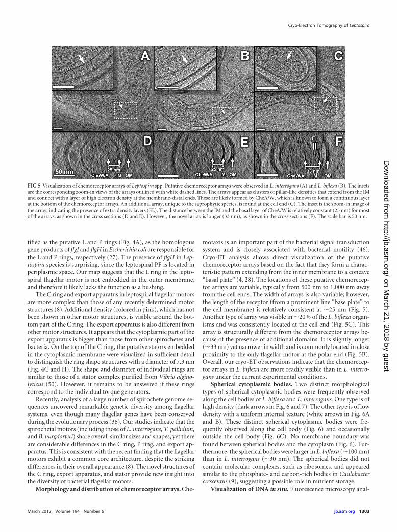
Morphology and distribution of chemoreceptor arrays. Che-
motaxis is an important part of the bacterial signal transductionsystem and is closely associated with bacterial motility (46).Cryo-ET analysis allows direct visualization of the putativechemoreceptor arrays based on the fact that they form a charac-teristic pattern extending from the inner membrane to a concave“basal plate” (4, 28). The locations of these putative chemorecep-tor arrays are variable, typically from 500 nm to 1,000 nm awayfrom the cell ends. The width of arrays is also variable; however,the length of the receptor (from a prominent line “base plate” tothe cell membrane) is relatively consistent at �25 nm (Fig. 5).Another type of array was visible in �20% of the L. biflexa organ-isms and was consistently located at the cell end (Fig. 5C). Thisarray is structurally different from the chemoreceptor arrays be-cause of the presence of additional domains. It is slightly longer(�33 nm) yet narrower in width and is commonly located in closeproximity to the only flagellar motor at the polar end (Fig. 5B).Overall, our cryo-ET observations indicate that the chemorecep-tor arrays in L. biflexa are more readily visible than in L. interro-gans under the current experimental conditions.
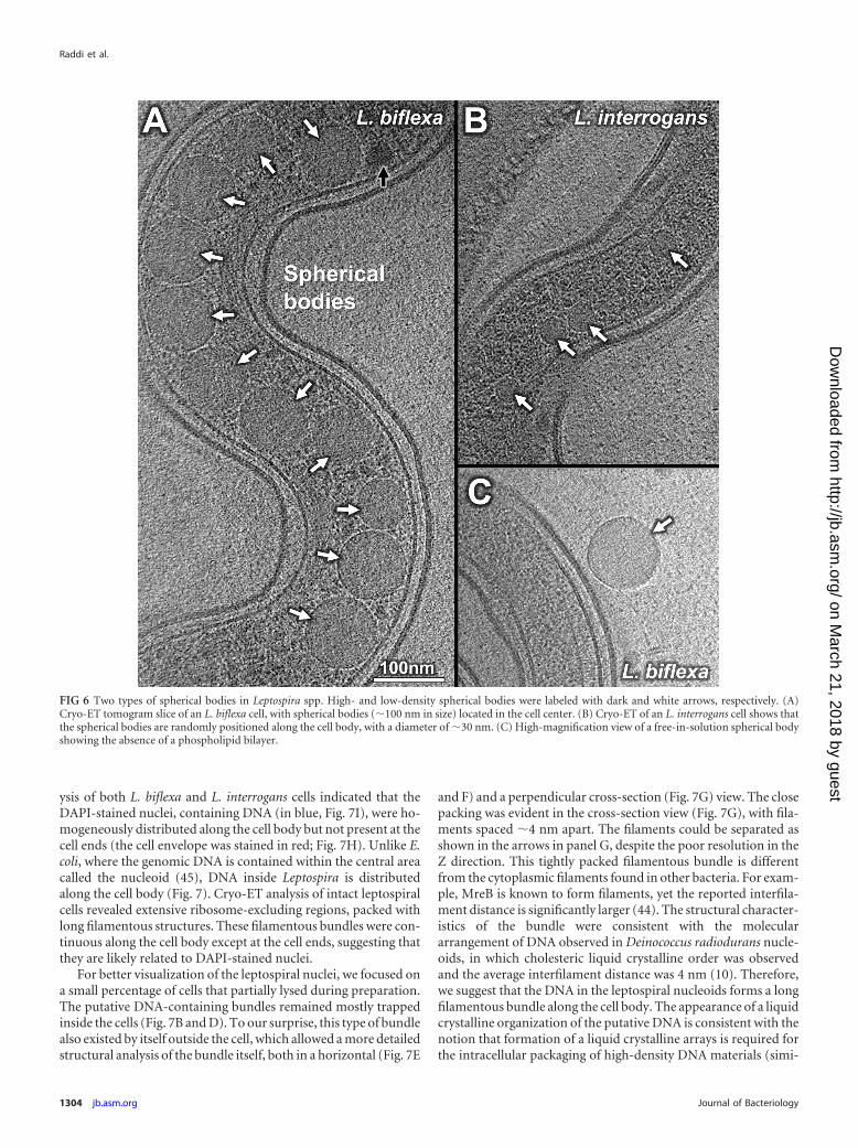
Spherical cytoplasmic bodies. Two distinct morphologicaltypes of spherical cytoplasmic bodies were frequently observedalong the cell bodies of L. biflexa and L. interrogans. One type is ofhigh density (dark arrows in Fig. 6 and 7). The other type is of lowdensity with a uniform internal texture (white arrows in Fig. 6Aand B). These distinct spherical cytoplasmic bodies were fre-quently observed along the cell body (Fig. 6) and occasionallyoutside the cell body (Fig. 6C). No membrane boundary wasfound between spherical bodies and the cytoplasm (Fig. 6). Fur-thermore, the spherical bodies were larger in L. biflexa (�100 nm)than in L. interrogans (�30 nm). The spherical bodies did notcontain molecular complexes, such as ribosomes, and appearedsimilar to the phosphate- and carbon-rich bodies in Caulobactercrescentus (9), suggesting a possible role in nutrient storage.
Visualization of DNA in situ. Fluorescence microscopy anal-
FIG 5 Visualization of chemoreceptor arrays of Leptospira spp. Putative chemoreceptor arrays were observed in L. interrogans (A) and L. biflexa (B). The insetsare the corresponding zoom-in views of the arrays outlined with white dashed lines. The arrays appear as clusters of pillar-like densities that extend from the IMand connect with a layer of high electron density at the membrane-distal ends. These are likely formed by CheA/W, which is known to form a continuous layerat the bottom of the chemoreceptor arrays. An additional array, unique to the saprophytic species, is found at the cell end (C). The inset is the zoom-in image ofthe array, indicating the presence of extra density layers (EL). The distance between the IM and the basal layer of CheA/W is relatively constant (25 nm) for mostof the arrays, as shown in the cross sections (D and E). However, the novel array is longer (33 nm), as shown in the cross sections (F). The scale bar is 50 nm.
Cryo-Electron Tomography of Leptospira
March 2012 Volume 194 Number 6 jb.asm.org 1303
on March 21, 2018 by guest
http://jb.asm.org/
Dow
nloaded from
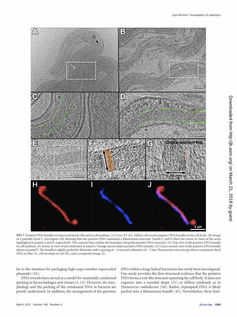
ysis of both L. biflexa and L. interrogans cells indicated that theDAPI-stained nuclei, containing DNA (in blue, Fig. 7I), were ho-mogeneously distributed along the cell body but not present at thecell ends (the cell envelope was stained in red; Fig. 7H). Unlike E.coli, where the genomic DNA is contained within the central areacalled the nucleoid (45), DNA inside Leptospira is distributedalong the cell body (Fig. 7). Cryo-ET analysis of intact leptospiralcells revealed extensive ribosome-excluding regions, packed withlong filamentous structures. These filamentous bundles were con-tinuous along the cell body except at the cell ends, suggesting thatthey are likely related to DAPI-stained nuclei.
For better visualization of the leptospiral nuclei, we focused ona small percentage of cells that partially lysed during preparation.The putative DNA-containing bundles remained mostly trappedinside the cells (Fig. 7B and D). To our surprise, this type of bundlealso existed by itself outside the cell, which allowed a more detailedstructural analysis of the bundle itself, both in a horizontal (Fig. 7E
and F) and a perpendicular cross-section (Fig. 7G) view. The closepacking was evident in the cross-section view (Fig. 7G), with fila-ments spaced �4 nm apart. The filaments could be separated asshown in the arrows in panel G, despite the poor resolution in theZ direction. This tightly packed filamentous bundle is differentfrom the cytoplasmic filaments found in other bacteria. For exam-ple, MreB is known to form filaments, yet the reported interfila-ment distance is significantly larger (44). The structural character-istics of the bundle were consistent with the moleculararrangement of DNA observed in Deinococcus radiodurans nucle-oids, in which cholesteric liquid crystalline order was observedand the average interfilament distance was 4 nm (10). Therefore,we suggest that the DNA in the leptospiral nucleoids forms a longfilamentous bundle along the cell body. The appearance of a liquidcrystalline organization of the putative DNA is consistent with thenotion that formation of a liquid crystalline arrays is required forthe intracellular packaging of high-density DNA materials (simi-
FIG 6 Two types of spherical bodies in Leptospira spp. High- and low-density spherical bodies were labeled with dark and white arrows, respectively. (A)Cryo-ET tomogram slice of an L. biflexa cell, with spherical bodies (�100 nm in size) located in the cell center. (B) Cryo-ET of an L. interrogans cell shows thatthe spherical bodies are randomly positioned along the cell body, with a diameter of �30 nm. (C) High-magnification view of a free-in-solution spherical bodyshowing the absence of a phospholipid bilayer.
Raddi et al.
1304 jb.asm.org Journal of Bacteriology
on March 21, 2018 by guest
http://jb.asm.org/
Dow
nloaded from
lar to the situation for packaging high-copy-number supercoiledplasmids) (41).
DNA toroids have served as a model for maximally condensedpacking in bacteriophages and viruses (3, 13). However, the mor-phology and the packing of the condensed DNA in bacteria arepoorly understood. In addition, the arrangement of the genomic
DNA within a long, helical bacterium has rarely been investigated.Our study provides the first structural evidence that the putativeDNA forms a rod-like structure spanning the cell body. It does notorganize into a toroidal shape (21) or diffuse randomly as inDeinococcus radiodurans (10). Rather, leptospiral DNA is likelypacked into a filamentous bundle (41). Nevertheless, these find-
FIG 7 Putative DNA bundles in intact and lysed cells and in cell medium. (A) Cryo-ET of L. biflexa cell reveals putative DNA bundles in the cell body. (B) Imageof a partially lysed L. interrogans cell, showing that the putative DNA maintains a filamentous structure. Panels C and D show the zoom-in views of the areashighlighted in panels A and B, respectively. The contour lines outline the boundary along the putative DNA structure. (E) Top view of the putative DNA bundlein cell medium. (F) Zoom-in view of area indicated in panel E. Orange arrows depict putative DNA strands. (G) Cross-section view of the putative DNA bundleshown in panel F. The bundle is tightly packed by filaments with a spacing of �4 nm and a diameter of �2 nm. Fluorescence microscopy shows continuous dyedDNA in blue (I), cell envelope in red (H), and a composite image (J).
Cryo-Electron Tomography of Leptospira
March 2012 Volume 194 Number 6 jb.asm.org 1305
on March 21, 2018 by guest
http://jb.asm.org/
Dow
nloaded from

ings raise important questions. How do spirochetes package DNAinto this morphology? Why is DNA restricted from the cell end?How is the DNA bundle changed during different growth phasesof Leptospira and other spirochetes? Much work will be requiredto answer these questions.
ACKNOWLEDGMENTS
We thank Steven Norris, Angel Paredes, Feng Xue, Dinara Yangirova, andBrian Poindexter for their comments and suggestions. We are grateful toFeng Xue for bacterial cultures. We particularly thank the reviewers fornumerous critical comments and suggestions.
This work was supported in part by grants R01AI087946 (to J. Liu) and5R01AI034431 (to D. A. Haake) from the National Institute of Allergy andInfectious Diseases (NIAID), by grant AU-1714 from the Welch Founda-tion (to J. Liu), and by VA Medical Research funds (to D. A. Haake).
REFERENCES1. Beck M, et al. 2009. Visual proteomics of the human pathogen Leptospira
interrogans. Nat. Methods 6:817– 823.2. Bharti AR, et al. 2003. Leptospirosis: a zoonotic disease of global impor-
tance. Lancet Infect. Dis. 3:757–771.3. Bloomfield VA. 1996. DNA condensation. Curr. Opin. Struct. Biol.
6:334 –341.4. Briegel A, et al. 2009. Universal architecture of bacterial chemoreceptor
arrays. Proc. Natl. Acad. Sci. U. S. A. 106:17181–17186.5. Bromley DB, Charon NW. 1979. Axial filament involvement in the mo-
tility of Leptospira interrogans. J. Bacteriol. 137:1406 –1412.6. Bulach DM, et al. 2006. Genome reduction in Leptospira borgpetersenii
reflects limited transmission potential. Proc. Natl. Acad. Sci. U. S. A. 103:14560 –14565.
7. Charon NW, et al. 2009. The flat-ribbon configuration of the periplasmicflagella of Borrelia burgdorferi and its relationship to motility and mor-phology. J. Bacteriol. 191:600 – 607.
8. Chen S, et al. 2011. Structural diversity of bacterial flagellar motors.EMBO J. 30:2972–2981.
9. Comolli LR, Kundmann M, Downing KH. 2006. Characterization ofintact subcellular bodies in whole bacteria by cryo-electron tomographyand spectroscopic imaging. J. Microsc. 223:40 –52.
10. Eltsov M, Dubochet J. 2005. Fine structure of the Deinococcus radio-durans nucleoid revealed by cryoelectron microscopy of vitreous sections.J. Bacteriol. 187:8047– 8054.
11. Goldstein SF, Charon NW. 1988. Motility of the spirochete Leptospira.Cell Motil. Cytoskeleton 9:101–110.
12. Haake DA, Matsunaga J. 2010. Leptospira: a spirochete with a hybridouter membrane. Mol. Microbiol. 77:805– 814.
13. Hud NV, Vilfan ID. 2005. Toroidal DNA condensates: unraveling the finestructure and the role of nucleation in determining size. Annu. Rev. Bio-phys. Biomol. Struct. 34:295–318.
14. Izard J, Hsieh CE, Limberger RJ, Mannella CA, Marko M. 2008. Nativecellular architecture of Treponema denticola revealed by cryo-electron to-mography. J. Struct. Biol. 163:10 –17.
15. Izard J, et al. 2009. Cryo-electron tomography elucidates the moleculararchitecture of Treponema pallidum, the syphilis spirochete. J. Bacteriol.191:7566 –7580.
16. Kennedy MJ, Rosey EL, Yancey RJ, Jr. 1997. Characterization of flaA-and flaB- mutants of Serpulina hyodysenteriae: both flagellin subunits,FlaA and FlaB, are necessary for full motility and intestinal colonization.FEMS Microbiol. Lett. 153:119 –128.
17. Ko AI, Goarant C, Picardeau M. 2009. Leptospira: the dawn of themolecular genetics era for an emerging zoonotic pathogen. Nat. Rev. Mi-crobiol. 7:736 –747.
18. Kremer JR, Mastronarde DN, McIntosh JR. 1996. Computer visualization ofthree-dimensional image data using IMOD. J. Struct. Biol. 116:71–76.
19. Kudryashev M, et al. 2009. Comparative cryo-electron tomography ofpathogenic Lyme disease spirochetes. Mol. Microbiol. 71:1415–1434.
20. Levett PN. 2001. Leptospirosis. Clin. Microbiol. Rev. 14:296 –326.21. Levin-Zaidman S, et al. 2003. Ringlike structure of the Deinococcus
radiodurans genome: a key to radioresistance? Science 299:254 –256.22. Li L, Ojcius DM, Yan J. 2007. Comparison of invasion of fibroblasts and
macrophages by high- and low-virulence Leptospira strains: colonization
of the host-cell nucleus and induction of necrosis by the virulent strain.Arch. Microbiol. 188:591–598.
23. Li Z, Jensen GJ. 2009. Electron cryotomography: a new view into micro-bial ultrastructure. Curr. Opin. Microbiol. 12:333–340.
24. Liu J, et al. 2010. Cellular architecture of Treponema pallidum: novelflagellum, periplasmic cone, and cell envelope as revealed by cryo electrontomography. J. Mol. Biol. 403:546 –561.
25. Liu J, et al. 2009. Intact flagellar motor of Borrelia burgdorferi revealed bycryo-electron tomography: evidence for stator ring curvature and rotor/C-ring assembly flexion. J. Bacteriol. 191:5026 –5036.
26. Lucic V, Forster F, Baumeister W. 2005. Structural studies by electrontomography: from cells to molecules. Annu. Rev. Biochem. 74:833– 865.
27. Macnab RM. 2003. How bacteria assemble flagella. Annu. Rev. Microbiol.57:77–100.
28. Malmstrom J, et al. 2009. Proteome-wide cellular protein concentrationsof the human pathogen Leptospira interrogans. Nature 460:762–765.
29. McBride AJ, Athanazio DA, Reis MG, Ko AI. 2005. Leptospirosis. Curr.Opin. Infect. Dis. 18:376 –386.
30. Milne JL, Subramaniam S. 2009. Cryo-electron tomography of bacteria: prog-ress, challenges and future prospects. Nat. Rev. Microbiol. 7:666–675.
31. Motaleb MA, et al. 2000. Borrelia burgdorferi periplasmic flagella haveboth skeletal and motility functions. Proc. Natl. Acad. Sci. U. S. A. 97:10899 –10904.
32. Murphy GE, Leadbetter JR, Jensen GJ. 2006. In situ structure of thecomplete Treponema primitia flagellar motor. Nature 442:1062–1064.
33. Murphy GE, Matson EG, Leadbetter JR, Berg HC, Jensen GJ. 2008.Novel ultrastructures of Treponema primitia and their implications formotility. Mol. Microbiol. 67:1184 –1195.
34. Nahori MA, et al. 2005. Differential TLR recognition of leptospiral lipidA and lipopolysaccharide in murine and human cells. J. Immunol. 175:6022– 6031.
35. Nascimento AL, et al. 2004. Comparative genomics of two Leptospirainterrogans serovars reveals novel insights into physiology and pathogen-esis. J. Bacteriol. 186:2164 –2172.
36. Pallen MJ, Penn CW, Chaudhuri RR. 2005. Bacterial flagellar diversity inthe post-genomic era. Trends Microbiol. 13:143–149.
37. Paster BJ, et al. 1991. Phylogenetic analysis of the spirochetes. J. Bacteriol.173:6101– 6109.
38. Pettersen EF, et al. 2004. UCSF Chimera—a visualization system forexploratory research and analysis. J. Comput. Chem. 25:1605–1612.
39. Picardeau M, Brenot A, Saint Girons I. 2001. First evidence for gene replace-ment in Leptospira spp. Inactivation of L. biflexa flaB results in non-motile mu-tants deficient in endoflagella. Mol. Microbiol. 40:189–199.
40. Picardeau M, et al. 2008. Genome sequence of the saprophyte Leptospirabiflexa provides insights into the evolution of Leptospira and the patho-genesis of leptospirosis. PLoS One 3:e1607.
41. Reich Z, Wachtel EJ, Minsky A. 1994. Liquid-crystalline mesophases ofplasmid DNA in bacteria. Science 264:1460 –1463.
42. Ren SX, et al. 2003. Unique physiological and pathogenic features ofLeptospira interrogans revealed by whole-genome sequencing. Nature422:888 – 893.
43. Sadziene A, Thomas DD, Bundoc VG, Holt SC, Barbour AG. 1991. Aflagella-less mutant of Borrelia burgdorferi. Structural, molecular, and invitro functional characterization. J. Clin. Invest. 88:82–92.
44. Swulius MT, et al. 2011. Long helical filaments are not seen encirclingcells in electron cryotomograms of rod-shaped bacteria. Biochem. Bio-phys. Res. Commun. 407:650 – 655.
45. Tchamedeu Kameni AP, Couture-Tosi E, Saint-Girons I, Picardeau M.2002. Inactivation of the spirochete recA gene results in a mutant with lowviability and irregular nucleoid morphology. J. Bacteriol. 184:452– 458.
46. Wadhams GH, Armitage JP. 2004. Making sense of it all: bacterial che-motaxis. Nat. Rev. Mol. Cell Biol. 5:1024 –1037.
47. Werts C, et al. 2001. Leptospiral lipopolysaccharide activates cellsthrough a TLR2-dependent mechanism. Nat. Immunol. 2:346 –352.
48. Winkler H, Taylor KA. 2006. Accurate marker-free alignment with si-multaneous geometry determination and reconstruction of tilt series inelectron tomography. Ultramicroscopy 106:240 –254.
49. Xue F, Yan J, Picardeau M. 2009. Evolution and pathogenesis of Leptospira spp.:lessons learned from the genomes. Microbes Infect. 11:328–333.
50. Yonekura K, Maki-Yonekura S, Homma M. 2011. Structure of theflagellar motor protein complex PomAB: implications for the torque-generating conformation. J. Bacteriol. 193:3863–3870.
Raddi et al.
1306 jb.asm.org Journal of Bacteriology
on March 21, 2018 by guest
http://jb.asm.org/
Dow
nloaded from