18-PRESS-Assessment of pathogenic - NSM Journalnsmjournal.com/NSMJournal/updatedversion/Assessment...
12
Nigerian Journal of Microbiology 2015, 29: 3127-3138 Published online at www.nsmjournal.org 3127 | Page Assessment of pathogenic potentials of Colonial and filamentous Candida albicans in varied immunological state of animals 1 Uzoechi Anselem U. , 2 Orji Justina C., 1 Nwachukwu M.I., 1 Ohuocha P., 1 Nwachukwu I.O 1 Department of Microbiology, Imo State University Owerri Imo State 2 Department of Microbiology, Federal University of Technology Owerri Tel :08035562250 Abstrct: The infection process when C albicans grew in the filamentous and colonial forms were assessed in rats by determination of survival times, fungal burden from kidney and histological examination. Rats in the various immunological state survived C albicans in the Colonial form than in the filamentous form. Neutrophil deficient rats and rats depleted of all forms of immunity had the highest mortality with lowest mean survival times of 0.6±0.4 and 0.8 ±0.41 days respectively after infection with filamentous C albicans. There were no significant variations in the fungal kidney burden among the groups except for the Neutrophil deficient rats infected with filamentous C albicans that showed the lowest kidney burden 6.61 ± 0.73 cfu/g of kidney. Result indicates that there was variation in the relative invasion of the kidney by the C albicans when the organism grows in different morphological forms The result also mirrors the superiority of the immune status over morphological state of the organism and the consequences of certain regimen used in the treatment of candidiasis as shown on the effect of CPM, as this compound is an active substance in anti fungal drugs used in the treatment of infections caused by Candida albicans Key words : Mortality, Candidiasis, Anti fungal, Immunological, Filamentous, morphological INTRODUCTION andida albicans remains the main causative agent of invasive fungal infection in an expanding population of immunocompromised patients with associated high morbidity and mortality rates. Its pathogenesis is a complex phenomenon that results from a delicate balance between its intrinsic virulence attributes and host immune responses. This gives rise to the highly complex and dynamic nature of the *Corresponding author: [email protected] 1 Uzoechi Anselem U. Copyright © 2015 Nigerian Society for Microbiology host-fungus interaction that ultimately determines the outcome of an infection. Species of the genus Candida, as well as dimorphic fungi with a pathogenic yeast phase such as Histoplasma and Cryptococcus species cause systemic mycosis mainly in immunocompromised patients and following long-term therapy with broad-spectrum antibiotics and/or antifungal antibiotics. In recent years, our knowledge of the molecular biology of yeast pathogens has increased dramatically and is beginning to affect diagnostic and therapeutic strategies. A wealth of new information has appeared C
Transcript of 18-PRESS-Assessment of pathogenic - NSM Journalnsmjournal.com/NSMJournal/updatedversion/Assessment...
Nigerian Journal of Microbiology 2015, 29: 3127-3138 Published online at www.nsmjournal.org
3127 | P a g e
Assessment of pathogenic potentials of Colonial and filamentous Candida albicans in
varied immunological state of animals 1Uzoechi Anselem U. , 2Orji Justina C., 1Nwachukwu M.I., 1Ohuocha P.,1Nwachukwu I.O
1 Department of Microbiology, Imo State University Owerri Imo State 2 Department of Microbiology, Federal University of Technology Owerri
Tel :08035562250
Abstrct: The infection process when C albicans grew in the filamentous and colonial forms were assessed in rats by determination of survival times, fungal burden from kidney and histological examination. Rats in the various immunological state survived C albicans in the Colonial form than in the filamentous form. Neutrophil deficient rats and rats depleted of all forms of immunity had the highest mortality with lowest mean survival times of 0.6±0.4 and 0.8 ±0.41 days respectively after infection with filamentous C albicans. There were no significant variations in the fungal kidney burden among the groups except for the Neutrophil deficient rats infected with filamentous C albicans that showed the lowest kidney burden 6.61 ± 0.73 cfu/g of kidney. Result indicates that there was variation in the relative invasion of the kidney by the C albicans when the organism grows in different morphological forms The result also mirrors the superiority of the immune status over morphological state of the organism and the consequences of certain regimen used in the treatment of candidiasis as shown on the effect of CPM, as this compound is an active substance in anti fungal drugs used in the treatment of infections caused by Candida albicans Key words : Mortality, Candidiasis, Anti fungal, Immunological, Filamentous, morphological INTRODUCTION
andida albicans remains the main causative agent of invasive fungal infection in an expanding
population of immunocompromised patients with associated high morbidity and mortality rates. Its pathogenesis is a complex phenomenon that results from a delicate balance between its intrinsic virulence attributes and host immune responses. This gives rise to the highly complex and dynamic nature of the *Corresponding author: [email protected] 1Uzoechi Anselem U. Copyright © 2015 Nigerian Society for Microbiology
host-fungus interaction that ultimately determines the outcome of an infection. Species of the genus Candida, as well as dimorphic fungi with a pathogenic yeast phase such as Histoplasma and Cryptococcus species cause systemic mycosis mainly in immunocompromised patients and following long-term therapy with broad-spectrum antibiotics and/or antifungal antibiotics. In recent years, our knowledge of the molecular biology of yeast pathogens has increased dramatically and is beginning to affect diagnostic and therapeutic strategies. A wealth of new information has appeared
C
1Uzoechi et al.2015 Nigerian Journal of Microbiology, Vol. 29: 3127-3138
Published by Nigerian Society of Microbiology 3128 | P a g e
on the key virulence factors of yeast pathogens, including dimorphism, which is one of the striking features of most yeast pathogens. Molero et al; 1998, Ashman and Papadimitriou, 1987).
The ability of a pathogenic microorganism to detect and respond to environmental cues that signal an invasion opportunity is a potent survival mechanism. C. albicans is able to control its filamentation via its ability to respond to numerous of environmental signals and this in turn mediate and determine outcome of infections (Ashman et al; 2003).
While morphogenetic conversions, the ability to reversibly switch between yeast cell and filamentous forms, constitute one of the most important virulence attributes of this organism, the question then is at what form will infection be initiated most and within these forms what is the involvement of each immune cells Mitcheil 1998. It is also clear that all studies of putative C. albicans virulence factors, including dimorphism, must be analyzed in the context of the immune status of the host, since clinical experience teaches us that even the so-called virulent forms of the fungus are unlikely to cause infection in an immunocompetent host. MATERIALS AND METHODOLOGY Isolation and culture condition
Candida albicans used in this study was isolated from patients suffering from candidiasis from Imo State University Teaching Hospital. A sterile swab stick was used to collect a high virginal swab from patients that reported cases of candidiasis. The swab was streaked on a Petri dish containing potatoe dextrose agar (PDA). The plate
was incubated in an incubator at 37oC for 24 hours. After incubation, colonies with yeasty odour were isolated and purified by subculturing several times on PDA plates. Pure cultures on PDA slants were stored in a refrigerator at 4-8oC for further identification. Identification of Isolate
The test tubes containing the stored isolates were removed from the refrigerator and placed on the laboratory bench at room temperature (28+_2oC) for 2 hours. A loopfull of the isolate was spread on the surface of sterile PDA Plates. The plate was incubated at room temperature (28+_2oC) for 2 days. Thereafter, morphological and biochemical characterization of the isolate was done. The test carried out includes; Carbohydrate assimilation Chlamydospore formation and Germ tube formation as described by Mahn and Manusell 2000. Preparation of Nephlometry for the standardization of yeast suspension
Nephlometry is a method designed to standardize yeast cells suspension according to McFarland scale. Standardization was done as decribed by Ochei and Kolhatkar 2008. Study Animals
Outbreed female, four months old albino rats were purchased from Biochemistry department of University of Nigeria, Nsukka. The total number of animals used was one hundred (100). The rats were housed in a well ventilated cage with effective faecal removal system. The rats were fed with appropriate quantity of food, and allowed one week acclimatization
1Uzoechi et al.2015 Nigeria Journal of Microbiology 2015: 3127-3138
Published by Nigerian Society of Microbiology 3129 | P a g e
period before being used for the experiment. Determination of sub lethal dose of the C albicans
This was determined by inoculating four groups of rats each consisting of 5 normal rats with intact immune system, with varied dilutions of the test organism chosen from the McFarland scale. The population of organism chosen was 1.5 x 10⁵, 1.8 x 10⁶, 2.1 x 10⁷ and 2.4 x 10⁸ cfu/ml. Preparation of inoculum
A loopful of the purified organism identified as C albicans was transferred from the refrigerated stock into 100ml of sterile yeast potato dextrose broth contained in 250 ml conical flask. The culture was incubated on a rotary incubator maintained at room temperature (28+_2oC) for 2days. Thereafter, the yeast cells were harvested by centrifugation, washed twice in normal saline and resuspended in normal saline. Harvested cells were standardized using the Nephlometry method to obtain cell population corresponding to 1.5 x 10⁵, 1.8 x 10⁶, 2.1 x 10⁷ and 2.4 x 10⁸ cfu/ml respectively Infection of normal rats to determine sub lethal dose
A section of the abdomen of the rat was shaven with a sterile razor blade. The shaven area was swabbed with methylated spirit. Each group of rats (5 in number) was inoculated with 1ml cell suspension containing different population of organism. The animals were kept in wooden cage and observed for lethality within 48 hours.
Treatment of rats with immunosuppressive drugs Induction of Immunosuppression A section of the rat was carefully shaved and swabbed with methylated spirit. Using 27 gauge insulin needles 0.5ml of the immuno suppressive agents were collected and injected intradermally into the test animal. The immuno suppressive agents were administered 3 times at interval of 2 days. After the injection which lasted for 6 days, the rats were fed appropriately for another 3 days. While the final immunological states were ascertained using appropriate immunoassays. Immunoassay to determine the final immunological state of the animals
After inducing the animals with appropriate immunosuppressive agents immuno assays were done to ascertain the level of immunity operational in the animals before and after infections with the test organism. The immunoassays done include; (i) T and B cell estimation (Cytosphere Assay) (ii) Differential white blood cell count (peripheral blood count) all as described by Ochei and Kolhatkar (2008) Infection of the immunosupressed and normal experimental rats with C albicans Inoculum preparation
This was done as described previously but to obtain a population corresponding to 1.8 x 10⁶ Inoculation of the Rat Strains with test organism
The sublethal dose of C albicans earlier determined to be 1.8 x 10⁶ cfu/ml. was used as inoculum. Using a sterile 27 gauge insulin needle, 1ml of the yeast suspension containing 1.8 x 10⁶ cfu/ml of yeast cells was injected
1Uzoechi et al.2015 Nigerian Journal of Microbiology, Vol. 29: 3127-3138
Published by Nigerian Society of Microbiology 3130 | P a g e
intravenously through the lateral vein of the shaved swabbed caudal tail region of each of the test animal as described previously. In vivo Morphogenic conversion using DOX (DEOXYCYCLINE)
Each group of animal shown in Table 1 was further divided into two sub groups; with each subgroup comprising of 5 animals. One subgroup received DOX in their drinking water while the other did not. In animals fed with water containing DOX, the organism (C. albicans) was supposed to grow in the filamentous form while filamentation is regulated and prevented in those animals fed without DOX. DOX (2 mg/l) was administered to the organism in their drinking water three days prior to infection with the sublethal dose (1.8 x 10⁶ cfu/ml) of C. albicans. Examination of inoculated rats Monitoring for survival
The rats were fed and monitored for survival for 20 days post infection (DPI). Days on which rats died were recorded; moribund animals were euthanized and recorded as dying the following day. Fungal burden determination from the kidney organs.
The kidney were removed and analyzed separately for fungal burden. Using a sterile dissecting set, the kidney of each dead or sacrificed animal were extracted, and homogenized by grinding in a sterile mortar. A 10 fold serial dilution was done with the
homogenized kidneys. Cell count was taken from the 104 fold dilution plated on a Petri dish containing yeast potato dextrose agar and incubated at 37oC for 24 hours in an incubator Histological screening Animals sacrificed were held on a bond with masking tape. A surgical blade was used to lacerate regions closer to the organs to be screened. Two thin sections of the kidney were made using a sterile surgical blade. This was fixed in 10% buffered formalin and imbedded in paraffin, and thin tissue slices were removed and stained with methylamine-silver to visualize fungal element present in tissues and with eosin to evaluate necrosis and inflammation (Saville et al; 2003) Statistical Analysis
Results obtained were keyed into SPSS version 32. where analysis of Variance (ANOVA) and Chi square test were used to analyze the results. Result
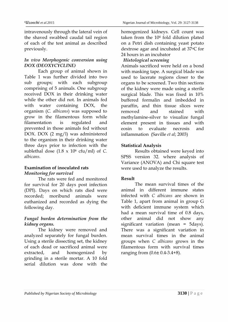
The mean survival times of the animal in different immune states infected with C albicans are shown in Table 1, apart from animal in group G with deficient immune system which had a mean survival time of 0.8 days, other animal did not show any significant variation (mean = 5days). There was a significant variation in mean survival times in the animal groups when C albicans grows in the filamentous form with survival times ranging from (0.6± 0.4-3.4+8).
1Uzoechi et al.2015 Nigeria Journal of Microbiology 2015: 3127-3138
Published by Nigerian Society of Microbiology 3131 | P a g e
Table 1 Mean survival time of rats in different immunological states after infection with different morphogenic form of C. albicans
Mean survival time (days) after infection Groups Immune state filamentous* colonial** A T cell deficient 1.5 ± 0.01 a5.00 B T cell active 3.4 ± 0.81 5.00 C B cell active 2.6 ± 0.71 5.00 D B cell active 2.0 ± 0.61 5.00 E Neutrophil deficient 0.8 ± 0.41 5.00 F SCID 1.4 ± 0.28 5.00 G CPM 0.6 ± 0.40 0.80 ± 0.42 H Normal 3.0 ± 0.11 5.00
* Test organism grown in vivo as filament ** Test organism grown in vivo as yeast a average mean survival time of each group is 5 days MST in days
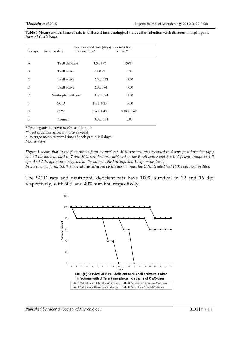
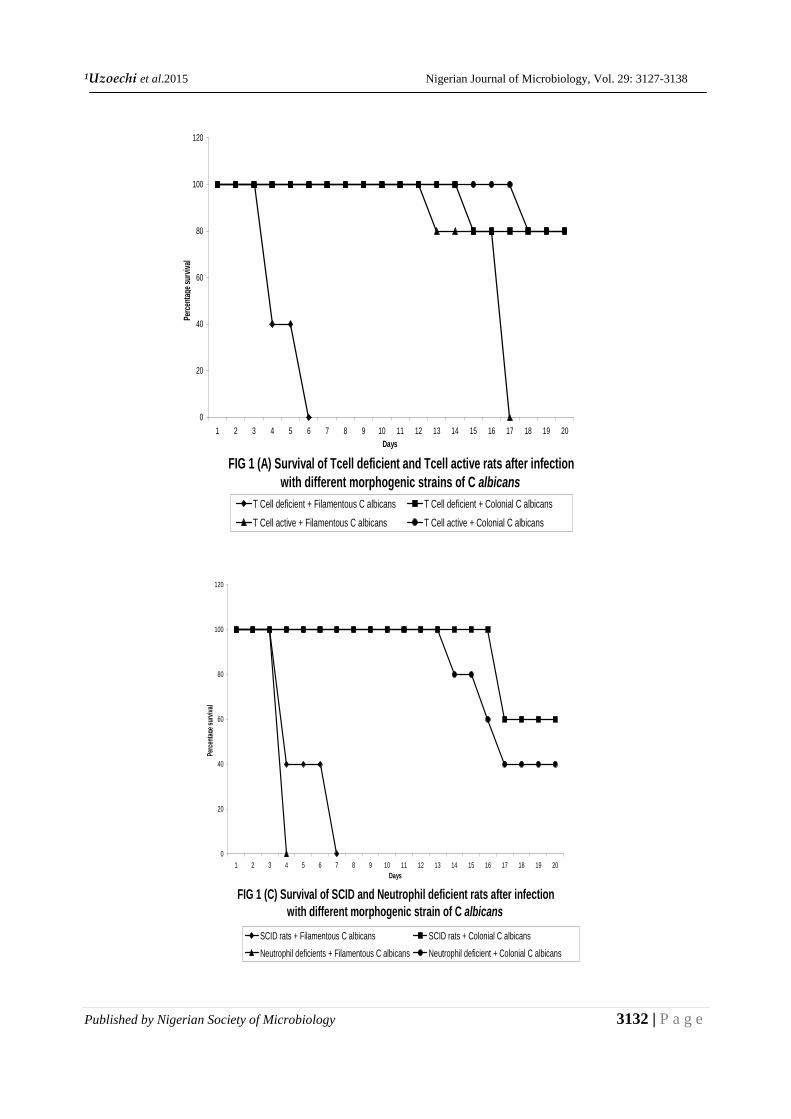
Figure 1 shows that in the filamentous form, normal rat 40% survival was recorded in 4 days post infection (dpi) and all the animals died in 7 dpi. 80% survival was achieved in the B cell active and B cell deficient groups at 4-5 dpi. And 2-10 dpi respectively and all the animals died in 3dpi and 10 dpi respectively. In the colonial form, 100% survival was achieved by the normal rats, the CPM treated had 100% survival in 4dpi. The SCID rats and neutrophil deficient rats have 100% survival in 12 and 16 dpi respectively, with 60% and 40% survival respectively.
FIG 1(B) Survival of B cell deficient and B cell active rats after infections with different morphogenic strains of C albicans
0
20
40
60
80
100
120
1 2 3 4 5 6 7 8 9 10 11 12 13 14 15 16 17 18 19 20Days
Per
cent
age
surv
ival
B Cell deficient + Filametous C albicans B Cell deficient + Colonial C albicans
B Cell active + Filamentous C albicans B Cell active + Colonial C albicans
1Uzoechi et al.2015 Nigerian Journal of Microbiology, Vol. 29: 3127-3138
Published by Nigerian Society of Microbiology 3132 | P a g e
FIG 1 (A) Survival of Tcell deficient and Tcell active rats after infection with different morphogenic strains of C albicans
0
20
40
60
80
100
120
1 2 3 4 5 6 7 8 9 10 11 12 13 14 15 16 17 18 19 20Days
Perc
enta
ge s
urvi
val
T Cell deficient + Filamentous C albicans T Cell deficient + Colonial C albicans
T Cell active + Filamentous C albicans T Cell active + Colonial C albicans
FIG 1 (C) Survival of SCID and Neutrophil deficient rats after infection with different morphogenic strain of C albicans
0
20
40
60
80
100
120
1 2 3 4 5 6 7 8 9 10 11 12 13 14 15 16 17 18 19 20Days
Perc
enta
ge s
urvi
val
SCID rats + Filamentous C albicans SCID rats + Colonial C albicans
Neutrophil deficients + Filamentous C albicans Neutrophil deficient + Colonial C albicans
1Uzoechi et al.2015 Nigeria Journal of Microbiology 2015: 3127-3138
Published by Nigerian Society of Microbiology 3133 | P a g e
Organ burden load as depicted in Table 2 shows that in the filamentous form of C albicans T cell deficient rates have the highest fungal load (7.5± 0.85 cfu/g of kidney). While neutrrophil have the least fungal load (6.6± 0.36 cfu/g of kidney). In the colonial form, CPM treated has the highest population of yeast (7.9±.11). however fungal load among the colonial groups is statically insignificant. Table 2 Kidney burdens of rats in different immunological states infected with C albicans in different morphological form.
Population of C albicans log (cfu/g of kidney)
Animal group immune state filamentous colonial
A T cell deficient 7.58 ± 0.85 7.0 ± 0.12 B T cell active 7.20 ± 0.41 7.12 ± 0.56 C B cell deficient 7.13 ± 0.2 7.19 ± 0.33 D B cell active 7.18 ± 0.34 7.20 ± 0.43 E Neutrophil deficient 6.61 ± 0.73 7.0 ± 0.51 F SCID 7.26 ± 0.73 7.30 ± 0.31 G CPM 7.14 ± 0.16 7.94 ± 0.11 H Normal 7.33 ± 0.40 7.16 ± 0.46
* Filamentious groups were feed with DOX ** Colonial forms were feed without DOX Inoculation of 1.8*106cfu
FIG 1 (D) Survival of CPM Treated rats and Normal rats in different morphogenic strains of the C albicans
0
20
40
60
80
100
120
1 2 3 4 5 6 7 8 9 10 11 12 13 14 15 16 17 18 19 20
Days
Perc
enta
ge su
rviva
l
CPM Treated rats + Filamentous C albicans CPM Treated rats + Filamentous C albicansNormal rats + Filamentous C albicans Normal rats + Colonial C albicans
1Uzoechi et al.2015 Nigerian Journal of Microbiology, Vol. 29: 3127-3138
Published by Nigerian Society of Microbiology 3134 | P a g e
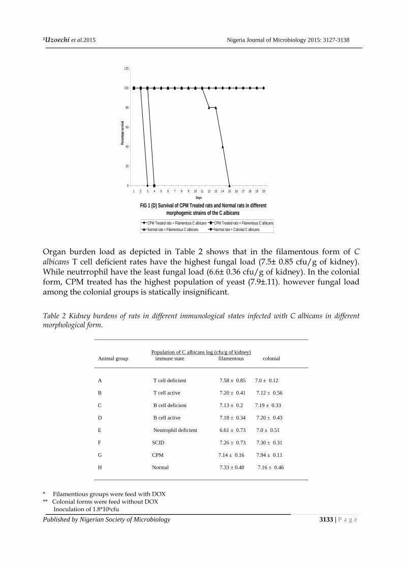
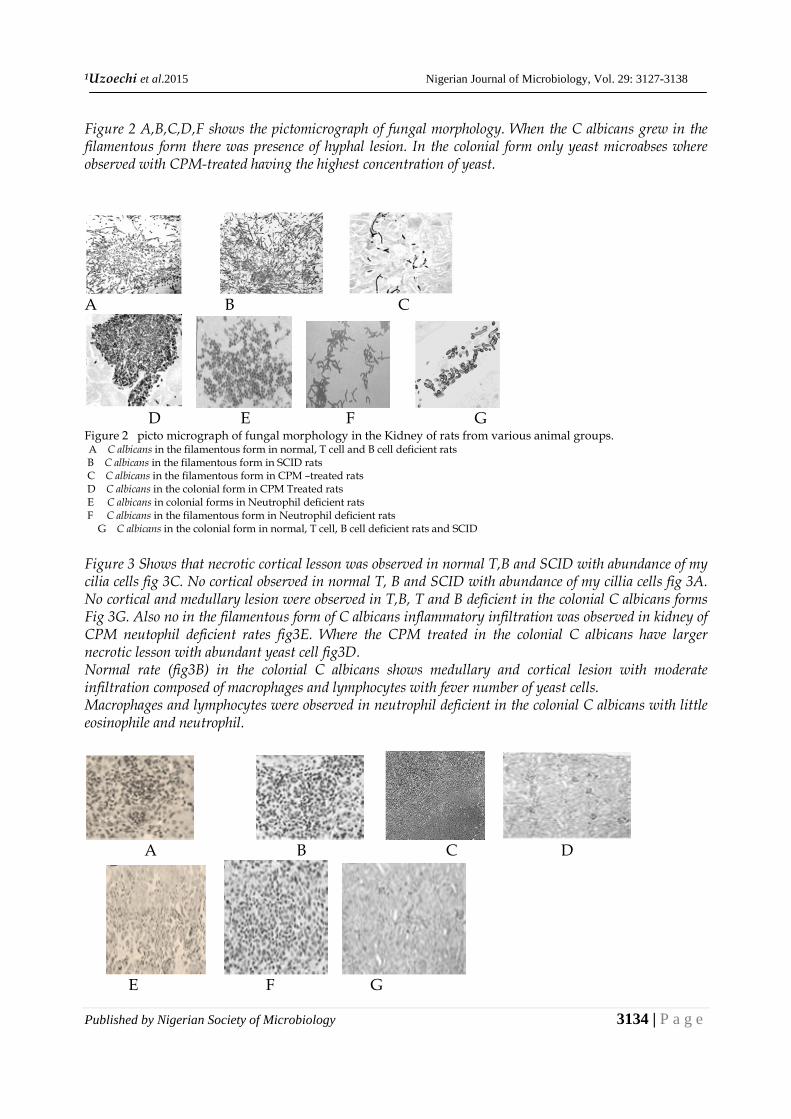
Figure 2 A,B,C,D,F shows the pictomicrograph of fungal morphology. When the C albicans grew in the filamentous form there was presence of hyphal lesion. In the colonial form only yeast microabses where observed with CPM-treated having the highest concentration of yeast.
A B C
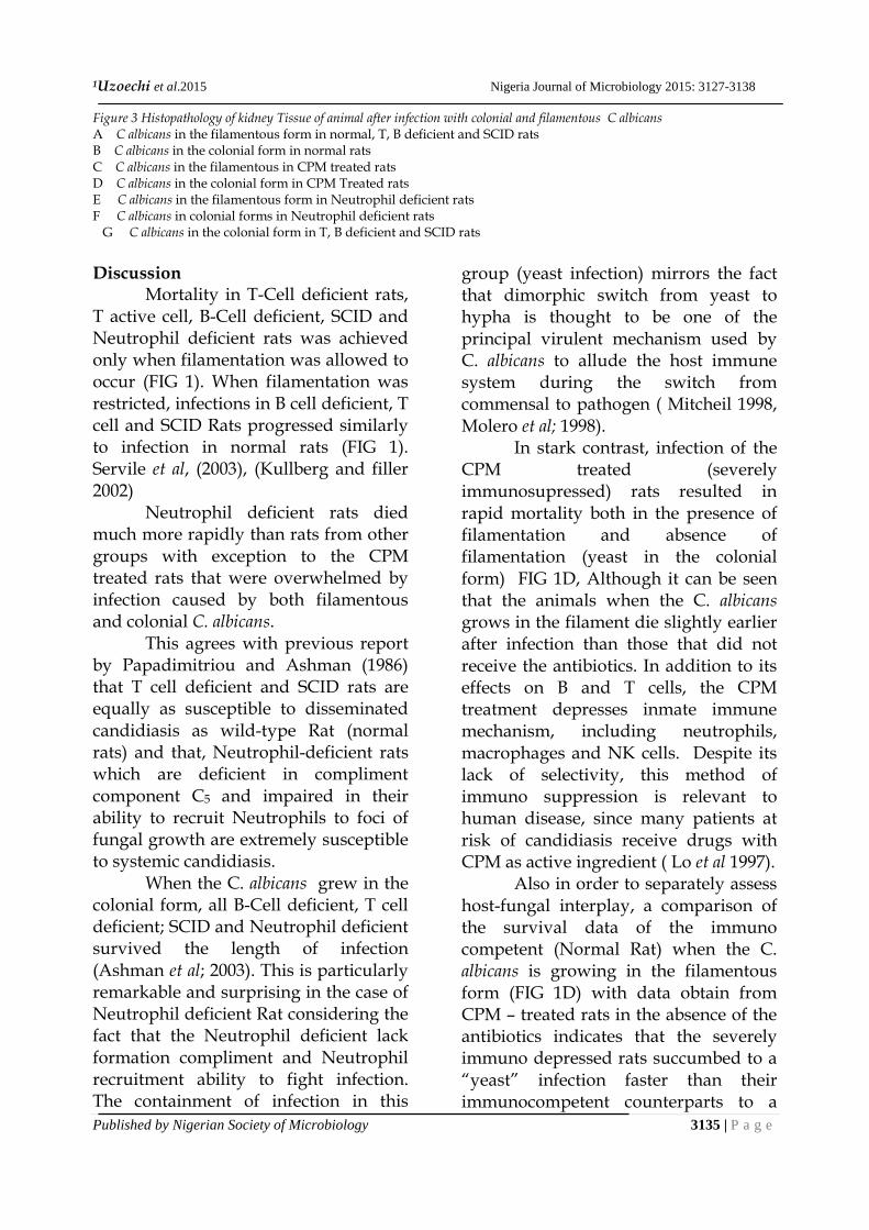
D E F G Figure 2 picto micrograph of fungal morphology in the Kidney of rats from various animal groups. A C albicans in the filamentous form in normal, T cell and B cell deficient rats B C albicans in the filamentous form in SCID rats C C albicans in the filamentous form in CPM –treated rats D C albicans in the colonial form in CPM Treated rats E C albicans in colonial forms in Neutrophil deficient rats F C albicans in the filamentous form in Neutrophil deficient rats G C albicans in the colonial form in normal, T cell, B cell deficient rats and SCID Figure 3 Shows that necrotic cortical lesson was observed in normal T,B and SCID with abundance of my cilia cells fig 3C. No cortical observed in normal T, B and SCID with abundance of my cillia cells fig 3A. No cortical and medullary lesion were observed in T,B, T and B deficient in the colonial C albicans forms Fig 3G. Also no in the filamentous form of C albicans inflammatory infiltration was observed in kidney of CPM neutophil deficient rates fig3E. Where the CPM treated in the colonial C albicans have larger necrotic lesson with abundant yeast cell fig3D. Normal rate (fig3B) in the colonial C albicans shows medullary and cortical lesion with moderate infiltration composed of macrophages and lymphocytes with fever number of yeast cells. Macrophages and lymphocytes were observed in neutrophil deficient in the colonial C albicans with little eosinophile and neutrophil.
A B C D
E F G
1Uzoechi et al.2015 Nigeria Journal of Microbiology 2015: 3127-3138
Published by Nigerian Society of Microbiology 3135 | P a g e
Figure 3 Histopathology of kidney Tissue of animal after infection with colonial and filamentous C albicans A C albicans in the filamentous form in normal, T, B deficient and SCID rats B C albicans in the colonial form in normal rats C C albicans in the filamentous in CPM treated rats D C albicans in the colonial form in CPM Treated rats E C albicans in the filamentous form in Neutrophil deficient rats F C albicans in colonial forms in Neutrophil deficient rats G C albicans in the colonial form in T, B deficient and SCID rats Discussion
Mortality in T-Cell deficient rats, T active cell, B-Cell deficient, SCID and Neutrophil deficient rats was achieved only when filamentation was allowed to occur (FIG 1). When filamentation was restricted, infections in B cell deficient, T cell and SCID Rats progressed similarly to infection in normal rats (FIG 1). Servile et al, (2003), (Kullberg and filler 2002)
Neutrophil deficient rats died much more rapidly than rats from other groups with exception to the CPM treated rats that were overwhelmed by infection caused by both filamentous and colonial C. albicans.
This agrees with previous report by Papadimitriou and Ashman (1986) that T cell deficient and SCID rats are equally as susceptible to disseminated candidiasis as wild-type Rat (normal rats) and that, Neutrophil-deficient rats which are deficient in compliment component C5 and impaired in their ability to recruit Neutrophils to foci of fungal growth are extremely susceptible to systemic candidiasis.
When the C. albicans grew in the colonial form, all B-Cell deficient, T cell deficient; SCID and Neutrophil deficient survived the length of infection (Ashman et al; 2003). This is particularly remarkable and surprising in the case of Neutrophil deficient Rat considering the fact that the Neutrophil deficient lack formation compliment and Neutrophil recruitment ability to fight infection. The containment of infection in this
group (yeast infection) mirrors the fact that dimorphic switch from yeast to hypha is thought to be one of the principal virulent mechanism used by C. albicans to allude the host immune system during the switch from commensal to pathogen ( Mitcheil 1998, Molero et al; 1998).
In stark contrast, infection of the CPM treated (severely immunosupressed) rats resulted in rapid mortality both in the presence of filamentation and absence of filamentation (yeast in the colonial form) FIG 1D, Although it can be seen that the animals when the C. albicans grows in the filament die slightly earlier after infection than those that did not receive the antibiotics. In addition to its effects on B and T cells, the CPM treatment depresses inmate immune mechanism, including neutrophils, macrophages and NK cells. Despite its lack of selectivity, this method of immuno suppression is relevant to human disease, since many patients at risk of candidiasis receive drugs with CPM as active ingredient ( Lo et al 1997).
Also in order to separately assess host-fungal interplay, a comparison of the survival data of the immuno competent (Normal Rat) when the C. albicans is growing in the filamentous form (FIG 1D) with data obtain from CPM – treated rats in the absence of the antibiotics indicates that the severely immuno depressed rats succumbed to a “yeast” infection faster than their immunocompetent counterparts to a
1Uzoechi et al.2015 Nigerian Journal of Microbiology, Vol. 29: 3127-3138
Published by Nigerian Society of Microbiology 3136 | P a g e
filamentous infection. This is a clear evidence of the fact that pathogenesis of candidiasis as determined by host-fungus interplay, the immune state of the host may take precedence over the virulence mechanism displayed by the fungus in determining the outcome of an infection (Kranse and Schaffer 1988; Nombella et al; 1989 and Ashman and Papadimitriou, 1987).
Between 1 to 2 days post infection, the kidneys had the highest fungal burden and this increases with time depending on the operational immune system (Fidel and Sobel, 1996; Kirsch and Whitney, 1991. In majority of the rats groups in different immunological state when the C albicans is growing in the filamentous form, tissue burdens were essentially identical to those displayed by normal rats, with the exception of Neutrophil deficient rats which displayed somewhat lower organ load (Table 3), this is attributed to their increased susceptibility to candidiasis and also of the fact that members of this group died earlier (within 72 hours of post infection) thereby not allowing the Candida albicans much time to multiply.
Also, the CPM – treated rats infected with C albicans growing in the colonial form exhibited higher fungal loads than both normal rats and CPM treated rats when the Candida albicans is growing in the filamentous form (Mitchell 1998). It is expected that the CPM treated rats when the C albicans was growing in the colonial form would have shown lower fungal burden than the normal rats in the colonial and filamentous C albicans and the CPM treated in the filamentous form of the C albicans but here the reverse was the case, this might be attributed to the
increase plating efficiency of yeast cell forms to their filamentous counterparts (Saville, 2003; Rooney and Klein, 2002).
The result depicted in FIG 2 demonstrated that tissue recovered from immuno deficient rats that succumbed to infection when the C albicans was growing in the filamentous form, revealed a predominately hyphal morphology with lesion similar to those normally observed during infection with wild-type Candida albicans strains. However, fungal elements almost exclusively with a yeast cell morphology were detected in CPM treated severely immuno suppressed rats when the yeast was growing in the colonial form, which still resulted in a lethal outcome without need for filamentation.
Analysis of kidneys recovered from infected animals at time of death or sacrifice was performed (FIG 3). Works by Fidel and Sobel (1996) and Fidel et al; (1994) using murine models have previously reported that in immunocompetent rats a mostly Neutrophil infiltrate is evident early on at the infection site, which is very similar to what was observed in normal rats infected with the C. albicans strain in the filamentous form (FIG 3A). Conversely, Normal rats survived the infection when the C albicans was growing in the colonial form, and histopathological examination at time of sacrifice revealed only moderate tissue damage and mild inflammatory infiltration (FIG 3B). As expected, in severely immunosuppressed CPM-treated mice, no inflammatory infiltration was present, irrespective of the filamentous or colonial yeast form (FIG 3C and D). Thus, it would seem that the inflammatory response to
1Uzoechi et al.2015 Nigeria Journal of Microbiology 2015: 3127-3138
Published by Nigerian Society of Microbiology 3137 | P a g e
infection in the kidney is completely absent in CPM-treated rats, although a possible explanation here is that these animals died very soon after infection. The same was true for the Neutrophil deficient rats when the C albicans was growing in the filamentous from, that also died very shortly (within hours) after infection .
In stark contrast, histopathological examination of kidneys recovered from Neutrophil deficient rats that survived the infection in the colonial Candida albicans form revealed an inflammatory infiltrate composed mainly of macrophages and lymphocytes (Eggimann et al; 2003) (FIG 3F). These results suggest that a yeast cell-only infection can be contained in the absence of functional Neutrophil, probably involving compensatory mechanisms and the participation of other components of the innate immune system (i.e., macrophages). Since the Neutrophil deficient rats feed when the C albicans was growing in the filamentous form died within 2 days (FIG 3E) of post infection it can be inferred that the oblation of Neutrophil contributed to lethality, hence no infiltration was observed as the infection was too lethal to allow such activity CONCLUSION
Overall, the results presented here reaffirm that the C. albicans strain represents a powerful tool to investigate different aspects of C. albicans pathogenesis. They also highlight the importance of properly integrating and taking into account the interplay between host immunity and mechanisms of fungal virulence, an area which has been largely overlooked in the study of C. albicans pathogenesis, as we try to better understand the clinical
scenario in patients suffering from these insidious infections. A large body of evidence, in preclinical settings and in humans, supports the model of specific immunity to opportunistic fungal infections as being highly susceptible to cytokine influences and reciprocally
regulated by cells of the innate immune system. Therefore, finding cells and cytokines that are essential to control fungal infectivity or oppose fungus-associated immunopathology represents
an intense area of research, the results of which may have important implications in the development of vaccines, and immunodiagnosis of and therapy for BNOP.UO
Reference Ashman, R. B and Papadimitriou, J. M.
(1987). Murine Candidiasi Pathogenesis and host responses in genetically distinct inbred mice Immunol. Cell Biol. 65:163-171.
Ashman, R. B., Papadimitriou J. M., Fulurija
A., Drysdale K. E., Farah C. S, Naidoo O., and Gotjamanos T.. (2003). Role ofcomplement C5 and T lymphocytes in pathogenesis of disseminated and Mucosal Candidiasis in susceptible DBA/2 mice. Microb.Pathog. 34:103-113.
Mahon R. C and George Manuselis
(2000) A text book of Diagnostic microbiology Saunders philadelphia
`Eggimann P, Garbino J, Pittet D. (2003).
Epidemiology of Candida species infections in critically ill non-immunosuppressed patients. Lancet Infect Dis; 3: 685–702.
Fidel P.L., Jr., Sobel J.D. (1996)
Immunopathogenesis of Recurrent
1Uzoechi et al.2015 Nigerian Journal of Microbiology, Vol. 29: 3127-3138
Published by Nigerian Society of Microbiology 3138 | P a g e
Vulvovaginal Candidiasis. Clin Micro Rev. 9;(1):335-348.
Fidel P.L. Jr, Lynch M.E., Sobel J.D. (1994)
Effects of preinduced Candida- specific systemic cell-mediated immunity on experimental vaginal candidiasis. Infect Immun. Mar; 62(3):1032-8.
Krause M, Schaffner A (1988) Comparison
of immune mechanisms against Candida albicans in models of localized and systemic infections in mice. 28th. Inters. Conf. Antimicrob. Agents Chemother, Los Angeles, pp 341–347
Kirsch D.R, Whitney R.R (1991)
Pathogenicity of Candida albicans auxotrophic mutants in experimental infections. Infect Immun 59:3297–3300
Lo H.J., Kohler J.R., DiDomenico B.,
Loeberg D., Cacciaputi A., Fink G.R. (1997) Nonfilamentous Candida albicans Mutants are Avirulent. Cell 90:939-949.
Mitchell A.P. (1998) Dimorphism and
Virulence in Candida albicans. Curr Opin Microbiol. 1:687-692.
Molero G., Diez-Orejas R., Navarro-Garcia
F., Monteoliva L., Pla J., Gil C., Sanchez-Perez M., Nombela (1998) C. Candida albicans: Genetics, Dimorphism, and Pathogenecity. Internatl. Microbiol. 1:95-106
Nombela C, Pomés R, Gil C, Herreros E,
Sánchez M (1989) Genetics of pathogenic fungi: Candida albicans. Rev Iber Micol 6:47–53
Ochei J. and Kolhatkar (2008) Medical
Labouratory Science; Theory and
Practice . TaTa McGraw-Hill Publishing Company. New Delhi
Rooney, P. J., and B. S. Klein. (2002).
Linking fungal morphogenesis with virulence.
Cel Microbiol. 4:127-137. Saville S.P., Lazzel A.L. Monteagudo C.,
Lopez-Ribot J.L. (2003) Engineered Control of Cell Morphology in vivo Reveal Distinct Roles for Yeast and Filamentous Forms of Candida albicans During Infection. Eukaryot Cell. 2:1053-1060.