TheEffect of Estrogen on Bile Formation the Rat · should be observable as enhanced excretion of...
10
The Effect of Estrogen on Bile Formation in the Rat E. L. FORKER From the Department of Physiology and Biophysics and the Gastroenterology Research Laboratories, Department of Medicine, University of Iowa, Iowa City, Iowa 52240 A B ST R A CT Female rats given large doses of estrone developed increased permeability of the biliary tree as determined by an increase in the biliary clearances of sucrose and mannitol. Spontaneous bile flow and the choleretic response to dehydrocholate declined. Estrone reduced the clearance of sulfobromophthalein (BSP) at low plasma concentrations as well as the absolute rate of BSP excretion when plasma levels were above those required to saturate active transport. The findings are consistent with a postulate that estrogen cholestasis may involve enhanced diffusion of materials from bile to blood in addition to inhibition of active transport in the opposite direction. INTRODUCTION Sporadic occurrence of recurrent jaundice and/or pru- ritus in the last trimester of pregnancy (1-3), abnormal removal of sulfobromophthalein (BSP) during other- wise uncomplicated pregnancy (4, 5), and the predis- position of multiparous women to form gallstones all point to a possible association between high levels of circulating estrogen and altered bile formation. Scat- tered reports (6-9) of cholestasis following the use of oral contraceptives have heightened this suspicion as have recent suggestions (10-12), that impaired hepatic excretion in the newborn may reflect, at least in part, a deleterious effect of maternal estrogen on the fetus. However one regards the circumstantial evidence impli- cating sex hormones in these clinical situations, it is clear that large doses of estrogen regularly impair BSP excretion in both rats (13, 14) and people (10, 15). Among a variety of natural and synthetic estrogens which share this capacity in the rat, estrone is reported to be most active (13). Judging from the observation Dr. Forker is a Markle Scholar in Academic Medicine. Received for publication 19 August 1968 and in revised form 22 Octobcr 1968. that conjugation with glutathione is normal under these circumstances, while the maximum rate of BSP excre- tion is reduced, it is probable that estrogens interfere primarily with the processes responsible for excreting BSP and its conjugates into bile rather than with the delivery of BSP to the hepatic parenchyma or with its in- tracellular metabolism. Beyond this, however, nothing is known about the underlying mechanism of the estro- gen effect. This is a report of experiments designed to explore the possibility that changes in passive permeability of the canalicular membrane might help to explain the ef- fect of estrone on bile formation in rats. The specific hypothesis is as follows. To the extent that reduced ex- cretion of BSP is attributable to increased diffusion of BSP from bile to plasma rather than to impaired active transport of BSP in the opposite direction, this effect should be observable as enhanced excretion of inert sol- utes assumed to enter bile passively. As predicted by this hypothesis, biliary clearances of radioactive mannitol and sucrose increased after treatment with estrone, whereas bile flow and BSP excretion declined. Possible interpretations of these findings are discussed in light of assumptions about the site of the estrogen effect and the normal pathway for biliary excretion of inert solutes. METHODS Virgin female Sprague-Dawley rats (250 g body weight) received seven daily subcutaneous injections of estrone (2.5 mg, Sigma Chemical Corp.) dissolved in propylene glycol (0.5 ml). Control rats received identical volumes of pro- pylene glycol alone. Total dose and method of administration were similar to those employed by Gallagher, Mueller, and Kappas (13) except that the solubilizing agent, N,N-di- methylacetamide, was omitted in favor of warming the solu- tion before injection. Experiments were conducted alternately with estrone-treated rats and controls fasted for 18 hr after the last injection. After anesthesia with intraperitoneal al- lobarbital and urethane (Dial-urethane, Ciba), the renal pedicles were ligated and small polyethylene catheters in- 654 The Journal of Clinical Investigation Volume 48 1969
Transcript of TheEffect of Estrogen on Bile Formation the Rat · should be observable as enhanced excretion of...
The Effect of Estrogen on
Bile Formation in the Rat
E. L. FORKER
From the Department of Physiology and Biophysics and the GastroenterologyResearch Laboratories, Department of Medicine, University of Iowa,Iowa City, Iowa 52240
A B ST R A CT Female rats given large doses of estronedeveloped increased permeability of the biliary tree asdetermined by an increase in the biliary clearances ofsucrose and mannitol. Spontaneous bile flow and thecholeretic response to dehydrocholate declined. Estronereduced the clearance of sulfobromophthalein (BSP) atlow plasma concentrations as well as the absolute rateof BSP excretion when plasma levels were above thoserequired to saturate active transport. The findings areconsistent with a postulate that estrogen cholestasismay involve enhanced diffusion of materials from bileto blood in addition to inhibition of active transport inthe opposite direction.
INTRODUCTION
Sporadic occurrence of recurrent jaundice and/or pru-ritus in the last trimester of pregnancy (1-3), abnormalremoval of sulfobromophthalein (BSP) during other-wise uncomplicated pregnancy (4, 5), and the predis-position of multiparous women to form gallstones allpoint to a possible association between high levels ofcirculating estrogen and altered bile formation. Scat-tered reports (6-9) of cholestasis following the use oforal contraceptives have heightened this suspicion ashave recent suggestions (10-12), that impaired hepaticexcretion in the newborn may reflect, at least in part, adeleterious effect of maternal estrogen on the fetus.However one regards the circumstantial evidence impli-cating sex hormones in these clinical situations, it isclear that large doses of estrogen regularly impair BSPexcretion in both rats (13, 14) and people (10, 15).Among a variety of natural and synthetic estrogenswhich share this capacity in the rat, estrone is reportedto be most active (13). Judging from the observation
Dr. Forker is a Markle Scholar in Academic Medicine.Received for publication 19 August 1968 and in revised
form 22 Octobcr 1968.
that conjugation with glutathione is normal under thesecircumstances, while the maximum rate of BSP excre-tion is reduced, it is probable that estrogens interfereprimarily with the processes responsible for excretingBSP and its conjugates into bile rather than with thedelivery of BSP to the hepatic parenchyma or with its in-tracellular metabolism. Beyond this, however, nothingis known about the underlying mechanism of the estro-gen effect.
This is a report of experiments designed to explorethe possibility that changes in passive permeability ofthe canalicular membrane might help to explain the ef-fect of estrone on bile formation in rats. The specifichypothesis is as follows. To the extent that reduced ex-cretion of BSP is attributable to increased diffusion ofBSP from bile to plasma rather than to impaired activetransport of BSP in the opposite direction, this effectshould be observable as enhanced excretion of inert sol-utes assumed to enter bile passively. As predicted by thishypothesis, biliary clearances of radioactive mannitoland sucrose increased after treatment with estrone,whereas bile flow and BSP excretion declined. Possibleinterpretations of these findings are discussed in lightof assumptions about the site of the estrogen effect andthe normal pathway for biliary excretion of inert solutes.
METHODSVirgin female Sprague-Dawley rats (250 g body weight)received seven daily subcutaneous injections of estrone (2.5mg, Sigma Chemical Corp.) dissolved in propylene glycol(0.5 ml). Control rats received identical volumes of pro-pylene glycol alone. Total dose and method of administrationwere similar to those employed by Gallagher, Mueller, andKappas (13) except that the solubilizing agent, N,N-di-methylacetamide, was omitted in favor of warming the solu-tion before injection. Experiments were conducted alternatelywith estrone-treated rats and controls fasted for 18 hr afterthe last injection. After anesthesia with intraperitoneal al-lobarbital and urethane (Dial-urethane, Ciba), the renalpedicles were ligated and small polyethylene catheters in-
654 The Journal of Clinical Investigation Volume 48 1969
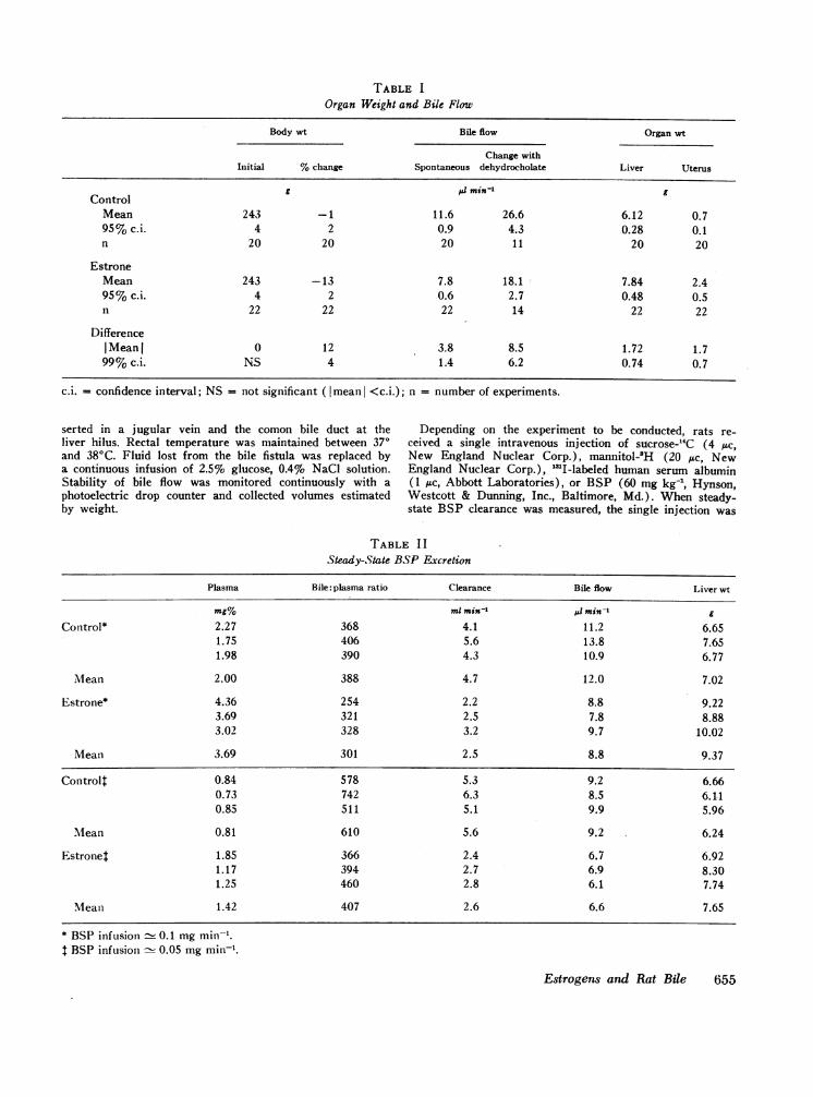
TABLE IOrgan Weight and Bile Flow
Body wt Bile flow Organ wt
Change withInitial %change Spontaneous dehydrocholate Liver Uterus
g Al min-' gControl
Mean 243 -1 11.6 26.6 6.12 0.795% c.i. 4 2 0.9 4.3 0.28 0.1n 20 20 20 11 20 20
EstroneMean 243 -13 7.8 18.1 7.84 2.495% c.i. 4 2 0.6 2.7 0.48 0.5n 22 22 22 14 22 22
DifferenceI Mean I 0 12 3.8 8.5 1.72 1.799%c.i. NS 4 1.4 6.2 0.74 0.7
c.i. = confidence interval; NS = not significant ( mean <c.i.); n = number of experiments.
serted in a jugular vein and the comon bile duct at the Depending on the experiment to be conducted, rats re-liver hilus. Rectal temperature was maintained between 37° ceived a single intravenous injection of sucrose-14C (4 ,uc,and 380C. Fluid lost from the bile fistula was replaced by New England Nuclear Corp.), mannitol-'H (20 Asc, Newa continuous infusion of 2.5%o glucose, 0.4% NaCl solution. England Nuclear Corp.), "MI-labeled human serum albuminStability of bile flow was monitored continuously with a (1 /Ac, Abbott Laboratories), or BSP (60 mg kg-', Hynson,photoelectric drop counter and collected volumes estimated Westcott & Dunning, Inc., Baltimore, Md.). When steady-by weight. state BSP clearance was measured, the single injection was
TABLE I ISteady-State BSP Excretion
Plasma Bile: plasma ratio Clearance Bile flow Liver wt
mg% ml min-' ;6 min-' g
Control* 2.27 368 4.1 11.2 6.651.75 406 5.6 13.8 7.651.98 390 4.3 10.9 6.77
Mean 2.00 388 4.7 12.0 7.02
Estrone* 4.36 254 2.2 8.8 9.223.69 321 2.5 7.8 8.883.02 328 3.2 9.7 10.02
Mean 3.69 301 2.5 8.8 9.37
Controlt 0.84 578 5.3 9.2 6.660.73 742 6.3 8.5 6.1.10.85 511 5.1 9.9 5.96
Mlean 0.81 610 5.6 9.2 6.24
Estronet 1.85 366 2.4 6.7 6.921.17 394 2.7 6.9 8.301.25 460 2.8 6.1 7.74
Mean 1.42 407 2.6 6.6 7.65
* BSP infusion - 0.1 mg min-.t BSP infusion - 0.05 mg min'.
Estrogens and Rat Bile 655
0.16
7 0.14
t 0.12
t 0.10
Q 0.08
i 0.06
i0.04
0.02
RESULTS
10 20 30MINUTE
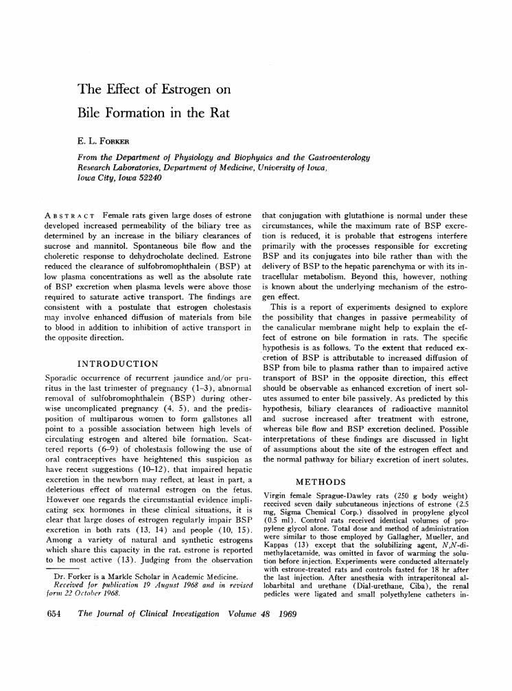
FIGURE 1 The time course of BWvenous injection of 60 mg kg-' toport. Mean values +SE, n = 5 for (
replaced by a continuous 2 hr infu0.9% NaCl solution. Steady-state csucrose were measured 2.5-3 hr afrats again after a stable maximuthv infising sodium dehvdrocholate
Control The large dose of estrone employed in these experi-ments led to a moderate loss of body weight, an increasein liver weight, and striking enlargement of the uterus.
I-F HEstrone Estrone-treated rats appeared healthy, however, andexcept for diminished bile flow could not be distinguishedby inspection from rats that received propylene glycolalone. The changes in organ weight and bile flow aresummarized in Table I.
The results of two studies in which BSP was givenby continuous infusion at nominal rates of 0.1 or 0.05mg min-' appear in Table II. After 2 hr biliary excretion
60mgSP doatse of BSP was approximately 85% of the infusion rate inboth estrone-treated rats and controls. Doubling the rateof BSP infusion caused a similar increase in bile flowin both groups, but this procedure did not produce sig-
, , nificant changes in plasma clearance. Mean values for40 50 60 six determinations of clearance were 50% less in es-
trone-treated rats (2.6 ±0.6 ml min-) than in controlsSP excretion after intra- (5.1 +1.3 ml min-'). The decline in clearance was as-saturate excretory trans- sociated with mean decrements of 20% in bile flow andeach curve.eachcurve.
30% in bile: plasma concentration ratio.Five additional rats given estrone and five controls
ision of BSP dissolved in received a single injection of BSP (60 mg kg-1) to satu-learances of mannitol and rate the active transport of this dye. The time courseEter injection and in some*** *choerinestisn was sodued of BSP excretion after this procedure is shown in Fig.
(Dolme Chemicas. Tncd 1. Peak excretion occurred in both groups after 40 minJJV1_ 1 LU3111r OVU1UA1l %_LyL V %_11V 1CLL %,-V IAI% sss~so Ls..%'
New York) for an additional 40 min at a rate of 0.6 mgmin-. Concentrations of labeled albumin in plasma and livertissue were determined 20 min after injection. Blood wasobtained during an experiment by cutting the tip of the tailor, at the end of an experiment, from the abdominal aorta.To minimize variations in liver weight all rats were ex-sanguinated.
Concentrations of sucrose and mannitol were determinedin a liquid scintillation counter after dissolving 20-50 ul ofplasma, bile, or the supernatant fluid from liver homogenatein Bray's solution (16). Small corrections required forvariable quenching were determined by internal standardi-zation. Liver homogenates were prepared by grinding ap-proximately half of the liver with 10 ml of 3% trichloraceticacid in a microhomogenizer (Ivan Sorvall, Inc., Norwalk,Conn.). Radioactivity in liver water was calculated from theactivity in the supernatant and the fractional water con-tent of the liver measured by drying the remaining half toconstant weight. Radioactivity from "31I was counted di-rectly in liver tissue with a NaI crystal. BSP was measuredby the method of Seligson, Marino, and Dodson (17) in aBeckman DU spectrophotometer at 580 mit. Na+ and K+were determined by flame photometry. Plasma concentra-tions of total and direct reacting bilirubin, glutamic-oxala-cetic transaminase, and alkaline phosphatase were deter-mined, respectively, according to the methods of Malloy andEvelyn (18), Reitman and Frankel (19), and Bessey,Lowrey, and Brock (20).
Unless otherwise noted, results are expressed as themean value + the 95% confidence interval. Statistical sig-nificance is indicated by citing the value of P determinedby Student's t test or the 99% confidence interval for thedifference between mean values (21).
and remained virtually constant during the next 15 min.In controls peak excretion (0.170 mg min') was similarto a previously reported value for "transport maximum"in normal rats determined by a continuous infusionmethod (22). Peak excretion by estrone-treated ratswas 20% lower (P < 0.05). This change was associ-
t 0.8
{-;i,0.6
0.4-4 %0.2
"p
zi-1
.8 *0
- 0 00
- ~~~~S0 00
I -2 3BILE FLOW (pl min6lg1l)
- 0%
7*:* A000I. ° 80°lp l
10 20BILE FLOW(PI mifri)
30
FIGURE 2 Sucrose clearance and spontaneous bile flow be-fore and after division by wet liver weight. Estrone (0);control (0).
656 E. L. Forker
ated with mean decrements of 12% in bile flow and 8%in BSP concentration. In both Fig. 1 and in Table IIthe rates of BSP excretion, plasma clearance, and bileflow have been expressed in absolute terms. Since ineach case liver weights in the control group were lessthan those in the group receiving estrone, expressingthe results as rates per gram of liver has the effect ofexaggerating the differences.
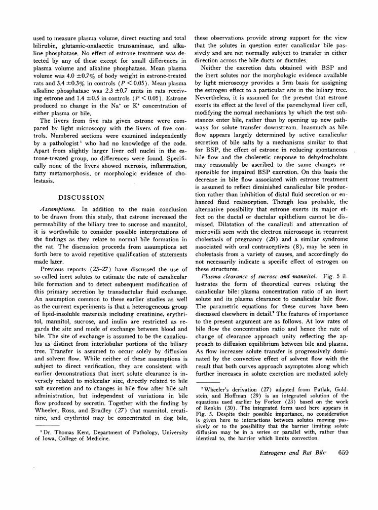
The effect of estrone on sucrose clearance duringspontaneous bile flow is presented in Fig. 2. The sameresults are given in two ways to illustrate the effect ofnormalizing the data to take account of differences inliver weight. Dividing by liver weight exaggerates thedifferences in bile flow while diminishing the differ-ences in plasma clearance, but in either case it is clearthat sucrose clearance was on the average at least twiceas fast in rats receiving estrone as in controls, despitean opposite change in bile flow.
Fig. 3 shows the effect of estrone on the bile: plasmaconcentration ratio for sucrose during spontaneous flowand dehydrocholate choleresis. In control rats this ra-tio was approximately 0.14 over a four-fold range ofbile flow, but in rats given estrone, choleresis led to anabrupt fall in this ratio from an average value of 0.56to levels approaching those observed in the control group.Concentration ratios in Fig. 3 are plotted against bileflow per gram of liver weight to emphasize that themaximum choleretic response of estrone-treated ratswas uniformly less than that of the control group.Though less pronounced, the same effect was evidentfrom a comparison of mean increments in bile flow with-out regard to liver weight (Table I).
When a smaller solute, mannitol, was used to assesspermeability changes, the effect of estrone was ap-parent only during choleresis. Results from 11 experi-
0.8
R 0.7 -
.t 6
0.4-
0.3-
00
1 2 3 4 5 6 7 8 9 10BILE FLOW (plI mi91g1)
FIGURE 3 Sucrose bile: plasma concentration ratios duringspontaneous bile flow and dehydrocholate choleresis. Linesconnect points determined in one rat. Estrone (0); control(0).
~11I4'4
00
0
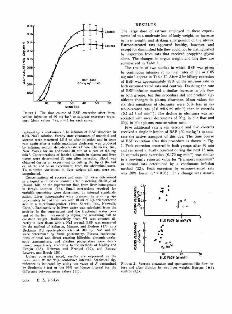
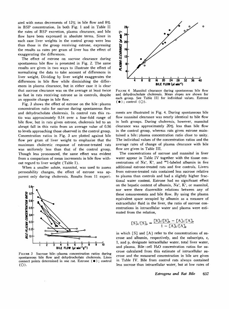
BILE FLOW(p1 .i")FIGURE 4 Mannitol clearance during spontaneous bile flowand dehydrocholate choleresis. Mean slopes are shown foreach group. See Table III for individual values. Estrone( * ); control (0).
ments are illustrated in Fig. 4. During spontaneous bileflow mannitol clearance was nearly identical to bile flowin both groups. During choleresis, however, mannitolclearance was approximately 20% less than bile flowin the control group, whereas rats given estrone main-tained a bile: plasma concentration ratio close to unity.The individual values of the concentration ratios and theaverage rates of change of plasma clearance with bileflow are given in Table III.
The concentrations of sucrose and mannitol in liverwater appear in Table IV together with the tissue con-centrations of Na+, K+, and '3I-labeled albumin in fiveadditional estrone-treated rats and five controls. Liversfrom estrone-treated rats contained less sucrose relativeto plasma than controls and had a slightly higher frac-tional water content. Estrone had no significant effecton the hepatic content of albumin, Na', K+, or mannitol,nor were there discernible relations between any ofthese measurements and bile flow. By using the plasmaequivalent space occupied by albumin as a measure ofextracellular fluid in the liver, the ratio of sucrose con-centrations in intracellular water and plasma were esti-mated from the relation,
[5] /[S]_- [S]i/[S]p -A]i/EA]p1 -EAiEp
in which [S] and [A] refer to the concentrations of su-crose and albumin, respectively, and the subscripts, c,1, and p, designate intracellular water, total liver water,and plasma. Bile: cell H20 concentration ratios for su-crose calculated from this estimate of intracellular su-crose and the measured concentration in bile are givenin Table IV. Bile from control rats always containedless sucrose than intracellular water, but at low rates of
Estrogens and Rat Bile 657
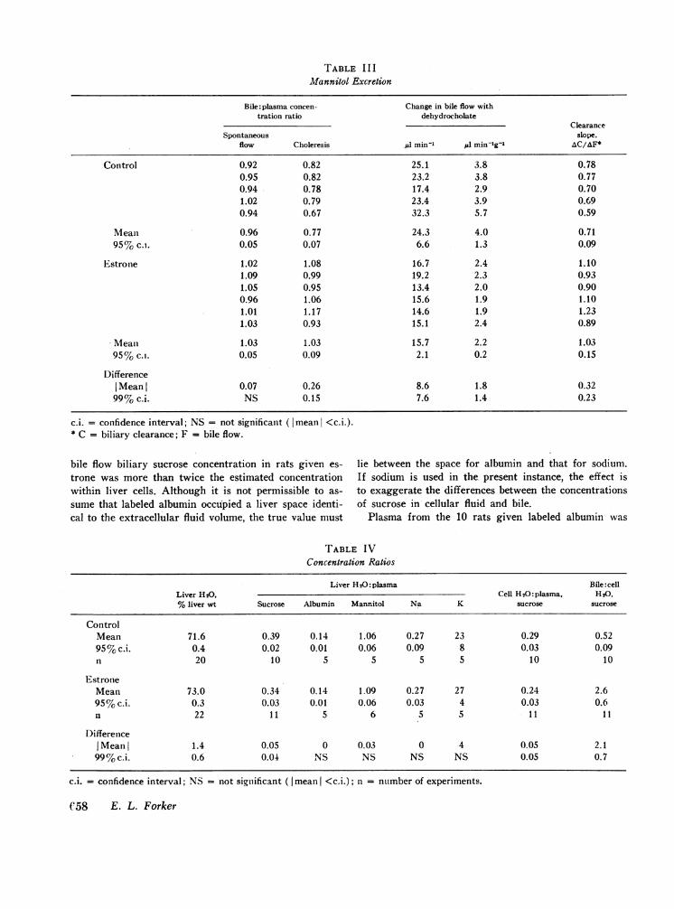
TABLE I I IMannitol Excretion
Bile: plasma concen- Change in bile flow withtration ratio dehydrocholate
ClearanceSpontaneous slope,
flow Choleresis ,d1 min-' ju min-lg-1 AC/A&F*
Control 0.92 0.82 25.1 3.8 0.780.95 0.82 23.2 3.8 0.770.94 0.78 17.4 2.9 0.701.02 0.79 23.4 3.9 0.690.94 0.67 32.3 5.7 0.59
Mean 0.96 0.77 24.3 4.0 0.7195% c.i. 0.05 0.07 6.6 1.3 0.09
Estrone 1.02 1.08 16.7 2.4 1.101.09 0.99 19.2 2.3 0.931.05 0.95 13.4 2.0 0.900.96 1.06 15.6 1.9 1.101.01 1.17 14.6 1.9 1.231.03 0.93 15.1 2.4 0.89
Mean 1.03 1.03 15.7 2.2 1.0395% c.i. 0.05 0.09 2.1 0.2 0.15
DifferenceI Mean l 0.07 0.26 8.6 1.8 0.3299% c.i. NS 0.15 7.6 1.4 0.23
c.i. = confidence interval; NS = not significant (Imean <c.i.).* C = biliary clearance; F = bile flow.
bile flow biliary sucrose concentration in rats given es- lie between the space for albumin and that for sodium.trone was more than twice the estimated concentration If sodium is used in the, present instance, the effect iswithin liver cells. Although it is not permissible to as- to exaggerate the differences between the concentrationssume that labeled albumin occupied a liver space identi- of sucrose in cellular fluid and bile.cal to the extracellular fluid volume, the true value must Plasma from the 10 rats given labeled albumin was
TABLE IVConcentration Ratios
Liver H20: plasma Bile:cellLiver H20, Cell H20: plasma. H20,%liver wt Sucrose Albumin Mannitol Na K sucrose sucrose
ControlMean 71.6 0.39 0.14 1.06 0.27 23 0.29 0.5295%c.i. 0.4 0.02 0.01 0.06 0.09 8 0.03 0.09n 20 10 5 5 5 5 10 10
EstroneMean 73.0 0.34 0.14 1.09 0.27 27 0.24 2.695%c.i. 0.3 0.03 0.01 0.06 0.03 4 0.03 0.6n 22 11 5 6 5 5 11 11
DifferenceI Mean 1.4 0.05 0 0.03 0 4 0.05 2.199%c.i. 0.6 0.04 NS NS NS NS 0.05 0.7
c.i. = confidence interval; NS = not significant ( mean <c.i.); n = number of experiments.
C58 E. L. Forker
used to measure plasma volume, direct reacting and totalbilirubin, glutamic-oxalacetic transaminase, and alka-line phosphatase. No effect of estrone treatment was de-tected by any of these except for small differences inplasma volume and alkaline phosphatase. Mean plasmavolume was 4.0 +0.7% of body weight in estrone-treatedrats and 3.4 +0.3% in controls (P < 0.05). Mean plasmaalkaline phosphatase was 2.3 ±0.7 units in rats receiv-ing estrone and 1.4 ±0.5 in controls (P < 0.05). Estroneproduced no change in the Na+ or K+ concentration ofeither plasma or bile.
The livers from five rats given estrone were com-pared by light microscopy with the livers of five con-trols. Numbered sections were examined independentlyby a pathologist1 who had no knowledge of the code.Apart from slightly larger liver cell nuclei in the es-trone-treated group, no differences were found. Specifi-cally none of the livers showed necrosis, inflammation,fatty metamorphosis, or morphologic evidence of cho-lestasis.
DISCUSSION
Assumptions. In addition to the main conclusionto be drawn from this study, that estrone increased thepermeability of the biliary tree to sucrose and mannitol,it is worthwhile to consider possible interpretations ofthe findings as they relate to normal bile formation inthe rat. The discussion proceeds from assumptions setforth here to avoid repetitive qualification of statementsmade later.
Previous reports (23-27) have discussed the use ofso-called inert solutes to estimate the rate of canalicularbile formation and to detect subsequent modification ofthis primary secretion by transductular fluid exchange.An assumption common to these earlier studies as wellas the current experiments is that a heterogeneous groupof lipid-insoluble materials including creatinine, erythri-tol, mannitol, sucrose, and inulin are restricted as re-gards the site and mode of exchange between blood andbile. The site of exchange is assumed to be the canalicu-lus as distinct from interlobular portions of the biliarytree. Transfer is assumed to occur solely by diffusionand solvent flow. While neither of these assumptions issubject to direct verification, they are consistent withearlier demonstrations that inert solute clearance is in-versely related to molecular size, directly related to bilesalt excretion and to changes in bile flow after bile saltadministration, but independent of variations in bileflow produced by secretin. Together with the finding byWheeler, Ross, and Bradley (27) that mannitol, creati-nine, and erythritol may be concentrated in dog bile,
1Dr. Thomas Kent, Department of Pathology, Universityof Iowa, College of Medicine.
these observations provide strong support for the viewthat the solutes in question enter canalicular bile pas-sively and are not normally subject to transfer in eitherdirection across the bile ducts or ductules.
Neither the excretion data obtained with BSP andthe inert solutes nor the morphologic evidence availableby light microscopy provides a firm basis for assigningthe estrogen effect to a particular site in the biliary tree.Nevertheless, it is assumed for the present that estroneexerts its effect at the level of the parenchymal liver cell,modifying the normal mechanisms by which the test sub-stances enter bile, rather than by opening up new path-ways for solute transfer downstream. Inasmuch as bileflow appears largely determined by active canalicularsecretion of bile salts by a mechanisms similar to thatfor BSP, the effect of estrone in reducing spontaneousbile flow and the choleretic response to dehydrocholatemay reasonably be ascribed to the same changes re-sponsible for impaired BSP excretion. On this basis thedecrease in bile flow associated with estrone treatmentis assumed to reflect diminished canalicular bile produc-tion rather than inhibition of distal fluid secretion or en-hanced fluid reabsorption. Though less probable, thealternative possibility that estrone exerts its major ef-fect on the ductal or ductular epithelium cannot be dis-missed. Dilatation of the canaliculi and attenuation ofmicrovilli seen with the electron microscope in recurrentcholestasis of pregnancy (28) and a similar syndromeassociated with oral contraceptives (8), may be seen incholestasis from a variety of causes, and accordingly donot necessarily indicate a specific effect of estrogen onthese structures.
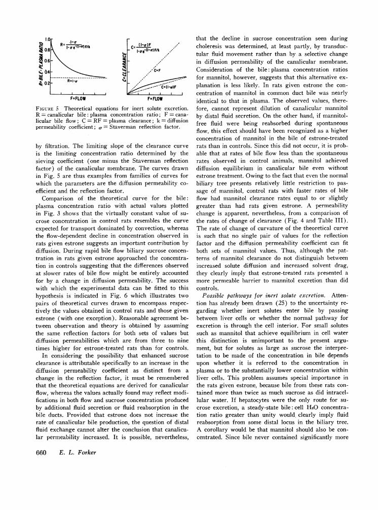
Plasma clearance of sucrose and mannitol. Fig. 5 il-lustrates the form of theoretical curves relating thecanalicular bile: plasma concentration ratio of an inertsolute and its plasma clearance to canalicular bile flow.The parametric equations for these curves have beendiscussed elsewhere in detail.' The features of importanceto the present argument are as follows. At low rates ofbile flow the concentration ratio and hence the rate ofchange of clearance approach unity reflecting the ap-proach to diffusion equilibrium between bile and plasma.As flow increases solute transfer is progressively domi-nated by the convective effect of solvent flow with theresult that both curves approach asymptotes along whichfurther increases in solute excretion are mediated solelv
2 Wheeler's derivation (27) adapted from Patlak, Gold-stein, and Hoffman (29) is an integrated solution of theequations used earlier by Forker (23) based on the workof Renkin (30). The integrated form used here appears inFig. 5. Despite their possible importance, no considerationis given here to interactions between solutes moving pas-sively or to the possibility that the barrier limiting solutediffusion may be in a series or parallel with, rather thanidentical to, the barrier which limits convection.
Estrogens and Rat Bile 659
1.0
1 0.8
I 0.6
%;, 0.4
P 0.2
I-a eiI-1F/k
F FLOW FaFLOW
FIGURE 5 Theoretical equations for inert solute excretion.R = canalicular bile: plasma concentration ratio; F = cana-licular bile flow; C = RF = plasma clearance; k = diffusionpermeability coefficient; a= Staverman reflection factor.
by filtration. The limiting slope of the clearance curveis the limiting concentration ratio determined by thesieving coefficient (one minus the Staverman reflectionfactor) of the canalicular membrane. The curves drawnin Fig. 5 are thus examples from families of curves forwhich the parameters are the diffusion permeability co-efficient and the reflection factor.
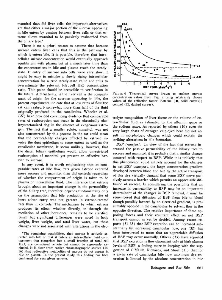
Comparison of the theoretical curve for the bile:plasma concentration ratio with actual values plottedin Fig. 3 shows that the virtually constant value of su-crose concentration in control rats resembles the curveexpected for transport dominated by convection, whereasthe flow-dependent decline in concentration observed inrats given estrone suggests an important contribution bydiffusion. During rapid bile flow biliary sucrose concen-tration in rats given estrone approached the concentra-tion in controls suggesting that the differences observedat slower rates of bile flow might be entirely accountedfor by a change in diffusion permeability. The successwith which the experimental data can be fitted to thishypothesis is indicated in Fig. 6 which illustrates twopairs of theoretical curves drawn to encompass respec-tively the values obtained in control rats and those givenestrone (with one exception). Reasonable agreement be-tween observation and theory is obtained by assumingthe same reflection factors for both sets of values butdiffusion permeabilities which are from three to ninetimes higher for estrone-treated rats than for controls.
In considering the possibility that enhanced sucroseclearance is attributable specifically to an increase in thediffusion permeability coefficient as distinct from achange in the reflection factor, it must be rememberedthat the theoretical equations are derived for canalicularflow, whereas the values actually found may reflect modi-fications in both flow and sucrose concentration producedby additional fluid secretion or fluid reabsorption in thebile ducts. Provided that estrone does not increase therate of canalicular bile production, the question of distalfluid exchange cannot alter the conclusion that canalicu-lar permeability increased. It is possible, nevertheless,
that the decline in sucrose concentration seen duringcholeresis was determined, at least partly, by transduc-tular fluid movement rather than by a selective changein diffusion permeability of the canalicular membrane.Consideration of the bile: plasma concentration ratiosfor mannitol, however, suggests that this alternative ex-planation is less likely. In rats given estrone the con-centration of mannitol in common duct bile was nearlyidentical to that in plasma. The observed values, there-fore, cannot represent dilution of canalicular mannitolby distal fluid secretion. On the other hand, if mannitol-free fluid were being reabsorbed during spontaneousflow, this effect should have been recognized as a higherconcentration of mannitol in the bile of estrone-treatedrats than in controls. Since this did not occur, it is prob-able that at rates of bile flow less than the spontaneousrates observed in control animals, mannitol achieveddiffusion equilibrium in canalicular bile even withoutestrone treatment. Owing to the fact that even the normalbiliary tree presents relatively little restriction to pas-sage of mannitol, control rats with faster rates of bileflow had mannitol clearance rates equal to or slightlygreater than had rats given estrone. A permeabilitychange is apparent, nevertheless, from a comparison ofthe rates of change of clearance (Fig. 4 and Table III).The rate of change of curvature of the theoretical curveis such that no single pair of values for the reflectionfactor and the diffusion permeability coefficient can fitboth sets of mannitol values. Thus, although the pat-terns of mannitol clearance do not distinguish betweenincreased solute diffusion and increased solvent drag,they clearly imply that estrone-treated rats presented amore permeable barrier to mannitol excretion than didcontrols.
Possible pathways for inert solute excretion. Atten-tion has already been drawn (25) to the uncertainty re-garding whether inert solutes enter bile by passingbetween liver cells or whether the normal pathway forexcretion is through the cell interior. For small solutessuch as mannitol that achieve equilibrium in cell waterthis distinction is unimportant to the present argu-ment, but for solutes as large as sucrose the interpre-tation to be made of the concentration in bile dependsupon whether it is referred to the concentration inplasma or to the substantially lower concentration withinliver cells. This problem assumes special importance inthe rats given estrone, because bile from these rats con-tained more than twice as much sucrose as did intracel-lular water. If hepatocytes were the only route for su-crose excretion, a steady-state bile: cell H20 concentra-tion ratio greater than unity would clearly imply fluidreabsorption from some distal locus in the biliary tree.A corollary would be that mannitol should also be con-centrated. Since bile never contained significantly more
660 E. L. Forker
mannitol than did liver cells, the important alternativesare that either a major portion of the sucrose appearingin bile enters by passing between liver cells or that es-trone allows mannitol to be passively reabsorbed fromthe biliary tree.'
There is no a priori reason to assume that becausesucrose enters liver cells that this is the pathway bywhich it enters bile. It is possible, therefore, that intra-cellular sucrose concentration would eventually approachequilibrium with plasma but at a much later time thanthe concentrations in bile and plasma reach the steady-state. If entry of sucrose into cells were very slow, itmight be easy to mistake a slowly rising intracellularconcentration for a true steady-state value and thus tooverestimate the relevant bile: cell H20 concentrationratio. This point should be accessible to verification inthe future. Alternatively, if the liver cell is the compart-ment of origin for the sucrose appearing in bile, thepresent experiments indicate that at low rates of flow therat can reabsorb somewhat more than half of the fluidoriginally produced in the canaliculus. Wheeler et al.(27) have provided convincing evidence that comparablerates of reabsorption can occur in the chronically cho-lecystectomized dog in the absence of exogenous estro-gen. The fact that a smaller solute, mannitol, was notalso concentrated by this process in the rat could meanthat the permeability changes effected by estrone in-volve the duct epithelium to some extent as well as thecanalicular membrane. It seems unlikely, however, thatthe distal biliary epithelium would allow unrestrictedreabsorption of mannitol yet present an effective bar-rier to sucrose.
In any event, it is worth emphasizing that at com-parable rates of bile flow, rats given estrone excretedmore sucrose and mannitol than did controls regardlessof whether the compartment of origin is taken to beplasma or intracellular fluid. The inference that estronebrought about an important change in the permeabilityof the biliary tree, therefore, depends fundamentally onlyon the assumption that bile production at the site ofinert solute entry was not greater in estrone-treatedrats than in controls. The mechanism by which estroneproduces its effect, whether directly or through themediation of other hormones, remains to be clarified.Small but significant differences were noted in bodyweight, liver weight, and total liver water, but thesechanges were not associated with alterations in the elec-
3The remaining possibiliites, that sucrose is actively se-creted into bile or that it enters an intracellular fluid com-partment that comprises but a small fraction of total cellH20, are considered remote but cannot be rigorously ex-cluded. It is clear from earlier chromatographic studies (25,26) that radioactive metabolites of sucrose do not appear inbile or plasma. In the present study this finding has beenconfirmed for rats given estrone.
N0 21-%
\
C4I ' 01c-4-a0.051
1 2 3 4 5 6 7 8 9 10BILE FLOW(,1i Miele'I)
FIGURE 6 Theoretical curves drawn to enclose sucroseconcentration ratios from Fig. 2 using arbitrarily chosenvalues of the reflection factor. Estrone ( 0, solid curves)control (0, dashed curves).
trolyte composition of liver tissue or the volume of ex-tracellular fluid as estimated by the albumin space orthe sodium space. As reported by others (10) even thevery large doses of estrogen employed here did not re-sult in morphologic changes which could explain thestriking alterations in bile formation.
BSP transport. In view of the fact that estrone in-creased the passive permeability of the biliary tree tosucrose and mannitol, it is probable that a similar changeoccurred with respect to BSP. While it is unlikely thatthis phenomenon could entirely account for the changesin net BSP transport, the large concentration gradientdeveloped between blood and bile by the active transportof this dye virtually demand that some BSP move pas-sively across a barrier which permits an appreciable dif-fusion of sucrose. In considering the possibilitythat anincrease in permeability to BSP may be an importantdeterminant of the changes in BSP removal, it must beremembered that diffusion of BSP from bile to bloodthough possibly favored by an electrical gradient, is pre-sumably opposed in the canaliculus by solvent flow in theopposite direction. The relative importance of these op-posing forces and their resultant effect on net BSPtransport cannot as yet be decided. Among recent re-ports (31-33) that BSP excretion can be increased sub-stantially by increasing canalicular flow, one (32) hasbeen interpreted to mean that an appreciable diffusionof BSP may occur normally. Others (33) have reportedthat BSP excretion is flow-dependent only at high plasmalevels of BSP, a finding more in keeping with the sug-gestion of O'Maille, Richards, and Short (31) that fora given rate of canalicular bile flow maximum dye ex-cretion is limited by the absolute concentration in bile
Estrogens and Rat Bile 661
rather than by the gradient from bile to blood or frombile to the cell interior.
Earlier reports (4, 5, 10, 13, 15, 34) of the deleteriouseffect of pregnancy or exogenous estrogens on BSPdisposal have suggested a primary defect involving thefinal rate-limiting step in the transport sequence withouta change in the capacity of liver cells to conjugate BSPwith glutathione. There are conflicting reports (4, 5,10, 34) about changes, if any, in the dual processes ofuptake and intracellular accumulation as defined by therelative storage capacity of Wheeler et al. (35, 36).All of these studies appear to have been performed withdoses of BSP in excess of those required to achievemaximum BSP excretion. If BSP diffusion occurs,however, its contribution to the deficit in net transfermust be greater at low plasma levels than when activetransport is saturated. This minimum condition is met,since estrone causes pronounced inhibition of BSP trans-port when the excretion rate is substantially less thanthat which the liver can perform. A number of other fac-tors, not considered here, require further study, however,before the relative importance of passive BSP transfercan be determined. These include in addition to the ef-fect of canalicular bile flow and the choleretic action ofBSP itself, the possible influence of estrogens on hepaticblood flow and cellular uptake, as well as the appearancein bile of several conjugates of BSP, each of which mayhave somewhat different physical characteristics.
The present results do measure one additional param-eter of the system which may be important. Kreek et al.(14) have reported that in rats ethinyl estradiol pro-foundly delays the appearance of BSP at the commonduct. In the experiments reported here the initial ap-pearance of BSP was somewhat later in estrone-treatedrats than in controls, but this change was too small tobe verified statistically and much less than that reportedby Kreek. In any event, a more reliable index of the timerelationships is provided by the mean excretory transittime, i.e., the average time taken by BSP to traverse theliver cells and the biliary tree. Though not mentioned inKreek's report, this time can be estimated from thecurves in Fig. 1 by methods presented elsewhere (37).Mean values obtained after small corrections for delayin the common duct cannula were 16 min and 17 minfor estrone-treated rats and controls respectively. Sincethe delay in BSP excretion is largely determined by thesize of the intracellular pool relative to the final excre-tion rate, the present results suggest that this ratio wassimilar in both estrone-treated rats and controls, an un-expected finding if the effect of estrone were limited toinhibition of the active transport system responsible formoving dye from the cell into bile but consistent withthe view that BSP may move from canalicular bileback to blood along a different pathway.
ACKNOWLEDGMENTSTechnical assistants were Mrs. Phyllis Anderson and Mrs.Kathleen Maddox.
This work was supported by U. S. Public Health ServiceGrant Am-09892.
REFERENCES1. Haemmerli, U. P. 1967. Jaundice During Pregnancy.
Springer-Verlag New York Inc., New York.2. Sherlock, S. 1968. Jaundice in pregnancy. Brit. Med.
Bull. 24: 39.3. Kreek, M. J., E. Weser, M. H. Sleisenger, and G. H.
Jeffries. 1967. Idiopathic cholestasis of pregnancy. N.Eng. J. Med. 277: 1391.
4. Combes, B., H. Shibata, R. Adams, B. D. Mitchell, andV. Trammell. 1963. Alterations in sulfobromophthaleinsodium-removal mechanisms from blood during normalpregnancy. J. Clin. Invest. 42: 1431.
5. Tindall, V. R., and J. M. Beazley. 1965. An assessmentof changes in liver function during normal pregnancy-using a modified bromsulphthalein test. J. Obstet.Gynaecol. Brit. Empire. 72: 717.
6. Orellana-Alcalde, J. M., and J. P. Dominguez. 1966.Jaundice and oral contraceptive drugs. Lancet. 11: 1278.
7. Eisalo, A., P. A. Jarvinen, and T. Luukkainen. 1965.Liver-function tests during intake of contraceptive tab-lets in pre-menopausal women. Brit. Med. J. 1: 1416.
8. Larsson-Cohn, U., and U. Stenram. 1967. Liver ultra-structure and function in icteric and non-icteric womenusing oral contraceptive agents. Acta Med. Scand. 181:257.
9. Boake, W. C., S. G. Schade, J. F. Morrissey, and F.Schaffner. 1965. Intrahepatic cholestatic jaundice ofpregnancy followed by Enovid-induced cholestatic jaun-dice. Ann. Intern. Med. 63: 302.
10. Mueller, M. N., and A. Kappas. 1964. Estrogen phar-macology. I. The influence of estradiol and estriol onhepatic disposal of sulfobromophthalein (BSP) in man.J. Clin. Invest. 43: 1905.
11. Kottra, J., and A. Kappas. 1967. Steroid effects onhepatic function: recent observations. Annu. Rev. Med.18: 325.
12. Kappas, A. 1968. Studies in endocrine pharmacology.Biologic actions of some natural steroids on the liver.N. Eng. J. Med. 278: 378.
13. Gallagher, T. F., M. N. Mueller, and A. Kappas. 1966.Estrogen pharmacology. IV. Studies on the structuralbasis for estrogen-induced impairment of liver function.Medicine. 45: 471.
14. Kreek, M. J., R. E. Peterson, M. H. Sleisenger, andG. H. Jeffries. 1967. Influence of ethinyl estradiol-in-duced cholestasis on bile flow and biliary excretion ofestradiol and bromosulfophthalein by the rat. J. Clin. In-vest. 46: 1080.
15. Kottra, L. L., and A. Kappas. 1966. Estrogen pharma-cology. III. Effect of estradiol on plasm disappear-ance rate of sulfobromophthalein in man. Arch. Inttern.Med. 117: 373.
16. Bray, G. A. 1960. A simple efficient liquid scintillatorfor counting aqueous solutions in a liquid scintillationcounter. Anal. Biochen. 1: 279.
17. Seligson, D., J. Marino, and E. Dodson. 1957. Deter-mination of sulfobromophthalein in serum. Clin. Chem.3: 638.
662 E. L. Forker
18. Malloy, H. T., and K. A. Evelyn. 1937. The determina-tion of bilirubin with the photoelectric colorimeter. J.Biol. Chem. 119: 481.
19. Reitman, S., and S. Frankel. 1957. Colorimetric methodfor the determination of serum glutamic oxalacetic andglutamic pyruvic transaminases. Amer. J. Clin. Pathol.28: 56.
20. Bessey, 0. A., 0. H. Lowry, and M. J. Brock. 1946. Amethod for rapid determination of alkaline phosphatasewith five cubic millimeters of serum. J. Biol. Chem. 164:321.
21. Natrella, M. G. 1963. Experimental Statistics. U. S.Government Printing Office, Washington, D. C.
22. Klaassen, C. D., and G. L. Plaa. 1967. Species variationin metabolism, storage, and excretion of sulfobromoph-thalein. Amer. J. Physiol. 213: 1322.
23. Forker, E. L. 1967. Two sites of bile formation as de-termined by mannitol and erythritol clearance in theguinea pig. J. Clin. Invest. 46: 1189.
24. Forker, E. L., T. Hicklin, and H. Sornson. 1967. Theclearance of mannitol and erythritol in rat bile. Proc.Soc. Exp. Biol. Med. 126: 115.
25. Forker, E. L. 1968. Bile formation in guinea pigs: analy-sis with inert solutes of graded molecular radius. Amer.J. Physiol. 215: 56.
26. Schanker, L. S., and C. A. M. Hogben. 1961. Biliaryexcretion of inulin, sucrose, and mannitol: analysis ofbile formation. Amer. J. Physiol. 200: 1087.
27. Wheeler, H. O., E. D. Ross, and S. E. Bradley. 1968.Canalicular bile production in dogs. Amer. J. Physiol.214: 866.
28. Eliakim, M., E. Sadovsky, 0. Stein, and Y. G. Shenkar.1966. Recurrent cholestatic jaundice of pregnancy. Arch.IWtern. Med. 117: 696.
29. Patlak, C. S., D. A. Goldstein, and J. F. Hoffman. 1963.The flow of solute and solvent across a two-membranesystem. J. Theor. Biol. 5: 426.
30. Renkin, E. M. 1954. Filtration, diffusion, and molecularsieving through porous cellulose membranes. J. Gen.Physiol. 38: 225.
31. O'Maille, E. R. L., T. G. Richards, and A. H. Short.1966. Factors determining the maximal rate of organicanion secretion by the liver and further evidence on thehepatic site of action of the hormone secretin. J. Physiol.186: 424.
32. Ritt, D. J., and B. Combes. 1967. Enhancement of ap-parent excretory maximum of sulphobromophthalein so-dium (BSP) by taurocholate and dehydrocholate. J. Clin.Invest. 46: 1108.
33. Gronwall, R., and C. E. Cornelius. 1966. Biliary excre-tion of sulfobromophthalein in sheep. Fed. Proc. 25: 576.
34. Kleiner, G. J., L. Kresch, and I. M. Arias. 1965. Studiesof hepatic excretory function. II. The effect of nor-ethynodrel and mestranol on bromosulfalein sodium me-tabolism in women of childbearing age. N. Eng. J. Med.273: 420.
35. Wheeler, H. O., R. M. Epstein, R. R. Robinson, andE. S. Snell. 1960. Hepatic storage and excretion of sul-fobromophthalein sodium in the dog. J. Clin. Invest. 39:236.
36. Wheeler, H. O., J. I. Meltzer, and S. E. Bradley. 1960.Biliary transport and hepatic storage of sulfobromoph-thalein sodium in the unanesthetized dog, in normal man,and in patients with hepatic disease. J. Clin. Invest. 39:1131.
37. Forker, E. L., and C. A. M. Hogben. 1967. Diodrasttransit time in guinea pig biliary tree. Amer. J. Physiol.212: 104.
Estrogens and Rat Bile 663