The Urinary 14 CHAPTER OUTLINE - Universitas...
16
CHAPTER OUTLINE P eggy and Rob recently moved from their college town in coastal Washington State to northern New Mexico. Not long afterward, Peggy began to feel dizzy and noticed that she was urinating less than before and didn’t feel quite as sharp mentally. To herself, she blamed these symptoms on the stress of moving and setting up their home in a strange community far from their families. But Peggy did not feel any better as she got established in her new life. In fact, within weeks, she had some near- blackouts when rising from a chair. Eventually, Rob asked Peggy about her fluid intake. Their new environment was much warmer and drier than their old one, and al- though he did not notice much extra perspira- tion, he felt constantly thirsty and was toting a water bottle along on errands. In the middle of the night, he’d even wake up and take a long drink from the bedside water glass. When he thought about it, he noticed that his urine was more colorful than what he remembered from Washington. And he realized that when he wore a backpack, his shirt got soaked in the ar- eas where air could not circulate. Peggy, however, was not drinking more than usual and had unwittingly pushed herself toward dehydration. By changing her body’s fluid balance, she had opened herself up to systemic symptoms that could even be life- threatening. What organ system is responsible for maintaining fluid balance? How does the body know when to conserve water and when to excrete it? These are the focus of our chapter on the urinary system. 444 The Urinary System ■ The Urinary System Filters, Transports, and Stores Waste Products p. 000 ■ Urine Is Transported to the Bladder for Storage p. 000 ■ The Urinary System Maintains the Body’s Water–Salt Balance p. 000 ■ The Kidneys Help Maintain the Blood’s Acid-Base Balance p. 000 ■ Life-Threatening Diseases Affect the Urinary System p. 000 ■ Urine Is Formed Through Filtration and Osmosis p. 000 14 human_ch14_444-475v2.qxd 25-01-2007 16:31 Page 444
Transcript of The Urinary 14 CHAPTER OUTLINE - Universitas...

CHAPTER OUTLINE
Peggy and Rob recently moved from their
college town in coastal Washington State
to northern New Mexico. Not long afterward,
Peggy began to feel dizzy and noticed that she
was urinating less than before and didn’t feel
quite as sharp mentally. To herself, she blamed
these symptoms on the stress of moving and
setting up their home in a strange community
far from their families. But Peggy did not feel
any better as she got established in her new
life. In fact, within weeks, she had some near-
blackouts when rising from a chair.
Eventually, Rob asked Peggy about her
fluid intake. Their new environment was much
warmer and drier than their old one, and al-
though he did not notice much extra perspira-
tion, he felt constantly thirsty and was toting a
water bottle along on errands. In the middle of
the night, he’d even wake up and take a long
drink from the bedside water glass. When he
thought about it, he noticed that his urine was
more colorful than what he remembered from
Washington. And he realized that when he
wore a backpack, his shirt got soaked in the ar-
eas where air could not circulate.
Peggy, however, was not drinking more
than usual and had unwittingly pushed herself
toward dehydration. By changing her body’s
fluid balance, she had opened herself up to
systemic symptoms that could even be life-
threatening. What organ system is responsible
for maintaining fluid balance? How does the
body know when to conserve water and when
to excrete it? These are the focus of our
chapter on the urinary system.
444
The Urinary System
■ The Urinary System Filters, Transports,and Stores Waste Products p. 000
■ Urine Is Transported to the Bladder for Storage p. 000
■ The Urinary System Maintains the Body’sWater–Salt Balance p. 000
■ The Kidneys Help Maintain the Blood’sAcid-Base Balance p. 000
■ Life-Threatening Diseases Affect the Urinary System p. 000
■ Urine Is Formed Through Filtration andOsmosis p. 000
14
human_ch14_444-475v2.qxd 25-01-2007 16:31 Page 444
helen walden
Note
p. 446
helen walden
Note
p. 452
helen walden
Note
p. 458
helen walden
Note
p. 462
helen walden
Note
Hyphen should be en dash p. 466
helen walden
Note
p. 468
446 CHAPTER 14 The Urinary System The Urinary System Filters, Transports, and Stores Waste Products 447
The Urinary System Filters, Transports, and Stores Waste Products
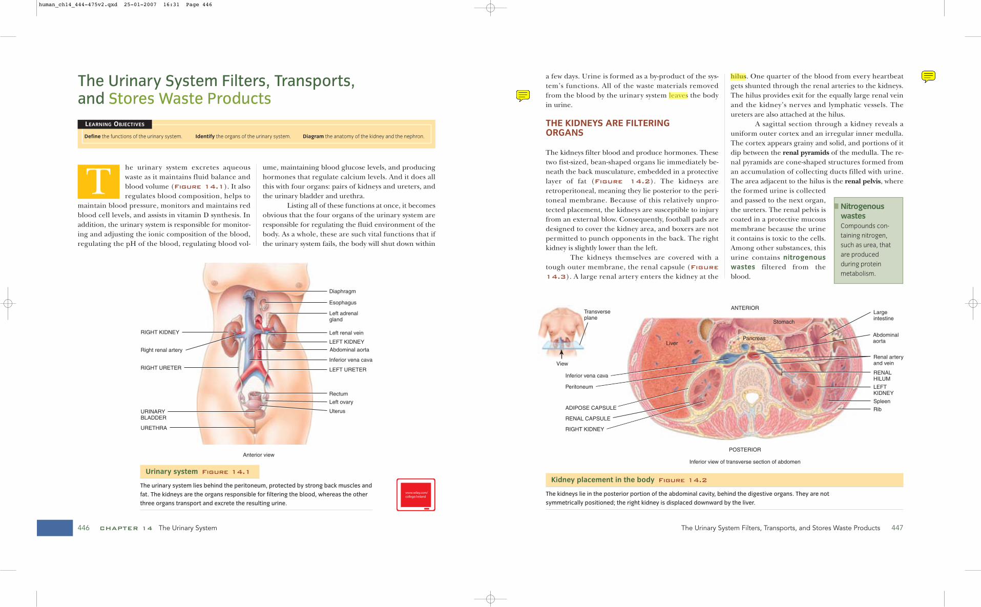
he urinary system excretes aqueouswaste as it maintains fluid balance andblood volume (Figure 14.1). It alsoregulates blood composition, helps to
maintain blood pressure, monitors and maintains redblood cell levels, and assists in vitamin D synthesis. Inaddition, the urinary system is responsible for monitor-ing and adjusting the ionic composition of the blood,regulating the pH of the blood, regulating blood vol-
a few days. Urine is formed as a by-product of the sys-tem’s functions. All of the waste materials removedfrom the blood by the urinary system leaves the bodyin urine.
THE KIDNEYS ARE FILTERING ORGANS
The kidneys filter blood and produce hormones. Thesetwo fist-sized, bean-shaped organs lie immediately be-neath the back musculature, embedded in a protectivelayer of fat (Figure 14.2). The kidneys areretroperitoneal, meaning they lie posterior to the peri-toneal membrane. Because of this relatively unpro-tected placement, the kidneys are susceptible to injuryfrom an external blow. Consequently, football pads aredesigned to cover the kidney area, and boxers are notpermitted to punch opponents in the back. The rightkidney is slightly lower than the left.
The kidneys themselves are covered with atough outer membrane, the renal capsule (Figure
14.3). A large renal artery enters the kidney at the
Tume, maintaining blood glucose levels, and producinghormones that regulate calcium levels. And it does allthis with four organs: pairs of kidneys and ureters, andthe urinary bladder and urethra.
Listing all of these functions at once, it becomesobvious that the four organs of the urinary system areresponsible for regulating the fluid environment of thebody. As a whole, these are such vital functions that ifthe urinary system fails, the body will shut down within
Diaphragm
Esophagus
Left adrenal gland
Abdominal aorta
Inferior vena cava
Rectum
RIGHT KIDNEY
Right renal artery
RIGHT URETER LEFT URETER
LEFT KIDNEY
Left renal vein
URINARY BLADDER
URETHRA
Left ovary
Uterus
Anterior view
LEARNING OBJECTIVES
Define the functions of the urinary system. Identify the organs of the urinary system. Diagram the anatomy of the kidney and the nephron.
Liver
ANTERIOR
POSTERIOR
Inferior view of transverse section of abdomen
Rib
RIGHT KIDNEY
Spleen
RENAL CAPSULE
LEFT KIDNEY
ADIPOSE CAPSULE
Renal arteryand vein
Peritoneum
Inferior vena cava
Abdominalaorta
Largeintestine
Transverseplane
Pancreas
Stomach
RENAL HILUM
View
Kidney placement in the body Figure 14.2
The kidneys lie in the posterior portion of the abdominal cavity, behind the digestive organs. They are not
symmetrically positioned; the right kidney is displaced downward by the liver.
hilus. One quarter of the blood from every heartbeatgets shunted through the renal arteries to the kidneys.The hilus provides exit for the equally large renal veinand the kidney’s nerves and lymphatic vessels. Theureters are also attached at the hilus.
A sagittal section through a kidney reveals auniform outer cortex and an irregular inner medulla.The cortex appears grainy and solid, and portions of itdip between the renal pyramids of the medulla. The re-nal pyramids are cone-shaped structures formed froman accumulation of collecting ducts filled with urine.The area adjacent to the hilus is the renal pelvis, wherethe formed urine is collectedand passed to the next organ,the ureters. The renal pelvis iscoated in a protective mucousmembrane because the urineit contains is toxic to the cells.Among other substances, thisurine contains nitrogenouswastes filtered from theblood.
NitrogenouswastesCompounds con-
taining nitrogen,
such as urea, that
are produced
during protein
metabolism.
Urinary system Figure 14.1
The urinary system lies behind the peritoneum, protected by strong back muscles and
fat. The kidneys are the organs responsible for filtering the blood, whereas the other
three organs transport and excrete the resulting urine.
www.wiley.com/college/ireland
human_ch14_444-475v2.qxd 25-01-2007 16:31 Page 446
helen walden
Note
leaves should be leave (singular) (EA)
helen walden
Highlight
helen walden
Highlight
helen walden
Note
Hilus should be a key term, or else remove it from the list at the end of the chapter.

The Urinary System Filters, Transports, and Stores Waste Products 449448 CHAPTER 14 The Urinary System
The kidneys are com-posed of millions ofnephrons, packed togetherunder the renal capsule (Fig-
ure 14.4). The large bloodsupply that enters the kidneysis diverted through smallerand smaller arteries and arte-rioles until it winds its way to aknotted vessel at the begin-ning of each nephron. Theblood vessel leaving eachnephron then breaks into per-itubular capillaries, which
wind around the entire nephron before being collectedinto venules. The venous system of the kidneys followsthe same route as the arterial system, eventually leavingthe kidney in the large renal veins. At the nephron, theblood is filtered, and the necessary ions and nutrientsare returned to the circulatory system. The waste mate-rial remains in the fluid within the tubules of thenephron.
Beyond cleaning blood, the kidneys also pro-duce the hormones calcitriol and erythropoietin, whichregulate the concentration of calcium and formed ele-ments in blood. Calcitriol, the active form of vitamin D,helps maintain blood calcium levels. Erythropoietinstimulates production of new red blood cells.
Kidney anatomy and kidney blood flow Figure 14.4
Pro
cess
Dia
gra
m
Blood supply of the nephron
Renal vein
Interlobar veins
Arcuate veins
Interlobular veins
Peritubular capillaries
Efferent arterioles
Glomerular capillaries
Afferent arterioles
Interlobular arteries
Arcuate arteries
Interlobar arteries
Segmental arteries
Renal artery
Interlobularartery
Arcuateartery
Interlobarartery
Segmental artery
Renal artery
Renal vein
Frontal section of right kidney
Renal capsule
Renal cortex
Renal pyramidin renal medulla
Interlobar vein
Arcuate vein
Interlobular vein
Frontalplane
Path of blood flow
Efferentarteriole
Peritubularcapillary
Afferentarteriole
Glomerulus
Interlobularvein
Vasa recta
A B
Blood flow through the kidneys must be highly regulated.
A full quarter of every heartbeat is filtered through the
kidneys rather than sent to body tissues. ●1 Blood enters
the kidneys via the renal artery. ●2 The renal artery
branches into the segmental arteries that supply each renal
pyramid of the kidneys. ●3 Segmental arteries give rise to
the interlobar arteries that dive between renal pyramids.
●4 These arteries then loop over the renal pyramids in
arcuate arteries. ●5 The interlobular arteries take the blood
to the renal cortex, where ●6 it is further divided into
afferent arterioles leading to the glomerular capillaries.
●7 Filtered blood leaves the glomerulus through the
efferent arteriole, where it moves to the peritubular
capillaries and is then collected by the ●8 interlobular
veins. From here, the pathway is a reverse of the arterial
flow, moving consecutively through ●9 arcuate veins,
●10 interlobar veins, and finally ●11 leaving the kidneys via
the renal vein.
Renal cortex
Renal medulla
Renal column
Renal pyramidin renal medulla
Renal sinus
Renal papilla
Fat in renal sinus
Renal capsule
Renal hilum
Nephron
Path of urine drainage:
Papillary duct in renal pyramid
Minor calyx
Major calyx
Renal pelvisRenal vein
Renal artery
Ureter
Urinary bladder
Frontal section of right kidney
Collecting duct
Internal anatomy of the kidney Figure 14.3
The renal cortex houses a large blood supply, and it is here that filtration occurs. The renal medulla is
involved in the fine tuning of this filtrate, and the renal pelvis transports the final waste product,
the urine, from the kidneys.
Peritubular capillaries(peri � around;
tubular � nephron
tubules)
Capillaries that
surround
the nephron.
NephronThe filtering unit of
the kidney.
www.wiley.com/college/ireland
human_ch14_444-475v2.qxd 25-01-2007 16:31 Page 448
helen walden
Highlight
helen walden
Note
Some ambiguity as to what this sentence means?
Urine is Formed Through Filtration and Osmosis 451450 CHAPTER 14 The Urinary System
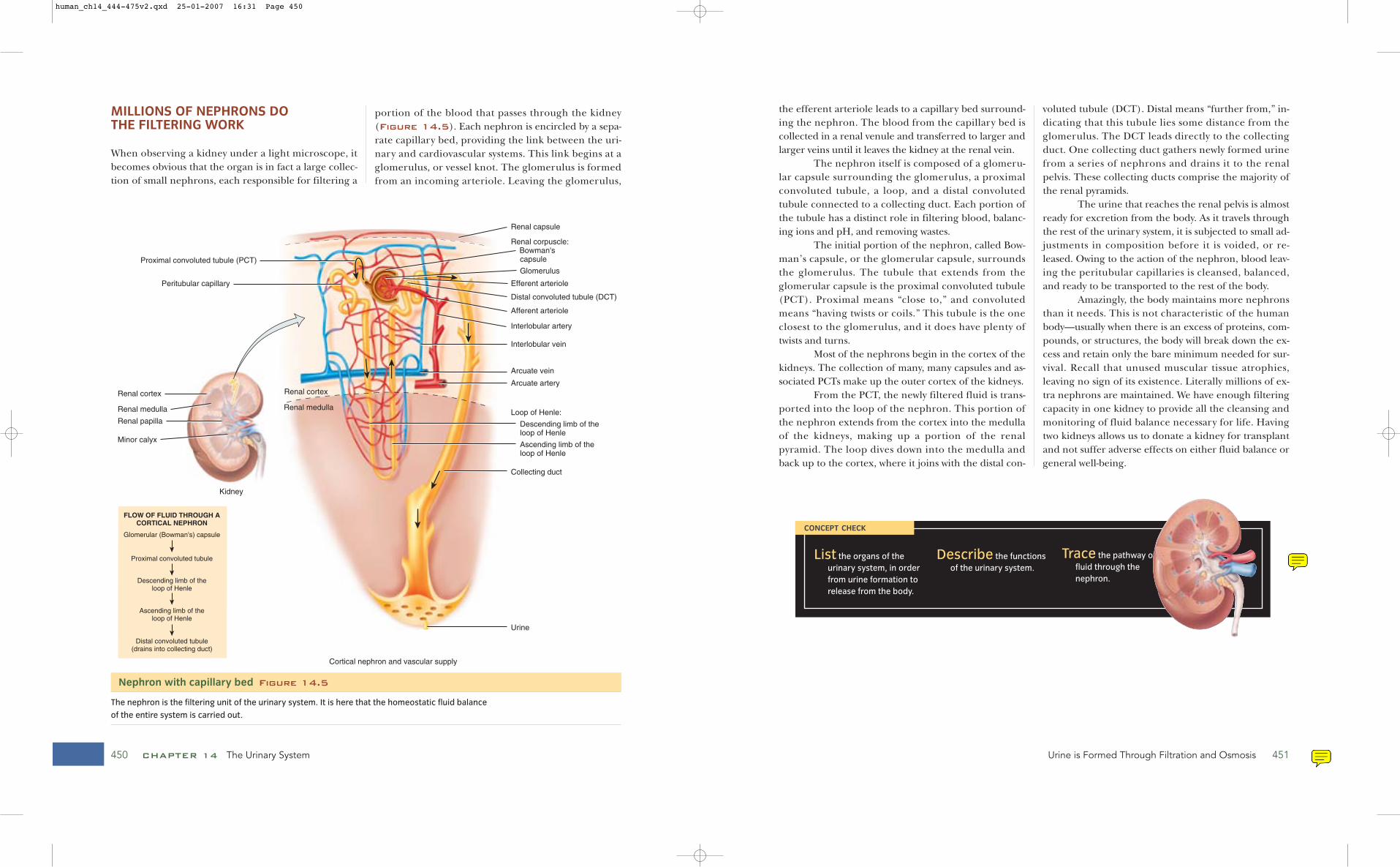
MILLIONS OF NEPHRONS DO THE FILTERING WORK
When observing a kidney under a light microscope, itbecomes obvious that the organ is in fact a large collec-tion of small nephrons, each responsible for filtering a
Minor calyx
Cortical nephron and vascular supply
Kidney
Renal cortex
Renal medulla
Renal papilla
Renal cortex
Renal medulla
Renal capsule
Renal corpuscle: Bowman's
capsule
Glomerulus
Efferent arteriole
Distal convoluted tubule (DCT)
Proximal convoluted tubule (PCT)
Afferent arteriole
Interlobular artery
Interlobular vein
Arcuate vein
Arcuate artery
Loop of Henle:
Descending limb of theloop of Henle
Ascending limb of theloop of Henle
Collecting duct
Urine
Peritubular capillary
Distal convoluted tubule(drains into collecting duct)
FLOW OF FLUID THROUGH ACORTICAL NEPHRON
Glomerular (Bowman's) capsule
Proximal convoluted tubule
Descending limb of theloop of Henle
Ascending limb of theloop of Henle
Nephron with capillary bed Figure 14.5
The nephron is the filtering unit of the urinary system. It is here that the homeostatic fluid balance
of the entire system is carried out.
the efferent arteriole leads to a capillary bed surround-ing the nephron. The blood from the capillary bed iscollected in a renal venule and transferred to larger andlarger veins until it leaves the kidney at the renal vein.
The nephron itself is composed of a glomeru-lar capsule surrounding the glomerulus, a proximalconvoluted tubule, a loop, and a distal convolutedtubule connected to a collecting duct. Each portion ofthe tubule has a distinct role in filtering blood, balanc-ing ions and pH, and removing wastes.
The initial portion of the nephron, called Bow-man’s capsule, or the glomerular capsule, surroundsthe glomerulus. The tubule that extends from theglomerular capsule is the proximal convoluted tubule(PCT). Proximal means “close to,” and convolutedmeans “having twists or coils.” This tubule is the oneclosest to the glomerulus, and it does have plenty oftwists and turns.
Most of the nephrons begin in the cortex of thekidneys. The collection of many, many capsules and as-sociated PCTs make up the outer cortex of the kidneys.
From the PCT, the newly filtered fluid is trans-ported into the loop of the nephron. This portion ofthe nephron extends from the cortex into the medullaof the kidneys, making up a portion of the renalpyramid. The loop dives down into the medulla andback up to the cortex, where it joins with the distal con-
voluted tubule (DCT). Distal means “further from,” in-dicating that this tubule lies some distance from theglomerulus. The DCT leads directly to the collectingduct. One collecting duct gathers newly formed urinefrom a series of nephrons and drains it to the renalpelvis. These collecting ducts comprise the majority ofthe renal pyramids.
The urine that reaches the renal pelvis is almostready for excretion from the body. As it travels throughthe rest of the urinary system, it is subjected to small ad-justments in composition before it is voided, or re-leased. Owing to the action of the nephron, blood leav-ing the peritubular capillaries is cleansed, balanced,and ready to be transported to the rest of the body.
Amazingly, the body maintains more nephronsthan it needs. This is not characteristic of the humanbody—usually when there is an excess of proteins, com-pounds, or structures, the body will break down the ex-cess and retain only the bare minimum needed for sur-vival. Recall that unused muscular tissue atrophies,leaving no sign of its existence. Literally millions of ex-tra nephrons are maintained. We have enough filteringcapacity in one kidney to provide all the cleansing andmonitoring of fluid balance necessary for life. Havingtwo kidneys allows us to donate a kidney for transplantand not suffer adverse effects on either fluid balance orgeneral well-being.
CONCEPT CHECK
List the organs of theurinary system, in orderfrom urine formation torelease from the body.
Describe the functionsof the urinary system.
Trace the pathway offluid through thenephron.
portion of the blood that passes through the kidney(Figure 14.5). Each nephron is encircled by a sepa-rate capillary bed, providing the link between the uri-nary and cardiovascular systems. This link begins at aglomerulus, or vessel knot. The glomerulus is formedfrom an incoming arteriole. Leaving the glomerulus,
human_ch14_444-475v2.qxd 25-01-2007 16:31 Page 450
helen walden
Note
is should be Is
helen walden
Note
Move box a bit left, and move art a bit right to clear text (PE)

Urine is Formed Through Filtration and Osmosis 453452 CHAPTER 14 The Urinary System
NET FILTRATION PRESSURE (NFP) = GBHP – CHP – BCOP = 55 mmHg – 15 mmHg – 30 mmHg = 10 mmHg
Proximal convoluted tubule
The now thicker blood pulls water backinto it from the more watery filtrate inthe nephron. The net filtration pressureis the sum of these three forces, #1 pushing fluid into the nephron, while #s 2and 3 pull water back from the nephron
The fluid already in the nepron exertsan opposite force, a back pressure preventinga huge influx of filtrate
The force of the blood in theglomerular vessel causes fluidto leak into the nephron
Capsularspace
Bowman'scapsule
Efferent arteriole
Afferent arteriole
1 2
3
systolic blood pressure. This increase is partially causedby the kinking and twisting of the glomerular vessels.You have experienced this in your garden hose if youhave ever bent it. The pressure increases because thewater must travel past the obstructions. A similar phe-nomenon occurs in the glomerular vessels. In addition,the incoming (afferent) arterioles have a larger diame-ter than the outgoing (efferent) glomerular arterioles.This increases pressure in the glomerulus by creatingback pressure. The total pressure on the blood forcesmost of the fluid into the capsule. In order to actuallyfilter the blood, the blood pressure has to overcomethe pressure of the fluid already in the capsule (capsu-lar pressure) as well as the osmotic pressure of theblood itself.
Because the glomerular system relies on pres-sure, there is a lower limit to its functioning (Figure
14.6). If your systolic pressure drops below 60 mil-limeters Hg, blood in the glomerulus will not beforced through the glomerular wall because glomeru-lar pressure will not rise high enough to force plasmafrom the blood vessels. This leads to serious complica-tions because the aqueous portion of the blood can-
not filter into the nephron and therefore cannot becleansed.
During filtration, the formed elements andplasma proteins remain in the glomerular vessel be-cause they are too large to pass through the fenestra-tions of the cells that line the glomerulus (Figure
14.7). The proteins left in the blood of the capillaryare essential because they set up the osmotic gradientthat later pulls most of the water from the filtrate backinto the blood. Every day, approximately 180 liters offluid are filtered from the blood, but only a small frac-tion of that is excreted. Imagine how different lifewould be if we lost 180 liters of fluid every day! That isequal to 60 times the total plasma volume of the body.Not only would we have todrink constantly, but we wouldmost likely also have a differ-ent social custom surroundingthe need to urinate, because itwould occur almost constantly.In the body as in the bios-phere, recycling makes a realdifference.
Parietal layer of glomerular (Bowman’s) capsule
Capsular space
Proximal convoluted tubule
Podocyte (Bowman’s) capsule
Afferent arteriole
Efferent arteriole
Ascending limbof loop of Henle
Endothelium of glomerulus
Renal corpuscle (internal view)
Renal corpuscle(external view)
Renal corpuscle histology Figure 14.7
The blood is forced through the walls of the glomerulus via increased blood pressure. Water, ions, nutrients, and waste
materials pass through the fenestrations of the podocytes (specialized cells comprising the glomerular walls).
FenestrationsWindows or open-
ings between cells
in the lining of the
glomerulus.
Urine is Formed Through Filtration and OsmosisLEARNING OBJECTIVES
Define glomerular filtration.
Explain the functions of the PCT, loop, and DCT.
rine formation begins in the glomerulusand is finalized in the renal pelvis,through the processes of filtration, ac-tive transport, and osmosis. As blood
passes through the glomerulus of the nephron, most ofthe liquid is forced out of the arteriole and into the lu-
U
men of the nephron. This is glomerular filtration. Thenephron capsule completely surrounds the glomeru-lus, looking like a sock balled up around the blood ves-sel. Water, nitrogenous wastes, nutrients, and salts areall forced from the blood at this point.
LIQUID IS FORCED INTO THE LUMEN OF THE GLOMERULUS
To understand how this filtration occurs, it may help toreview the material on osmosis and pressure fromChapter 3. Glomerular blood pressure is higher than
www.wiley.com/college/ireland
Glomerular pressure Figure 14.6
Pro
cess Diag
ram
There are three situations that must be met in order to filter
the constituents of the blood plasma through the
glomerulus. ●1 Blood pressure must be high enough to
force plasma out of the glomerular vessel walls; ●2 the fluid
already in the glomerulus must have a low enough presure
that more fluid can be forced in to the nephron tubules; and
●3 the osmotic pressure of the blood in the peritubular
capillaries must be high enough to draw water back into the
capillaries from the nephron tubule. If these conditions are
not met, the neprhon cannot filter the blood, and the urinary
system will fail.
human_ch14_444-475v2.qxd 25-01-2007 16:31 Page 452
helen walden
Note
Cap I in is
helen walden
Note
Here's mmHg closed up again, in the art (note that it was mm Hg in previous chapters in the text)
helen walden
Highlight
helen walden
Highlight
helen walden
Highlight
helen walden
Note
nephron (EA)
helen walden
Note
pressure (EA)
helen walden
Note
into (EA)
helen walden
Note
is should be Is

TUBULAR REABSORPTION RECYCLESWATER TO THE BLOOD
As filtrate passes through the nephron, ions and waterare returned to the peritubular capillaries in a processcalled tubular reabsorption. Approximately 80% of thefiltered water is returned to the blood immediately atthe PCT. Glucose, amino acids, and salts are also re-turned to the bloodstream. The walls of the proximalconvoluted tubule have a large surface area to accom-
modate all this reabsorption.The cells that line the PCT arecovered with microvilli. Thesecells are adjacent to the en-dothelial cells of the peritubu-lar capillaries, creating a thinlayer that allows diffusion fromthe tubule to the blood.
Essential ions and waterare sent back to the blood viaosmosis and diffusion (Figure
14.8). Glucose is sent back tothe bloodstream using facili-tated diffusion. The walls ofthe PCT have a finite number of
glucose receptors to pick up glucose from the filtrate.Normally, there are enough receptors to remove all theglucose from the filtrate and return it to the blood. Butif there is too much glucose in the blood, it will over-run these receptors and drop from the PCT into theloop of the nephron. Once glucose moves beyond thePCT, it cannot be returned to the bloodstream. It is saidto “spill” into the urine because it literally spills into theloop. One symptom of diabetes mellitus is glucosespilling as a result of very highlevels of blood glucose in theoriginal filtrate.
Waste products and otherunwanted substances too large tofilter out of the blood at theglomerulus, such as steroids anddrug breakdown products, areactively secreted into the filtrate at the distal convo-luted tubule (Figure
14.9).
454 CHAPTER 14 The Urinary System Urine is Formed Through Filtration and Osmosis 455
Health
, Welln
ess, and
Disease
How much water should I drink?
Water. It’s just about the most popular nutrient around,
and these days, hydration is almost a personal virtue. Ad-
equate hydration is necessary for the functioning of the
kidneys and most other body systems. Water maintains
the blood’s volume, helping it transport nutrients and re-
move waste. When water is scarce, homeostasis is threat-
ened, and especially so during illness or physical exer-
tion.
Hydration, combined with adequate sodium levels in
the interstitial fluids, helps keep the osmolarity the same
on both sides of the cell membrane. Underhydration, if
not corrected by the urinary system through mechanisms
discussed in the main text, can cause water to osmose
from the cells into interstitial fluid. Overhydration, some-
times called “water intoxication,” can cause water to os-
mose into cells, creating swelling, or edema.
So how much water should we drink? Historically, the
standard advice was to drink eight 8-ounce glasses of wa-
ter each day. Oddly, this widely trumpeted health advice
did not have a scientific basis. In 2004, the National Acad-
emy of Sciences (NAS) came up with some scientifically
driven drinking advice. Instead of performing experi-
ments, the academy’s experts concluded that healthy
people who have access to food and water are almost
never chronically dehydrated. So they set the water re-
quirement by measuring the water intake of healthy, well-
hydrated people.
The NAS researchers found that each day, healthy
women in the United States consume about 2.7 liters of
water from food and beverages, whereas men consume
about 3.7 liters. About 80 percent of that amount comes
from water and other beverages, and the rest from mois-
ture in food. In general, the NAS experts said, hydration
is not something to worry about because “thirst provides
the body feedback that we are getting dehydrated, so
that we can consume more fluids.”
If this recommendation is accurate, the 8 by 8 advice
(1.9 liters) seriously understated the need for water. Ac-
cording to the NAS recommendations, a man needs to in-
gest 3.0 liters of fluids (not counting moisture from food)
to stay hydrated.
In hot, dry conditions, or during strenuous exercise,
fluid consumption must increase. And that raises one fi-
nal question: When is water a curse rather than a cure? In
rare situations, athletes have died from hyponatremia, a
low blood sodium concentration, after drinking too much
water during long endurance competitions. The condi-
tion resembles water intoxication: Cells swell and can
burst, as water responds to the higher sodium concentra-
tion by undergoing osmosis, moving from interstitial fluid
into the cells. So instead of guzzling gallons of water dur-
ing athletic events, it may be better to swill sports drinks,
which contain sodium and other electrolytes, or even eat
some potato chips along with your water if possible.
K+
H2O H2O
Peritubular capillaryFluidin tubulelumen
Proximalconvolutedtubule cell
Mg2+Ca2+
Diffusion
Osmosis
Cl–
Ca2+
Urea
K+
Mg2+
Urea
Cl–
PCT function Figure 14.8
Reabsorption of water, bicarbonate ions, organic solutes
including glucose and amino acids, and important ions such as
sodium, calcium, and potassium, occur in the PCT.
DCT functioning Figure 14.9
The cells surrounding the DCT are especially susceptible to
hormonal controls, allowing a final adjustment of blood
composition.
SecretedMoved from the
blood to the filtrate,
using energy.
Facilitated diffusion Moving
substances from
high concentration
to low with the
assistance of a
carrier molecule.
MicrovilliSmall hair-like
projections
extending from
the free surface of
epithelial cells.
Diffusion
Sodium-potassium pump
Key:
Leakage channels
Peritubular capillary
Drug breakdownproducts
Fluidin tubulelumen
Principalcell
Na+
K+
Na+
Interstitialfluid
Na+
Na+
K+K+
ADP
ATP
human_ch14_444-475v2.qxd 25-01-2007 16:31 Page 454
helen walden
Note
is should be Is

Urine is Formed Through Filtration and Osmosis 457
This process requires ATP, and provides delicate con-trol over fluid homeostasis. Tubular secretion providesa final fine-tuning of the dissolved compounds in theblood. It is this process that provides clues as to theamount and type of drugs that are traveling throughthe body at any one time. Most of the breakdown prod-ucts of drugs, both pharmaceutical and recreational,are large and must be secreted into the nephron. TheEthics and Issues box in the last section of the chapterdiscusses this in detail.
The loop of the nephron and the collectingduct help remove even more water from the filtrate,serving to precisely regulate fluid loss (Figures
14.10 and 14.11). Interestingly, the descendingarm of the loop of the nephron is permeable to water,but the ascending limb is not. Therefore, water leavesthe filtrate as it moves down the loop of the nephron,and salts leave the filtrate as it flows up the ascendingarm, creating a salt gradient inthe medulla of the kidney. This isreferred to as countercurrentmultiplication, or CCM, becausethe ascending and descendingloop flow opposite one another.Each current affects the waterand salt concentration in theother. The collecting ducts passright through the salt gradient setup by the CCM, providing one lastopportunity to remove water fromthe urine before sending it on tothe ureters. Water is a vital fluid and as such must becarefully monitored. The Health Wellness and Diseasebox, “How much water should I drink?” takes a look atthe role of water in personal health.
456 CHAPTER 14 The Urinary System
2Cl–
Vasa rectaThick ascendinglimb cell
K+
K+
Ca2+
Mg2+
Na+
2Cl–
Interstitial fluid is morenegative than fluid intubule lumen
Cations
2Cl–
Fluidin tubulelumen
2Cl–
K+
Na+
Na+
Apical membrane(impermeableto water)
Cations:
ADP
Na+
Na+
Na+
Na+– K+– 2Cl– symporter
Leakage channels
Sodium-potassium pump
Diffusion
Key:
ATP
Loop functioning Figure 14.10
The countercurrent multiplication activity of the nephron loop
can be easily seen in this diagram. Because of this action, the
medulla of the kidney is a very salty area.
CONCEPT CHECK
List two ways that bloodpressure is increased inthe glomerulus.
What is the mainfunction of the PCT?
Countercurrentmultiplication(CCM)Increasing the
diffusion rate by
flowing solutions in
opposite directions
on either side of
the diffusion
membrane.
Urine formation Figure 14.11
Urine formation begins in the glomerulus,
where blood plasma is filtered and collected by
the renal corpuscle. At the PCT, most of the
water and many ions and nutrients are re-
absorbed by the blood. The loop serves to
remove more water and ions, setting up a salt
gradient in the medulla of the kidney. The distal
convoluted tubule allows for final adjustments
in the composition of urine and blood by
actively secreting larger substances from the
blood to the urine.
How does the DCT helpmonitor and adjust bloodcomposition?
DISTAL CONVOLUTED TUBULE
Reabsorption (into blood) of:
Water 10–15% (osmosis)
Na+ 5%
CI– 5%
Ca2+ variable
LAST PART OF DISTAL TUBULEAND COLLECTING DUCT
Reabsorption (into blood) of:
Water 5–9% (insertion of water channels stimulated by ADH)
Na+ 1–4%
Urea variable
Secretion (into urine) of:
K+ variable amount to adjust for dietary intake (leakage channels)
Tubular fluid leaving the collecting duct is dilutewhen ADH level is low and concentrated whenADH level is high.
RENAL CORPUSCLE
Glomerular filtration rate: 105–125 mL/min
Filtered substances: water and all solutes present in blood (except proteins) including ions, glucose, amino acids, creatinine
HCO3– variable amount
H+ variable amounts to maintain acid–base homeostasis (H+ pumps)
PROXIMAL CONVOLUTED TUBULE
Reabsorption (into blood) of filtered:
Water 65% (osmosis)
Na+ 65%
K+ 65%
Glucose 100%
Amino acids 100%
CI– 50%
HCO3– 80–90%
Urea 50%
Secretion (into urine) of:
H+ variable
Ca2+, Mg2+ variable
Ammonia variable
Urea variable
Creatinine small amount
LOOP OF HENLE
Reabsorption (into blood) of:
Water 15% (osmosis in descending limb)
Na+ 20–30% (ascending limb)
K+ 20–30% (ascending limb)
CI– 35% (ascending limb)
HCO3– 10–20%
Secretion (into urine) of:
Urea variable
Ca2+, Mg2+ variable
Urine
human_ch14_444-475v2.qxd 25-01-2007 16:31 Page 456
helen walden
Note
Insert commas after Health and Wellness (EA)
helen walden
Note
is should be Is
helen walden
Note
Center box horizontally

THE URINARY BLADDER STORES URINEBEFORE RELEASE
The urinary bladder is a hollow, variable-sized organ(Figure 14.14). It lies in the pelvic cavity, posteriorto the pubic bones and the pubic symphysis. The baseof the bladder has a triangular area where the twoureters enter and the urethra exits. This area is calledthe trigone. The bladder is lined with transitional ep-ithelium to allow for expansion without tearing or
nce the filtrate has passed through thenephron and collecting ducts, andreaches the renal pelvis, it is referred toas urine. Most of the fine-tuning of ion
concentration and water content is completed by thispoint (Table 14.1). Water can still be removed asthe urine sits in the remainder of the organs of the uri-nary system, but the salt content is relatively stable.
While in the renal pelvis, water can continue toleave the urine, concentrating the salts in the urine.This can lead to the formation of kidney stones. Theserock-like masses, usually composed of calcium oxalate,can grow large enough to block renal flow (Figure
14.12). Kidney stones are extremely painful as theymove through the urinarypelvis and can become lodgedin the kidney or the ureters.Some kidney stones are jaggedor pointy, making them evenmore likely to jam in the sys-tem. Removal of kidney stonesrarely requires medical assis-tance. Drinking lots of waterand resting as the stone moves
LEARNING OBJECTIVES
Outline the functions of the bladder and the urethra. Understand why females are more susceptible to urinary tract infections.
Urine is Transported to the Bladder for Storage
458 CHAPTER 14 The Urinary System Urine is Transported to the Bladder for Storage 459
Filtered* Reabsorbed Substance (enters renal tubule) (returned to blood) Excreted in Urine
Water 180 liters 178–179 liters 1–2 liters
Chloride ions (CI�) 640 g 633.7 g 6.3 g
Sodium ions (Na�) 579 g 575 g 4 g
Bicarbonate ions (HCO3�) 275 g 274.97 g 0–0.03 g
Glucose 162 g 162 g 0
Urea 54 g 24 g 30 g†
Potassium ions (K�) 29.6 g 29.6 g 2.0 g‡
Othrough the renal pelvis and the ureter often do thetrick, but some stones are too large to pass. These maybe broken apart by ultrasound waves, to allow the frag-ments to be excreted. Because kidney stones often reap-pear, patients are asked to avoid foods high in calcium,eat less protein (to decrease urine acidity), and drinkmore fluids, especially water.
Calcium oxalateA chemical
compound com-
posed of calcium
ions bound to
the oxalate ion
(C2O42�).
* Assuming glomerular filtration is 180 liters per day.† In addition to being filtered and reabsorbed, urea is secreted.‡ After virtually all filtered K� is reabsorbed in the convoluted tubules and loop of Henle, a variable amount of K� is secreted in the collecting duct.
Ureters
Bladder
Bladder Figure 14.14
Transitional epithelium lines the walls of this distensible organ.
These cells also secrete mucus to protect the bladder from the
toxic compounds in the urine.
Kidney stone Figure 14.12
A typical small kidney stone is seen here on the tip of a finger.
Stones can be as large as a pearl or even, rarely, a golf ball.
Ureters Figure 14.13
Ureters carry urine from the renal pelvis to the urinary bladder.
They are approximately 20 centimeters long, and curl behind
the urinary bladder to enter from the trigone, or base of the
bladder. These tubes are ringed with smooth muscle, which
helps propel the urine to the bladder.
From the renal pelvis, urine travels down theureter to the urinary bladder (Figure 14.13). Theureters are approximately 20-centimeter-long, thinmuscular tubes lined with mucosa. The ureters loop be-hind the urinary bladder and enter it at the base. Thisallows the bladder to expand upward without dislodg-ing the ureters.
With every heartbeat, blood is pushed into theglomerulus and filtered. The nephrons constantly formurine, so the tubes and ducts of the urinary system arealways full of fluid. As more urine is produced, it pusheswhat is already formed down the ureters and into thebladder, where small contractions move the urine to-ward the bladder.
Substances Filtered, Reabsorbed, and Excreted in Urine per Day Table 14.1
human_ch14_444-475v2.qxd 25-01-2007 16:32 Page 458
helen walden
Note
is should be Is
helen walden
Note
Table title should be initial cap only
helen walden
Note
is should be Is

destroying the integrity of the inner lining. The emptybladder is the size of a walnut, but can stretch to holdup to 800 ml of fluid in males, and slightly less in fe-males.
Discharging urine from the bladder is called uri-nating, voiding, or micturition. This reflex involves bothsmooth and skeletal muscles. Urine is constantly beingformed and drained into the bladder. When the bladdercontains approximately 300 ml, pressure in the bladder
stimulates stretch receptorsthat send nerve impulses to themicturition center. The mic-turition reflex causes contrac-tion of the walls of the bladderand relaxation of the internalurethral sphincter muscle.Urine moves down into theurethra, pressing on the exter-nal sphincter muscle. At thispoint, you can consciously con-trol the opening of the exter-nal urinary sphincter. Shouldyou choose not to void the con-tents of the bladder, the urgeto urinate will subside until thenext 300 ml is collected in the
bladder.As we mature, we learn to anticipate and con-
trol this reflex, but we cannot delay micturition indefi-nitely. The bladder continues to expand, and a secondreflex will begin shortly. Just as we are not able to holdour breath until we die, we cannot retain urine untilthe bladder bursts. When the bladder reaches 700 to800 ml, micturition occurs despite our best efforts tocontrol the external urethral sphincter.
THE URETHRA TRANSPORTS URINE OUT OF THE BODY
When micturition occurs, the urine leaves the body viathe urethra, a single tube extending from the trigoneof the bladder to the exterior. In females, the urethra isa short 5 centimeters, emptying in front of the vaginal
460 CHAPTER 14 The Urinary System
INCONTINENCE IS THE LOSS OF CONTROL OVER VOIDING
As we age, many things change, including our ability tocontrol micturition when the urge arises. Incontinencecan and does occur in all age brackets, genders, and so-cial levels, but it is far more common in elderly women.Perhaps the stress of bearing children weakens the mus-cles of the pelvic floor, leading to greater difficulty con-trolling these muscles in later years.
An estimated 12 million Americans suffer in-continence, and most do not require surgery. Inconti-
nence can be a symptom ofmany different pathologies butis not a pathology in its ownright. Causes include chronicurinary tract infections, side ef-fects of medication, muscularweakness, an enlarged prostategland in males, constipation, orneuromuscular disease. There
Sagittal section showing the female urethra
Rectum
Vagina
Urinary bladderPubic symphysis
Urethra
External urethralorifice
Uterus
Sagittalplane
A
RectumProstaticurethra
Membranousurethra
TestisScrotum
Urinary bladder
Pubic symphysis
Deep muscles ofperineum
Spongy urethraPenis
Prostate
External urethral orifice
Sagittalplane
Sagittal section showing the male urethraB
Comparison between female and male urethrasFigure 14.15
Internal urethral sphincterRing of involuntary
smooth muscle that
keeps the urethra
closed.
External sphincter muscleRing of voluntary
skeletal muscle that
closes the urethra.opening. The male urethra is almost four times longerbecause it runs the length of the penis (Figure
14.15). The urinary and reproductive systems join inthe male, sharing the male urethra. In the female, thetwo systems are separate. The female urethra carriesonly urine, and the female reproductive tract opens atthe vagina.
Because the distance from the exterior to thebladder is shorter in females, they suffer far more uri-nary tract infections (UTIs). Bacteria outside the bodycan travel the short distance up the urethra and colo-nize the bladder, resulting in painful urination oftenaccompanied by bleeding from the irritated bladderwalls. (If the urine contains glucose, the bacteria multi-ply even faster.) UTIs are serious infections that mustbe cleared up. If the bacteria are allowed to remain inthe bladder, they will eventually travel up the uretersand colonize the pelvis and tubules of the kidney. Kid-ney infections are painful and serious because theyblock normal kidney function and can lead to kidneyfailure.
are three types of incontinence determined by the un-derlying cause of the problem, each with the same re-sult. Stress incontinence is the leaking of urine duringphysical exertion. Urge incontinence is the inability toquell the urge to urinate. Overflow incontinence is theoverflowing of the urinary bladder caused by waitingtoo long before urinating, as happens in young chil-dren who are learning to control their sphincter mus-cles. Treatment for incontinence is tailored to thecause. Muscular strengthening exercises or behavioralmodification may be recommended.
IncontinenceThe inability to
prevent urine
leakage.
Constipationdifficult or
infrequent
defecation, leading
to dry, potentially
painful fecal
evacuation
CONCEPT CHECK
What is the micturitionreflex?
How does the urethra differin males and females?
The Urinary System Maintains the Body’s Water–Salt Balance 461
human_ch14_444-475v2.qxd 25-01-2007 16:32 Page 460
helen walden
Note
Wrong rf: should be Urine is Transported to the Bladder for Storage (PE)
462 CHAPTER 14 The Urinary System The Urinary System Maintains the Body’s Water–Salt Balance 463
Bowman's capsuleAfferentarteriole
Efferentarteriole
Loop of Henle
Papillaryduct
Collectingduct
Glomerulus
Proximalconvoluted
tubule
Diluteurine
Interstitialfluid inrenalmedulla
Interstitialfluid inrenalcortex
100
90
65900
550750
350300
Distal convolutedtubule
150
550 350 80
300
70
750
350
550
750
350
550
300
65
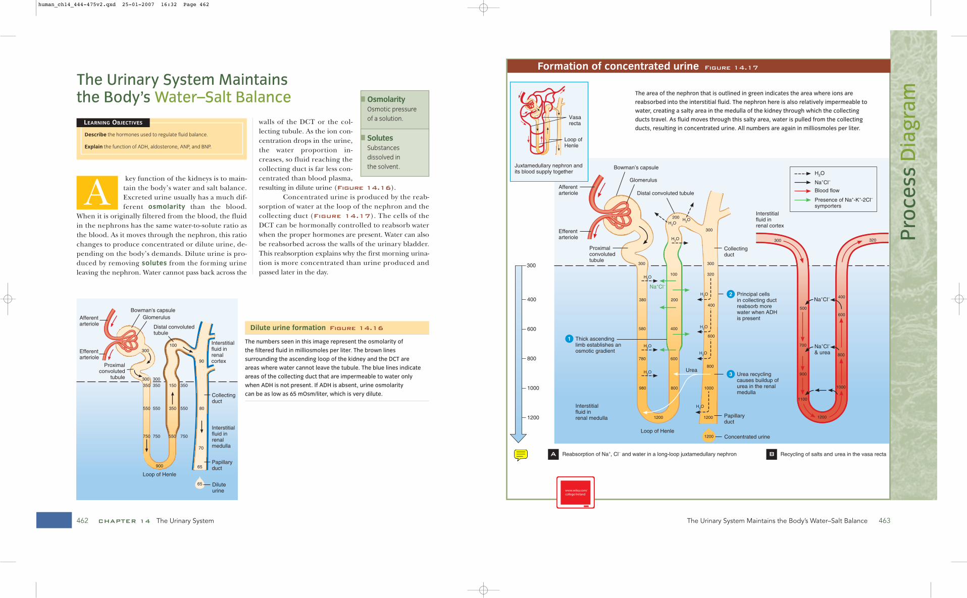
Dilute urine formation Figure 14.16
The numbers seen in this image represent the osmolarity of
the filtered fluid in milliosmoles per liter. The brown lines
surrounding the ascending loop of the kidney and the DCT are
areas where water cannot leave the tubule. The blue lines indicate
areas of the collecting duct that are impermeable to water only
when ADH is not present. If ADH is absent, urine osmolarity
can be as low as 65 mOsm/liter, which is very dilute.
key function of the kidneys is to main-tain the body’s water and salt balance.Excreted urine usually has a much dif-ferent osmolarity than the blood.
When it is originally filtered from the blood, the fluidin the nephrons has the same water-to-solute ratio asthe blood. As it moves through the nephron, this ratiochanges to produce concentrated or dilute urine, de-pending on the body’s demands. Dilute urine is pro-duced by removing solutes from the forming urineleaving the nephron. Water cannot pass back across the
The Urinary System Maintains the Body’s Water–Salt Balance
LEARNING OBJECTIVES
Describe the hormones used to regulate fluid balance.
Explain the function of ADH, aldosterone, ANP, and BNP.
A
walls of the DCT or the col-lecting tubule. As the ion con-centration drops in the urine,the water proportion in-creases, so fluid reaching thecollecting duct is far less con-centrated than blood plasma,resulting in dilute urine (Figure 14.16).
Concentrated urine is produced by the reab-sorption of water at the loop of the nephron and thecollecting duct (Figure 14.17). The cells of theDCT can be hormonally controlled to reabsorb waterwhen the proper hormones are present. Water can alsobe reabsorbed across the walls of the urinary bladder.This reabsorption explains why the first morning urina-tion is more concentrated than urine produced andpassed later in the day.
OsmolarityOsmotic pressure
of a solution.
SolutesSubstances
dissolved in
the solvent.
Formation of concentrated urine Figure 14.17
Pro
cess
Dia
gra
m
Recycling of salts and urea in the vasa rectaReabsorption of Na+, Cl– and water in a long-loop juxtamedullary nephron
1
Interstitialfluid inrenal medulla
Bowman’s capsule
Afferentarteriole
Efferentarteriole
Glomerulus
Distal convoluted tubule
Proximalconvolutedtubule
Thick ascendinglimb establishes anosmotic gradient
300
1200
1000
800
600
400
Na+Cl–
H2OH2O
H2O
200
1200
980
600780
400580
200380
300
100H2O
H2O
H2O
Loop of Henle
3
2
1200 Concentrated urine
300
300
320
400
600
800
1000
1200
800
H2O
H2O
H2O
H2O
Urea
Papillaryduct
Urea recyclingcauses buildup ofurea in the renalmedulla
Collectingduct
Principal cellsin collecting ductreabsorb morewater when ADHis present
H2O
300
500
700
900
1100
1200
400
800
1000
600
Na+Cl–
Na+Cl–
Blood flow
Presence of Na+-K+-2Cl–
symporters
Na+Cl–
& urea
Interstitialfluid inrenal cortex
320
Juxtamedullary nephron andits blood supply together
Vasarecta
Loop ofHenle
A B
The area of the nephron that is outlined in green indicates the area where ions are
reabsorbed into the interstitial fluid. The nephron here is also relatively impermeable to
water, creating a salty area in the medulla of the kidney through which the collecting
ducts travel. As fluid moves through this salty area, water is pulled from the collecting
ducts, resulting in concentrated urine. All numbers are again in milliosmoles per liter.
www.wiley.com/college/ireland
human_ch14_444-475v2.qxd 25-01-2007 16:32 Page 462
helen walden
Note
Insert series comma in front of and

Output Increased releaseof ADH
Return to homeostasis
Loop of nephronand collectingduct both allowwater to returnto the bloodstream
Decrease in ionconcentration of plasma
Effectors
Control center
Fluid balanceHypothalamus directsposterior pituitary to secrete ADH
Hypothalamusdetects an increasein osmolarity of plasma
Input Nerve impulses
Receptors
Ion concentration of plasma and interstitial fluid
Increasing
Some stimulus disruptshomeostasis by
H2O
ADH
464 CHAPTER 14 The Urinary System The Urinary System Maintains the Body’s Water–Salt Balance 465
I WO
ND
ER ...
Why do they keep telling me to cut down on salt?
Table salt is sodium chloride, and sodium is about as es-sential as electrolytes get because it helps control osmosisthroughout the body. But eating a lot of salt can raise theblood pressure by causing a subtle swelling of the tissues.Over long periods, hypertension can cause deadly or dis-abling strokes, heart attacks, heart failure, or kidney failure.
For years, we’ve been told to cut down on the salt. Butthat is easier said than done. As mentioned in the text, thekidney is built to recycle salt, enabling some people to sur-vive on less than 1 gram per day. Saltimproves the taste of food, and evolu-tion has forced us to crave salt.Sodium chloride is so vital that in thehot, dry Sahel region bordering theSahara Desert, salt was once traded—gram for gram—for gold. You can cer-tainly live without gold, but you willdie without salt.
In 2004, the National Academy ofSciences waded into the salt debateby suggesting that people aged 19 to50 ingest at least 1.5 grams of sodium(3.75 grams of salt) per day. This in-take, the group’s experts said, wouldreplace losses due to sweating andhelp ensure an adequate supply ofother nutrients.
At the other end of the spectrum,the National Academy recommendeda maximum of 2.3 grams of sodium(5.75 grams salt), and mentioned thedanger of hypertension associatedwith increased levels. For many Ameri-
cans and Canadians who eat processed food, that maxi-mum poses a problem. Salt is liberally added to mostprocessed food because it improves shelf life and peoplelike the taste.
Interestingly, the “cut the salt” advice is hotly debated.Some large clinical trials have associated lower salt levelswith lower blood pressure, which is usually desirable. Butsome experts note that only about one-third of people geta strong hypertensive response to high sodium intake. And
in some studies, the cardiovascularbenefits of reducing salt intake showup only among people who were over-weight when the study began.
Adding to the confusion, a studyreported in 2006 found a higher, notlower, rate of cardiovascular deathsamong people who ate less than 2.3grams of sodium per day. This kind ofresult leads some researchers to ques-tion the traditional advice about cut-ting down on salt. Even if reducing saltdoes reduce blood pressure in theshort term, there is no proof that it re-duces mortality over the long term. Ifthese benefits exist, a definitive clini-cal trial has yet to identify them.
Obviously if you want to reducechronic hypertension, salt reduction isnot your only option. Exercise helps,as do antihypertensive medicines. Re-ducing stress and learning to relax willalso reduce blood pressure, withoutmaking your food taste bland.
CONCEPT CHECK
List the sections of the nephronin order from glomerulus tocollecting duct.
What is the effectof ADH?
Which hormonesact in oppositionto ADH?
Water can be reabsorbed into the bloodstreamat the DCT and collecting duct with help from the hor-mone ADH (antidiuretic hormone) (Figure
14.18). A diuretic increases the volume of urine pro-duced, whereas an antidiuretic decreases the volume ofurine produced. ADH will therefore decrease urine vol-ume. ADH is secreted by the posterior lobe of the pitu-itary gland, located on the undersurface of the brain,in response to blood volume. ADH in the blood causesthe cells surrounding the collecting duct and the DCTto remove more water from the urine, returning it tothe depleted bloodstream.
Surprisingly, sodium is conserved almost asstingily as water. It is important to remember that wheresodium goes, water follows. If you live in a humid area,
you already know this. In the Deep South, the Midwestin summer, the southern shorelines of the East andWest Coasts, and on Pacific islands, humidity can clogsalt shakers because sodium chloride draws water mole-cules from the air, causing clumps in the salt shaker. Ifyou add a few grains of uncooked rice to the salt shaker,they will absorb the water from the salt, preventing thesalt crystals from sticking together.
In the nephron, this attraction between waterand sodium is used to good advantage. More than 99percent of the sodium filtered from the blood at theglomerulus is returned before the urine leaves thenephron. Two-thirds of this reabsorption occurs at thePCT. Another 25 percent of the filtered sodium is re-moved from the forming urine at the ascending limb of
ADH feedback system Figure 14.18
If you drink less water than you need, ADH will be secreted to preserve the volume
of water in your body. A small volume of more concentrated urine will be produced.
Conversely, if you drink a lot of water, ADH will not be secreted and more fluid will
be lost through the urinary system.
the loop of the nephron. This loop sets up a sodiumgradient in the medulla of the kidney by removingsodium from the filtrate. Sodium is also reabsorbedfrom the DCT and the collecting duct, so sodium levelsare strictly maintained. (For more on salt consumption,see the I Wonder box.)
Several hormones are involved in salt regula-tion. Aldosterone, atrial natriuretic peptide (ANP), andbrain natriuretic peptide (BNP) all regulate sodium re-absorption at the distal convoluted tubule. Aldosteronecauses the excretion of potassium ions and the reab-sorption of sodium ions, so water will leave the filtratewith the sodium ions rather than leave with the potas-sium ions. ANP and BNP both oppose ADH. When ei-ther is present, the kidneys produce lots of dilute urine.
Ingested chemicals can also affect nephronfunction. Caffeine and alcohol both increase urine pro-duction, apparently through de-creased ADH production. Whencaffeine is ingested in quantitiesbelow 350 mg, we experience cen-tral nervous system stimulation,decreased sleepiness, and possibleincreases in athletic performance.But the side effects include a po-tential headache and drowsinessas the caffeine wears off, and in-somnia. The diuretic effects of caf-feine are not particularly helpful.Dehydrated muscle cramps moreeasily and is less likely to be re-paired after injury. Your body can-not achieve peak function withoutgood hydration.
human_ch14_444-475v2.qxd 25-01-2007 16:32 Page 464
helen walden
Note
Insert ellipses after Wonder?
ody pH must be held within a narrowrange (7.35 to 7.45). This is done pri-marily through the bicarbonate buffersystem of the respiratory system, with
help from the urinary system. This pH stability isachieved through the maintenance of chemical equilib-rium. In the body, all three of the product sets shownbelow are in equilibrium, balanced as if on a teeter-tot-ter. Adding water to the body will increase the reactantsat the right (H2O � CO2), causing the amount ofH2CO3 and H� � HCO3
� to increase proportionatelyas the reactions return to equilibrium. When this hap-pens, we say it is “pushing the reaction to the left.”
H� � HCO3� 1 H2CO3 1 H2O � CO2
When carbon dioxide is exhaled, the above re-actions are “pushed to the right.” In order to maintainan equal concentration of reactants on either side ofthe arrows, more hydrogen ions are picked up by thebicarbonate ion (HCO3
�) and removed from theblood, returning the reaction to a point where all threecompartments are equal (Figure 14.19). Thishomeostatic function is so vital that the rate and depthof breathing respond to the level of carbon dioxide in
466 CHAPTER 14 The Urinary System The Kidneys Help Maintain the Blood’s Acid-Base Balance 467
The Kidneys Help Maintain the Blood’s Acid-Base Balance
B
LEARNING OBJECTIVES
Define the carbonate buffering system of the blood.
Explain the role of the kidneys in maintaining blood pH.
Return to homeostasis when response brings blood pH or H+ con- centration back to normal
Diaphragmcontracts more force-fully and frequently so more CO2 is exhaled
As less H2CO3 formsand fewer H+ are present, blood pH increases (H+
concentration decreases)
Effectors
Output Nerveimpulses
Inspiratory area inmedulla oblongata
Control center
Blood pH (increase in H+ concentration)
Input Nerveimpulses
Chemo-receptors inmedullaoblongata
Chemo-receptorsin aorticandcarotid bodies
Receptors
Decreasing
Some stimulus disruptshomeostasis by
Fluidin tubulelumen
ADP
Interstitialfluid
Intercalated cellin collecting duct
Absorbed intoperitubular capillary
H+ + HCO3–
H2CO3
CO2
Cl–
+ H2O
HPO42– + H+
NH3 + H+
NH4+
H2PO4 –
HCO3–
Cl–
HCO3–
(new) H+
H+
ADP
Proton pump (H+ ATPase) in apical membrane
Key:
Diffusion
HCO3– /Cl– antiporter in basolateral membrane
Secretion of H+ Buffering of H+ in urine
CAATP
ATP
A B
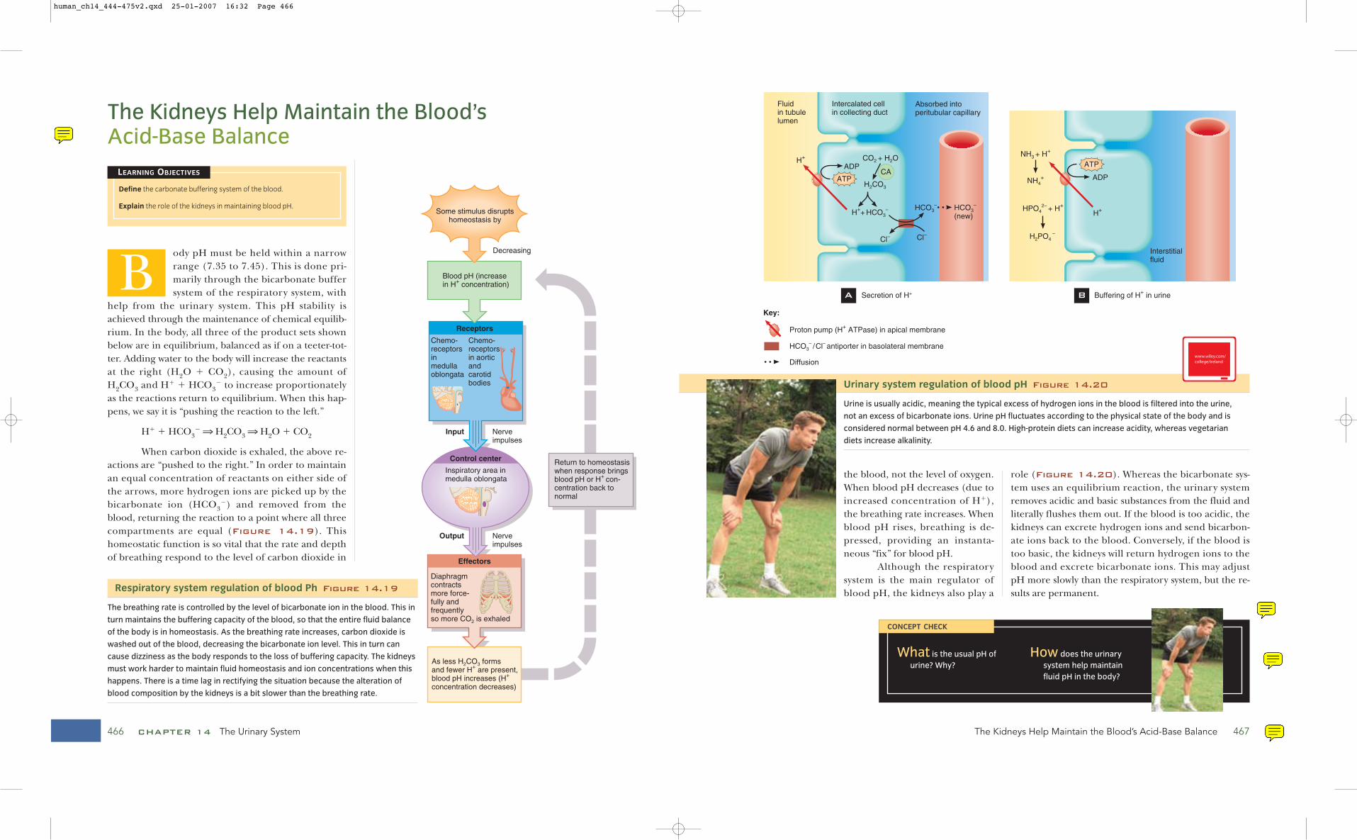
Urinary system regulation of blood pH Figure 14.20
Urine is usually acidic, meaning the typical excess of hydrogen ions in the blood is filtered into the urine,
not an excess of bicarbonate ions. Urine pH fluctuates according to the physical state of the body and is
considered normal between pH 4.6 and 8.0. High-protein diets can increase acidity, whereas vegetarian
diets increase alkalinity.
CONCEPT CHECK
What is the usual pH ofurine? Why?
How does the urinarysystem help maintainfluid pH in the body?
the blood, not the level of oxygen.When blood pH decreases (due toincreased concentration of H�),the breathing rate increases. Whenblood pH rises, breathing is de-pressed, providing an instanta-neous “fix” for blood pH.
Although the respiratorysystem is the main regulator ofblood pH, the kidneys also play a
role (Figure 14.20). Whereas the bicarbonate sys-tem uses an equilibrium reaction, the urinary systemremoves acidic and basic substances from the fluid andliterally flushes them out. If the blood is too acidic, thekidneys can excrete hydrogen ions and send bicarbon-ate ions back to the blood. Conversely, if the blood istoo basic, the kidneys will return hydrogen ions to theblood and excrete bicarbonate ions. This may adjustpH more slowly than the respiratory system, but the re-sults are permanent.
www.wiley.com/college/ireland
Respiratory system regulation of blood Ph Figure 14.19
The breathing rate is controlled by the level of bicarbonate ion in the blood. This in
turn maintains the buffering capacity of the blood, so that the entire fluid balance
of the body is in homeostasis. As the breathing rate increases, carbon dioxide is
washed out of the blood, decreasing the bicarbonate ion level. This in turn can
cause dizziness as the body responds to the loss of buffering capacity. The kidneys
must work harder to maintain fluid homeostasis and ion concentrations when this
happens. There is a time lag in rectifying the situation because the alteration of
blood composition by the kidneys is a bit slower than the breathing rate.
human_ch14_444-475v2.qxd 25-01-2007 16:32 Page 466
helen walden
Note
Hyphen should be en dash
helen walden
Note
Hyphen should be en dash
helen walden
Note
Center box horizontally
helen walden
Note
Use a different photo to avoid twice on one page?

468 CHAPTER 14 The Urinary System Life-Threatening Diseases Affect the Urinary System 469
Life-Threatening Diseases Affect the Urinary System
hemical analysis of urine can reveal anumber of serious diseases as well as theuse of illegal drugs (see Ethics and Is-sues box). Urinalysis (UA) is a simple,
common test that is routinely done in the doctor’s of-fice to produce a view of your internal health. It is non-invasive, meaning that instruments or sensing equip-ment are not placed in or on your body. Because urineis the by-product of filtered blood, any unusual com-pounds or incorrect levels of normal blood constituentswill appear in the urine.
WARNING SIGNALSFROM URINALYSIS
Abnormal components in urinecan include albumin, hemoglo-bin, red blood cells, white bloodcells, glucose, and casts. Eachcan indicate a specific problem.
See Table 14.2 for a listing of the normal and ab-normal constituents of urine.
• Albumin is a small protein that must be enter-ing the nephrons at the glomerulus. This couldreflect high blood pressure in the glomerulusthat forces proteins through the podocyte walls,or tears in glomerular arterioles. Normally, al-bumin remains in the blood to provide an os-motic force to draw excess water back from thefiltrate to the blood. Albumin or other less com-mon proteins in the urine are diagnosed as pro-teinurea, but this may not indicate pathology.Serious weight training puts tremendous pres-
C
LEARNING OBJECTIVES
List what health facts can be learned from routine urinalysis.
Explain the process of conventional dialysis and hemodialysis.
Eth
ics
and
Issu
es
How does a urine test prove drug abuse?
Urinalysis (UA) is a noninvasive way to get a holistic picture
of the immediate physiological events occurring in the body.
In a sense, urine tells us about the chemical processes in
the body, just as examining the chemicals in a river tells us
about events in the river’s watershed. If a person has been
taking illicit drugs, prescribed medications, or even diet sup-
plements, those compounds, or their breakdown products,
will show up in the urine.
Urinalysis is most often used as a screening and diag-
nostic tool. UAs are performed when people complain of ab-
dominal pain, back pain, painful or frequent urination, blood
in the urine, or other symptoms of a urinary tract infection,
which may show up as elevated levels of white blood cells .
It is also a routine part of regular physical examinations.
The test detects substances in the urine associated
with many metabolic and kidney disorders. An abnormal UA
can be an early warning of trouble, because substances like
protein or glucose will begin to appear in the urine before a
person is aware of a problem. . The health care provider
must correlate the urinalysis results with physical com-
plaints and clinical findings to make a diagnosis.
Have you ever wondered what happens to your urine
sample? The medical professional first examines its physical
characteristics,
such as clarity,
color, odor,
and specific
gravity.
The next
step is a chemi-
cal analysis ,
usually with a
“dipstick” test
that includes
many pads soaked with indicator substances. The strip is
dipped into the sample, so the urine can interact with the
chemicals in each pad. After a specific time (usually 30 sec-
onds) the color of each pad is compared to a reference
chart. This comparison is now automated for greater accu-
racy.
Urine usually contains urochrome, which gives it that
yellow color; nitrogenous wastes like ammonia and urea
from metabolic processes; water; ions; and cast-off cells
from the epithelial lining of the bladder and urethra. In addi-
tion, many large molecules enter the urine when blood in
the peritubular capillaries passes the distal convoluted
tubule. These molecules can include breakdown products of
sure on the capillaries and can force proteininto the urine.
• Hemoglobin indicates bleeding in the upperurinary tract because the red blood cells havebeen present in the urine long enough to breakopen and release hemoglobin. Intact red bloodcells would indicate bleeding closer to the lowerend of the urinary tract, perhaps in the urethra.White blood cells in the urine indicate that animmune response is occurring, usually in re-sponse to an infection of the urinary tract, oroccasionally the kidney.
CastsSmall structures
formed by mineral
or fat deposits
on the walls of
the renal tubules.
Normal Constituent Description
Volume One to two liters in 24 hours but variesconsiderably.
Color Yellow or amber but varies with urine con-centration and diet. Color is due tourochrome (pigment produced frombreakdown of bile) and urobilin (frombreakdown of hemoglobin). Concentratedurine is darker in color. Diet (reddish col-ored urine from beets), medications, andcertain diseases affect color. Kidney stonesmay produce blood in urine.
Turbidity Transparent when freshly voided but be-comes turbid (cloudy) upon standing.
Odor Mildly aromatic but becomesammonia–like upon standing. Some peo-ple inherit the ability to form methylmer-captan from digested asparagus that givesurine a characteristic odor. Urine of dia-betics has a fruity odor due to presence ofketone bodies.
pH Ranges between 4.6 and 8.0; average 6.0;varies considerably with diet. High-proteindiets increase acidity; vegetarian diets in-crease alkalinity.
Specific gravity Specific gravity (density) is the ratio of theweight of a volume of a substance to theweight of an equal volume of distilled wa-ter. In urine, it ranges from 1.001 to 1.035.The higher the concentration of solutes,the higher the specific gravity.
legal and illegal drugs, vitamin and mineral supplements, or
even various environmental contaminants.
The pH of urine should be between 4.6 and 8, and the
specific gravity between 1.002 and 1.028. For reference, dis-
tilled water has a specific gravity of 1.000, and the Pacific
Ocean has an average specific gravity of 1.023.
When urinalysis is being used to test for the presence
of drugs, the sample is first put through a fast, inexpensive,
and inexact screening test. Samples that test positive are
then put into an analytical machine called the gas chromato-
graph-mass spectrometer (GC/MS). The GC/MS first sepa-
rates compounds based on their mass, and then uses detec-
tor chemicals to identify certain compounds. The machine is
expensive but is so sensitive that it can easily detect traces
of compounds in concentrations of 1 part per billion, or
even less.
The GC/MS produces a graph showing all chemical com-
pounds detected in the sample. If peaks on the graph indi-
cate the presence of illicit drugs or their metabolites, the
test is said to be positive and the person is considered a
user of illicit drugs.
Drug testing is often sold as a cure-all for detecting
drug use, especially among students, athletes, or potential
employees. But urinalysis is not perfect. A test result can
mistake metabolites of over-the-counter or prescription
drugs for those of illicit drugs, forcing test administrators to
interpret results carefully. Poppy seeds, found on bagels
and pastries, can break down into compounds that resem-
ble metabolites from opiate drugs, because opiates and
poppy seeds both come from the opium poppy. Urinalysis is
more effective at detecting some drugs than others. Mari-
juana and other drugs are detectable in the urine for long
periods, whereas alcohol and cocaine are cleared quickly
from the body. Finally, drug testing is expensive, and some
studies suggest that the knowledge that urinalysis will be
performed on a regular basis has little effect on employee
performance.
The most common “street” advice for fooling a urinaly-
sis, or passing as “clean” despite having recently introduced
illicit drugs into your system, is to dilute the urine by drink-
ing massive quantities of water before filling the sample
cup. But the GC/MS is so sensitive that it can usually pick up
traces of drug metabolites in very dilute urine. Deliberately
ingesting compounds that will interfere with drug tests also
raises moral questions. And these “interferences” are identi-
fied in the test, raising a red flag. The best, most surefire
way to test drug-free is to live drug-free.
Specific gravityA ratio of
the density
of asubstance
to the density
of pure water.
Normal and abnormal constituents of urineTable 14.2a
human_ch14_444-475v2.qxd 25-01-2007 16:32 Page 468
helen walden
Note
Insert the after see?
helen walden
Note
Delete extra period (EA)
helen walden
Note
Insert space: a substance (PE)

nausea, dizziness, fatigue, and mem-ory loss. If the ion and fluid balanceis not restored, death can result.
Dialysis is the exchange ofaqueous substances between two so-lutions through a membrane. In ef-fect, the entire nephron performsdialysis with the peritubular capillar-ies on a continuous basis. When thekidneys shut down, dialysis must con-tinue somehow, or the blood will be-come toxic to the cells of the body.Dialysis machines permit dialysis tooccur outside the body.
Hemodialysis is dialysis be-tween blood and another fluid (Fig-
ure 14.21). This is a relativelycommon procedure used to compensate for impairedkidney function. It can be done for extended periods,such as when the kidneys have failed and no matchingdonor kidneys are available.
In hemodialysis, blood is withdrawn from anartery and passed across a dialysis membrane. Toxins inthe blood diffuse into the prepared solution, while nec-essary blood plasma components are either (1) pre-vented from diffusing by putting the same concentra-tion of these components in the dialysis fluid as in theblood or (2) added to the blood by increasing their lev-els in the dialysis fluid. The dialyzed blood is then sentback to the body. The procedure takes three to fourhours and must be done three times a week.
Hemodialysis is tough on the blood cells be-cause they are passed through tubes and across mem-branes under pressure. If the patient requires dialysisfor a long period, peritoneal dialysis may be recom-mended. In this procedure, two liters of dialysis fluid areput directly into the abdominal cavity, left to diffuse for
a period, andthen removed.The peritoneumser ves as thedialysis mem-brane. As withhemodialysis, this procedure must beperformed regularly; in the case ofperitoneal dialysis, the procedure iscompleted several times a day to sus-tain life.
Kidney transplants are rela-tively common, second only tocorneal transplants in numbers per-formed per year. Because of the kid-neys’ retroperitoneal placement, theyare easy to reach surgically. The kid-
neys have essentially one artery and one vein. Theselarge vessels are easily cut and sutured to the donor kid-ney. The suturing can be completed using laparo-scopes, and the kidney removed and/or replacedthrough a three-inch incision on the side of the ab-domen. Kidney transplants are highly successful trans-plant operations, with a near 80 percent patient and or-gan survival rate after one year. Living transplants,transplanting organs obtained by removing one kidneyfrom a living, healthy donor rather than an accidentvictim, have success rates above 90 percent.
Obtaining nutrients, absorbing the necessarycompounds, getting rid of waste products, and main-taining fluid homeostasis are all imperative to survival.With the digestive and urinary systems handling thesevital functions, we humans can turn our attention toother occupations. One of the more interesting of theseis reproduction, the topic of the next chapter. Perpetu-ating the species is the underlying biological drive thatkeeps us going on this planet.
• Glucose in the urine signifies diabetes mellitus.As described previously, glucose spills into theurine due to a high concentration in the blood.
• Casts are plugs of material, shaped like thenephron tubules, that build up in the tubulesand then get forced out by pressure. Casts canbe formed from minerals that enter the filtrateand clog the PCT and nephron loop, or theycan be composed of proteins and cells that findtheir way into the system. Casts always indicateserious kidney trouble, such as nephritis orglomerulonephritis.
KIDNEY DISEASE IS LIFE THREATENING
Without functioning kidneys, blood composition can-not be maintained and homeostasis will be lost. Threeof the most common kidney diseases are nephritis,glomerulonephritis, and polycystic kidney disease. Of
these, only polycystic kidneydisease is inherited. This dis-ease causes cysts to form in thekidneys, destroying normalkidney tissue. In severe cases,the patient may require dialy-sis or even a kidney trans-plant.
Nephritis and glome-rulonephritis are both inflammations of the filteringunit of the kidney. Because the kidney is covered by therenal capsule, any inflammation within the kidney in-creases pressure, compresses kidney tissues and slows orhalts filtration at the glomerulus. The kidney is shutdown until the swelling is resolved.
Glomerulonephritis is a general term for block-ing of kidney circulation, and swelling and shutdown ofthe nephrons. When the kidneys cannot filter blood,toxins build up and the blood becomes filled with meta-bolic wastes. Blood volume and composition are dis-turbed, and the patient becomes ill. Symptoms include
470 CHAPTER 14 The Urinary System
Abnormal Constituent Description
Albumin A normal constituent of plasma, it usually appears in only very small amounts in urine because it is too largeto pass through capillary fenestrations. The presence of excessive albumin in the urine—albuminuria—indicates an increase in the permeability of filtration membranes due to injury or disease, increased bloodpressure, or irritation of kidney cells by substances such as bacterial toxins, ether, or heavy metals.
Glucose The presence of glucose in the urine is called glucosuria and usually indicates diabetes mellitus. Occasion-ally it may be caused by stress, which can cause excessive amounts of epinephrine to be secreted. Epineph-rine stimulates the breakdown of glycogen and liberation of glucose from the liver.
Red blood cells (erythrocytes) The presence of red blood cells in the urine is called hematuria and generally indicates a pathological condi-tion. One cause is acute inflammation of the urinary organs as a result of disease or irritation from kidneystones. Other causes include tumors, trauma, and kidney disease, or possible contamination of the sampleby menstrual blood.
Ketone bodies High levels of ketone bodies in the urine, called ketonuria, may indicate diabetes mellitus, anorexia, starva-tion, or simply too little carbohydrate in the diet.
Bilirubin When red blood cells are destroyed by macrophages, the globin portion of hemoglobin is split off and theheme is converted to biliverdin. Most of the biliverdin is converted to bilirubin, which gives bile its majorpigmentation. An above normal level of bilirubin in urine is called bilirubinuria.
Urobilinogen The presence of urobilinogen (breakdown product of hemoglobin) in urine is called urobilinogenuria.Trace amounts are normal, but elevated urobilinogen may be due to hemolytic or pernicious anemia, infec-tious hepatitis, biliary obstruction, jaundice, cirrhosis, congestive heart failure, or infectious mononucleosis.
Casts Casts are tiny masses of material that have hardened and assumed the shape of the lumen of the tubule inwhich they formed. They are then flushed out of the tubule when filtrate builds up behind them. Casts arenamed after the cells or substances that compose them or based on their appearance.
Microbes The number and type of bacteria vary with specific infections in the urinary tract. One of the most commonis E. coli. The most common fungus to appear in urine is the yeast Candida albicans, a cause of vaginitis. Themost frequent protozoan seen is Trichomonas vaginalis, a cause of vaginitis in females and urethritis in males.
DialysisSubstance
exchange via
diffusion across
a membrane,
artificially
mimicking
the kidney.
PeritoneumMembrane lining
the abdominal
cavity.
A patient undergoing dialysisFigure 14.21
CONCEPT CHECK
List four problem signs in aurinalysis, and describe whatpathology each indicates.
Differentiate betweenpolycystic kidney diseaseand nephritis.
How doeshemodialysiswork?
Life-Threatening Diseases Affect the Urinary System 471
Normal and abnormal constituents of urine Table 14.2b
human_ch14_444-475v2.qxd 25-01-2007 16:32 Page 470
helen walden
Note
Insert comma after tissues (EA)

1The Urinary System Filters,Transports, and StoresWaste Products
The urinary system is responsible for
maintaining fluid homeostasis, ion balance,
and blood calcium concentration and for re-
moving fluid waste from the body. The sys-
tem includes the paired kidneys, the paired
ureters, the urinary bladder, and the ure-
thra. The nephron is composed of a
glomerular capsule surrounding the
glomerulus, a proximal convoluted tubule, a
loop, and a distal convoluted tubule con-
nected to a collecting duct. Each portion of
the tubule has a distinct role in filtering
blood, balancing ions and pH, and removing
wastes.
4The Urinary SystemMaintains the Body’sWater–Salt Balance
Hormones regulate the amount of wa-
ter and ions excreted with the urine. ADH
prevents the loss of water, causing the pro-
duction of concentrated urine. Aldosterone
regulates sodium reabsorption, effectively
removing water and sodium from the body.
ANP and BNP work in opposition to ADH,
causing the formation of dilute urine.
2Urine Is Formed ThroughFiltration and Osmosis
The nephron is the functional unit of
the urinary system. It is here that 180 liters
of fluid are filtered and maintained per day.
Glomerular filtration depends on blood
pressure, capsular hydrostatic pressure,
and osmotic pressure of the blood. The fil-
trate is captured in Bowman’s capsule and
passed to the PCT, where most of the nec-
essary nutrients and water are reabsorbed.
The loop of the nephron extends into the
middle of the kidney, and assists in removal
of salts and water. The DCT is involved in se-
cretion from the blood to the forming urine.
The collecting ducts remove urine from the
kidney.
472 CHAPTER 14 The Urinary System
6Life-ThreateningDiseases Affect theUrinary System
Dialysis is the exchange of aque-
ous substances between two solu-
tions through a membrane. In effect,
the entire nephron performs dialysis
with the peritubular capillaries on a
continuous basis. When the kidneys
shut down, dialysis must continue
somehow, or the blood will become
toxic to the cells of the body. Dialysis
machines permit dialysis to occur
outside the body.
Critical Thinking Questions 473
KEY TERMS
1. Imagine that you contracted a urinary tract infection and did
not treat it. Trace the pathway of the bacteria as it moves up
the urinary system. What structures would you expect to be
damaged by the bacteria once they reached the kidneys?
2. Many home pregnancy tests look for a specific protein in the
urine. This compound is present only in pregnant women. Why
do the tests recommend using first morning urine? How is it dif-
ferent from urine produced and excreted at midday?
3. Caffeine and alcohol both block the secretion of ADH from the
posterior pituitary gland. Explain what this does to fluid bal-
ance. Does it make sense to drink caffeine before an athletic
event? Explain why a cold beer may not be such a great idea on
a hot afternoon.
4. What are the differences between hemodialysis and peritoneal
dialysis? What are the benefits of each? What are the draw-
backs?
5. Assume you were given the following results from a series of
urinalysis tests. What would each test indicate?
Cloudy urine, above-normal specific gravity, high white
blood cell count, many transitional epithelial cells.
Presence of protein, casts, and hemoglobin.
Presence of glucose and ketones (ketones are a by-product
of the digestion of body proteins)
Pale yellow color, pH 6.3, specific gravity 1.015, no RBCs, no
proteins.
CRITICAL THINKING QUESTIONS
■ arteriole p. 000
■ calcium oxalate p. 000
■ casts p. 000
■ constipation p. 000
■ countercurrent multiplication p. 000
■ dialysis p. 000
■ external sphincter muscle p. 000
■ facilitated diffusion p. 000
■ fenestrations p. 000
■ hilus p. 000
■ incontinence p. 000
■ internal urethral sphincter p. 000
■ microvilli p. 000
■ nephron p. 000
■ nitrogenous wastes p. 000
■ osmolarity p. 000
■ peritoneum p. 000
■ peritubular capillaries p. 000
■ secretion p. 000
■ solutes p. 000
■ specific gravity p. 000
CHAPTER SUMMARY
3Urine Is Transported to theBladder for Storage
Urine is stored in the bladder until
voided. After 300 ml of urine fills the blad-
der, the micturition reflex is stimulated. The
voluntary, external urinary sphincter deter-
mines when voiding takes place. Inconti-
nence is the loss of this control. The female
urethra is shorter than the male urethra,
leading to a higher incidence of urinary tract
infection in females.
5The Kidneys Help Maintain the Blood’s Acid-Base Balance
The kidneys and respira-
tory system combine to con-
trol blood pH. The urinary sys-
tem can remove acidic and
basic substances from the
fluid and flush them from the
body, with permanent results.
If the blood is too acidic, the
kidneys can excrete hydrogen
ions and send bicarbonate
ions back to the blood. If the
blood is too basic, the kidneys
will return hydrogen ions to
the blood, and excrete bicar-
bonate ions.
human_ch14_444-475v2.qxd 25-01-2007 16:32 Page 472
helen walden
Note
Hyphen should be en dash (EA)
helen walden
Note
secretion is given as secreted in the key term box on p. 455
helen walden
Note
No arteriole key term in this chapter. The other page numbers in this column are 458, 468, 461, 456, 470, 460
helen walden
Note
hilus is not set as key term in text (on p. 447) and there is no key term box for it. Either add the box or delete from this list.
helen walden
Note
Second column page numbers (including hilus) are 455, 453, 447, 461, 460, 455, 448
helen walden
Note
Third column page numbers are 447, 462, 471, 448, 455, 462, 469

6. The function of the structure indicated as B is to
a. filter blood.
b. collect filtrate.
c. reabsorb necessary nutrients.
d. secrete unwanted large waste products.
7. In the above figure, label E indicates the
a. PCT.
b. loop of Henle.
c. DCT.
d. glomerulus.
8. The portion(s) of the nephron that are usually found in the cor-
tex of the kidney are the
a. PCT, capsule, and loop of Henle.
b. PCT and DCT.
c. loop of Henle and collecting duct.
d. PCT, capsule, and DCT.
e. capsule only.
9. When blood is filtered through the glomerulus, the two forces
opposing movement into the nephron are
a. A and B.
b. B and C.
c. A and C.
d. All of these forces oppose movement into the capsule.
SELF TEST
474 CHAPTER 14 The Urinary System
10. Most of the filtered water, and hopefully all of the filtered glu-
cose is returned to the bloodstream at the
a. PCT.
b. DCT.
c. loop of Henle.
d. collecting duct.
11. The portion of the nephron that sets up the salt gradient in the
medulla of the kidney is the
a. PCT.
b. DCT.
c. loop of Henle.
d. collecting duct.
12. The structure shown below is formed when
a. overly dilute urine is produced.
b. overly concentrated urine is produced.
c. kidney failure is experienced.
d. too many calcium-rich foods are consumed.
16. True or false: Both caffeine and alcohol serve as diuretics, caus-
ing the production of copius dilute urine.
17. The compound most important in
driving respiration rates and
depth of breathing shown in
the feedback loop in this
figure is
a. oxygen.
b. bicarbonate ions.
c. carbon dioxide.
d. hydrogen ions.
Self Test 475
A
B B
A
C
D
A
Renal corpuscle: Bowman's
capsule
E
Efferent arteriole
C
D
D
B
F
BA
C
Return to homeostasis when response brings blood pH or H+ con- centration back to normal
Diaphragmcontracts more force-fully and frequently so more CO2 is exhaled
As less H2CO3 formsand fewer H+ are present, blood pH increases (H+
concentration decreases)
Effectors
Output Nerveimpulses
Inspiratory area inmedulla oblongata
Control center
Blood pH (increase in H+ concentration)
Input Nerveimpulses
Chemo-receptors inmedullaoblongata
Chemo-receptorsin aorticandcarotid bodies
Receptors
Decreasing
Some stimulus disruptshomeostasis by
1. Which of the following is NOT a specific function of the urinary
system?
a. Production of urine
b. Maintenance of blood pH
c. Maintenance of blood volume
d. Maintenance of red blood cell levels
2. The function of the structure indicated as C is
a. filtration of blood.
b. transport of urine within the body.
c. transport of urine from the body.
d. storage of produced urine.
3. In the above diagram, the urethra is indicated by label
a. A
b. B
c. C
d. D
4. The renal pyramids are composed of
a. glomeruli.
b. peritubular capillaries.
c. collecting ducts.
d. renal capsules.
5. The correct sequence of blood vessels through the kidney is
a. Renal artery S arcuate artery S afferent arteriole S effer-
ent arteriole S peritubular capillaries S renal vein
b. Renal vein S renal artery S peritubular capillaries S arcu-
ate artery S interlobar veins
c. Renal artery S peritubular capillaries S efferent arteriole
S afferent arteriole S renal vein
d. Renal artery S efferent arteriole S peritubular capillaries
S interlobar artery S renal vein
13. True or false: The urethra in the image below carries both urine
and reproductive fluids.
14. Dilute urine is produced when
a. ADH is present.
b. ADH is absent.
c. solutes are added to the forming urine.
d. water is removed from the collecting ducts.
15. If you drink more water than you need, __________ will be se-
creted and you will lose water through the urinary system.
a. ADH
b. ANP
c. aldosterone
d. Both b and c will be secreted.
18. Urinary analysis is able to detect all of the following EXCEPT:
a. vitamin supplementation.
b. illegal drug use.
c. viral infection.
d. metabolic kidney disorders.
19. White blood cells in the urine indicate
a. normal urinary tract functioning.
b. a possible UTI.
c. bleeding in the kidneys or ureters.
d. diabetes mellitus.
20. During this procedure, if a
physician wants to remove
excess potassium from
the blood, she or he must
include __________ of
potassium in the dialysis
fluid than in the blood.
a. higher levels
b. lower levels
human_ch14_444-475v2.qxd 25-01-2007 16:32 Page 474
helen walden
Note
Make a, b parts consistent as to initial cap and period (or not) at end
helen walden
Note
Delete extra space between c and d (PE)
helen walden
Note
Insert comma after glucose (EA)
helen walden
Note
copius should be copious (EA)
helen walden
Note
Instead of saying "During this procedure" say "During the procedure shown in this photo"?