The transcription factor Sp3 regulates the expression of a ...
12
RIGHT: URL: CITATION: AUTHOR(S): ISSUE DATE: TITLE: The transcription factor Sp3 regulates the expression of a metastasis-related marker of sarcoma, actin filament- associated protein 1-like 1(AFAP1L1)( Dissertation_全文 ) Kajita, Yoichiro Kajita, Yoichiro. The transcription factor Sp3 regulates the expression of a metastasis-related marker of sarcoma, actin filament-associated protein 1-like 1(AFAP1L1). 京都大学, 2013, 博士(医学) 2013-05-23 https://doi.org/10.14989/doctor.k17780
Transcript of The transcription factor Sp3 regulates the expression of a ...

RIGHT:
URL:
CITATION:
AUTHOR(S):
ISSUE DATE:
TITLE:The transcription factor Sp3 regulates the expression of ametastasis-related marker of sarcoma, actin filament-associated protein 1-like 1(AFAP1L1)( Dissertation_全文 )
Kajita, Yoichiro
Kajita, Yoichiro. The transcription factor Sp3 regulates the expression of a metastasis-related marker of sarcoma, actinfilament-associated protein 1-like 1(AFAP1L1). 京都大学, 2013, 博士(医学)
2013-05-23
https://doi.org/10.14989/doctor.k17780
The Transcription Factor Sp3 Regulates the Expression ofa Metastasis-Related Marker of Sarcoma, Actin Filament-Associated Protein 1-Like 1 (AFAP1L1)Yoichiro Kajita1,2, Tomohisa Kato, Jr.1, Sakura Tamaki1, Moritoshi Furu1,3, Ryo Takahashi4,
Satoshi Nagayama4¤a, Tomoki Aoyama1, Hiroyuki Nishiyama2¤c, Eijiro Nakamura2, Toyomasa Katagiri5¤b,
Yusuke Nakamura5, Osamu Ogawa2, Junya Toguchida1,3,6*
1 Department of Tissue Regeneration, Institute for Frontier Medical Sciences, Kyoto University, Kyoto, Japan, 2 Department of Urology, Graduate School of Medicine,
Kyoto University, Kyoto, Japan, 3 Department of Orthopaedic Surgery, Graduate School of Medicine, Kyoto University, Kyoto, Japan, 4 Department of Surgery, Graduate
School of Medicine, Kyoto University, Kyoto, Japan, 5 Laboratory of Molecular Medicine, Human Genome Center, Institute of Medical Science, The University of Tokyo,
Tokyo, Japan, 6 Center for iPS Cell Research and Application, Kyoto University, Kyoto, Japan
Abstract
We previously identified actin filament-associated protein 1-like 1 (AFAP1L1) as a metastasis-predicting marker from thegene-expression profiles of 65 spindle cell sarcomas, and demonstrated the up-regulation of AFAP1L1 expression to be anindependent risk factor for distant metastasis in multivariate analyses. Little is known, however, about how the expression ofAFAP1L1 is regulated. Luciferase reporter assays showed tandem binding motives of a specificity protein (Sp) located at 285to 275 relative to the transcriptional start site to be essential to the promoter activity. Overexpression of Sp1 and Sp3proteins transactivated the proximal AFAP1L1 promoter construct, and electrophoretic mobility shift assays showed thatboth Sp1 and Sp3 were able to bind to this region in vitro. Chromatin immunoprecipitation experiments, however, revealedthat Sp3 is the major factor binding to the proximal promoter region of the AFAP1L1 gene in AFAP1L1- positive cells.Treatment with mithramycin A, an inhibitor of proteins binding to GC-rich regions, prevented Sp3 from binding to theproximal promoter region of AFAP1L1 and decreased its expression in a dose-dependent manner. Finally, knocking downSp3 using small inhibitory RNA duplex (siRNA) reduced AFAP1L1 expression significantly, which was partially restored byexpressing siRNA-resistant Sp3. These findings indicate a novel role for Sp3 in sarcomas as a driver for expression of themetastasis-related gene AFAP1L1.
Citation: Kajita Y, Kato T Jr, Tamaki S, Furu M, Takahashi R, et al. (2013) The Transcription Factor Sp3 Regulates the Expression of a Metastasis-Related Marker ofSarcoma, Actin Filament-Associated Protein 1-Like 1 (AFAP1L1). PLoS ONE 8(1): e49709. doi:10.1371/journal.pone.0049709
Editor: Wei-Guo Zhu, Peking University Health Science Center, China
Received April 2, 2012; Accepted October 12, 2012; Published January 9, 2013
Copyright: � 2013 Kajita et al. This is an open-access article distributed under the terms of the Creative Commons Attribution License, which permitsunrestricted use, distribution, and reproduction in any medium, provided the original author and source are credited.
Funding: This work was supported by Grants-in-aid for Scientific Research from the Ministry of Education, Culture, Sports, Science and Technology. The fundershad no role in study design, data collection and analysis, decision to publish, or preparation of the manuscript.
Competing Interests: The authors have declared that no competing interests exist.
* E-mail: [email protected]
¤a Current address: Gastroenterology Center, The Cancer Institute Hospital of Japanese Foundation of Cancer Research, Tokyo, Japan¤b Current address: Division of Genome Medicine, Institute for Genome Research, The University of Tokushima, Tokoshima, Japan¤c Current address: Department of Urology, Faculty of Medicine, University of Tsukuba, Tsukuba, Japan
Introduction
Soft tissue sarcoma (STS) is a malignant neoplasm that can arise
in fat, muscle, fibrous tissue, blood vessels, or other supporting
tissue in any part of the body. STSs are divided into two groups
based on morphology; small round cell sarcomas and spindle cell
sarcomas. The former include rhabdomyosarcomas and extra-
skeletal Ewing’s tumors, against which chemotherapy and
radiotherapy are effective at least in the initial stages, and
therefore treatment other than surgery is usually the first choice.
STSs in the latter group, such as leiomyosarcomas and malignant
fibrous histiocytomas, however, are radio- and chemoresistant in
most cases and therefore wide resection with proper surgical
margins is the only way to control local tumors. In spite of proper
treatment for local disease, approximately half of patients develop
metastasis in distant organs, particularly in the lungs. Although
recent studies have demonstrated a beneficial effect of chemo-
therapy, the improvement is far from satisfactory. Considering the
associated side effects, it is desirable to identify high-risk patients,
to whom additional treatments should be administered.
AFAP1L1 was previously identified as a metastasis-predicting
marker from the gene-expression profiles of 65 spindle cell
sarcomas by our group [1]. In univariate and multivariate
analyses, higher expression of AFAP1L1 was found to contribute
to the occurrence of distant metastases, along with patient age and
tumor grade. Knocking down of the AFAP1L1 gene in sarcoma
cells reduced cell invasiveness and forced expression of AFAP1L1
in immortalized human mesenchymal stem cells increased
anchorage-independent cell growth as well as cell invasiveness.
These results suggest that the molecular mechanism up-regulating
the expression of AFAP1L1 is a key to the progression of sarcomas.
In this study, we explored the transcriptional regulation of
AFAP1L1 in order to find factors responsible for the up-regulation
PLOS ONE | www.plosone.org 1 January 2013 | Volume 8 | Issue 1 | e49709
of AFAP1L1 expression, which will help us to understand how
sarcoma cells gain the malignant phenotype.
Materials and Methods
Cell Lines, antibodies and reagentsHuman osteosarcoma cell lines (U2OS, MG63, and Saos2) and
a human fibrosarcoma cell line (HT1080) were obtained from
American Type Culture Collection (ATCC, Manassas, VA). PC-3
(human prostate cancer) and 293T were also obtained from
ATCC. SYO-1 (human synovial sarcoma cell line) [2] was
provided by Dr. A. Kawai (National Cancer Center, Japan), and
293T was described elsewhere [3]. Informed consent was obtained
from the patient with written consent, and the procedure was
approved by the Ethics Committee of Graduate School of
Medicine and Dentistry, Okayama University. Cells were cultured
in DMEM (for U2OS, MG63, Saos2, 293T, HT1080 and SYO-1)
or RPMI (for PC-3) supplemented with 10% fetal bovine serum,
0.1 mg/ml streptomycin, and 100 units/ml penicillin under 5%
CO2 at 37uC. The anti-AFAP1L1 polyclonal antibody was
produced in our laboratory as described previously [1]. The
anti-Sp1 antibodies (1C6 and PEP2) and anti-Sp3 antibody (D-20)
were purchased from Santa Cruz Biotechnology (Santa Cruz, CA).
The anti-b-tubulin antibody was obtained from Thermo Fisher
Scientific Inc. (Waltham, MA), and anti-acetylated H3K9 (06-
942), from Millipore Corp (Billerica, MA). The anti-Flag antibody
and mithramycin A were purchased from Sigma-Aldrich (St.
Louis, MO).
Semiquantitative reverse-transcription (RT)-PCR andquantitative real-time RT-PCR (qPCR)
The procedures for extracting total RNA and RT-PCR have
been described previously [4]. Sets of primers for RT-PCR and
qPCR are listed in Table S1. To quantitate AFAP1L1 expression,
qPCR was performed in triplicate using TaqMan Universal
Master Mix (Applied Biosystems, Foster City, CA) and a thermal
cycler (ABI 7300 Real-Time PCR System, Applied Biosystems).
qPCR for ChIP assays was done using SYBR GREEN reagent
(Applied Biosystems) and a set of primers used in RT-PCR.
Conditions for PCR and qPCR are available upon request.
Plasmid constructsInformation on the 59 flanking regulatory region of the
AFAP1L1 gene was obtained from GenBank (NC_000005.9). A
2,325-bp DNA fragment from 22250 to +75 relative to the
transcription start site (TSS) was amplified by PCR using a sense
primer with a XhoI site and an antisense primer with a HindIII
site. DNA synthesis was performed with Primestar DNA
polymerase (Takara, Shiga, Japan). The product was digested by
XhoI and HindIII and cloned into a luciferase reporter plasmid,
PGV-basic (Toyo Ink, Tokyo, Japan), to obtain PGV-(22250).
Other reporter vectors harboring a shorter DNA fragment
(21039, 2778, 2688, 2601, 2410, 2224, 271, 253 or 246
to +75) were generated by a PCR-based method using PGV-
(22250) as a template. The primers used to amplify each fragment
are listed in Table S1. Plasmids harboring mutations in the Sp-
binding site (SBS) or Ets-binding site (EBS) were created by PCR-
based mutagenesis using PGV-(2224) as a template. Briefly, PCR
was performed with pairs of primers containing mutations in SBS1
(286 -GGGCGGGGCGG- 276 to GTTCGGTTCGG), SBS2
(2102 -GGGCGG- 297 to GTTCGG), EBS1 (260 -ATCCT-
256 to ATAAT) and EBS2 (2121 -TTCCG- 2117 to TTAAG).
The PCR product was digested by DpnI (TOYOBO, Osaka,
Japan), transformed to competent cells and propagated. pEVR2/
Sp1 and pRC/Sp3 were kindly provided by Dr. G. Suske
(Marburg University, Marburg, Germany). Because pRC/Sp3
lacks the N-terminal part of the Sp3 gene [5] , a vector that
includes a full-length version of the Sp3 gene was created as
described previously [4]. Briefly, a PCR-amplified EcoRI-NotI
fragment of the N-terminal part of Sp3 and a NotI-XhoI fragment
from pRC/Sp3 were sequentially cloned into pcDNA3.1(+)
(Invitrogen, Carlsbad, CA), yielding pcDNA/Sp3(li-1), which
contained a long isoform of the Sp3 gene [5]. Using this vector
as a template, another type of long isoform (li-2) [5] and two types
of short isoform (si-1 and si-2) [5] were created by a PCR-based
method and subcloned into pcDNA3.1(+) vectors, yielding
pcDNA/Sp3(li-2), pcDNA/Sp3(si-1), and pcDNA/Sp3(si-2). Se-
quences of all the cDNAs were confirmed by sequencing. Plasmid
vectors for Ets1, Ets2, ELK1, SAP1, PEA3 and dominant negative
Ets (DN-Ets) were kindly provided by Dr. E. Hara (The Cancer
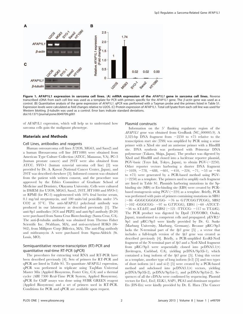
Figure 1. AFAP1L1 expression in sarcoma cell lines. (A) mRNA expression of the AFAP1L1 gene in sarcoma cell lines. Reversetranscribed cDNA from each cell line was used as a template for PCR with primers specific for the AFAP1L1 gene. The b-actin gene was used as acontrol. (B) Quantitative analysis of the gene expression of AFAP1L1. qPCR was performed with a Taqman probe and the primers listed in Table S1.Expression levels were calculated as fold changes relative to U2OS. (C) Protein expression of AFAP1L1. Total cell lysate from each cell line was used forWestern blotting. b-tubulin was used as a control. Error bars indicate standard deviations.doi:10.1371/journal.pone.0049709.g001
Sp3 Regulates a Sarcoma-Related Gene AFAP1L1
PLOS ONE | www.plosone.org 2 January 2013 | Volume 8 | Issue 1 | e49709
Institute of Japanese Foundation for Cancer Research, Tokyo,
Japan).
Luciferase assaysCells (26104) in 24-well dishes were transfected with 0.5 mg of
each reporter plasmid and 2 ng of pRL-TK control vector (Toyo
Ink) using Lipofectamine LTX (Invitrogen) according to the
manufacturer’s instructions. In the co-transfection experiments,
the total amount of plasmid was adjusted with pcDNA3.1(+) to
2 mg. Cells were harvested at 24 h after transfection and luciferase
assays were performed with the Dual Luciferase Assay Reporter
System (Promega, Madison, WI) as described previously [4].
Electrophoretic Mobility Shift Assay (EMSA)Single-stranded oligonucleotides (ONDs) corresponding to sense
and antisense sequences of the wild-type or mutated SBS1 site
were synthesized (Table S1), and mutated ONDs (25 pmol) were
end-labeled at 37uC for 30 min in a 50-ml reaction mixture
containing 1 ml of [c-32P]ATP and 10 units of T4 Polynucleotide
Kinase (New England Biolabs, Ipswich, MA). Sense and antisense
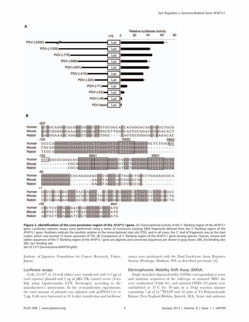
Figure 2. Identification of the core promoter region of the AFAP1L1 gene. (A) Transcriptional activity of the 59-flanking region of the AFAP1L1gene. Luciferase reporter assays were performed using a series of constructs carrying DNA fragments derived from the 59-flanking region of theAFAP1L1 gene. Numbers indicate the position relative to the transcriptional start site (TSS), and in all cases, the 39 end of fragments was at the startcodon, which was located 75 bases upstream of TSS. (B) Comparison of 59-flanking region of the AFAP1L1 gene among species. Human, mouse andrabbit sequences of the 59-flanking region of the AFAP1L1 gene are aligned, and conserved sequences are shown in gray boxes. EBS, Ets-binding site;SBS, Sp1-binding site.doi:10.1371/journal.pone.0049709.g002
Sp3 Regulates a Sarcoma-Related Gene AFAP1L1
PLOS ONE | www.plosone.org 3 January 2013 | Volume 8 | Issue 1 | e49709

ONDs of each pair were mixed and annealed by heating at 98uCfor 1 min and cooling off at room temperature for 1 h in a block
incubator. Double-stranded ONDs, designated SBS1WT and
SBS1MUT respectively, were purified with illustra ProbeQuantTM
G-50 Micro Colum’s (GE Healthcare, Little Chalfont, United
Kingdom). Nuclear extracts were prepared from cells by using
NE-PER Nuclear and Cytoplasmic Extraction Kit (Thermo Fisher
Scientific Inc.). The radio-labeled DNA probe was incubated for
15 minutes at room temperature with the reaction mixtures,
containing nuclear extract of U2OS (12 mg), 2 ml of 106 binding
buffer (Thermo Fisher Scientific Inc.), 1 mg of poly(dI-dC), 2.5%
glycerol, 5 mmol/L MgCl2, 1 mmol/L dithiothreitol and
0.5 mmol/L ZnCl2. DNA-protein complexes were loaded on a
6% nondenaturing polyacrylamide gel and electrophoresed at
200 V for 70 min. In the supershift assay, nuclear extracts were
mixed with the anti-Sp1 antibody (1C6), the anti-Sp3 antibody (D-
20), or rabbit non-immunized control IgG (Dako, Tokyo, Japan) in
the reaction mixture and incubated for 30 min on ice before the
formation of DNA-protein complexes. In the competition exper-
iment, excess amounts of unlabeled ONDs were added to the
reaction mixtures before the incubation with the labeled DNA
probe.
Chromatin Immunoprecipitation (ChIP) assayCells in a semi-confluent state in a 150-mm dish were fixed with
formaldehyde at a final concentration of 1.0% for 10 min at room
temperature to cross-link protein to DNA. Cells were washed with
ice-cold PBS and lysed in 300 ml of lysis buffer (10 mM Tris-HCl
pH 8.0, 300 mM NaCl, 1 mM EDTA, 0.5 mM EGTA, 0.1%
sodium deoxycholate and 0.5% N-laurolysarcosine), then sonicat-
ed on ice. Triton-X 100 was added at a final concentration of 10%
to dissolve the protein-DNA complexes. A soluble fraction was
obtained after centrifugation at 20,0006 g for 10 min at 4uC.
Fifteen microliters of supernatant (one-twentieth of the total
volume) was saved as an input, and the rest was divided into three
and mixed with Dynabeads (Invtirogen) at 4uC overnight with
rotation, which were pre-incubated with 2 mg of anti-Sp1 (PEP2)
or -Sp3 (D-20) antibodies, or rabbit non-immune IgG at 4uCovernight. The next day, immunoprecipitated complexes were
Figure 3. Identification of Sp1-binding sites as essential sequences for AFAP1L1 transcription. (A) Identification of core domains fortranscriptional activity. Open and closed circles represent wild-type and mutated EBS and open and closed rectangles represent wild-type andmutated SBS. PGV-vectors containing various segments of the AFAP1L1 promoter were transfected into U2OS cells, and their luciferase activities weremeasured. (B) The effect of exogenous Sp1 and Sp3 on the transcriptional activity of the core promoter region of the AFAP1L1 gene. The luciferaseactivity of the core promoter region (2224 to +75) was evaluated after Sp1 or Sp3-expressing vectors were co-transfected into U2OS cells. The totalamount of transfected plasmid DNA was equalized by the addition of pcDNA3.1(+), an empty vector. Error bars indicate standard deviations.doi:10.1371/journal.pone.0049709.g003
Sp3 Regulates a Sarcoma-Related Gene AFAP1L1
PLOS ONE | www.plosone.org 4 January 2013 | Volume 8 | Issue 1 | e49709

washed with low salt, high salt, LiCl RIPA buffer and finally with
TE (pH 8.0) buffer containing 50 mM NaCl. The complexes were
eluted from Dynabeads by treatment with the elution buffer
(50 mM Tris-HCl pH 8.0, 10 mM EDTA and 1% SDS) and
boiling at 65uC for 15 min. After centrifugation at 16,0006 g for
15 min at room temperature, DNA-protein cross-links were
reversed by incubating overnight at 65uC. Following RNaseA
and proteinase K treatment, DNA was purified and precipitated
with the phenol-chloroform method. PCR was performed to
amplify a DNA fragment spanning from -136 to +142 including
two SBSs and EBSs by KOD Plus polymerase (TOYOBO) and a
set of primers listed in Table S1.
Western blot analysesWestern blotting was performed as described previously [1].
Membranes were probed with anti-AFAP1L1 (1:2000), anti-Sp1
(1C6, 1:1000), anti-Sp3 (1:1000), anti-b-tubulin (1:1000), anti-
acetyl H3K9 (1:2500) and anti-Flag (1:2000) antibodies. An NE-
PER Kit (Thermo Fisher Scientific Inc.) was used to prepare
nuclear protein before the Western blotting.
Small inhibitory RNAs (siRNAs)siRNA duplexes were transfected into cells (1.56106 cells) using
RNAiMAX (Invitrogen) at a concentration of 20 nM. RNA and
protein were extracted 48 h and 72 h after transfection, respec-
tively. To knock down the Sp1 and Sp3 genes, two different
siRNAs were used (siSp1#1 and siSp1#2 for Sp1; siSp3#1 and
siSp3#2 for Sp3). siSp1#1 and siSp3#1were purchased from
Dharmacon (Thermo Fisher Scientific Inc.) and had been used in
our previous study [6]. Luciferase GL2 siRNA (siGL2) and GL3
siRNA (siGL3) were also purchased from Dharmacon. siSp1#2,
siSp3#2, and an siRNA sequence targeting Sp4 gene (siSp4) were
synthesized by Sigma-Aldrich (Table S1).
siRNA-resistant Sp3 geneA vector that harbors the Sp3(li-1) gene resistant to both
siSp3#1 and siSp3#2 was generated by a mutagenesis-based
method. Primers for mutagenesis were designed to harbor silent
mutations at the third nucleotide of every codon in the target
sequence (Table S1). pcDNA/Sp3(li-1) was sequentially mutated
using the two sets of primers, and the construct was transferred to
Figure 4. Binding of Sp transcription factors to the core-promoter region of the AFAP1L1 gene in vitro. EMSA was performed to analyzethe binding ability of putative transcription binding sites. Nuclear extracts were prepared from U2OS cells. Cold competitor experiments wereconducted by the addition of 25- and 50-fold excess amounts of unlabeled SBS1WT or SBS1MUT to nuclear extracts before incubating with labeledSBS1WT (lanes c–f). Supershift experiments were conducted by the addition of anti-Sp1 or anti-Sp3 antibody to protein-OND complexes (lanes g andh). Non-immune IgG was used as a control (lane i). Open and closed arrowheads indicate the Sp3-OND and Sp1-OND complex, respectively. Singleand double asterisks indicate bands supershifted by the addition of Sp1 or Sp3 antibody, respectively.doi:10.1371/journal.pone.0049709.g004
Sp3 Regulates a Sarcoma-Related Gene AFAP1L1
PLOS ONE | www.plosone.org 5 January 2013 | Volume 8 | Issue 1 | e49709

pLenti6/V5-DEST (Invitrogen). pLenti6/V5-GW/lacZ (Invitro-
gen) and pLenti6/V5-DEST/EGFP were used as lentiviral
controls. Using the ViraPower Lentiviral Expression System
(Invitrogen), U2OS cells were infected with viral supernatant
containing the siRNA-resistant Sp3(li-1) or control gene according
to the manufacturer’s instructions.
Matrigel invasion assayAt 48 h after siRNA treatment, cells were collected and cultured
in BioCoat Matrigel Invasion Chambers (BD Biosciences) and 8-
mm pore Control Cell Culture Inserts (BD Biosciences) as
described previously [1]. Cells (56104) were seeded in each
chamber in triplicate and incubated for 22 h. Then cells were
fixed and migrating cells were counted in five random fields under
the microscope at 6100 magnification.
Results
AFAP1L1 mRNA expression in sarcoma cell linesFirst, we checked AFAP1L1 expression in sarcoma cell lines by
RT-PCR and qPCR. AFAP1L1 was expressed strongly in U2OS
and MG63 cells, very weakly in SYO-1 and Saos2 cells, and not at
all in HT1080 cells (Fig. 1A–B). In the Western blot analysis,
AFAP1L1 was detected in U2OS and MG63 cells but undetect-
able in SYO-1, Saos2 and HT1080 cells (Fig. 1C), indicating that
the expression of AFAP1L1 was regulated differently among
sarcomas at the transcriptional level.
AFAP1L1 promoter activity depends on the proximalconserved region
To identify the transcriptional regulatory elements of the
AFAPL1 gene, DNA fragments with various segments of the
AFAP1L1 promoter were cloned into the PGV-basic vector as
described in the section of Materials and Methods. They were
transfected into U2OS cells expressing endogenous AFAP1L1 and
their luciferase activities were measured (Fig. 2A). The longest
fragment showed the strongest promoter activity and shorter ones
less, but the decrease was not remarkable until the fragment lost
the region between 2224 and 271 relative to TSS (Fig. 2A). By
searching the CONSITE database [7], we found that the sequence
from 2150 to 240 was highly conserved in three species (Fig. 2B).
Of note, within that conserved region two Ets-binding motifs (59-
(A/C)GGA(A/T)-39) and two Sp1-binding motifs (59-GGGCGG-
39) were identified. The proximal (260 to 256) and distal (2102
to 297) Ets-binding motifs were designated Ets-binding site 1
(EBS1) and 2 (EBS2), respectively. The proximal Sp1-binding site
(286 to 276) contained two overlapping consensus sequences
(286 to 281 and 281 to 276) and was conserved completely in
Figure 5. Identification of Sp3 as a major transcription factor for AFAP1L1. (A) and (B) Binding of Sp transcription factors to the core-promoter region of the AFAP1L1 gene in vitro. ChIP assays were performed using anti-Sp1 and anti-Sp3 antibodies or control IgG and the precipitatedDNA was PCR-amplified using a pair of primers located in the core-promoter region (Table S1) (A), and the precipitated genome was quantified byqPCR (B). (C) The effect of mithramycin A treatment on Sp3 binding. U2OS cells were treated with mithramycin A or DMSO for 48 h, andimmunoprecipitated DNA by Sp3 antibody was quantified by qPCR. (D) The effect of mithramycin A on the expression of the AFAP1L1 gene. RNA wasextracted from U2OS cells treated with mithramycin A or DMSO for 48 h, and RT-PCR was performed to semi-quantify the expression of each gene.The b-actin and GAPDH genes were used as a control. Error bars indicate standard deviations.doi:10.1371/journal.pone.0049709.g005
Sp3 Regulates a Sarcoma-Related Gene AFAP1L1
PLOS ONE | www.plosone.org 6 January 2013 | Volume 8 | Issue 1 | e49709

all three species, and was designated SBS1. The distal Sp1-binding
site (SBS2) spanning 2102 to 297 was found only in the human
genome. Several studies have shown that Ets and Sp proteins
function together in the transcription of target genes [8,9], and
therefore we focused on Ets and Sp transcription factors.
The Proximal Sp1-binding site is essential to AFAP1L1transcription
To investigate the role of Ets and Sp transcription factors in the
promoter activity, four types of luciferase reporters with mutations
in the conserved sequence of each binding site were constructed
using PGV-(2224) as a template and designated PGV-mtEBS1,
PGV-mtEBS2, PGV-mtSBS1, and PGV-mtSBS2. When EBS1
was mutated, the promoter activity was reduced by 50%
compared to PGV-(2224), although PGV-(271) which retained
EBS1 also showed reduced activity (Fig. 3A). However, the effect
was most remarkable when SBS1 was mutated, which resulted in a
75% reduction in promoter activity (Fig. 3A). This level was
almost equivalent to that of PGV-(253), which retained no EBSs
or SBSs. Mutations in EBS2 or SBS2 had less significant effects on
the promoter activity (Fig. 3A). These results suggested that
although both Sp and Ets proteins might play roles in
transcriptional regulation of the AFAP1L1 gene, the Sp protein
binding to SBS1 is the main factor driving the expression of
AFAP1L1. Therefore, we focused on Sp proteins.
Sp1 and Sp3 transactivate the proximal AFAP1L1promoter
To determine whether Sp1 and/or Sp3 transactivate the
promoter activity of the AFAP1L1 gene, a luciferase assay was
carried out using the Sp1 (pEVR2/Sp1) and Sp3 (pcDNA/Sp3(li-
1)) expression vectors, which produce each protein effectively in
transfected cells (Fig. S1). Co-transfection of the Sp1 or Sp3(li-1)
expression vector increased the promoter activity of PGV-(2224)
in a dose-dependent manner (Fig. 3B), suggesting Sp1 and Sp3 to
function in the transactivation of AFAP1L1. Interestingly, co-
transfection of the vector expressing a short form of Sp3, Sp3(si-1),
significantly reduced the promoter activity of PGV-(2224) (Fig.
S2). No significant effects were observed on the co-transfection of
the Sp3(li-2) or Sp3(si-2) expression vector (data not shown).
Sp1 and Sp3 bind to AFAP1L1’s proximal promoter regionTo elucidate whether Sp1 and Sp3 bind to SBS1 in vitro, EMSA
was conducted using labeled SBS1 OND and U2OS nuclear
extract. Using wild-type ONDs (SBS1WT), several shifted bands
were observed (Fig. 4, lane b), among which three showed a
decrease in intensity in competition with unlabeled SBS1WT in a
dose-dependent manner (Fig. 4, lanes c–d). These three bands were
not detected when labeled SBS1MUT was used instead of SBSWT
for the assay (Fig. S3, lanes f–h). When unlabeled SBS1MUT was
used as a competitor, no reduction in intensity was observed (Fig. 4,
lanes e and f), suggesting that the bands were specific to SBS1
complexes. When the anti-Sp1 antibody was added to the OND/
protein mixture, the intensity of the uppermost band decreased
and a supershifted band was identified, whereas no remarkable
changes were observed in the other two bands (Fig. 4, lane g; Fig.
S3, lane c). The intensity of the uppermost band showed no change
when an anti-Sp3 antibody was used but the other two bands
showed a clear difference (Fig. 4, lane h; Fig. S3, lane d). The
intensity of the middle band decreased and the lower band almost
disappeared, which was associated with the appearance of two
supershifted bands (Fig. 4, lane h). These changes were not
observed when labeled SBS1MUT was used in the assay (Fig. S3,
lanes g–h). No remarkable change was observed with the addition of
control IgG (Fig. 4, lane i). These results suggested that the
uppermost and lower two bands corresponded to Sp1- and Sp3-
OND complexes, respectively, and therefore both Sp1 and Sp3
are able to bind to the proximal Sp1-binding site in vitro. Similar
results were obtained when nuclear extracts were prepared from
Figure 6. Linking of Sp3 with AFAP1L1 by siRNA experiments.(A) The specificity of siRNA. U2OS cells were treated with siRNAtargeting Sp1, Sp3, or Sp4 for 48 h, and the expression of these genes aswell as the AFAP1L1 gene was analyzed by PCR. Two different siRNAstargeting the Sp1 and Sp3 genes were designed and used. b-actin wasused as a control. (B) Down-regulation of AFAP1L1 expression by siRNAtargeting the Sp3 gene at the mRNA level. U2OS cells were treated withsiRNAs targeting each gene for 48 h and the expression of AFAP1L1 wasanalyzed by qPCR and indicated as fold changes relative to that inuntreated cells. (C) Down-regulation of AFAP1L1 expression by siRNAtargeting the Sp3 gene at the protein level. U2OS cells were treatedwith siRNA targeting each gene for 72 h and proteins were extractedand used for Western blotting. b-tubulin was used as a control.doi:10.1371/journal.pone.0049709.g006
Sp3 Regulates a Sarcoma-Related Gene AFAP1L1
PLOS ONE | www.plosone.org 7 January 2013 | Volume 8 | Issue 1 | e49709
MG63 cells, which were strongly positive for AFAP1L1 (Fig. S4,
lanes h–n). Interestingly, similar results were also obtained when
nuclear extracts were prepared from SYO-1 cells, which were very
weakly positive for AFAP1L1 (Fig. S4, lanes a–g). These results
suggested that the expression of AFAP1L1 in vivo was regulated by
not only the cis-element but also other factors such as chromatic
modification.
Sp3 regulates the transcription of the AFAP1L1 gene bybinding to the endogenous promoter region
To investigate whether Sp1 and/or Sp3 bind to SBS1 in vivo,
ChIP assays were conducted using four cell lines in which the gene
expression of AFAP1L1 differed considerably; U2OS (strong),
MG63 (strong), SYO-1 (very weak), Saos2 (very weak) and
HT1080 (null) (Fig. 1A). We found that Sp3 bound to the
AFAP1L1 promoter region strongly in U2OS and MG63 cells
(Fig. 5A), but weakly in SYO-1 and Saos2 cells. No binding of Sp3
to the proximal promoter region was detected in HT1080 cells.
Binding of Sp1 was below the significant level by as determined by
qPCR (data not shown). Quantitative analyses showed a clear
correlation between the binding of Sp3 and the expression level of
AFAP1L1 (Fig. 1A and Fig. 5B). To exclude the possibility that this
difference in the binding of Sp3 to the promoter is due to
mutations in binding sites, we checked the genomic DNA of
U2OS, MG63, SYO-1 and HT1080. No mutations were found in
the proximal promoter including EBS1, EBS2, SBS1 and SBS2 in
any of the cell lines investigated (data not shown).
Mithramycin A is an aureolic acid antibiotic, which inhibits
gene expression by displacing transcriptional activators like the Sp
protein family that bind to GC-rich regions of promoters [10,11].
Treatment with mithramycin A inhibited the binding of Sp3 to the
promoter region of the AFAP1L1 gene in a dose-dependent
manner (Fig. 5C). Consistent with this finding, the treatment with
Mithramycin A reduced the mRNA expression of AFAP1L1
without changing that of Sp3 in U2OS cells (Fig. 5D). Similar
results were observed in another AFAP1L1-positive cell line,
MG63 cells (Fig. S5). These results indicate that the binding of Sp3
to SBS1 is a prerequisite for AFAP1L1 transcription, the level of
which is regulated by the extent of the binding. Total and nuclear
protein levels of Sp3 are almost the same in these four cell lines
(Fig. S6A–B), suggesting the existence of undiscovered mecha-
nisms that regulate the binding of Sp3 to SBS1. The luciferase
assays suggested the involvement of the Ets protein family in the
regulation of AFAP1L1 transcription (Fig. 3A). Transfection of a
dominant-negative Ets vector significantly reduced AFAP1L1
promoter activity, also suggesting the Ets family to participate in
the transcription of AFAP1L1 (Fig. S7A). Interestingly, transfection
of ELK1, another member of the Ets family, reduced AFAP1L1
promoter activity (Fig. S7A), and we found that forced expression
of ELK1 up-regulated the two short isoforms of Sp3 (Fig. S7B),
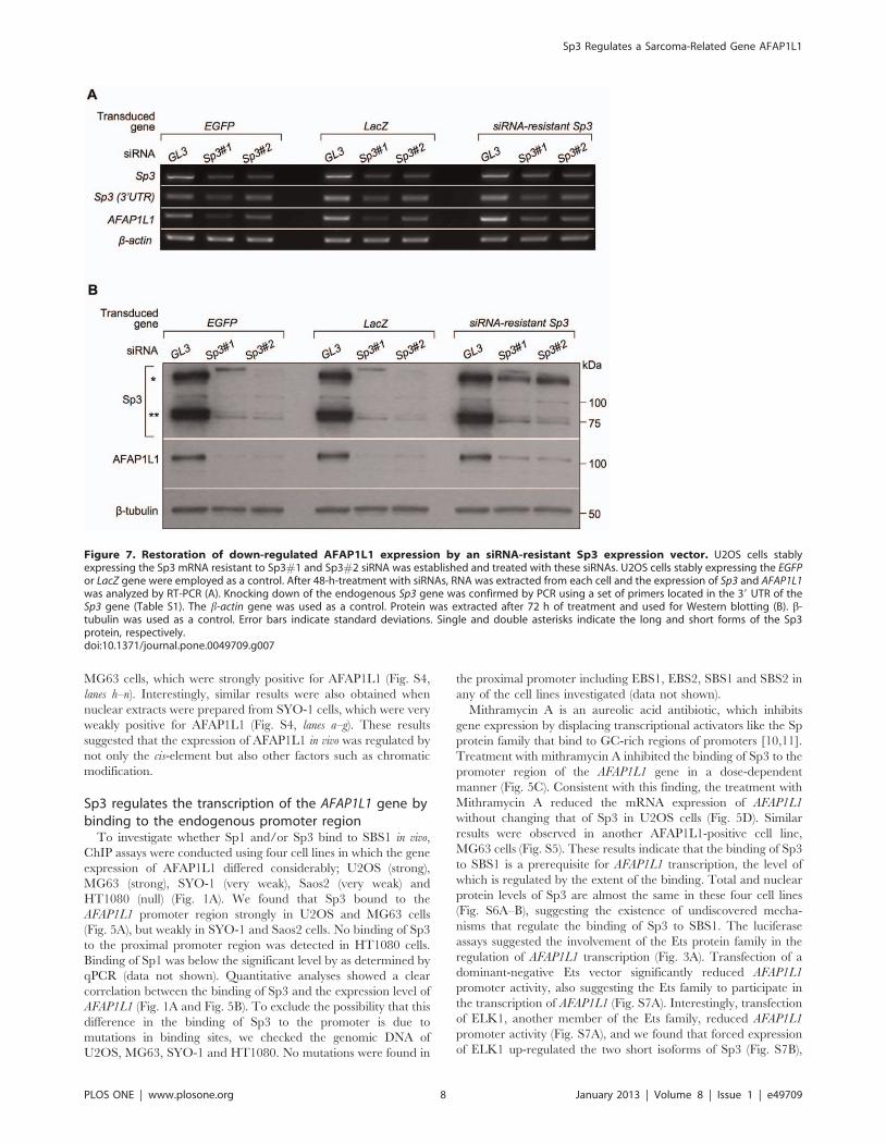
Figure 7. Restoration of down-regulated AFAP1L1 expression by an siRNA-resistant Sp3 expression vector. U2OS cells stablyexpressing the Sp3 mRNA resistant to Sp3#1 and Sp3#2 siRNA was established and treated with these siRNAs. U2OS cells stably expressing the EGFPor LacZ gene were employed as a control. After 48-h-treatment with siRNAs, RNA was extracted from each cell and the expression of Sp3 and AFAP1L1was analyzed by RT-PCR (A). Knocking down of the endogenous Sp3 gene was confirmed by PCR using a set of primers located in the 39 UTR of theSp3 gene (Table S1). The b-actin gene was used as a control. Protein was extracted after 72 h of treatment and used for Western blotting (B). b-tubulin was used as a control. Error bars indicate standard deviations. Single and double asterisks indicate the long and short forms of the Sp3protein, respectively.doi:10.1371/journal.pone.0049709.g007
Sp3 Regulates a Sarcoma-Related Gene AFAP1L1
PLOS ONE | www.plosone.org 8 January 2013 | Volume 8 | Issue 1 | e49709
which may be responsible for the reduction in promotor activity,
based on the results of co-transfection experiments (Fig. S2).
mRNA expression levels of ELK1 and ELK4 showed no significant
differences among sarcoma cell lines irrespective of the AFAP1L1
expression level (Fig. S7C).
Sp3 is essential to the expression of AFAP1L1Finally, siRNA was employed to investigate the role of Sp3 in
AFAP1L1 transcription in vivo. In U2OS cells, siRNA targeting
each of Sp1, Sp3, and Sp4 significantly reduced the expression of
the targeted gene, but only the siRNA targeting Sp3 consistently
reduced the expression of the AFAP1L1 gene (Fig. 6A), which was
confirmed by quantitative analyses (Fig. 6B). Specific reduction of
AFAP1L1 expression by siRNA against Sp3 was further confirmed
at the protein level (Fig. 6C). These effects of siRNA against Sp3
were also confirmed in other cell lines (MG63 and SYO-1) at the
mRNA level (Fig. S8A–D). This phenomenon was also observed in
prostate cancer PC-3 cells (Fig. S9), indicating that the transcrip-
tional role of Sp3 for the AFAP1L1 gene is not restricted to
sarcoma cells. To exclude the off-target effect of siRNA, a rescue
experiment was carried out. Pre-induction of siRNA-resistant Sp3
using a lentivirus partially rescued AFAP1L1 expression after Sp3
siRNA treatment (Fig. 7A–B), indicating that the reduction in
AFAP1L1 expression cause by siRNA for Sp3 is due to a direct
effect on the Sp3 gene.
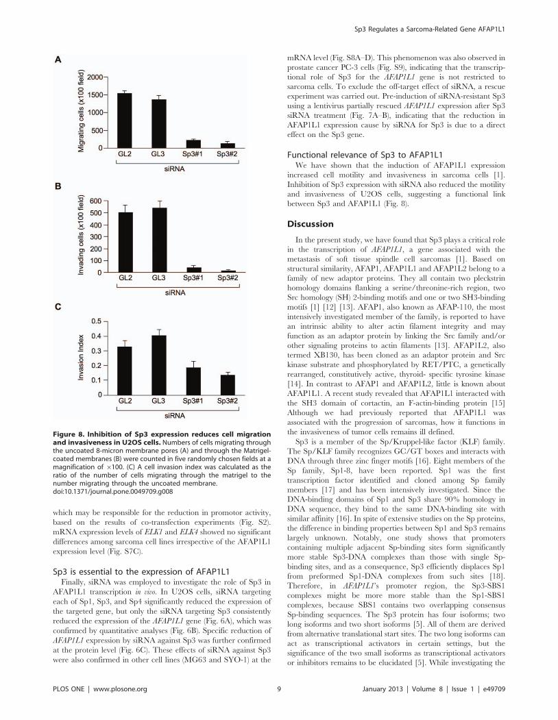
Functional relevance of Sp3 to AFAP1L1We have shown that the induction of AFAP1L1 expression
increased cell motility and invasiveness in sarcoma cells [1].
Inhibition of Sp3 expression with siRNA also reduced the motility
and invasiveness of U2OS cells, suggesting a functional link
between Sp3 and AFAP1L1 (Fig. 8).
Discussion
In the present study, we have found that Sp3 plays a critical role
in the transcription of AFAP1L1, a gene associated with the
metastasis of soft tissue spindle cell sarcomas [1]. Based on
structural similarity, AFAP1, AFAP1L1 and AFAP1L2 belong to a
family of new adaptor proteins. They all contain two pleckstrin
homology domains flanking a serine/threonine-rich region, two
Src homology (SH) 2-binding motifs and one or two SH3-binding
motifs [1] [12] [13]. AFAP1, also known as AFAP-110, the most
intensively investigated member of the family, is reported to have
an intrinsic ability to alter actin filament integrity and may
function as an adaptor protein by linking the Src family and/or
other signaling proteins to actin filaments [13]. AFAP1L2, also
termed XB130, has been cloned as an adaptor protein and Src
kinase substrate and phosphorylated by RET/PTC, a genetically
rearranged, constitutively active, thyroid- specific tyrosine kinase
[14]. In contrast to AFAP1 and AFAP1L2, little is known about
AFAP1L1. A recent study revealed that AFAP1L1 interacted with
the SH3 domain of cortactin, an F-actin-binding protein [15]
Although we had previously reported that AFAP1L1 was
associated with the progression of sarcomas, how it functions in
the invasiveness of tumor cells remains ill defined.
Sp3 is a member of the Sp/Kruppel-like factor (KLF) family.
The Sp/KLF family recognizes GC/GT boxes and interacts with
DNA through three zinc finger motifs [16]. Eight members of the
Sp family, Sp1-8, have been reported. Sp1 was the first
transcription factor identified and cloned among Sp family
members [17] and has been intensively investigated. Since the
DNA-binding domains of Sp1 and Sp3 share 90% homology in
DNA sequence, they bind to the same DNA-binding site with
similar affinity [16]. In spite of extensive studies on the Sp proteins,
the difference in binding properties between Sp1 and Sp3 remains
largely unknown. Notably, one study shows that promoters
containing multiple adjacent Sp-binding sites form significantly
more stable Sp3-DNA complexes than those with single Sp-
binding sites, and as a consequence, Sp3 efficiently displaces Sp1
from preformed Sp1-DNA complexes from such sites [18].
Therefore, in AFAP1L1’s promoter region, the Sp3-SBS1
complexes might be more more stable than the Sp1-SBS1
complexes, because SBS1 contains two overlapping consensus
Sp-binding sequences. The Sp3 protein has four isoforms; two
long isoforms and two short isoforms [5]. All of them are derived
from alternative translational start sites. The two long isoforms can
act as transcriptional activators in certain settings, but the
significance of the two small isoforms as transcriptional activators
or inhibitors remains to be elucidated [5]. While investigating the
Figure 8. Inhibition of Sp3 expression reduces cell migrationand invasiveness in U2OS cells. Numbers of cells migrating throughthe uncoated 8-micron membrane pores (A) and through the Matrigel-coated membranes (B) were counted in five randomly chosen fields at amagnification of 6100. (C) A cell invasion index was calculated as theratio of the number of cells migrating through the matrigel to thenumber migrating through the uncoated membrane.doi:10.1371/journal.pone.0049709.g008
Sp3 Regulates a Sarcoma-Related Gene AFAP1L1
PLOS ONE | www.plosone.org 9 January 2013 | Volume 8 | Issue 1 | e49709
role of Sp3 and Ets in the AFAP1L1 promoter’s activity, we found
that forced expression of ELK1, an Ets transcription factor,
induced up-regulation of the two short isoforms of Sp3 and
resulted in decreased AFAP1L1 promoter activity (Fig. S7B). As
forced expression of a short isoform (si-1) reduced the AFAP1L1
promoter activity induced by endogenous factors (Fig. S2), si-1
may have a negative effect on the transcription of AFAP1L1.
Sp1 and Sp3 have been shown to be expressed ubiquitously and
reported to regulate basal and constitutive expression of genes
both in normal and cancerous tissues [19]. Several reports have
referred to a correlation between Sp1 and Sp3 and tumor
development, growth and metastasis. Sp1 is reported to be
overexpressed and regulate vascular endothelial growth factor
(VEGF) in gastric and shown to be linked to a poor prognosis [20].
Up-regulation of Sp1 expression has been also observed in thyroid
[21] and colorectal cancer [22]. Sp3 enhances the growth of
pancreatic cancer cells by suppressing p27 expression through
interaction with GC-rich promoter elements [23]. In breast
cancer, Sp3 accelerates tumor cell growth by acting as a repressor
of TGF signaling [24]. A recent report demonstrated the
expression of Sp3 to be an independent prognostic factor for the
poor survival of head and neck cancer patients [25]. Of note, in
the web database ONCOMINE (http://www.oncomine.org),
upregulation of Sp3 expression in soft tissue sarcomas compared
to normal connective tissue has been confirmed [26] [27].
Because the cause of sarcoma patients’ death is uncurable
distant metastasis in most cases, methods of both predicting and
treating metastasis are urgently needed. Our findings may provide
new insight regarding this clinical difficulty. Considering that Sp3
is expressed at higher levels in soft tissue sarcomas and
transactivates the AFAP1L1 gene, targeting Sp3 could be a
powerful approach to treating advanced soft tissue sarcomas.
Supporting Information
Figure S1 Expression of exogenous Sp1 or Sp3 proteinin 293T cells. 293T cells were transfected with each plasmid, as
described in Materials and Methods, and the expression of the Sp1
or Sp3 protein was analyzed 24 h later. pRC/Sp3 lacks N-
terminal part of the Sp3 gene as described in Experimental Procedures.
b-tubulin was used as an internal control. Single and double
asterisks indicate the long and short forms of the Sp3 protein,
respectively.
(TIF)
Figure S2 Isoform-dependent activity of Sp3 onAFAP1L1 promoter. The luciferase reporter assay was per-
formed as described in Fig. 3B. Reporter plasmids were co-
transfected with either an empty, Sp1 or Sp3 expression vector.
Error bars indicate the standard deviations.
(TIF)
Figure S3 Binding of Sp transcription factors to thewild-type, but not mutated Sp-binding site in vitro.Nuclear extracts were prepared from U2OS cells and used for
EMSA with radiolabeled SBS1WT (lane a–d) or SBS1MUT (lanes
e–h). A supershifted assay was performed with anti-Sp1 (lane c and
g) or anti-Sp3 (lane d and h) antibody. Open and closed arrowheads
indicate an Sp3-OND and Sp1-OND complex.
(TIF)
Figure S4 EMSA using nuclear extracts from cellsexpressing the AFAP1L1 gene very weakly (SYO-1) andstrongly (MG63). Nuclear extracts were prepared from SYO-1
and MG63 cells, and EMSA was performed as described in
Figure 4. Open and closed arrowheads indicate Sp3-OND and
Sp1-OND complex, respectively. Single and double asterisks
indicate bands supershifted by the addition of Sp1 or Sp3
antibody, respectively.
(TIF)
Figure S5 The effect of mithramycin in MG63 cells.RNA was extracted from MG63 cells treated with mithramycin A
at the indicated dose or DMSO for 48 h, and subjected to RT-
PCR. The b-actin gene was used as a control.
(TIF)
Figure S6 Western blot analyses of AFAP1L1, Sp1 andSp3 in sarcoma cell lines. Total cell lysate (A) or nuclear
extract (B) was prepared from each cell line and used for Western
blotting. b-tubulin and acetylated H3K9 were used as the internal
control for total cell lysate and nuclear extract, respectively. Single
and double asterisks indicate the long and short forms of the Sp3
protein, respectively.
(TIF)
Figure S7 The effect of Ets transcription factors on theexpression of AFAP1L1. (A) The effect of Ets transcription
factors on luciferase activity. Luciferase assays were performed in
U2OS cells 48 h after the co-transfection of various expression
vectors containing an Ets transcription factor with PGV-(2224).
(B) The effect of ELK1 on the expression of Sp3. 293T cells were
transfected with indicated plasmids and proteins were analyzed at
24 h by Western blotting. b-tubulin was used as an internal
control. Single and double asterisks indicate the long and short
forms of Sp3, respectively. DN-Ets represents dominant negative
Ets. (C) Expression of ELK family gene in sarcoma cells. RNA was
extracted from cells and RT-PCR was performed.
(TIF)
Figure S8 Down-regulation of AFAP1L1 expression bysiRNA targeting the Sp3 gene in SYO-1 and MG63 cells.(A) and (D) The specificity of siRNA. SYO-1 (A) and MG63 (D) cells
were treated with siRNA targeting Sp1, Sp3, or Sp4 for 48 h, and the
expression of these genes as well as the AFAP1L1 gene was analyzed
by PCR. Two different siRNAs targeting the Sp1 and Sp3 genes
were designed and used. b-actin was used as a control. (B) and (E)
Down-regulation of AFAP1L1 expression by siRNA targeting the
Sp3 gene at the mRNA level. SYO-1 (B) and MG63 (E) cells were
treated with siRNAs targeting each gene for 48 h and the expression
of AFAP1L1 was analyzed by qPCR and indicated as fold changes
relative to that in untreated cells. (C) and (F) Down-regulation of
AFAP1L1 expression by siRNA targeting the Sp3 gene at the
protein level. SYO-1 (C) and MG63 (F) cells were treated with
siRNAs targeting each gene for 72 h and proteins were extracted
and used for Western blotting. b-actin was used as a control.
(TIF)
Figure S9 Down-regulation of Sp3 expression causesdown-regulation of AFAP1L1 expression in prostatecancer cells. (A) The specificity of siRNA. PC-3 cells were
treated with siRNA targeting Sp1, Sp3, or Sp4 for 48 h, and the
expression of these genes as well as the AFAP1L1 gene was
analyzed by PCR. Two different siRNAs targeting the Sp1 or Sp3
gene were designed and used. b-actin was used as a control. (B)
Down-regulation of AFAP1L1 expression by siRNA targeting the
Sp3 gene at the protein level. PC-3 cells were treated with siRNA
targeting each gene for 72 h and proteins were extracted and used
for Western blotting. b-tubulin was used as a control. Single and
double asterisks indicate the long and short forms of Sp3,
respectively.
(TIF)
Sp3 Regulates a Sarcoma-Related Gene AFAP1L1
PLOS ONE | www.plosone.org 10 January 2013 | Volume 8 | Issue 1 | e49709

Table S1 Sequences for primers and other oligonucle-otides used in this study.(XLS)
Acknowledgments
We thank Dr. Y. Jin and Mrs. Y. Kobayashi for technical support and Dr.
M. Ikeya for editing the manuscript.
Author Contributions
Conceived and designed the experiments: YK HN EN OO JT. Performed
the experiments: YK MF ST TK RT SN TA. Analyzed the data: YK JT.
Contributed reagents/materials/analysis tools: TK YN JT. Wrote the
paper: YK JT.
References
1. Furu M, Kajita Y, Nagayama S, Ishibe T, Shima Y, et al. (2011) Identification of
AFAP1L1 as a prognostic marker for spindle cell sarcomas. Oncogene 30: 4015–4025.
2. Kawai A, Naito N, Yoshida A, Morimoto Y, Ouchida M, et al. (2004)
Establishment and characterization of a biphasic synovial sarcoma cell line,SYO-1. Cancer Lett 204: 105–113.
3. Kato, Jr T, Gotoh Y, Hoffmann A., Ono Y (2008) Negative regulation ofconstitutive NF-kB and JNK signaling by PKN1–mediated phosphorylation of
TRAF1: Genes to Cells; 13: 509–520.
4. Kohno Y, Okakmoto T, Ishibe T, Nagayama S, Shima Y, et al. (2006)Expression of claudin7 is tightly associated with epithelial structures in synovial
sarcomas and regulated by an Ets family transcription factor, ELF3. J Biol Chem281: 38941–38950.
5. Sapetschnig A, Koch F, Rischitor G, Mennenga T, Suske G (2004) Complexityof translationally controlled transcription factor Sp3 isoform expression. J Biol
Chem 279: 42095–42105.
6. Aoyama T, Okamoto T, Kohno Y, Fukiage K, Otsuka S, et al. (2008) Cell-specific epigenetic regulation of ChM-I gene expression: crosstalk between DNA
methylation and histone acetylation. Biochem Biophys Res Commun 365: 124–130.
7. Sandelin A, Wasserman WW, Lenhard B (2004) ConSite: web-based prediction
of regulatory elements using cross-species comparison. Nucleic Acids Res 32:W249–252.
8. Giatzakis C, Batarseh A, Dettin L, Papadopoulos V (2007) The role of Etstranscription factors in the basal transcription of the translocator protein (18
kDa). Biochemistry 46: 4763–4774.9. Shirasaki F, Makhluf HA, LeRoy C, Watson DK, Trojanowska M (1999) Ets
transcription factors cooperate with Sp1 to activate the human tenascin-C
promoter. Oncogene 18: 7755–7764.10. Blume SW, Snyder RC, Ray R, Thomas S, Koller CA, et al. (1991)
Mithramycin inhibits SP1 binding and selectively inhibits transcriptional activityof the dihydrofolate reductase gene in vitro and in vivo. J Clin Invest 88: 1613–
1621.
11. Ray R, Snyder RC, Thomas S, Koller CA, Miller DM (1989) Mithramycinblocks protein binding and function of the SV40 early promoter. J Clin Invest
83: 2003–2007.12. Xu J, Bai XH, Lodyga M, Han B, Xiao H, et al. (2007) XB130, a novel adaptor
protein for signal transduction. J Biol Chem 282: 16401–16412.
13. Flynn DC, Leu TH, Reynolds AB, Parsons JT (1993) Identification andsequence analysis of cDNAs encoding a 110-kilodalton actin filament-associated
pp60src substrate. Mol Cell Biol 13: 7892–7900.
14. Lodyga M, De Falco V, Bai XH, Kapus A, Melillo RM, et al. (2009) XB130, a
tissue-specific adaptor protein that couples the RET/PTC oncogenic kinase toPI 3-kinase pathway. Oncogene 28: 937–949.
15. Snyder BN, Cho Y, Qian Y, Coad JE, Flynn DC, et al. (2011) AFAP1L1 is a
novel adaptor protein of the AFAP family that interacts with cortactin andlocalizes to invadosomes. Eur J Cell Biol 90: 376–389.
16. Suske G (1999) The Sp-family of transcription factors. Gene 238: 291–300.17. Kadonaga JT, Carner KR, Masiarz FR, Tjian R (1987) Isolation of cDNA
encoding transcription factor Sp1 and functional analysis of the DNA binding
domain. Cell 51: 1079–1090.18. Yu B, Datta PK and Bagchi S (2003) Stability of the Sp3-DNA is promoter-
specific: Sp3 efficiently competes with Sp1 for binding to promoters containingmultiple Sp-sites. Nucleic Acids Res 31: 5368–5376
19. Safe S, Abdelrahim M (2005) Sp transcription factor family and its role incancer. Eur J Cancer 41: 2438–2448.
20. Wang L, Wei D, Huang S, Peng Z, Le X, et al. (2003) Transcription factor Sp1
expression is a significant predictor of survival in human gastric cancer. ClinCancer Res 9: 6371–6380.
21. Chiefari E, Brunetti A, Arturi F, Bidart JM, Russo D, et al. (2002) Increasedexpression of AP2 and Sp1 transcription factors in human thyroid tumors: a role
in NIS expression regulation? BMC Cancer 2: 35.
22. Hosoi Y, Watanabe T, Nakagawa K, Matsumoto Y, Enomoto A, et al. (2004)Up-regulation of DNA-dependent protein kinase activity and Sp1 in colorectal
cancer. Int J Oncol 25: 461–468.23. Abdelrahim M, Smith R 3rd, Burghardt R, Safe S (2004) Role of Sp proteins in
regulation of vascular endothelial growth factor expression and proliferation ofpancreatic cancer cells. Cancer Res 64: 6740–6749.
24. Wright C, Angus B, Napier J, Wetherall M, Udagawa Y, et al. (1987) Prognostic
factors in breast cancer: immunohistochemical staining for SP1 and NCRC 11related to survival, tumour epidermal growth factor receptor and oestrogen
receptor status. J Pathol 153: 325–331.25. Essafi-Benkhadir K, Grosso S, Puissant A, Robert G, Essafi M, et al. (2009) Dual
role of Sp3 transcription factor as an inducer of apoptosis and a marker of
tumour aggressiveness. PLoS One 4: e4478.26. Barretina J, Taylor BS, Banerji S, Ramos AH, Lagos-Quintana M, et al. (2010)
Subtype-specific genomic alterations define new targets for soft-tissue sarcomatherapy. Nat Genet 42: 715–721.
27. Detwiller KY, Fernando NT, Segal NH, Ryeom SW, D’Amore PA, et al. (2005)
Analysis of hypoxia-related gene expression in sarcomas and effect of hypoxia onRNA interference of vascular endothelial cell growth factor A. Cancer Res 65:
5881–5889.
Sp3 Regulates a Sarcoma-Related Gene AFAP1L1
PLOS ONE | www.plosone.org 11 January 2013 | Volume 8 | Issue 1 | e49709