The ETS-domain transcription factor Elk-1 regulates the expression ...
36
Kasza et al. 1 The ETS-domain transcription factor Elk-1 regulates the expression of its partner protein, SRF. Aneta Kasza, Amanda O’Donnell, Karen Gascoigne, Leo A.H. Zeef, Andy Hayes and Andrew D. Sharrocks* Faculty of Life Sciences, University of Manchester, Michael Smith building, Oxford Road, Manchester, M13 9PT, UK. *Corresponding author: A.D. Sharrocks Tel: 0044-161 275 5979 Fax: 0044-161 275 5082 E-mail: [email protected] Running title: Elk-1 regulates SRF expression. Keywords: Elk-1, ETS-domain, MAP kinase, SRF, Transcription factor. JBC Papers in Press. Published on November 4, 2004 as Manuscript M411161200 Copyright 2004 by The American Society for Biochemistry and Molecular Biology, Inc. by guest on February 11, 2018 http://www.jbc.org/ Downloaded from
Transcript of The ETS-domain transcription factor Elk-1 regulates the expression ...
Kasza et al.
1
The ETS-domain transcription factor Elk-1 regulates the expression of its
partner protein, SRF.
Aneta Kasza, Amanda O’Donnell, Karen Gascoigne, Leo A.H. Zeef, Andy Hayes
and Andrew D. Sharrocks*
Faculty of Life Sciences, University of Manchester, Michael Smith building, Oxford Road,
Manchester, M13 9PT, UK.
*Corresponding author: A.D. Sharrocks
Tel: 0044-161 275 5979
Fax: 0044-161 275 5082
E-mail: [email protected]
Running title: Elk-1 regulates SRF expression.
Keywords: Elk-1, ETS-domain, MAP kinase, SRF, Transcription factor.
JBC Papers in Press. Published on November 4, 2004 as Manuscript M411161200
Copyright 2004 by The American Society for Biochemistry and Molecular Biology, Inc.
by guest on February 11, 2018http://w
ww
.jbc.org/D
ownloaded from

Kasza et al.
2
Abstract
The ternary complex factors (TCF) are a subfamily of ETS-domain transcription factors that
bind and activate serum response elements (SREs) in the promoters of target genes in a
ternary complex with a second transcription factor, serum response factor (SRF). Here, we
have identified the SRF gene as a target for the TCFs, thereby providing a positive feedback
loop whereby TCF activation leads to the enhancement of the expression of its partner protein
SRF. The binding of the TCF Elk-1 to the SRF promoter and subsequent regulation of SRF
expression occurs in a ternary complex-dependent manner. Our data therefore reveal that SRF
is an important target for the Erk and Rho signaling pathways that converge on a ternary
TCF-SRF complex at the SRE on the SRF promoter.
by guest on February 11, 2018http://w
ww
.jbc.org/D
ownloaded from
Kasza et al.
3
Introduction
Elk-1, SAP-1 and SAP-2/ERP/Net comprise the ternary complex factor (TCF)
subfamily of ETS-domain transcription factors (reviewed in 1, 2). These proteins form ternary
complexes on target promoters together with the MADS-box protein, serum response factor
(SRF). Both protein-DNA and protein-protein interactions with SRF are required to form
ternary complexes at the promoters of target genes like c-fos. The conserved B-box region of
the TCFs plays a pivotal role in mediating these protein-protein interactions (3, 4, 5, 6, 7, 8).
The TCFs can be phosphorylated within their transcription activation domains (TADs) by
members of all three of the major MAPK pathways present in mammals; ERK, JNK and p38
(reviewed in 1, 2, 9). In the case of Elk-1, this phosphorylation leads to the enhancement of its
transactivation properties both by the recruitment and activation of the coactivator proteins
Sur-2 and p300/CBP (10, 11) and also the loss of corepressor complexes containing HDAC-2
(12). Thus, Elk-1 provides an adaptor protein for SRF that can link it to the MAP kinase
signalling pathways.
In addition to binding to the TCFs, SRF has recently been shown to be able to bind to
members of the MAL/myocardin family of coactivator proteins (13, 14, 15, 16, 17, 18). In the
case of MAL, this interaction permits linkage of the MAL-SRF complex to the Rho signaling
pathway and subsequent activation of SRF-dependent gene expression (13, 16). The SRF
gene itself is thought to be one target gene for the MAL/myocardin-SRF complex. The
interaction of the TCFs and MAL/myocardin proteins with SRF is mutually exclusive, where
the Elk-1 B-box inhibits MAL/myocardin recruitment by SRF (19, 20). This suggests either
the existence of two different classes of SRF target genes (21) or the possibility of sequential
(13) or mutually exclusive interactions of different coregulatory proteins with the same SRF
target genes (20).
by guest on February 11, 2018http://w
ww
.jbc.org/D
ownloaded from
Kasza et al.
4
To date the number of TCF target genes identified is limited and comprises well
studied immediate-early genes such as c-fos and egr-1 (reviewed in 1, 2, 9). On these latter
targets, SRF appears to aid TCF recruitment, but the reciprocal situation appears to operate on
other genes such as nur77 where the TCFs recruit SRF to the promoter (22, 23). It is currently
unclear if the TCFs act in an SRF-independent manner although evidence has been gathered
to suggest such a role on genes such as TNF-α (24) and 9E3/cCAF (25).
Due to the possibility of functional redundancy amongst the TCFs, suggested by the
minimal phenotypes obtained in mouse knockout studies (23, 26, 27), we developed a cell
line encoding an inducible repressive form of Elk-1 (Elk-1 fused to the engrailed repression
domain; Elk-En) to probe the potential role of TCFs in regulating gene expression (28). The
induction of Elk-En caused apoptosis and one key target gene identified was the anti-
apoptotic gene Mcl-1. Importantly, the activity of Elk-En was B-box dependent demonstrating
that it functions in a SRF-dependent manner. Here, we have extended these studies and show
that the gene encoding the TCF partner protein SRF is also a target for the TCF Elk-1. Elk-1
works through a ternary complex with SRF on the SRF promoter, thereby providing a link to
the ERK signaling pathway in addition to the well characterized link to the Rho pathway that
acts through SRF on its own promoter.
by guest on February 11, 2018http://w
ww
.jbc.org/D
ownloaded from
Kasza et al.
5
Materials and Methods
Plasmid Constructs
pSRF-Luc(WT) (pAS2159) and pSRF-Luc(mETS-103) (pAS2160) contain a 322 bp
fragment of the mouse SRF promoter (-322- +1) upstream from the firefly luciferase gene,
with the wild-type promoter or containing a mutated ets site at position -103 respectively (29;
kindly provided by Ravi Misra), pCH110 (Pharmacia) contains an SV40 driven β-
galactosidase (LacZ) gene and is used to monitor transfection efficiency. pRSV-Elk-1-VP16
(pAS348) is an RSV promoter-driven vector encoding full-length wild-type Elk-1 fused to
residues 410-490 of VP16 C-terminal sequence (30). pAS1408 and pAS1411, encoding
respectively wild-type(WT) and L158P mutant derivatives of Elk-En (full-length Elk-1 fused
to the engrailed repression domain) have been described previously (28). pAS383 encodes
full-length wild-type Flag-tagged Elk-1 (31). pAS1801 encodes full-length mouse PEA3, and
was constructed by ligating a HindIII/SalI cleaved PCR product, (primers, A36/37) into
HindIII/XhoI cleaved pCDNA3. The plasmid pCGN (pAS2158) encodes full-length SRF
(kindly provided by Ravi Misra). pEFplink-MAL∆N encodes an N-terminally truncated,
constitutively active version of MAL (kindly provided by Richard Treisman; 16). pAS278
(encoding full-length His-Flag-tagged Elk-1) (31) and pAS58 (encoding GST fused to amino
acids 132-222 of SRF- coreSRF) (6) for expressing proteins in bacteria have been described
previously.
pAS197 and pAS489, encoding Elk-1(1-168)(WT) and Elk-1(1-168)(L158P) were
used in in vitro transcription/ translation of proteins and have been described previously (7).
by guest on February 11, 2018http://w
ww
.jbc.org/D
ownloaded from
Kasza et al.
6
Tissue culture, cell transfection and reporter gene assays
The EcR293 cell derivatives EcR293(Elk-En{WT})#1.3 and EcR293(Elk-
En{L158P})#L8, Hela and 293 cells were grown as described previously (28). Elk-En fusions
were induced in EcR293 derivatives by stimulating with 5 µM ponasterone A (PA).
Where indicated, cells were stimulated with 20% foetal calf serum (FCS) 10 nM PMA
or 0.5 µM jasplakinolide and where, required the inhibitors latrunculin B (LB) (0.5 µM) or
U0126 (10 µM) were added 30 and 60 mins respectively, prior to stimulation.
Transient transfection experiments were carried out using Polyfect transfection
reagent in 12 well plates (Qiagen). Luciferase assays carried out using the dual light reporter
gene assay system (Tropix) as described previously, using pCH110 as an internal control
(28).
Western Blot analysis
Western blotting was carried out using Supersignal West Dura Extended Duration
Substrate (Pierce) and the following primary antibodies; Anti-Elk-1 (Santa Cruz), anti-SRF
(Santa Cruz), anti-M2 FLAG antibody (Sigma). Data was visualised using a Biorad Fluor-S
MultiImager. Quantification of proteins was carried out using Quantity One software (Bio-
Rad).
Gel retardation assays
Gel retardation assays were carried out with a 32P-labelled 116 base-pair fragments of the
mouse SRF promoter generated by PCR on the templates pSRF-Luc(WT) and pSRF-
Luc(mETS-103) (primers; ADS1251, GCAGCGAGTTCGGTATGTC and ADS1252,
CCGCTCCTTATATGGCGAGC) as described previously (32). coreSRF was produced as a
GST-tagged protein in bacteria (6) C-terminally truncated Elk-1 was produced by in vitro
by guest on February 11, 2018http://w
ww
.jbc.org/D
ownloaded from

Kasza et al.
7
transcription and translation using a TNTTM-coupled reticulocyte lysate system. 35S-labelled
proteins were analysed by electrophoresis through a 0.1% SDS-12% polyacrylamide gel,
before visualisation and quantification using a phosphorimager and Quantity One software
(Bio-Rad). Protein-DNA complexes were analyzed on nondenaturing 5% polyacrylamide gels
cast in 0.5x Tris-borate-EDTA and visualized by autoradiography and phosphorimaging.
Northern and Microarray Analysis.
Total RNA extracted using the RNeasy kit (Qiagen) and Northern analysis, carried out as
described previously (28). Probes were made by random priming (Roche) using templates
derived from a human SRF and Elk-1 full-length cDNAs.
Microarray experiments were performed using Affymetrix “Human Focus” and
“Human U133A” GeneChip oligonucleotide arrays (Affymetrix, Inc.) as described previously
(28). Normalization and further analyses were carried out using RMAExpress software
(http://stat-www.berkeley.edu/users/bolstad/RMAExpress/RMAExpress.html). Microarray
data were generated using 3 independent samples from ponasterone A stimulated and 2 from
unstimulated EcR293(Elk-En)#1.3 cells.
Chromatin immunoprecipitation
Chromatin immunoprecipitations were carried out as described previously (28) and using
anti-Elk-1 (Santa Cruz) or non specific IgG (Upstate) antibodies. Promoter-specific primers
were used to amplify the DNA by PCR: human egr-1 (described in 28), human SRF promoter
(ADS1249, TGACAGCAACGAGTTCGGTA and ADS1250,
CCCCCATATAAAGAGATACAATGTT) and SRF intronic sequence (intron 3; +4986 to
by guest on February 11, 2018http://w
ww
.jbc.org/D
ownloaded from
Kasza et al.
8
+5167) (ADS1273, GCCACAGGGCAGTAGATGTT and ADS1274,
TCAGGCCCAAGTATCCACTC).
Induction of apoptosis and Hoechst Staining.
Apoptosis was scored by counting disrupted nuclei revealed by Hoechst stain (Sigma) as
described previously (28). Etoposide (0.2 mM) was added to 293 cells for 48 hours to induce
apoptosis.
by guest on February 11, 2018http://w
ww
.jbc.org/D
ownloaded from
Kasza et al.
9
Results
Repressive Elk-1 constructs downregulate the expression of SRF.
To study the effects of inhibiting TCF-mediated gene expression, and thereby uncover
novel TCF target genes, we created a 293-derived stable cell line that inducibly expresses a
fusion of Elk-1 with the powerful Engrailed (En) repression domain [EcR293(Elk-En)#1.3]
(28). Upon induction of its expression with ponasterone A (PA), this fusion protein is
expected to be recruited to all TCF-dependent promoters and hence ablate the expression of
genes normally controlled by TCFs. To identify potential target genes, we used Affymetrix
microarray analysis to compare the mRNA expression profiles of unstimulated EcR293(Elk-
En)#1.3 cells with the same cells stimulated with PA for 6 hours to induce the expression of
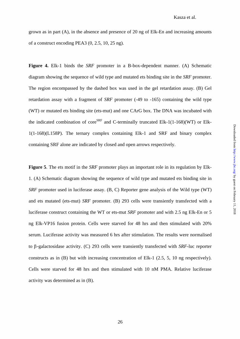
Elk-En. One of the downregulated genes identified was SRF (1.28 fold reduced). Northern
analysis confirmed the microarray data, demonstrating a clear reduction in SRF mRNA
following PA induction, which coincides with the induction of the expression of Elk-En (Fig.
1A). Similarly, SRF is also downregulated at the protein level following loss of the SRF
message (Fig. 1B). We also tested whether the prior induction of Elk-En could block the
activation of SRF expression by serum (FCS) stimulation. In comparison to the uninduced
cells, pretreatment of cells with PA to induce Elk-En expression led to a reduction in SRF
induction by serum (Fig. 2A). Thus, Elk-1 appears to be able to regulate the transcription of
the gene encoding its partner protein SRF.
Elk-En represses SRF expression in a B-box-dependent manner.
Elk-1 is thought to act primarily through ternary complexes in conjunction with SRF
on SREs (reviewed in 1, 2). However, it is possible that overexpression of Elk-En fusions
could result in the repression of genes usually regulated by different ETS-domain
by guest on February 11, 2018http://w
ww
.jbc.org/D
ownloaded from
Kasza et al.
10
transcription factors in an SRF-independent manner by virtue of their overlapping DNA
binding specificities. We therefore created a control cell line [EcR293(Elk-En{L158P})#L8]
that contains an Elk-En derivative that has a point mutation in its B-box region that blocks its
ability to bind to SRF and hence be recruited to DNA in an SRF-dependent manner (28). This
fusion protein should still inhibit transcription through high affinity binding sites. Control
experiments demonstrated that this was the case in reporter assays and that the expression
levels of the wild-type and L158P version of Elk-En were similar (28).
The prior induction of Elk-En(L158P) expression had little effect on serum-stimulated
SRF expression (Fig. 2B). This contrasts with the induction of wild-type (WT) Elk-En which
caused a clear reduction in serum-inducible SRF expression (Fig. 2A). We also examined the
protein levels of SRF in EcR293(Elk-En{L158P})#L8 cells following induction of Elk-
En(L158P), in contrast to the reductions in SRF levels observed upon induction of wild-type
Elk-En (Fig. 1B), no change in SRF expression was observed, even after 63 hours of
induction with PA (Fig. 2C).
To demonstrate that Elk-En could act directly on the SRF promoter, we carried out
transient transfection assays with increasing amounts of Elk-En and a SRF promoter-driven
luciferase reporter construct (Fig. 3A). Wild-type Elk-En efficiently repressed the activity of
the SRF promoter. In contrast, the mutant derivative, Elk-En(L158P), was unable to repress
the activity of the SRF promoter. This differential effect was not due to differences in
expression of the Elk-En fusions (Fig. 3B). Although Elk-En(L158P) is unable to affect the
activity SRF promoter, it is possible that other ETS-domain proteins might regulate this
promoter. Indeed, overexpression of a different ETS-domain protein, PEA3, also caused
upregulation of this promoter (data not shown). To establish the specificity of ETS-domain
protein action, we determined whether PEA3 could compete with Elk-En fusions in regulating
by guest on February 11, 2018http://w
ww
.jbc.org/D
ownloaded from

Kasza et al.
11
the SRF promoter. However, PEA3 was unable to compete with Elk-En (Fig. 3C), suggesting
that Elk-1 is an important regulator of this promoter.
Collectively, these data therefore demonstrate that Elk-1-mediated regulation of the
SRF promoter takes place in a B-box-dependent manner, suggesting that it is recruited by
SRF into a ternary, promoter-bound complex.
Elk-1 recruitment to the SRF promoter is mediated by an ets binding site.
The SRF promoter contains two CArG boxes that are bound by SRF and are important
for signal-mediated activation of SRF expression (33, 34). In addition, there is a functionally
important ets binding motif located upstream from these binding sites (29)(Fig. 4A). We
therefore tested whether Elk-1 can bind to this module in vitro using a DNA fragment
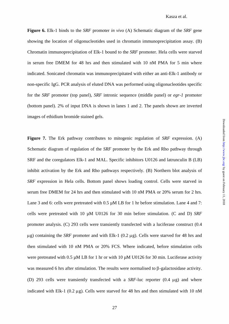
encompassing the upstream CArG box and the ets site (Fig. 4A). In the absence of SRF, Elk-1
was unable to bind to the SRF promoter (Fig. 4B, lane 3). However, in the presence of SRF,
the binding of Elk-1 was strongly stimulated and a ternary complex was formed on the
promoter (Fig. 4B, lane 4). This binding was dependent on the integrity of the B-box region
of Elk-1 as Elk-1(L158P) was unable to bind to the promoter (Fig. 4B, lane 5), demonstrating
the importance of interactions with SRF for its recruitment. Next, we tested the requirement
for the ets motif for recruitment of Elk-1 to the SRF promoter. Mutation of the ets motif
reduced the ability of Elk-1 to bind to the SRF promoter in a complex with SRF (Fig. 4B, lane
9). Thus, both protein-DNA contacts and protein-protein interactions with SRF are essential
for the efficient recruitment of Elk-1 to the SRF promoter
To extend these observations in vivo, we investigated the ability of Elk-1 derivatives
to regulate an SRF promoter-reporter construct that contains a mutation in the ets motif (Fig.
5A). The wild-type SRF promoter was repressed by Elk-En and activated by a constitutively
active Elk-VP16 fusion protein (Fig. 5B). In contrast, the activity of the mutated promoter,
by guest on February 11, 2018http://w
ww
.jbc.org/D
ownloaded from
Kasza et al.
12
SRF(ets-mut), was unaffected by either fusion protein (Fig. 5B). We also investigated the
ability of wild-type Elk-1 to activate the SRF promoter in the presence of PMA which
stimulates the MAP kinase pathway (Fig. 5C). Elk-1 activated the wild-type SRF promoter in
a dose-dependent manner. In contrast, its ability to activate the mutant SRF promoter was
severely reduced.
Collectively, these data demonstrate the importance of the ets motif within the SRF
promoter for the binding of Elk-1 and the subsequent ability of Elk-1 to regulate its
expression.
Elk-1 binds to the SRF promoter in vivo.
The above results indicate that Elk-1 can bind to the SRF promoter in vitro and
regulate its activity in vivo. To demonstrate that endogenous Elk-1 can occupy the SRF
promoter in vivo, we carried out a chromatin immunoprecipitation (ChIP) experiment in HeLa
cells. In the presence of Elk-1 antibodies, the SRF promoter was precipitated from
formaldehyde cross-linked total cell lysates (Fig. 6B, top panel, lanes 5 and 6). In contrast,
control antibodies precipitated background levels of the SRF promoter (Fig. 6B, lanes 3 and
4) and the Elk-1 antibody was unable to precipitate an intronic fragment of the SRF gene
above background levels (Fig. 6B, lower panel). The association of Elk-1 with the SRF
promoter was enhanced irrespective of the presence of PMA stimulation (Fig. 6B, lanes 5 and
6). Similarly, Elk-1 could be detected at the promoter of the well characterized target gene,
egr-1, both in the presence and absence of PMA stimulation. Thus, on the SRF and egr-1
promoters, it appears that Erk-pathway dependent activation of Elk-1 does not enhance
promoter occupancy, unlike previous in vitro studies that demonstrate phosphorylation-
enhanced recruitment of Elk-1 to the c-fos SRE (35, 36).
by guest on February 11, 2018http://w
ww
.jbc.org/D
ownloaded from
Kasza et al.
13
These data therefore demonstrate that endogenous Elk-1 binds to the SRF promoter in
vivo, thereby suggesting that the regulatory effects of Elk-1 on SRF expression are direct.
Regulation of SRF expression by the Erk and Rho pathways.
Previously, SRF has been identified as a target gene of the Rho signaling pathway
(21), and this regulation is mediated by coactivator proteins from the MAL family that act
through SRF bound to the promoter (Fig. 7A). The binding of MAL is mutually exclusive
with TCF binding to SRF (19), suggesting that regulation by TCFs and MAL might also be
mutually exclusive. However, it is also possible that the Erk pathway, through the TCFs, can
contribute to the activation of the SRF promoter. This would be important physiologically, as
under different conditions, signaling through either the Rho or Erk pathway alone or
simultaneous activation of several pathways might occur.
To investigate the convergence of these two pathways on the SRF promoter, we first
examined the expression of SRF in response to activating the Erk pathway with PMA or the
Erk and Rho pathways by serum (FCS). The Erk and Rho pathway inhibitors, U0126 and
Latrunculin B (LB) respectively, were used to identify the individual contributions of these
pathways to SRF induction. SRF expression was increased by PMA stimulation, and this
increase was selectively blocked with U0126, demonstrating that this activation was Erk
pathway dependent (Fig. 7B, lanes 2-4). Similarly, FCS stimulated SRF expression, and this
was inhibited by both LB and, to a lesser extent, U0126 (Fig. 7B, lanes 5-7). Similar
observations have been made in NIH3T3 cells (21). Thus, the Erk pathway can activate SRF
expression and contributes to its expression following serum induction.
We also tested whether the Erk and Rho pathways contributed directly to the
regulation of the SRF promoter using reporter gene assays. First, we overexpressed Elk-1 to
sensitise the SRF promoter to Erk-pathway mediated stimulation. Under these conditions,
by guest on February 11, 2018http://w
ww
.jbc.org/D
ownloaded from
Kasza et al.
14
both PMA and FCS treatment led to an increase in the activity of a SRF promoter-driven
luciferase reporter (Fig. 7C). The inhibition of the Erk pathway reduced the activation of the
SRF promoter by both treatments. However, LB was less effective in reducing the activation
of the SRF promoter by FCS, presumably due to Elk-1 competing with MAL recruitment. To
probe this possibility further, we tested the response of the SRF promoter to activation of the
Rho pathway by jasplakinolide in the presence and absence of exogenous Elk-1 (Fig. 7D).
The addition of Elk-1 inhibited the activation of the SRF promoter by jasplakinolide. This was
not a general inhibitory effect as the promoter now became more responsive to activation by
PMA. Thus, the level of Elk-1 in the cell can determine whether the Erk or Rho pathway can
activate the SRF promoter.
Conversely, we asked whether the overexpression of a constitutively active form of
MAL, MAL∆N (16) could interfere with Elk-1-mediated SRF promoter regulation. We
transfected 293 cells with either the wild-type (WT) or mutant (ets-mut) versions of the SRF-
luciferase reporter construct in the presence of low amount of Elk-En to dampen down the
activity of the promoter. A dose-dependent increase in the activity of the WT promoter was
observed upon transfection of increasing amounts of MAL∆N expression plasmid (Fig. 7E).
However, in contrast, the ets-mut promoter was activated to maximal levels at the lowest
concentration of MAL∆N expressed, with no further increases seen at higher levels of
MAL∆N (Fig.7E). Thus, the absence of the ets motif within the SRF promoter makes it more
sensitive to activation by MAL∆N, thereby demonstrating that occupancy of this ets motif in
vivo can inhibit activation via MAL.
Finally, we compared the ability of wild-type Elk-1 and an alternative ETS-domain
transcription factor, PEA3, to affect the activity of MAL∆N on the SRF promoter in serum
starved cells. Experiments were carried out under serum starved conditions where the ERK
pathway and hence the ETS-domain protein targets are not activated. Elk-1 inhibited the
by guest on February 11, 2018http://w
ww
.jbc.org/D
ownloaded from

Kasza et al.
15
action of MAL∆N in a dose-dependent manner, but PEA3 did not, and instead weakly
potentiated the activity of MAL∆N on the SRF promoter (Fig. 7F). This is consistent with a
model whereby Elk-1 binds through SRF interactions, thereby inhibiting MAL recruitment,
but that other ETS-domain proteins like PEA3 may in some circumstances function in an
SRF-independent manner to activate the SRF promoter.
Collectively, these data corroborate previous observations that indicate that TCF and
MAL/myocardin recruitment by SRF is mutually exclusive (19, 20). However importantly,
they demonstrate a clear role for the Erk pathway in regulating SRF expression and suggest
how multiple signaling inputs might lead to upregulation of the SRF promoter (see
discussion).
by guest on February 11, 2018http://w
ww
.jbc.org/D
ownloaded from

Kasza et al.
16
Discussion
The TCF transcription factors play an important role in transducing extracellular
signals into a nuclear response by acting as targets for the MAP kinase signaling pathways
(reviewed in 1, 2, 9). To fully understand the physiological role of the TCFs, it is important to
gain a fuller insight into the spectra of target genes that they regulate. To date, the focus has
been primarily on the immediate-early genes such as c-fos and egr-1 (reviewed in 1, 2, 9).
Here we have identified SRF as a direct TCF target gene. This provides an elegant positive
feedback loop whereby the TCFs can regulate the expression of the TCF partner protein SRF.
We initially identified SRF as a TCF target gene by demonstrating downregulation of
SRF expression in cell lines expressing the repressive Elk-En fusion protein (Fig. 1). By using
a combination of reporter gene and ChIP analyses, we have shown that the TCFs can directly
affect the SRF promoter and that endogenously expressed Elk-1 can be found on this
promoter in vivo (Figs. 3, 5 and 6). It is currently not known whether TCFs other than Elk-1
can participate in the regulation of SRF. Indeed, as overexpressed Elk-En can potentially
block regulation by all the TCFs due to their structural similarity, it will be important to probe
whether the other TCFs can work in the same way. It remains possible that other ETS-domain
proteins might act on the SRF promoter through the ets site. Indeed, overexpression of PEA3,
a target of the Erk pathway (37), can activate the SRF promoter in an ets site-dependent
manner (data not shown). However, PEA3 is unable to compete with Elk-1 for promoter
occupancy (Fig. 3C). Indeed, in we show that in cells containing Elk-1, the SRF promoter is
occupied by Elk-1 (Fig. 6). It remains possible that in other cell types, where low levels of
Elk-1 and high levels of ETS-domain proteins such as PEA3 exist, that different ETS-domain
proteins contribute to SRF regulation. However, the interplay with the Rho/MAL pathway
would likely differ, as interference with SRF-dependent MAL recruitment would not occur
(Fig. 7F).
by guest on February 11, 2018http://w
ww
.jbc.org/D
ownloaded from
Kasza et al.
17
Elk-1 appears to act to couple signals generated by the Erk pathway to the activation
of the SRF promoter (Fig. 7) whereas the coactivator MAL has previously been shown to
couple Rho pathway signals to this promoter (21). Signaling through the Erk pathway is of
importance where signaling does not activate the Rho pathway, here induced using the
mitogen PMA, and as a potential contributing pathway to more complex signaling triggers
such as serum. Importantly, we show that the ability of Elk-1 to regulate SRF expression is
dependent on the formation of a ternary complex with DNA-bound SRF (Figs. 2, 4 and 5).
Previous studies have shown that signaling via the Rho pathway also functions through SRF
bound to its own promoter, although in this case, the MAL/myocardin family protein MAL
represents the coregulatory partner that links to this pathway (13, 16). We demonstrate that a
functional antagonism can occur depending on the relative levels of MAL and the TCFs
within the cell (Fig. 7), which is consistent with previous data demonstrating that Elk-1 can
inhibit MAL recruitment in a B-box-dependent manner to SRF-regulated promoters (19, 20).
Thus depending on the relative levels of the TCF and MAL/myocardin family proteins, the
relative contributions of these two classes of coregulators might differ. Alternatively, there
may be a role for both coregulators in permitting convergence of the Erk and Rho pathways
through SRF bound to promoters. Our data suggest that the latter scenario may well exist at
the SRF promoter as inhibitor studies demonstrate a contribution of the Erk pathway to
mitogenic stimulation of SRF expression (Fig. 7B) that is consistent with results from a
previous study (21). In addition, it has been proposed that the TCFs might contribute to the
early induction phase of other genes like c-fos in response to mitogens while the
MAL/myocardin family contributes to the later phases (13). Furthermore, recent studies
demonstrate that promoters in several smooth muscle genes can be regulated through SRF by
either the TCFs or MAL/myocardin family members depending on the signaling pathways
that are active (20). Thus our data add further weight to an emerging model that SRF can
by guest on February 11, 2018http://w
ww
.jbc.org/D
ownloaded from

Kasza et al.
18
form a platform that can differentially recruit coregulatory transcription factors and permit
selective or combinatorial responses to different signaling pathways depending on the target
promoter.
Finally, our data further suggest an important role for SRF expression in cell survival.
Previously, SRF has been shown to be a target for degradation in response to apoptotic
pathways (38, 39) and also to regulate the expression of anti-apoptotic proteins like Bcl-2
(40). Recently we showed that the induction of Elk-En causes apoptosis and that one key anti-
apoptotic target gene was the antiapoptotic gene Mcl-1 (28). As SRF expression is also
downregulated upon Elk-En induction, then this too might play a role in triggering apoptosis.
Indeed, the downregulation of SRF transcript levels is a natural process observed upon
apoptotic induction caused by etoposide treatment (our unpublished data). However, although
we could partially rescue apoptosis induced by Elk-En (data not shown), we have been unable
to differentiate whether this was due to replacement of the downregulated endogenous SRF,
or merely titration of the Elk-En away from the promoters of antiapoptotic genes. Thus, the
TCFs can potentially promote cell survival through the regulation of multiple genes and
provide an important link to the prosurvival effects of the Erk pathway.
by guest on February 11, 2018http://w
ww
.jbc.org/D
ownloaded from

Kasza et al.
19
Acknowledgements
We would like to thank Anne Clancy for excellent technical support. We also would like to
thank Ravi Misra and Richard Treisman for reagents and Shen-Hsi Yang, and Alan
Whitmarsh for comments on the manuscript. This work was funded by grants from the
Wellcome Trust and the AICR. AK was supported by a Research Fellowship from the
Wellcome Trust and the Foundation for Polish Science.
by guest on February 11, 2018http://w
ww
.jbc.org/D
ownloaded from
Kasza et al.
20
References
1. Sharrocks, A. D. (2002) Biochem Soc Trans. 30, 1-9.
2. Shaw, P. E., and Saxton, J. (2003) Int. J. Biochem. Cell. Biol. 35, 1210-1226.
3. Janknecht, R. and Nordheim, A. (1992) Nucleic Acids Res. 20, 3317-3324.
4. Dalton, S. and Treisman, R. (1992) Cell 68, 597-612.
5. Treisman, R., Marais, R. and Wynne, J. (1992) EMBO J. 11, 4631-4640.
6. Shore, P. and Sharrocks, A. D. (1994) Mol. Cell. Biol. 14, 3283-3291.
7. Ling, Y., Lakey, J. H., Roberts, E.C. and Sharrocks. A .D. (1997) EMBO J. 16, 2431-2440.
8. Hassler, M. and Richmond, T.J. (2001) EMBO J. 20, 3018-3028.
9. Wasylyk, B., Hagman, J. And Gutierrez-Hartmann, A. (1998) Trends Biochem Sci. 23,
213-216.
10. Stevens, J. L., Cantin, G. T., Wang, G., Shevchenko, A., Shevchenko, A., and Berk, A. J.
(2002) Science 296, 755-758
by guest on February 11, 2018http://w
ww
.jbc.org/D
ownloaded from
Kasza et al.
21
11. Li, Q. J., Yang, S. H., Maeda, Y., Sladek, F. M., Sharrocks, A. D., and Martins-Green, M.
(2003) EMBO J. 22, 281-291
12. Yang, S. H. and Sharrocks, A. D. (2004) Mol.Cell 13, 611-617
13. Cen, B., Selvaraj, A., Burgess, R. C., Hitzler, J. K., Ma, Z., Morris, S. W., and Prywes, R.
(2003) Mol.Cell Biol. 23, 6597-6608
14. Wang, D., Chang, P. S., Wang, Z., Sutherland, L., Richardson, J. A., Small, E., Krieg, P.
A., and Olson, E. N. (2001) Cell 105, 851-862
15. Wang, D. Z., Li, S., Hockemeyer, D., Sutherland, L., Wang, Z., Schratt, G., Richardson, J.
A., Nordheim, A., and Olson, E. N. (2002) Proc.Natl.Acad.Sci.U.S.A 99, 14855-14860
16. Miralles, F., Posern, G., Zaromytidou, A. I., and Treisman, R. (2003) Cell 113, 329-342
17. Du, K. L., Ip, H. S., Li, J., Chen, M., Dandre, F., Yu, W., Lu, M. M., Owens, G. K., and
Parmacek, M. S. (2003) Mol.Cell Biol. 23, 2425-2437
18. Selvaraj, A. and Prywes, R. (2003) J.Biol.Chem. 278, 41977-41987
19. Murai, K. and Treisman, R. (2002) Mol.Cell Biol. 22, 7083-7092
by guest on February 11, 2018http://w
ww
.jbc.org/D
ownloaded from
Kasza et al.
22
20. Wang, Z., Wang, D. Z., Hockemeyer, D., McAnally, J., Nordheim, A., and Olson, E. N.
(2004) Nature 428, 185-189
21. Gineitis, D. and Treisman, R. (2001) J. Biol. Chem. 276, 24531-24539.
22. Latinkic, B. V., Zeremski, M. and Lau, L.F. (1996) Nucleic Acids Res. 24, 1345-1351.
23. Costello, P. S., Nicolas, R. H., Watanabe, Y., Rosewell, I. and Treisman, R. (2004) Nat.
Immunol. 5, 289-298.
24. Tsai, E., Falvo, J. V., Tsytsykova, A. V., Barczak, A. K., Reimold, A. M., Glimcher, L.
H., Fenton, M. J., Gordon, D. C., Dunn, I. F. and Goldfeld, A. E. (2000) Mol. Cell. Biol. 20,
6084-6094.
25. Li, Q. J., Vaingankar, S., Sladek, F.M. and Martins-Green, M. (2000) Blood 96, 3696-
3706.
26. Ayadi, A., Zheng, H., Sobieszczuk, P., Buchwalter, G., Moerman, P., Alitalo, K. and
Wasylyk, B. (2001) EMBO J. 20, 5139-5152.
27. Cesari, F., Brecht, S. Vintersten, K., Vuong, L. G., Hofmann, M., Klingel, K., Schnorr, J.
J., Arsenian, S., Schild, H., Herdegen, T., Wiebel, F. F. and Nordheim, A. (2004) Mol. Cell.
Biol. 24, 294-305.
by guest on February 11, 2018http://w
ww
.jbc.org/D
ownloaded from
Kasza et al.
23
28. Vickers, E. R., Kasza, A., Aksan-Kurnaz, I., Seifert, A., Zeef, L., O’Donnell, A., Hayes,
A. and Sharrocks, A. D. (2004) Mol. Cell. Biol. In Press.
29. Spencer, J. A., Major, M. L., and Misra, R. P. (1999) Mol.Cell Biol. 19, 3977-3988
30. Price, M. A., Rogers, A. E and Treisman, R. (1995) EMBO J. 14, 2589-2601.
31. Yang, S. H., Yates, P. R., Whitmarsh, A. J., Davis, R. J. and Sharrocks, A. D. (1998) Mol.
Cell. Biol. 18, 710-720.
32. Sharrocks, A. D., Gille, H. and Shaw, P. E. (1993) Mol. Cell. Biol. 13, 123-132.
33. Spencer, J. A. and Misra, R. P. (1996) J.Biol.Chem. 271, 16535-16543
34. Belaguli, N. S., Schildmeyer L. A. And Schwartz R. J. (1997) J. Biol. Chem. 272, 18222-
18231.
35. Gille, H., Sharrocks, A. D. and Shaw, P.E. (1992) Nature 358, 414-417.
36. Gille, H., Kortenjann, M., Thomae, O., Moomaw, C., Slaughter, C., Cobb, M. H., and
Shaw, P. E. (1995) EMBO J. 14, 951-62.
37. O'Hagan, R.C., Tozer, R.G., Symons, M., McCormick, F., and Hassell, J.A. (1996)
Oncogene 13, 1323-33.
by guest on February 11, 2018http://w
ww
.jbc.org/D
ownloaded from
Kasza et al.
24
38. Bertolotto, C., Ricci, J. E., Luciano, F., Mari, B. Chambard, J. C. and Auberger, P.
(2000) J.Biol.Chem. 275, 12941-12947.
39. Drewett, V., Devitt, A., Saxton, J., Portman, N., Greaney, P., Cheong, N. E., Alnemri, T.
F., Alnemri, E. and Shaw, P.E. (2001) J. Biol. Chem. 276, 33444-33451.
40. Schratt, G., Weinhold, B., Lundberg, A. S., Schuck, S., Berger, J., Schwarz, H.,
Weinberg, R., Ruther, U. And Nordheim, A. (2001) Mol. Cell. Biol. 21, 2933-2943.
by guest on February 11, 2018http://w
ww
.jbc.org/D
ownloaded from
Kasza et al.
25
Figure legends
Figure 1. SRF expression is downregulated by Elk-En. EcR293(Elk-En)#1.3 cells were
treated with 5µM ponasterone A (PA) for the indicated times. (A) The expression of SRF
mRNA was analysed by Northern blotting (A) and SRF protein by Western analysis (B). The
middle panels show the expression levels of Elk-En whereas the bottom panels show loading
controls (total RNA for Northern analysis and GAPDH for Western analysis).
Figure 2. SRF downregulation by Elk-En is B-box dependent. SRF expression was analysed
by Northern blotting in EcR293(Elk-En)#1.3 (A) and EcR293(Elk-En{L158P}) cells (B).
Cells were serum starved for 24 hrs, pretreated with 5µM ponasterone A (PA) for 19 hrs and
stimulated with 20% serum for 2 or 3 hrs. The middle panels show the expression levels of
Elk-En whereas the bottom panels show the total RNA loading control. (C) Western blot
analysis of SRF expression (top panel) in EcR293(Elk-En{L158P}) cells treated with 5µM
ponasterone A for the indicated times. The middle panel shows the expression levels of Elk-
En whereas the bottom panel shows GAPDH loading control.
Figure 3. Elk-En downregulates the SRF promoter. (A) 293 cells were transiently transfected
with a luciferase construct containing the SRF promoter and with increasing concentrations of
Elk-En(WT) and Elk-En(L158P) (0, 1, 5, 10, 25, 50 ng). Cells were starved for 48 hrs and
then stimulated with 20% serum. Luciferase activity was measured 6 hrs after stimulation.
The results were normalised to β-galactosidase activity. (B) Western blot analysis of Elk-
En(WT) and Elk-En(L158P) from samples derived from cells transfected with 10 ng (lanes 1
and 5), 25 ng (lanes 2 and 6), 50 ng (lanes 3 and 7) and 100 ng (lanes 4 and 8) of expression
vector. (C) Reporter gene analysis of the wild type (WT) SRF promoter (0.4 µg) in 293 cells
by guest on February 11, 2018http://w
ww
.jbc.org/D
ownloaded from
Kasza et al.
26
grown as in part (A), in the absence and presence of 20 ng of Elk-En and increasing amounts
of a construct encoding PEA3 (0, 2.5, 10, 25 ng).
Figure 4. Elk-1 binds the SRF promoter in a B-box-dependent manner. (A) Schematic
diagram showing the sequence of wild type and mutated ets binding site in the SRF promoter.
The region encompassed by the dashed box was used in the gel retardation assay. (B) Gel
retardation assay with a fragment of SRF promoter (-49 to -165) containing the wild type
(WT) or mutated ets binding site (ets-mut) and one CArG box. The DNA was incubated with
the indicated combination of coreSRF and C-terminally truncated Elk-1(1-168)(WT) or Elk-
1(1-168)(L158P). The ternary complex containing Elk-1 and SRF and binary complex
containing SRF alone are indicated by closed and open arrows respectively.
Figure 5. The ets motif in the SRF promoter plays an important role in its regulation by Elk-
1. (A) Schematic diagram showing the sequence of wild type and mutated ets binding site in
SRF promoter used in luciferase assay. (B, C) Reporter gene analysis of the Wild type (WT)
and ets mutated (ets-mut) SRF promoter. (B) 293 cells were transiently transfected with a
luciferase construct containing the WT or ets-mut SRF promoter and with 2.5 ng Elk-En or 5
ng Elk-VP16 fusion protein. Cells were starved for 48 hrs and then stimulated with 20%
serum. Luciferase activity was measured 6 hrs after stimulation. The results were normalised
to β-galactosidase activity. (C) 293 cells were transiently transfected with SRF-luc reporter
constructs as in (B) but with increasing concentration of Elk-1 (2.5, 5, 10 ng respectively).
Cells were starved for 48 hrs and then stimulated with 10 nM PMA. Relative luciferase
activity was determined as in (B).
by guest on February 11, 2018http://w
ww
.jbc.org/D
ownloaded from
Kasza et al.
27
Figure 6. Elk-1 binds to the SRF promoter in vivo (A) Schematic diagram of the SRF gene
showing the location of oligonucleotides used in chromatin immunoprecipitation assay. (B)
Chromatin immunoprecipitation of Elk-1 bound to the SRF promoter. Hela cells were starved
in serum free DMEM for 48 hrs and then stimulated with 10 nM PMA for 5 min where
indicated. Sonicated chromatin was immunoprecipitated with either an anti-Elk-1 antibody or
non-specific IgG. PCR analysis of eluted DNA was performed using oligonucleotides specific
for the SRF promoter (top panel), SRF intronic sequence (middle panel) or egr-1 promoter
(bottom panel). 2% of input DNA is shown in lanes 1 and 2. The panels shown are inverted
images of ethidium bromide stained gels.
Figure 7. The Erk pathway contributes to mitogenic regulation of SRF expression. (A)
Schematic diagram of regulation of the SRF promoter by the Erk and Rho pathway through
SRF and the coregulators Elk-1 and MAL. Specific inhibitors U0126 and latrunculin B (LB)
inhibit activation by the Erk and Rho pathways respectively. (B) Northern blot analysis of
SRF expression in Hela cells. Bottom panel shows loading control. Cells were starved in
serum free DMEM for 24 hrs and then stimulated with 10 nM PMA or 20% serum for 2 hrs.
Lane 3 and 6: cells were pretreated with 0.5 µM LB for 1 hr before stimulation. Lane 4 and 7:
cells were pretreated with 10 µM U0126 for 30 min before stimulation. (C and D) SRF
promoter analysis. (C) 293 cells were transiently transfected with a luciferase construct (0.4
µg) containing the SRF promoter and with Elk-1 (0.2 µg). Cells were starved for 48 hrs and
then stimulated with 10 nM PMA or 20% FCS. Where indicated, before stimulation cells
were pretreated with 0.5 µM LB for 1 hr or with 10 µM U0126 for 30 min. Luciferase activity
was measured 6 hrs after stimulation. The results were normalised to β-galactosidase activity.
(D) 293 cells were transiently transfected with a SRF-luc reporter (0.4 µg) and where
indicated with Elk-1 (0.2 µg). Cells were starved for 48 hrs and then stimulated with 10 nM
by guest on February 11, 2018http://w
ww
.jbc.org/D
ownloaded from

Kasza et al.
28
PMA or 0.5 µM jasplakinolide. Relative luciferase activity was determined as in (C). (E)
Reporter gene analysis of the wild type (WT) or ets mutated (ets-mut) SRF promoter (0.4 µg),
in the presence of 20 ng of Elk-En and increasing amounts of a construct encoding MAL∆N
(0, 10, 20, 50, 100 ng). (E) Reporter gene analysis of the wild type (WT) SRF promoter (0.4
µg) in serum starved 293 cells, in the absence and presence of 20ng of MAL∆N and
increasing amounts (0, 10, 20, 50 ng) of constructs encoding Elk-1 and PEA3.
by guest on February 11, 2018http://w
ww
.jbc.org/D
ownloaded from
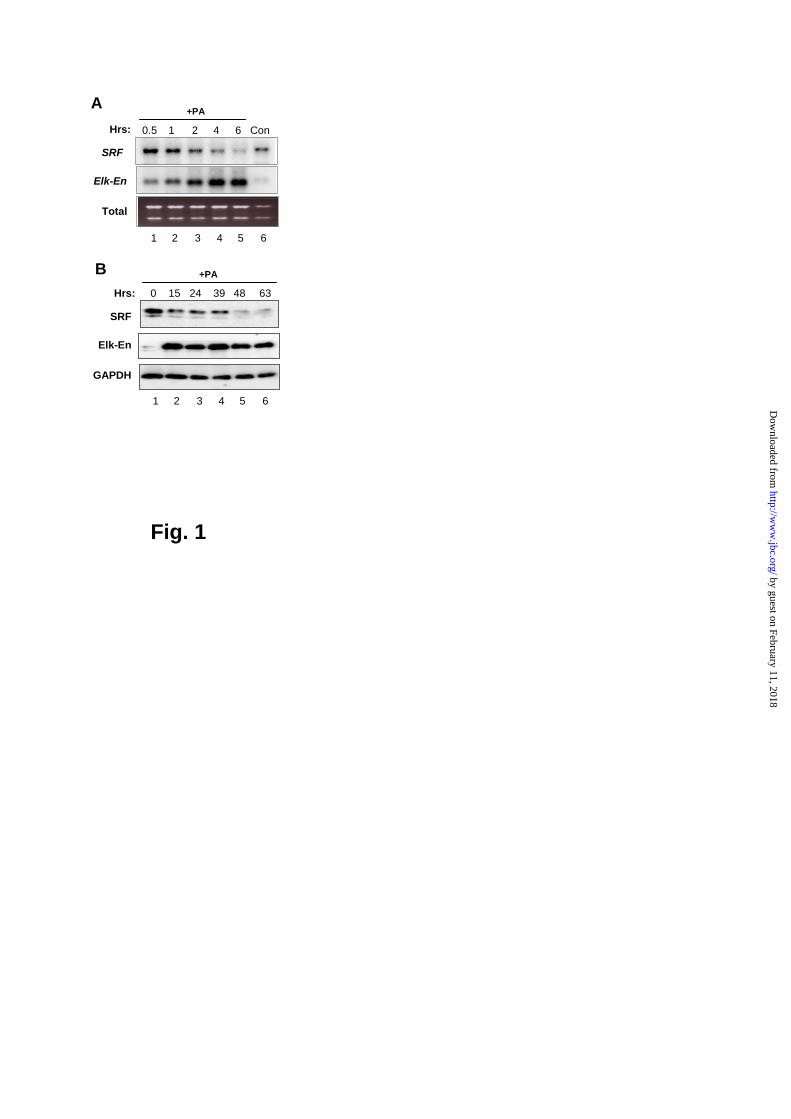
Fig. 1
A
1 2 3 4 5 6
0.5 1 2 4 6 ConHrs:
Total
SRF
Elk-En
+PA
BHrs:
GAPDH
SRF
Elk-En
0 15 24 39 48 63
+PA
1 2 3 4 5 6
by guest on February 11, 2018http://w
ww
.jbc.org/D
ownloaded from

Fig. 2
A
1 2 3 4 5 6B
FCS (Hrs):
Total
SRF
Elk-En
Total
SRF
Elk-En
0 2 3 0 2 3
-PA
1 2 3 4 5 6
+PA
EcR293(Elk-En{WT}):
FCS (Hrs): 0 2 3 0 2 3
-PA +PA
EcR293(Elk-En{L158P}):
C
Hrs:
GAPDH
SRF
Elk-En
0 15 24 39 48 63
+PA
1 2 3 4 5 6
EcR293(Elk-En{L158P}):
by guest on February 11, 2018http://w
ww
.jbc.org/D
ownloaded from

Fig. 3
A
B
1 2 3 4 5 6
Elk-En:
0.2
0.4
0.6
0.8
1.0
1.2
1.4
Rel
ativ
e Lu
c. A
c tiv
ity
0
WT L158P-
7 8
L158PWT
Elk-En
LucSRF
0
2
4
6
8
10
C
Elk-En: - + + + +--PEA3:
SRF(WT)-luc
Rel
. Luc
ifera
seac
tivity
by guest on February 11, 2018http://w
ww
.jbc.org/D
ownloaded from
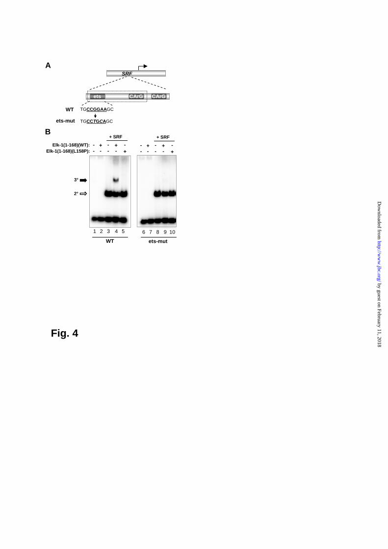
Fig. 4
A
1 2 3 4 5 6 7 8 9 10
- - - +Elk-1(1-168)(L158P):+
-- -+Elk-1(1-168)(WT): -
+ SRF
- - - ++
-- -+-
+ SRF
WT ets-mut
B
2°
3°
SRF
TGCCGGAAGC
ets CArG CArG
TGCCTGCAGC
WT
ets-mut
by guest on February 11, 2018http://w
ww
.jbc.org/D
ownloaded from

Fig. 5
ALucSRF
B
TGCCGGAAGC
ets CArG CArG
TGCCTGCAGC
WT
ets-mut
SRF(WT)-luc
Elk-1:0
2
4
6
8
10
- ---
Rel
ativ
e Lu
cife
rase
activ
ity
SRF(ets-mut)-luc
PMA PMA
C
0
1
2
3
4
Rel
ativ
e Lu
cife
rase
activ
ity
SRF(WT)-luc SRF(ets-mut)-luc
-
Elk-
VP16
Elk-
En -
Elk-
VP16
Elk-
En
by guest on February 11, 2018http://w
ww
.jbc.org/D
ownloaded from

Fig. 6
A
SRF
ets CArG CArG
ADS1249 ADS1250 ADS1273 ADS1274
Exon 1 Intron 3
1 2 3 4 5 6
SRF (promoter)
input
SRF (intron)
- + - -+ +PMA:
IgG Elk-1IP:B
Egr-1 (promoter)
by guest on February 11, 2018http://w
ww
.jbc.org/D
ownloaded from

B
1 2 3 4 5 6
C
Total
SRF
- LB U012
6
7
- LB U012
6
-
PMA FCS
0
1
2
3
4
Rel
ativ
e Lu
c. a
ctiv
ity
- LBU0
126- - LB
U012
6
PMA FCS
LucSRF
1
2
3
4
5
Rel
ativ
e lu
cife
rase
activ
ity
0Elk-1: - - -+ +
Jasp PMA
DLucSRF
E
SRFSRF SRF
Elk-1 MALRho pathwayErk Pathway
U0126 LB
A
ets CArG CArG
0
2
4
6
-MAL∆N
Rel
ativ
e Lu
c. a
ctiv
ity
-
LucSRF
ets CArG CArG
WT ets-mut
Fig. 7
0
20
40
60
80
-MAL∆N + + + + + + +-- -- -
-- -- -Elk-1PEA3
FLucSRF
Rel
ativ
e Lu
c. a
ctiv
ity
by guest on February 11, 2018http://w
ww
.jbc.org/D
ownloaded from
Andrew D. SharrocksAneta Kasza, Amanda O'Donnell, Karen Gascoigne, Leo A. H. Zeef, Andy Hayes and
protein, SRFThe ETS-domain transcription factor Elk-1 regulates the expression of its partner
published online November 4, 2004J. Biol. Chem.
10.1074/jbc.M411161200Access the most updated version of this article at doi:
Alerts:
When a correction for this article is posted•
When this article is cited•
to choose from all of JBC's e-mail alertsClick here
by guest on February 11, 2018http://w
ww
.jbc.org/D
ownloaded from