The Solid Phase of the Ehrlich Ascites Tumor in...
9
The Solid Phase of the Ehrlich Ascites Tumor in Mice RALPHN. BAILLIF (Department of Anatomy, Tulane University School of Medicine, New Orleans, La.) The Ehrlich mouse ascites tumor has become one of the widely used experimental cancers, par ticularly, as pointed out by Klein (6), in those studies dealing with quantitative aspects of tu mor biology. This tumor has been studied exten sively by a number of previous investigators. Loewenthal and Jahn (11) found that intra- peritoneal inoculation with organ brei containing the Ehrlich carcinoma produced not only a car- cinomatous peritonitis, but also an ascitic fluid rich in free neoplastic cells. These remained infec tive despite dilution and various types of deleteri ous treatment. The cytology of this fluid cancer has been described in Seeger's monograph (17), by Love, Koprowski, and Cox (12), and by Sugiura (19). Formation of an ascitic fluid rich in neoplastic cells is not the specific property of the so-called ascites tumors. Warren and Gates (20) were able to produce this condition in a variety of tumors by grinding the solid form, resuspending the frag ments, and injecting intraperitoneally the cell- containing fraction. The cell-containing ascitic fluid which resulted from this type of injection was then given intravenously to new hosts, and the fate of the tumor cells followed. More recent ly, Klein and Klein (7) were similarly able to con vert the Krebs carcinoma into an ascites tumor. Under favorable conditions the Ehrlich tumor readily forms métastases.Schairer (15), for ex ample, has observed metastasis formation in the lungs, and Schmidt (16) has found them in re gional lymph nodes after subcutaneous injection. This earlier work, as well as that of Lettré(9) and Klein, Kurnick, and Klein (5) indicates that, after intraperitoneal inoculation, the Ehrlich ascites tumor grows rapidly and produces an as citic suspension rich in malignant cells. The tumor is relatively nonspecific and can be maintained in most strains of Swiss mice. It also grows in rats and can be serially transplanted as long as the cell samples are taken before progressively in creasing host resistance causes their deterioration (10). The rate of accumulation of both cells and fluid Received for publication March 10, 1954. is directly related to the number of inoculated cells (5, 19). When the size of the inoculum is kept constant, it is possible to predict with considerable accuracy the length of time which mice of a given strain can be expected to survive (9). There are several reports concerning the effects of various substances on tumor growth rate. Koprowska and Koprowski (8) have used viruses as test materials; Lewis and Goland (10) have employed dyes; and Sugiura (19), chemicals from a wide variety of classes. Although the Ehrlich ascites tumor grows pri marily as a cell suspension in ascitic fluid, solid tumors also appear in the same animals. Owing, perhaps, to a preoccupation with quantitative studies on the tumor suspension, the solid phase of the neoplasm has not been thoroughly investi gated. An attempt has been made in the present study to supply some of the missing facets in its biology, including a possible relationship between solid and fluid tumors in the same animal. MATERIALS AND METHODS One hundred and forty CFW mice were each inoculated intraperitoneally with 10-15 million tumor cells in a volume of 0.1-0.15 cc. The number of cells used in each case was deter mined by counts made on the fluid in a blood cell counting chamber. Of these mice, 45 were inoculated with the tumor alone, while 95 were given both tumor and varying amounts of colloidal thorium dioxide (Thorotrast, Heyden), to determine if it would alter the pattern of solid tumor formation. Of those receiving tumor alone, 38 survived 10 days or more and were used in subsequent histológica!studies. Sixty-four of the second group survived 10 days or more and were used for microscopic studies. After inoculation, ascites became apparent in most cases within 5-7 days, and the animals exhibited marked ascites by 10-12 days. In four of the mice no ascites developed. In each case, however, wet smears obtained by imprint of the omentum show the presence of typical tumor cells, indicating that in oculation of tumor cells was made and that the cells persisted throughout the experimental period. Approximately one-half of the mice were allowed to die spontaneously; the other half were sacrificed on the 10th postinoculation day. In both cases, the ascitic fluid was drained as completely as possible with the aid of a hypodermic needle, and each animal subjected to detailed autopsy. Special search was made for solid tumors, especially those appearing in structures related to peritoneum. The volume of the ascitic suspension was measured, and a sample was used for making cell counts. A portion of the fluid was centrifuged, and the supernate discarded, the cell mass was resuspended in several 554 on May 24, 2018. © 1954 American Association for Cancer Research. cancerres.aacrjournals.org Downloaded from
Transcript of The Solid Phase of the Ehrlich Ascites Tumor in...

The Solid Phase of the Ehrlich Ascites Tumor in Mice
RALPHN. BAILLIF
(Department of Anatomy, Tulane University School of Medicine, New Orleans, La.)
The Ehrlich mouse ascites tumor has becomeone of the widely used experimental cancers, particularly, as pointed out by Klein (6), in thosestudies dealing with quantitative aspects of tumor biology. This tumor has been studied extensively by a number of previous investigators.Loewenthal and Jahn (11) found that intra-peritoneal inoculation with organ brei containingthe Ehrlich carcinoma produced not only a car-cinomatous peritonitis, but also an ascitic fluidrich in free neoplastic cells. These remained infective despite dilution and various types of deleterious treatment. The cytology of this fluid cancerhas been described in Seeger's monograph (17),
by Love, Koprowski, and Cox (12), and bySugiura (19).
Formation of an ascitic fluid rich in neoplasticcells is not the specific property of the so-calledascites tumors. Warren and Gates (20) were ableto produce this condition in a variety of tumorsby grinding the solid form, resuspending the fragments, and injecting intraperitoneally the cell-containing fraction. The cell-containing asciticfluid which resulted from this type of injectionwas then given intravenously to new hosts, andthe fate of the tumor cells followed. More recently, Klein and Klein (7) were similarly able to convert the Krebs carcinoma into an ascites tumor.
Under favorable conditions the Ehrlich tumorreadily forms métastases.Schairer (15), for example, has observed metastasis formation in thelungs, and Schmidt (16) has found them in regional lymph nodes after subcutaneous injection.
This earlier work, as well as that of Lettré(9)and Klein, Kurnick, and Klein (5) indicates that,after intraperitoneal inoculation, the Ehrlichascites tumor grows rapidly and produces an ascitic suspension rich in malignant cells. The tumoris relatively nonspecific and can be maintained inmost strains of Swiss mice. It also grows in ratsand can be serially transplanted as long as thecell samples are taken before progressively increasing host resistance causes their deterioration(10).
The rate of accumulation of both cells and fluid
Received for publication March 10, 1954.
is directly related to the number of inoculatedcells (5, 19). When the size of the inoculum is keptconstant, it is possible to predict with considerableaccuracy the length of time which mice of a givenstrain can be expected to survive (9). There areseveral reports concerning the effects of varioussubstances on tumor growth rate. Koprowska andKoprowski (8) have used viruses as test materials;Lewis and Goland (10) have employed dyes; andSugiura (19), chemicals from a wide variety ofclasses.
Although the Ehrlich ascites tumor grows primarily as a cell suspension in ascitic fluid, solidtumors also appear in the same animals. Owing,perhaps, to a preoccupation with quantitativestudies on the tumor suspension, the solid phaseof the neoplasm has not been thoroughly investigated. An attempt has been made in the presentstudy to supply some of the missing facets in itsbiology, including a possible relationship betweensolid and fluid tumors in the same animal.
MATERIALS AND METHODSOne hundred and forty CFW mice were each inoculated
intraperitoneally with 10-15 million tumor cells in a volumeof 0.1-0.15 cc. The number of cells used in each case was determined by counts made on the fluid in a blood cell countingchamber. Of these mice, 45 were inoculated with the tumoralone, while 95 were given both tumor and varying amounts ofcolloidal thorium dioxide (Thorotrast, Heyden), to determineif it would alter the pattern of solid tumor formation. Of thosereceiving tumor alone, 38 survived 10 days or more andwere used in subsequent histológica!studies. Sixty-four of thesecond group survived 10 days or more and were used formicroscopic studies.
After inoculation, ascites became apparent in most caseswithin 5-7 days, and the animals exhibited marked ascites by10-12 days. In four of the mice no ascites developed. In eachcase, however, wet smears obtained by imprint of the omentumshow the presence of typical tumor cells, indicating that inoculation of tumor cells was made and that the cells persistedthroughout the experimental period.
Approximately one-half of the mice were allowed to diespontaneously; the other half were sacrificed on the 10thpostinoculation day. In both cases, the ascitic fluid was drainedas completely as possible with the aid of a hypodermic needle,and each animal subjected to detailed autopsy. Special searchwas made for solid tumors, especially those appearing instructures related to peritoneum. The volume of the asciticsuspension was measured, and a sample was used for makingcell counts. A portion of the fluid was centrifuged, and thesupernate discarded, the cell mass was resuspended in several
554
on May 24, 2018. © 1954 American Association for Cancer Research. cancerres.aacrjournals.org Downloaded from

BAILLIF—SolidEhrlich Tumor in Mice 555
volumes of Benin's fluid and recentrifuged. The resulting con
centrate was treated as an ordinary tissue specimen. Observations were also made on cells in the fresh ascitic fluid suspension with the phase microscope.
The principal organs were examined grossly and samplesremoved for histologie study. After fixation in Bouin's fluid
or 10 per cent formalin, sections were stained in hematoxylinand eosin; check slides were also prepared using Mallory's
triple connective tissue stain. Studies on stromal pattern weremade according to Snook's modification of the Bielschowsky
technic (18).
RESULTSGrowth of the Ehrlich ascites tumor produced
toxic symptoms in the animals. Of the mice inoculated with the tumor alone, some died as earlyas the 9th day, while others lived as long as 22days. The average survival time was 15.0 days.In the series which received both tumor and colloid, the range of life-span was 8-21 days, withan average of 13.2 days.
Of the 102 animals which survived 10 days ormore after inoculation, 95 developed at least onesolid tumor; most were small in size and frequently hidden by abdominal organs. Of the mice inoculated with tumor alone, 92 per cent developedsolid tumors; 91 per cent of those with both tumor and colloid developed solid tumors. Intra-peritoneal fibrin clots permeated with tumor cellswere invariably found in mice with solid tumorsand in one instance where no solid tumor couldbe found. Previous studies on survival times andon percentages of solid tumors have been carriedout by Loewenthal and Jahn (11) and by Lettré(9). Particularly in the latter work, it has beenshown that survival time, changes in total bodyweight, and volume of ascitic fluid are accurateindicators of cell growth rate.
The basic type of free cell characteristic of theEhrlich ascites tumor has been carefully studiedand described by Seeger (17). It suffices here tonote that these cells are essentially spherical andcontain relatively large, highly chromatophilicnuclei with one or more prominent nucleoli. Thenuclear form is variable, ranging from a sphere toa lobulate condition simulating that seen in themegakaryocyte. The cytoplasm stains lightly andmay contain achromatophilic vacuoles. Signetcells were frequently encountered.
The cytologie features of tumor cells studiedin fresh state with the phase microscope werecompared with those in stained preparations fromcell concentrates and solid tumors. Such studiesrevealed no essential differences between cellssecured from the three sources mentioned above.The characteristics of solid tumor cells variedsomewhat depending on the region in which theygrew, but still resembled those cells seen in the
free state. Large solid tumors had regions of internal necrosis where cell study was impossible.The remaining parts, however, were composed ofcells of a single fundamental type similar to thefree tumor cells. Cells lying deeply within solidtumors were smaller than their more peripherallyplaced counterparts; the cytoplasm of the formerelement was more acidophilic and less burdenedwith vacuoles than the latter (Figs. 2, 4-7).
The more superficially placed regions of solidtumors were typically composed of cells that wereintermediate in form between deeply lying tumorcells and those in ascitic suspension. There was,however, great variation in diameter, even within the same tissue region (e.g., Figs. 2, 8).
By selecting suitable regions of the peritoneumfor microscopic study, it was possible to follow allphases of solid tumor development. In most cases,the center of a developing solid tumor was markedby small, highly chromatophilic tumor cells ofvariable shape, collected in the perivascular connective tissues (Fig. 2). At some distance fromthis focus, the tumor cells tended to be larger indiameter and scattered through the loose connective tissue (Fig. 1). When such tumor cells cameinto relationship with the peritoneal mesothelium,the latter became perforated and later disappeared over considerable areas (Figs. 3, 7). It wasin such denuded areas that fibrin clots invariablyappeared.
It soon became apparent from these observations that the solid phase of this tumor was represented by two rather dissimilar types of growing substance. In one instance the tumor developed within the stroma of various organs,while in the other it carried out the same activities while imbedded in a fibrinous feltwork formedby a localized intraperitoneal clot. The first typemight be termed the true solid tumor, while thesecond was the semisolid or gelatinous tumor.
Semisolid tumors were most frequently encountered in the angles of peritoneal attachment,folds of the mesentery, and in the greater andlesser omenta. They were invariably attached toand imbedded in the peritoneum. Such tumorswere plastic in character but of sufficient soliditythat with care they could be handled in the samemanner as ordinary tissue specimens. Histologi-cally, such tumors were rich in cells identical withthose in the ascitic suspension.
Solid tumors were encountered subcutaneously,intramuscularly, and subperitoneally in the anterior abdominal wall (Fig. 8). In these locationsinception was probably due to direct transfer oftumor cells from the inoculating needle, or to cellreflux through the wound. Structurally similar
on May 24, 2018. © 1954 American Association for Cancer Research. cancerres.aacrjournals.org Downloaded from
556 Cancer Research
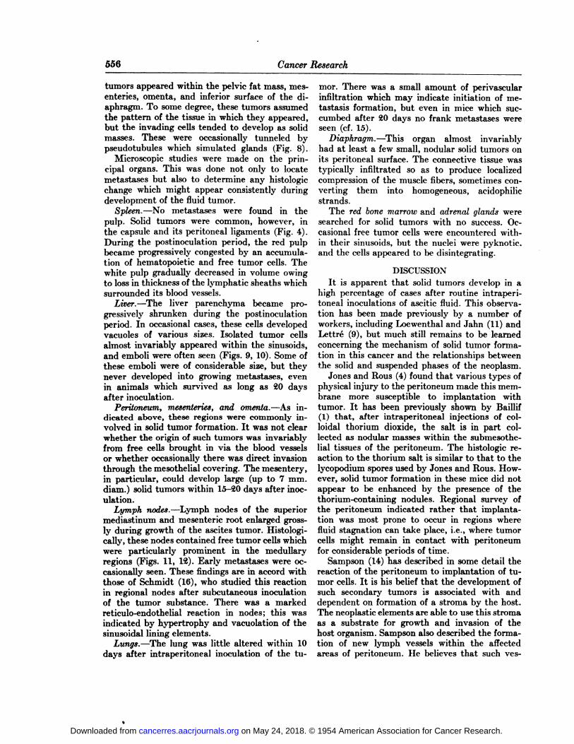
tumors appeared within the pelvic fat mass, mesenteries, omenta, and inferior surface of the diaphragm. To some degree, these tumors assumedthe pattern of the tissue in which they appeared,but the invading cells tended to develop as solidmasses. These were occasionally tunneled bypseudotubules which simulated glands (Fig. 8).
Microscopic studies were made on the principal organs. This was done not only to locatemétastases but also to determine any histologiechange which might appear consistently duringdevelopment of the fluid tumor.
Spleen.—No métastases were found in the
pulp. Solid tumors were common, however, inthe capsule and its peritoneal ligaments (Fig. 4).During the postinoculation period, the red pulpbecame progressively congested by an accumulation of hematopoietic and free tumor cells. Thewhite pulp gradually decreased in volume owingto loss in thickness of the lymphatic sheaths whichsurrounded its blood vessels.
Liver.—The liver parenchyma became pro
gressively shrunken during the postinoculationperiod. In occasional cases, these cells developedvacuoles of various sizes. Isolated tumor cellsalmost invariably appeared within the sinusoids,and emboli were often seen (Figs. 9, 10). Some ofthese emboli were of considerable size, but theynever developed into growing métastases, evenin animals which survived as long as 20 daysafter inoculation.
Peritoneum, mesenteries, and omenta.—As indicated above, these regions were commonly involved in solid tumor formation. It was not clearwhether the origin of such tumors was invariablyfrom free cells brought in via the blood vesselsor whether occasionally there was direct invasionthrough the mesothelial covering. The mesentery,in particular, could develop large (up to 7 mm.diam.) solid tumors within 15-20 days after inoc
ulation.Lymph nodes.—Lymph nodes of the superior
mediastinum and mesenteric root enlarged grossly during growth of the ascites tumor. Histologi-cally, these nodes contained free tumor cells whichwere particularly prominent in the medullaryregions (Figs. 11, 12). Early métastases were occasionally seen. These findings are in accord withthose of Schmidt (16), who studied this reactionin regional nodes after subcutaneous inoculationof the tumor substance. There was a markedreticulo-endothelial reaction in nodes; this wasindicated by hypertrophy and vacuolation of thesinusoidal lining elements.
Lungs.—The lung was little altered within 10
days after intraperitoneal inoculation of the tu
mor. There was a small amount of perivascularinfiltration which may indicate initiation of metastasis formation, but even in mice which succumbed after 20 days no frank métastases wereseen (cf. 15).
Diaphragm..—This organ almost invariably
had at least a few small, nodular solid tumors onits peritoneal surface. The connective tissue wastypically infiltrated so as to produce localizedcompression of the muscle fibers, sometimes converting them into homogeneous, acidophilicstrands.
The red bone marrow and adrenal glands weresearched for solid tumors with no success. Occasional free tumor cells were encountered within their sinusoids, but the nuclei were pyknotic.and the cells appeared to be disintegrating.
DISCUSSIONIt is apparent that solid tumors develop in a
high percentage of cases after routine intraperitoneal inoculations of ascitic fluid. This observation has been made previously by a number ofworkers, including Loewenthal and Jahn (11) andLettré (9), but much still remains to be learnedconcerning the mechanism of solid tumor formation in this cancer and the relationships betweenthe solid and suspended phases of the neoplasm.
Jones and Rous (4) found that various types ofphysical injury to the peritoneum made this membrane more susceptible to implantation withtumor. It has been previously shown by Baillif(1) that, after intraperitoneal injections of colloidal thorium dioxide, the salt is in part collected as nodular masses within the submesothe-lial tissues of the peritoneum. The histologie reaction to the thorium salt is similar to that to thelycopodium spores used by Jones and Rous. However, solid tumor formation in these mice did notappear to be enhanced by the presence of thethorium-containing nodules. Regional survey ofthe peritoneum indicated rather that implantation was most prone to occur in regions wherefluid stagnation can take place, i.e., where tumorcells might remain in contact with peritoneumfor considerable periods of time.
Sampson (14) has described in some detail thereaction of the peritoneum to implantation of tumor cells. It is his belief that the development ofsuch secondary tumors is associated with anddependent on formation of a stroma by the host.The neoplastic elements are able to use this stromaas a substrate for growth and invasion of thehost organism. Sampson also described the formation of new lymph vessels within the affectedareas of peritoneum. He believes that such ves-
on May 24, 2018. © 1954 American Association for Cancer Research. cancerres.aacrjournals.org Downloaded from
BAILLIF—SolidEhrlich Tumor in Mice 557
sels facilitate the escape of tumor cells into thelymphatic system of the host.
In the same manner, the development of as-cites tumor is associated with production of inflammation and development of granulation tissue in the peritoneum. Invading tumor cells advance through the submesothelial connective tissue spaces to extend the solid tumor (Fig. 1). Inregions where this is taking place, there is gradualloss of the mesothelial covering, allowing directcontinuity to be established between the tissuespaces and peritoneal cavity.
These denuded regions of peritoneum, however,do not typically remain naked; a precipitate offibrin appears and in some instances, at least,builds up a bulky tumor of semisolid consistency.This meshwork of fibrin threads becomes permeated with tumor cells. Enterline and Comans(2) have shown that a wide variety of tumor cellsare motile in tissue culture, this movement takingplace by ameboid activity. They have also foundthat small groups of cells are able to move assingle physical units. While no direct analogy canbe made between physiological conditions of invivo and in vitro experimentation, the histologiepattern of tumor cells found in both solid andsemisolid tumors indicates that migration maytake place. Solid tumors are composed primarilyof relatively small cells whose contours are molded by contiguity with their neighbors. Withinsemisolid tumors, the cells are larger, adspherical,and less crowded. Free cells in suspension arestill larger and essentially spherical in form.
As indicated above, these experiments offerno direct proof of cell migration from solid orsemisolid tumors into ascitic fluid. That some suchprocess may take place, however, is indicated byGoldie and Felix (3), who state: "It is common
knowledge that the presence of abdominal tumorsin cancer patients and the growth of peritonealimplants in laboratory animals may be associatedwith the presence of free tumor cells in the peritoneal fluid" (p. 73).
Histological examination of various organs inthese tumor-bearing mice indicated that metastasis occurs, often within 10 days after inoculation. Formation of métastaseshas been studiedpreviously by Schmidt (16), who centered his attention on lymph nodes, and by Schairer (15),who studied the lung. The mechanism of transport of such cells by the lymphatic system is wellknown. These observations, as well as those ofSchmidt (16), indicate that the lymphatic reticulo-endothelium is unable to effect complete destruction of the tumor cells and that some escape intothe blood-vascular system for further distribution.
Ascites tumors have been rather extensivelyused as a quantitative tool in estimating the rateof tumor cell growth under various conditions,for example by such workers as Klein (6), Lettré(9), and Patt, Blackford, and Drallmeier (13).In making such estimates the experimental method is subject to a number of variables which mayaffect the totals obtained. In addition to the inherent technical errors, evidence has been presented above which indicates that growth of tumor cells in ascitic fluid does not take place within a closed system. At present we have no usablequantitative method for determining the numberor percentage of cells added to ascitic fluid byescape from solid and semisolid tumors. Similarly,it is impossible to quantitate the degree of cellloss via lymphatic channels which drain the peritoneal cavity. It would be still more important ifa reliable method could be worked out to determine whether there is a marked individual variability in the types of cell transfer just mentioned.Notwithstanding these difficulties, the workersin quantitative tumor biology should be awareof these variables as well as the physiologic factors concerned in their production.
SUMMARYAfter the inoculation of 10-15 million Ehrlich
ascites tumor cells, over 90 per cent of the micedeveloped solid tumors. Whenever these tumorslay in tissues adjacent to the peritoneum, therewas established a region of direct continuity between these tissues and the peritoneal cavity.Particularly in regions of fluid stagnation withinthe peritoneum and of mesothelial denudation,semisolid tumors appeared. These masses wererich in tumor cells imbedded in a fibrin stroma.It is at least possible that the solid and semisolidphases of this tumor contribute cells which augment the number which appear in the ascitic suspension. The histologie identification of tumorcells in various viscera distant from the peritoneum indicates that tumor cells escape by way ofthe lymphatic drainage from this chamber and,in some locations, initiate métastases.
ACKNOWLEDGMENTSThis project was supported in part by grant C-1563 from
the National Cancer Institute of the United States PublicHealth Service. The Thorotrast used in these experiments wasa gift of the Heyden Chemical Corporation, New York. Tumor-bearing mice were given to me by Dr. M. R. Lewis, WistarInstitute of Anatomy and Biology, Philadelphia, Pa., andby Dr. Ana E. Carrera, Tulane University School of Medicine.
REFERENCES1. BAILLIF,R. N. Splenic Reactions to Colloidal Thorium
Dioxide in the Albino Rat. Am. J. Anat-, 92:55-115, 1953.
on May 24, 2018. © 1954 American Association for Cancer Research. cancerres.aacrjournals.org Downloaded from

WT"
558 Cancer Research
2. ENTERUNE, H. T., and COMAN,D. R. The AmeboidMotility of Human and Animal Neoplastia Cells. Cancer,3:1033-38,1950.
3. GOLDIE,H., and FELIX, M. D. Growth Characteristics ofFree Tumor Cells Transferred Serially in the PeritonealFluid of the Mouse. Cancer Research, 11:73-80, 1951.
4. JONES,F. S., and Rous, P. On the Cause of the Localization of Secondary Tumors at Points of Injury. J. Exper.Med., 20:404-12, 1914.
5. KLEIN, E.; KUBNICK,N. B.; and KLEIN, G. The Effect ofStorage on the Nucleic Acid Content and Virulence ofMouse Ascites Tumor. Exper. Cell Research, 1:127-34,1950.
6. KLEIN, G. The Use of the Ehrlich Ascites Tumor of Micefor Quantitative Studies on the Growth and Biochemistryof Neoplastic Cells. Cancer, 3:1052-61, 1950.
7. KLEIN, G., and KLEIN, E. The Transformation of a SolidTransplantable Mouse Carcinoma into an Ascites Tumor.Cancer Research, 11:466-69, 1951.
8. KOPROWSKA,I., and KOPROWSKI,H. Morphological andBiological Changes in Mouse Ascites Tumors FollowingInduced Infection with Certain Viruses. Fed. Proc., 11:420, 1952.
9. LETTRE,H. Einige Beobachtungen überdas Wachstum desMäuse-Ascites-Tumorsund seine Beeinflussung. Ztschr. f.physiol. Chem., 268:59-76, 1941.
10. LEWIS,M. R., and GOLAND,P. P. The Tumor-inhibitingActivity of Diaryl- and Triarylmethane Dyes. I. TheEhrlich Ascites Mouse Tumor. Cancer Research, 13:130-36, 1953.
11. LOEWENTHAL.H.,andjAHN, G. Übertragungsversuche mitcarcinomatöser Mäuse-Ascitesflüssigkeitund ihr Verhalten gegen physikalische und chemische Einwirkungen.Ztschr. f. Krebsforsch., 37:439-47, 1932.
12. LOVE,R.; KOPBOWSKI,H.; and Cox, H. R. CytologicalStudies on the Ehrlich Ascites Tumor before and afterInfection with Bunyamwera Virus. Cancer Research, 13:350-57, 1953.
13. PATT,H. M.; BLACKPOHD,M. E.; and DRALLMEIER,J. L.Growth Characteristics of the Krebs Ascites Tumor. Proc.Soc. Exper. Biol. & Med., 83:520-24, 1953.
14. SAMPSON,J. A. The Origin and Significance of NewlyFormed Lymph Vessels in Carcinomatous Peritoneal Implants. Am. J. Path., 12:437-67, 1936.
15. SCHAIRER,E. Überdas Wachstum des Mäuseascites-krebses in des Lungen. Ztschr. f. Krebsforsch., 44:296-307-1936.
16. SCHMIDT,W. Ãœberdie Bildung von Lymphknotenmetastasen beim Ascitescarcinom der Maus. Ztschr. f. Krebsforsch., 48:506-19, 1939.
17. SEEGER, P. G. Untersuchungen am Tumoraszites derMaus. 1. Mitteilung; Vitalfärbarkeit der Asziteszellen.Arch. f. exper. Zellforsch., 20:280-335, 1937.
18. SNOOK,T. The Guinea-Pig Spleen. Studies on the Structure and Connections of the Venous Sinuses. Anat. Ree.,89:413-27, 1944.
19. SUGIDRA,K. Effect of Various Compounds on the EhrlichAscites Carcinoma. Cancer Research, 13:431-41, 1953.
20. WARREN,S., and GATES,O. The Fate of IntravenouslyInjected Tumor Cells. Am. J. Cancer, 27:485-92, 1936.
The specimens were obtained from mice at autopsy 10days after intraperitoneal inoculation of the Ehrlich ascitestumor; they were fixed in Bouin's fluid and stained with
hematoxylin and eosin.FIG. 1.—Afold of tissue from the pelvic fat mass where the
solid tumor is becoming established. At the right, the meso-thelium has been destroyed and the denuded area is coveredwith fibrin clot which is richly infiltrated with tumor cells.X102.
FIG. 2.—Region near the surface of the greater omentumshowing a developing solid tumor. Small tumor cells abound intissues adjacent to the dilated blood vessels. There is variationin the size of tumor cells within the small area of tissues seenhere. X375.
FIG. 3.—Surfaceof the greater omentum in a region wherethere is localized destruction of the mesothelium. The arrows
point toward the omental surface. The upper right portion ofthe figure is composed of fibrin impregnated with tumor cells,while other cells lie on the surface of the clot. Note the variation in size of the tumor cells within the omentum proper(i.e., in solid tumor), in the meshes of the clot (semisolidtumor) and on the surface of the clot. X375.
FIG. 4.—Solidtumor attached to the capsule of the spleen.Splenic red pulp comprises the left half of the figure and solidtumor the right half, while the fibrous capsule runs diagonallydownward and toward the right between the two regions justdelineated. X375.
FIG. 5.—Solid tumor in fat of the mesentery. Note thedensity and pattern of the tumor cells as they invade the connective tissue between fat cells. X231.
FIG. 6.—Solidtumor which has invaded the pancreas andlargely destroyed its acini. X231.
on May 24, 2018. © 1954 American Association for Cancer Research. cancerres.aacrjournals.org Downloaded from

•<"fci/-v«W,v
on May 24, 2018. © 1954 American Association for Cancer Research. cancerres.aacrjournals.org Downloaded from

Fio. 7.—Solid tumor in a fold of the gastro-liciti tic omen-tum, a region where there is destruction of the covering meso-tlu'lium. Solid tumor comprises the dense tissue at the lowerside of the figure; most of this region is covered with a fibrinmeshwork. The upper and right regions show tumor cellswhich are either entirely free or partially ¡mix-tidedin fragments of the fibrin net. X375.
FIG. 8.—Solidtumor beneath the parietal peritoneum of theanterior ¡ibdoininalwall. Note the density of the cells andvariety in size. Near the top of the figure is a cell mass suggestive of pseudotubule formation. X 1,086.
FIG. 9.—Liverwith tumor cells lying free within the sinusoids. A mass of these cells has collected in one of these dilatedspaces; this region is outlined with Mack lines. Klsewhere,the tumor cells appear to be traversing the channels more orless singly. X210.
FIG. 10.—Higher magnification of the area outlined in thepreceding figure, to show relationships of tumor cells in thehepatic sinusoids. X 411.
FIG. 11.—Lymph ntxli- medulla with free tumor cells. Thelighter regions represent the nodal lymph sinusoids, variablyfilled with tumor cells. The tlarker regions are medullarylymphocytic cords, considerably fragmented by collections oftumor cells within the sinusoids. XilO.
FIG. 12.—Lymphnode medulla from a specimen similar tothat shown in the preceding figure. The higher magnificationclarifies the relationships between tumor cells and medullarycords. X411.
on May 24, 2018. © 1954 American Association for Cancer Research. cancerres.aacrjournals.org Downloaded from

* WPSVîa*,*.fytf\ a _:7 T
yaJF'-STOîï^.v"1!•/*.-fvÃAÃ'.f*>
on May 24, 2018. © 1954 American Association for Cancer Research. cancerres.aacrjournals.org Downloaded from

1954;14:554-558. Cancer Res Ralph N. Baillif The Solid Phase of the Ehrlich Ascites Tumor in Mice
Updated version
http://cancerres.aacrjournals.org/content/14/8/554
Access the most recent version of this article at:
E-mail alerts related to this article or journal.Sign up to receive free email-alerts
Subscriptions
Reprints and
To order reprints of this article or to subscribe to the journal, contact the AACR Publications
Permissions
Rightslink site. Click on "Request Permissions" which will take you to the Copyright Clearance Center's (CCC)
.http://cancerres.aacrjournals.org/content/14/8/554To request permission to re-use all or part of this article, use this link
on May 24, 2018. © 1954 American Association for Cancer Research. cancerres.aacrjournals.org Downloaded from