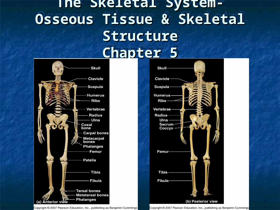
The Skeletal System- Osseous Tissue & Skeletal Structure Chapter 5.
30
The Skeletal System- The Skeletal System- Osseous Tissue & Skeletal Osseous Tissue & Skeletal Structure Structure Chapter 5 Chapter 5
-
Upload
shon-julius-washington -
Category
Documents
-
view
224 -
download
2
Transcript of The Skeletal System- Osseous Tissue & Skeletal Structure Chapter 5.
The Skeletal System-The Skeletal System-Osseous Tissue & Skeletal Osseous Tissue & Skeletal
StructureStructureChapter 5Chapter 5
The Skeletal System-The Skeletal System-Osseous Tissue & Skeletal Osseous Tissue & Skeletal
StructureStructureChapter 5Chapter 5
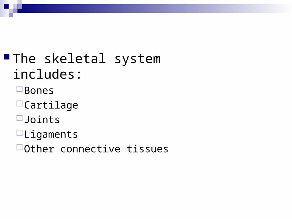
The skeletal system includes:BonesCartilageJointsLigamentsOther connective tissues
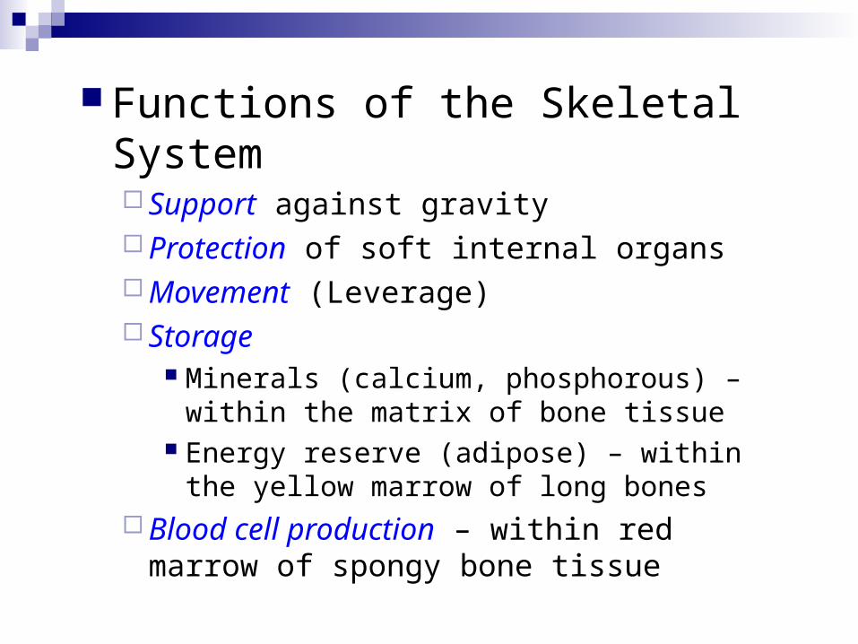
Functions of the Skeletal System Support against gravity Protection of soft internal organs Movement (Leverage) Storage
Minerals (calcium, phosphorous) – within the matrix of bone tissue
Energy reserve (adipose) – within the yellow marrow of long bones
Blood cell production – within red marrow of spongy bone tissue
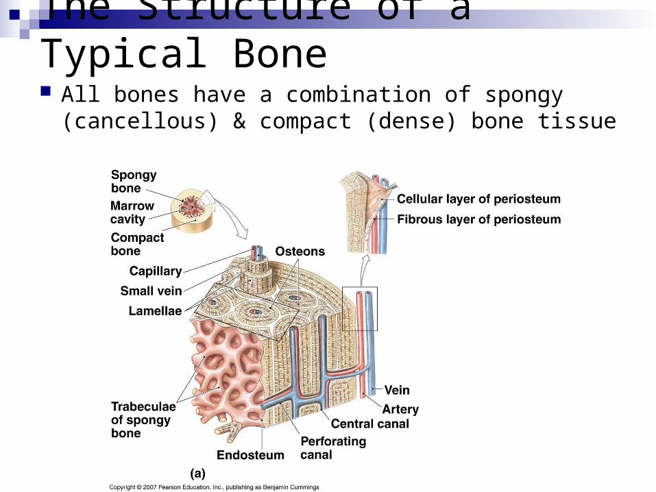
The Structure of a Typical Bone
All bones have a combination of spongy (cancellous) & compact (dense) bone tissue
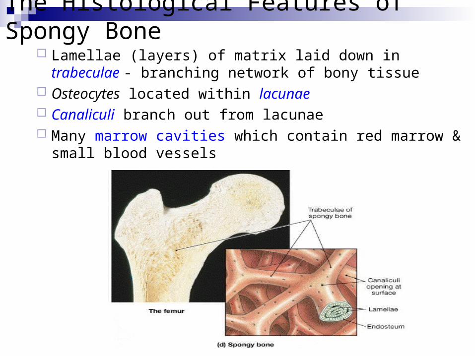
The Histological Features of Spongy Bone Lamellae (layers) of matrix laid down in trabeculae - branching
network of bony tissue Osteocytes located within lacunae Canaliculi branch out from lacunae Many marrow cavities which contain red marrow & small blood
vessels
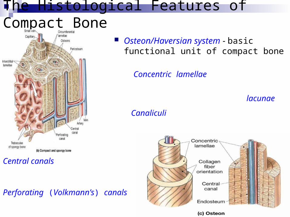
The Histological Features of Compact Bone
Osteon/Haversian system - basic functional unit of compact bone
Concentric lamellae (layers) of matrix surrounding central (Haversian) canal
Osteocytes located within lacunae
Canaliculi branch out radially from lacunae
Central canals (containing BVs) run vertically down the length of the bone
Perforating (Volkmann’s) canals (containing BVs) run horizontally across the width
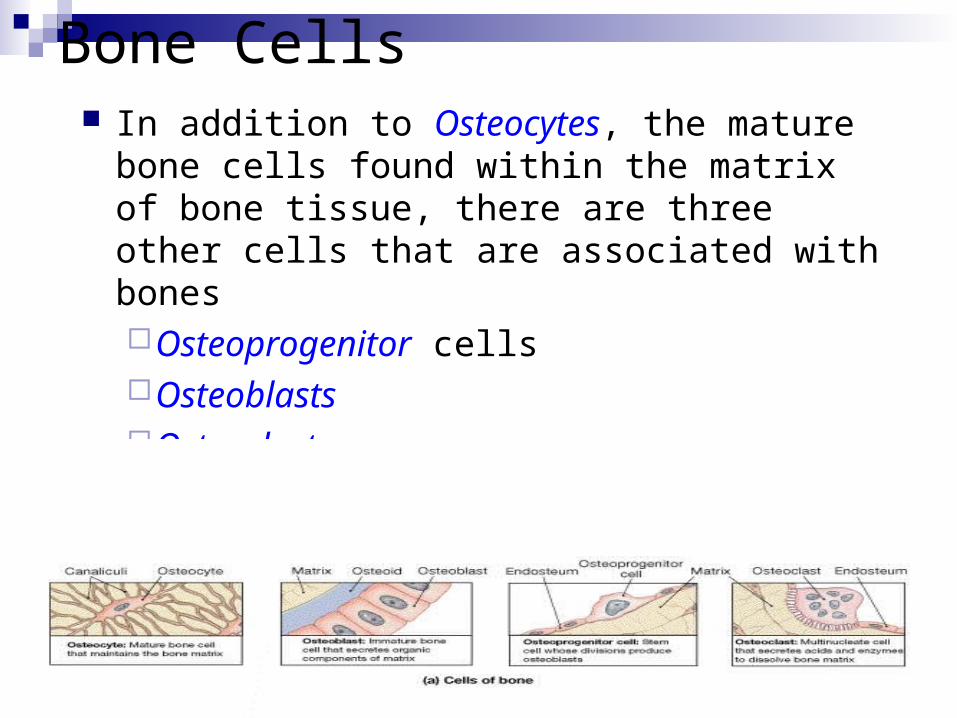
Bone Cells In addition to Osteocytes, the mature bone
cells found within the matrix of bone tissue, there are three other cells that are associated with bonesOsteoprogenitor cellsOsteoblastsOsteoclasts
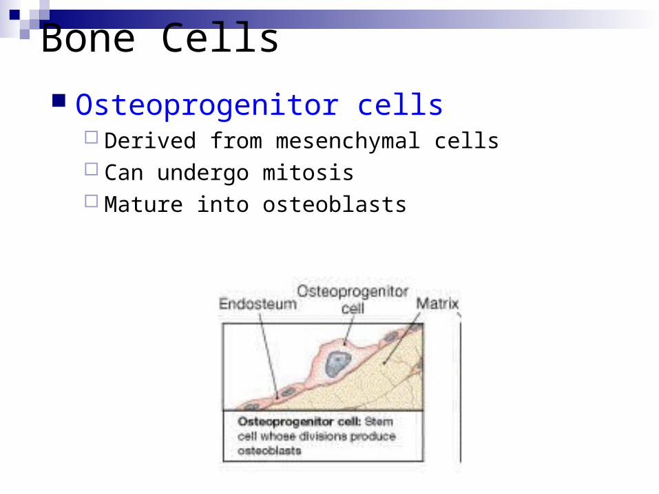
Bone Cells
Osteoprogenitor cells Derived from mesenchymal cells Can undergo mitosis Mature into osteoblasts
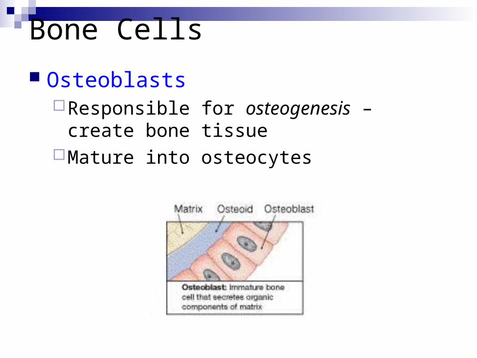
Bone Cells
OsteoblastsResponsible for osteogenesis – create bone
tissueMature into osteocytes
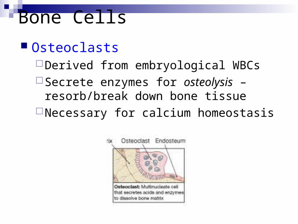
Bone Cells
OsteoclastsDerived from embryological WBCsSecrete enzymes for osteolysis –
resorb/break down bone tissueNecessary for calcium homeostasis
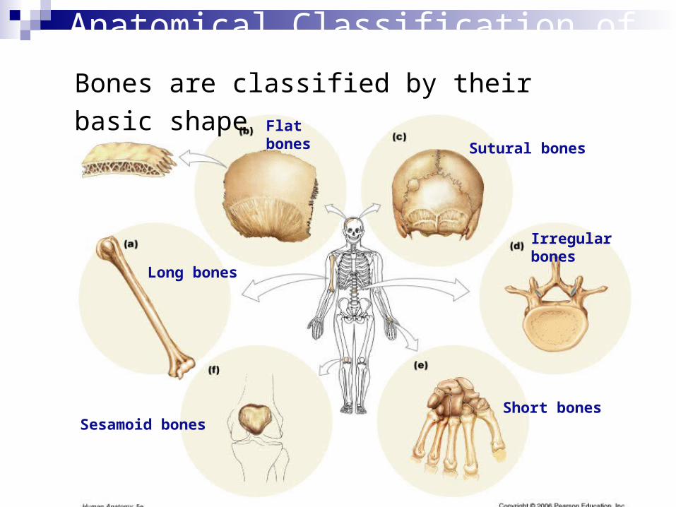
Long bones
Short bones
Flat bones
Irregular bones
Sesamoid bones
Sutural bones
Anatomical Classification of Bones
Bones are classified by their basic shape
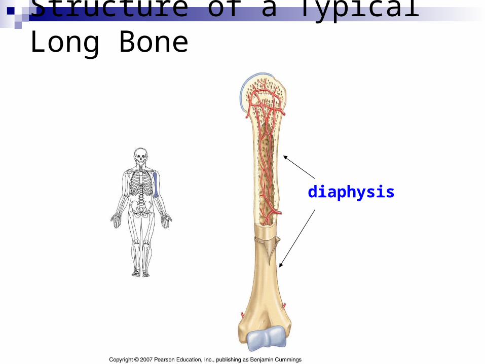
Structure of a Typical Long Bone
diaphysis (compact bone)
The Structure of a Long Bone
diaphysis
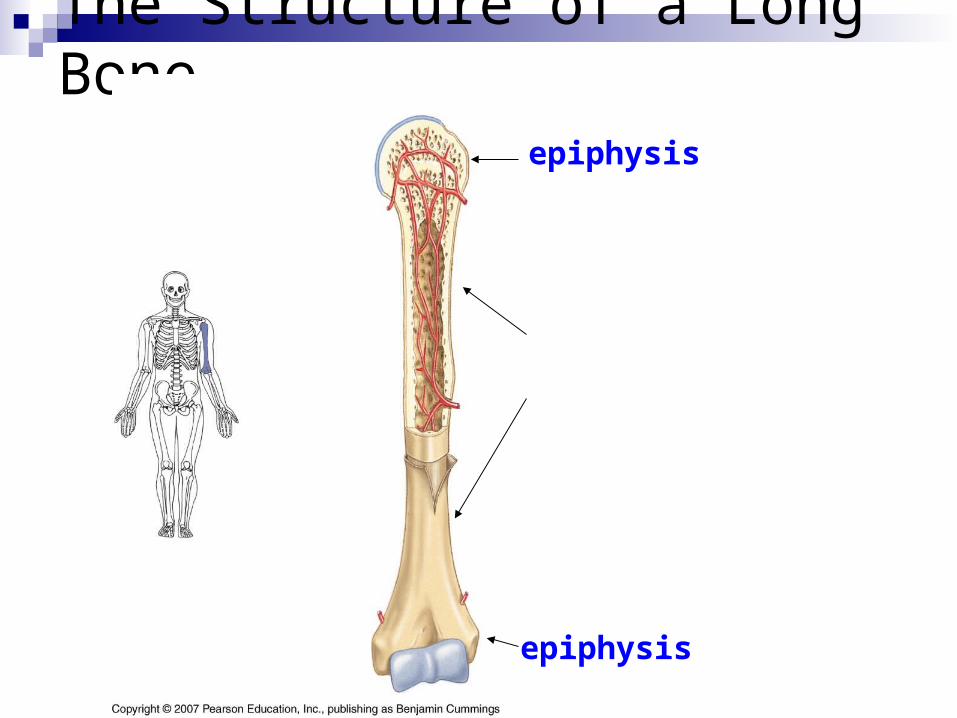
epiphysis (spongy bone)
epiphysis

The Structure of a Long Bone
diaphysis
epiphysis
epiphysis
articular cartilage
articular cartilage
The Structure of a Long Bone
epiphysis
epiphysis
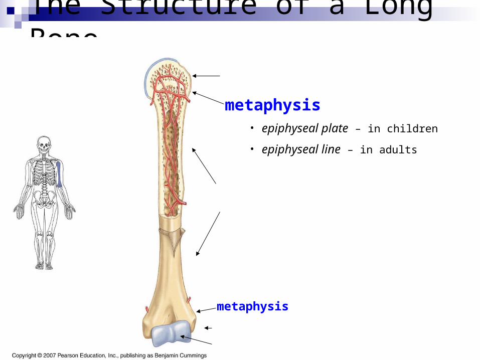
metaphysis – location of
• epiphyseal plate – in children
• epiphyseal line – in adults
diaphysis
metaphysis
articular cartilage

The Structure of a Long Bone
medullary cavity
• filled with yellow marrow in adults
• lined with endosteum
The Structure of a Long Bone
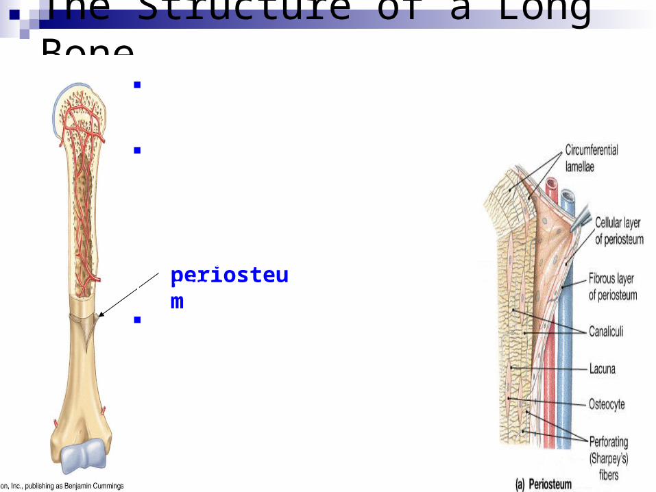
periosteum
Outer fibrous layer of dense irregular CT for attachment of tendons& ligaments; provides route for blood vessels & nerves; separates bone tissue from surrounding tissues
Double layered membrane surrounding bone except at articular cartilage
Inner cellular layer contains osteoprogenitor cells, osteoblasts, osteoclasts; therefore functions in bone growth & repair
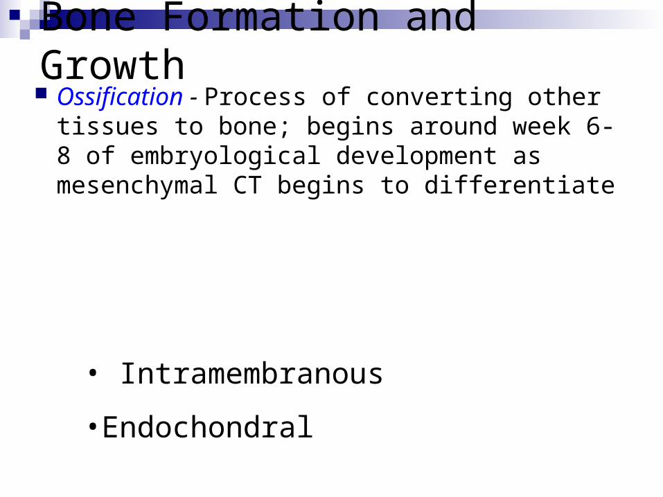
Bone Formation and Growth Ossification - Process of converting other
tissues to bone; begins around week 6-8 of embryological development as mesenchymal CT begins to differentiate
Two types of ossification processes occur during embryological formation:
• Intramembranous
•Endochondral
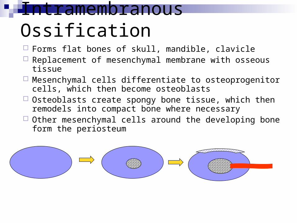
Intramembranous Ossification Forms flat bones of skull, mandible, clavicle Replacement of mesenchymal membrane with osseous tissue Mesenchymal cells differentiate to osteoprogenitor cells, which then
become osteoblasts Osteoblasts create spongy bone tissue, which then remodels into
compact bone where necessary Other mesenchymal cells around the developing bone form the
periosteum
Mesenchymal tissue forms
Osteoblasts begin to secrete osteoid forming spongy bone tissue
Blood vessels infiltrate tissue. Calcium salts deposit in osteoid. Periosteum develops
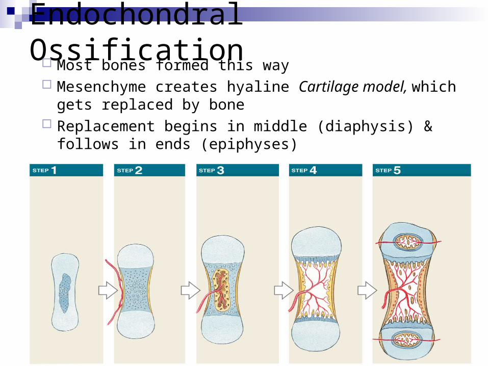
Endochondral Ossification Most bones formed this way Mesenchyme creates hyaline Cartilage model, which gets
replaced by bone Replacement begins in middle (diaphysis) & follows in ends
(epiphyses)
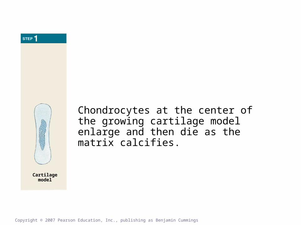
Chondrocytes at the center of the growing cartilage model enlarge and then die as the matrix calcifies.
Cartilagemodel
Copyright © 2007 Pearson Education, Inc., publishing as Benjamin Cummings
Endochondral Ossification
Cartilage model grows in length (interstitial growth) & in width (appositional growth)
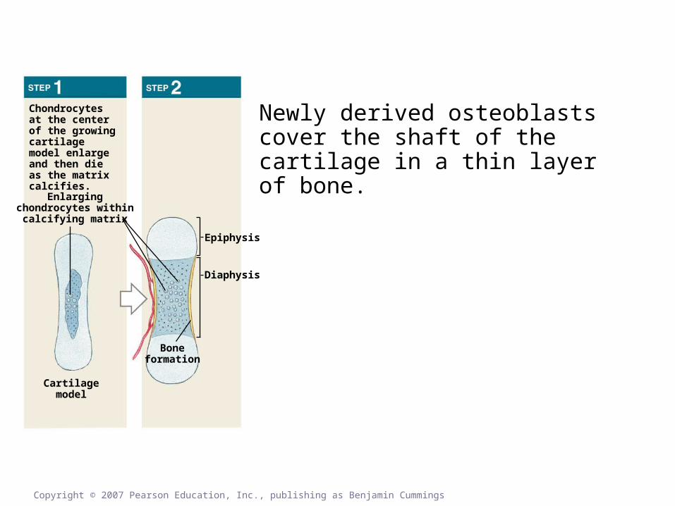
Figure 5.7
Enlargingchondrocytes within
calcifying matrix
Chondrocytes at the center of the growing cartilage model enlarge and then die as the matrix calcifies.
Newly derived osteoblasts cover the shaft of the cartilage in a thin layer of bone.
Cartilagemodel
Boneformation
Epiphysis
Diaphysis
Copyright © 2007 Pearson Education, Inc., publishing as Benjamin Cummings
The perichondrium, which surrounded the cartilage model, now must be referred to as the periosteum.
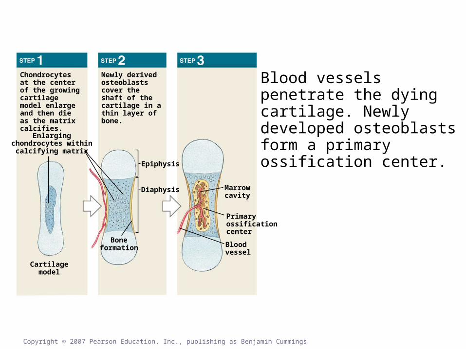
Figure 5.7
Enlargingchondrocytes within
calcifying matrix
Chondrocytes at the center of the growing cartilage model enlarge and then die as the matrix calcifies.
Newly derived osteoblasts cover the shaft of the cartilage in a thin layer of bone.
Blood vessels penetrate the dying cartilage. Newly developed osteoblasts form a primary ossification center.
Cartilagemodel
Boneformation
Epiphysis
Diaphysis Marrowcavity
Primaryossificationcenter
Bloodvessel
Copyright © 2007 Pearson Education, Inc., publishing as Benjamin Cummings
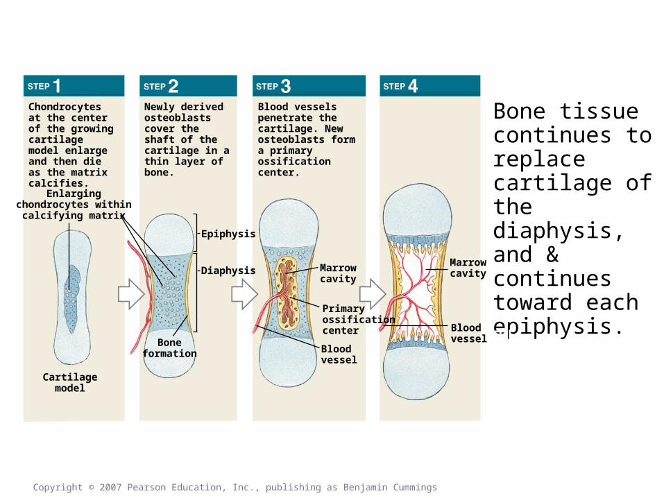
Figure 5.7
Enlargingchondrocytes within
calcifying matrix
Chondrocytes at the center of the growing cartilage model enlarge and then die as the matrix calcifies.
Newly derived osteoblasts cover the shaft of the cartilage in a thin layer of bone.
Blood vessels penetrate the cartilage. New osteoblasts form a primary ossification center.
Bone tissue continues to replace cartilage of the diaphysis, and & continues toward each epiphysis.
Cartilagemodel
Boneformation
Epiphysis
Diaphysis Marrowcavity
Primaryossificationcenter
Bloodvessel
Marrowcavity
Bloodvessel
Copyright © 2007 Pearson Education, Inc., publishing as Benjamin Cummings
The medullary cavity begins to hollow out
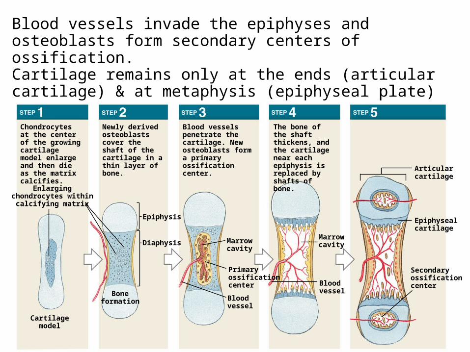
Blood vessels invade the epiphyses and osteoblasts form secondary centers of ossification.Cartilage remains only at the ends (articular cartilage) & at metaphysis (epiphyseal plate)
Enlargingchondrocytes within
calcifying matrix
Chondrocytes at the center of the growing cartilage model enlarge and then die as the matrix calcifies.
Newly derived osteoblasts cover the shaft of the cartilage in a thin layer of bone.
Blood vessels penetrate the cartilage. New osteoblasts form a primary ossification center.
The bone of the shaft thickens, and the cartilage near each epiphysis is replaced by shafts of bone.
Cartilagemodel
Boneformation
Epiphysis
Diaphysis Marrowcavity
Primaryossificationcenter
Bloodvessel
Marrowcavity
Bloodvessel
Secondaryossificationcenter
Epiphysealcartilage
Articularcartilage
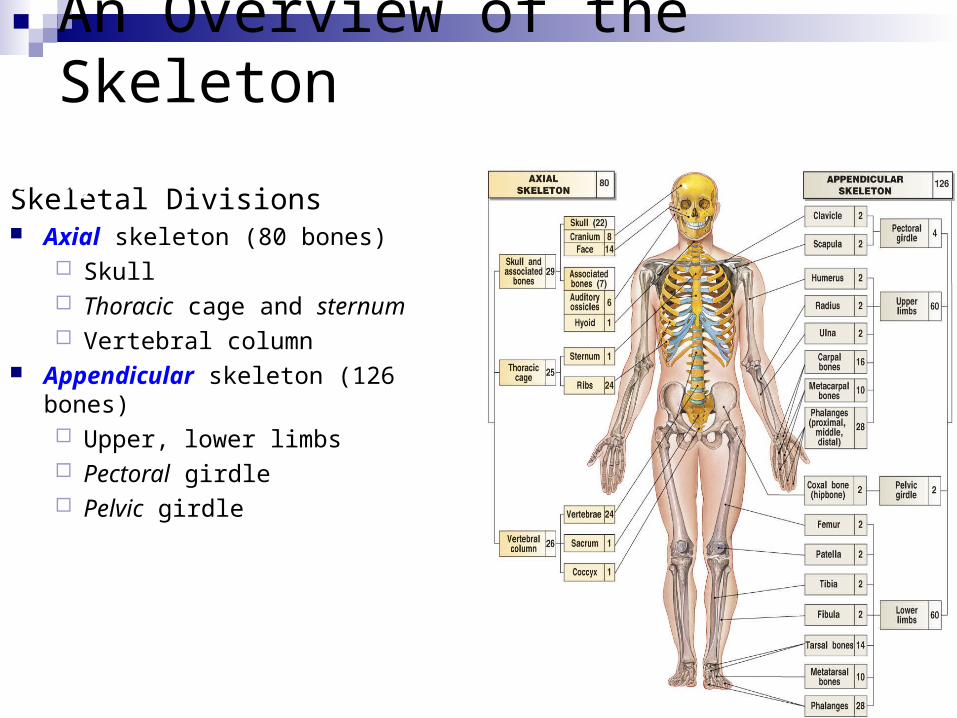
An Overview of the Skeleton
Skeletal Divisions Axial skeleton (80 bones)
Skull Thoracic cage and sternum Vertebral column
Appendicular skeleton (126 bones) Upper, lower limbs Pectoral girdle Pelvic girdle
There are 206 bones in the adult human body

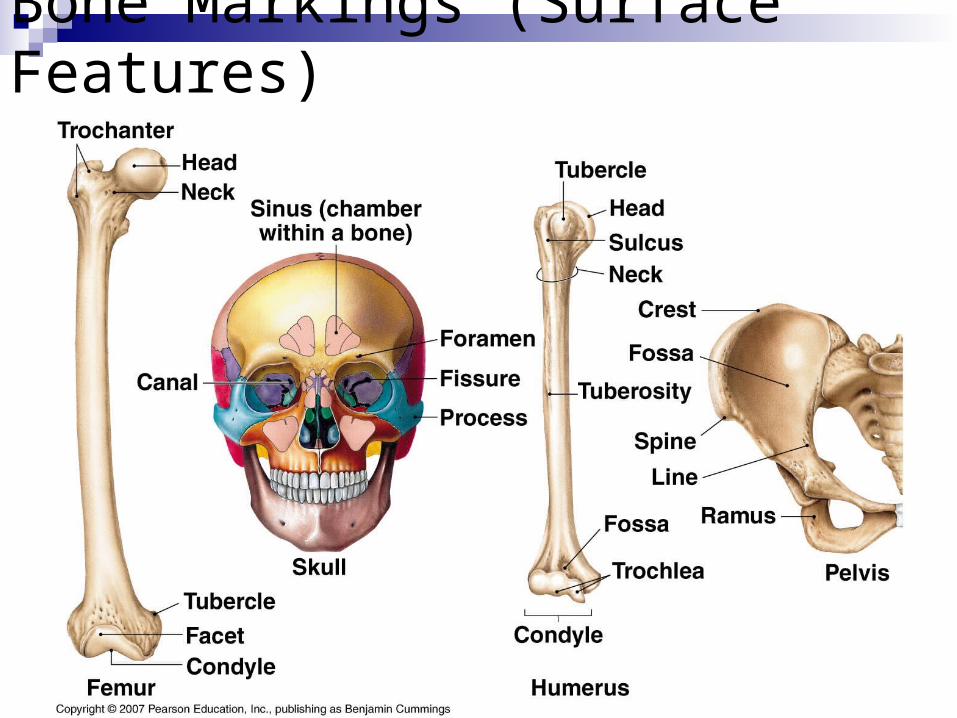
Bone Markings (Surface Features)
Markings for articulations: head condyle facet
General elevations & projections:
process
ramus
Table 5-1
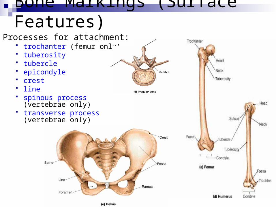
Bone Markings (Surface Features)Processes for attachment:
trochanter (femur only) tuberosity tubercle epicondyle crest line spinous process (vertebrae
only) transverse process (vertebrae
only)
Spinous process
Transverse process
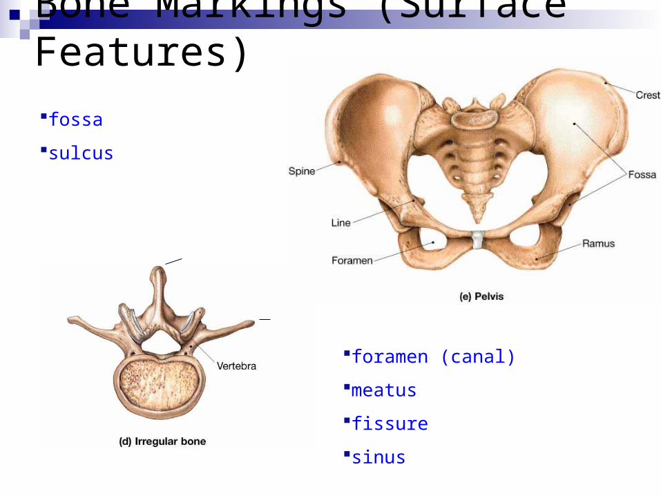
Bone Markings (Surface Features)
Spinous process
Transverse process
Depressions:
fossa
sulcus
Openings:
foramen (canal)
meatus
fissure
sinus
Bone Markings (Surface Features)