Chapter 7 Bone Tissue Tissues and organs of the skeletal system Histology of osseous tissue Bone...
47
Chapter 7 Bone Tissue • Tissues and organs of the skeletal system • Histology of osseous tissue • Bone development • Physiology of osseous tissue • Bone disorders
-
Upload
abner-cunningham -
Category
Documents
-
view
225 -
download
3
Transcript of Chapter 7 Bone Tissue Tissues and organs of the skeletal system Histology of osseous tissue Bone...

Chapter 7Bone Tissue
• Tissues and organs of the skeletal system
• Histology of osseous tissue
• Bone development
• Physiology of osseous tissue
• Bone disorders

Bone as a Tissue• Dynamic tissue that continually remodels itself
• Bones and bone tissue– bone or osseous tissue is a connective tissue with a
matrix hardened by minerals(calcium phosphate)
– bones make up the skeletal system • individual bones are made up of bone
tissue, marrow, cartilage & periosteum
• Functions of the skeletal system– support, protection, movement, blood formation,
mineral reservoir, pH balance & detoxification
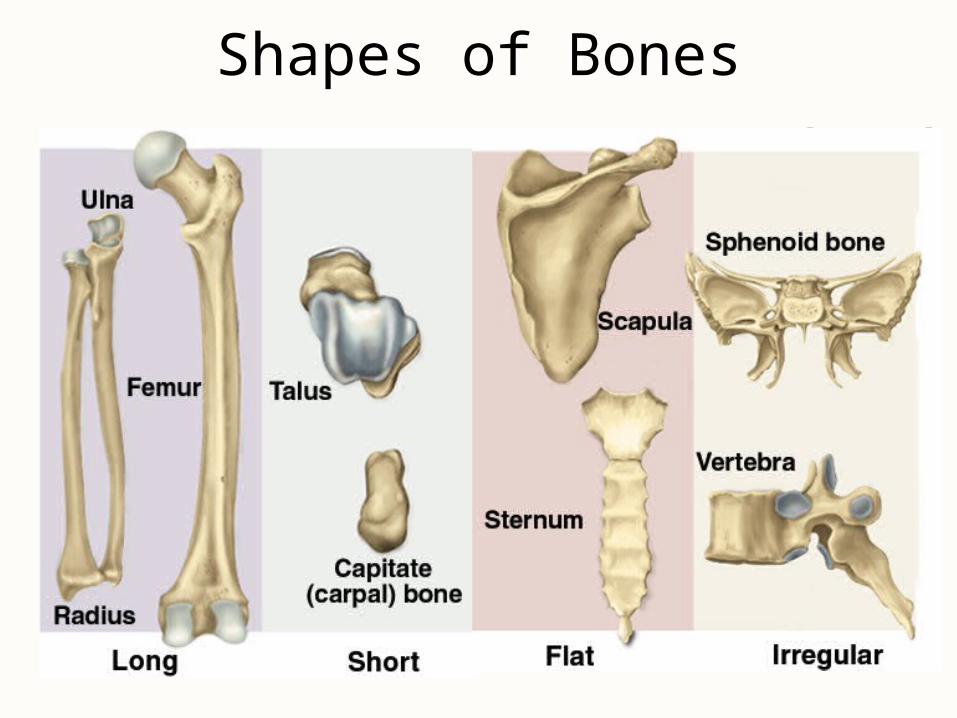
Shapes of Bones

Structure of a Flat Bone
• External and internal surfaces of flat bone are composed of compact bone
• Middle layer is spongy bone (diploe). No marrow cavity
• Blow to the skull may fracture outer layer and crush diploe, but not harm inner compact bone

Structure of a Long Bone
• Periosteum & articular cartilage
• Compact & spongy bone
• Endosteum• Yellow marrow

General Features of Bones• Shaft (diaphysis) is cylinder of compact bone containing
marrow cavity (medullary cavity) & lined with endosteum (layer of osteopenic cells and reticular connective tissue)
• Enlarged ends (epiphyses) are spongy bone covered with a layer of compact bone– enlarged to strengthen joint & provide for attachment of tendons
and ligaments
• Joint surface covered with articular cartilage (lubrication)• Remainder of bone covered with periosteum
– outer fibrous layer of collagen fibers continuous with tendons or perforating(Sharpey’s) fibers that penetrate into bone matrix
– inner osteogenic layer important for growth & healing
• Epiphyseal plate or line depends on age

Cells of Osseous Tissue (1)
• Osteogenic cells reside in endosteum, periosteum or central canals– arise from embryonic fibroblasts and become only source for new osteoblasts
– multiply continuously & differentiate into amitotic osteoblasts in response to stress or fractures
• Osteoblasts form and help mineralize organic matter of matrix
• Osteocytes are osteoblasts that have become trapped in the matrix they formed– cells in lacunae connected by gap junctions inside canaliculi
– signal osteoclasts & osteoblasts about mechanical stresses
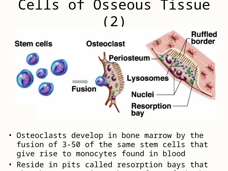
Cells of Osseous Tissue (2)
• Osteoclasts develop in bone marrow by the fusion of 3-50 of the same stem cells that give rise to monocytes found in blood
• Reside in pits called resorption bays that they have eaten into the surface of the bone
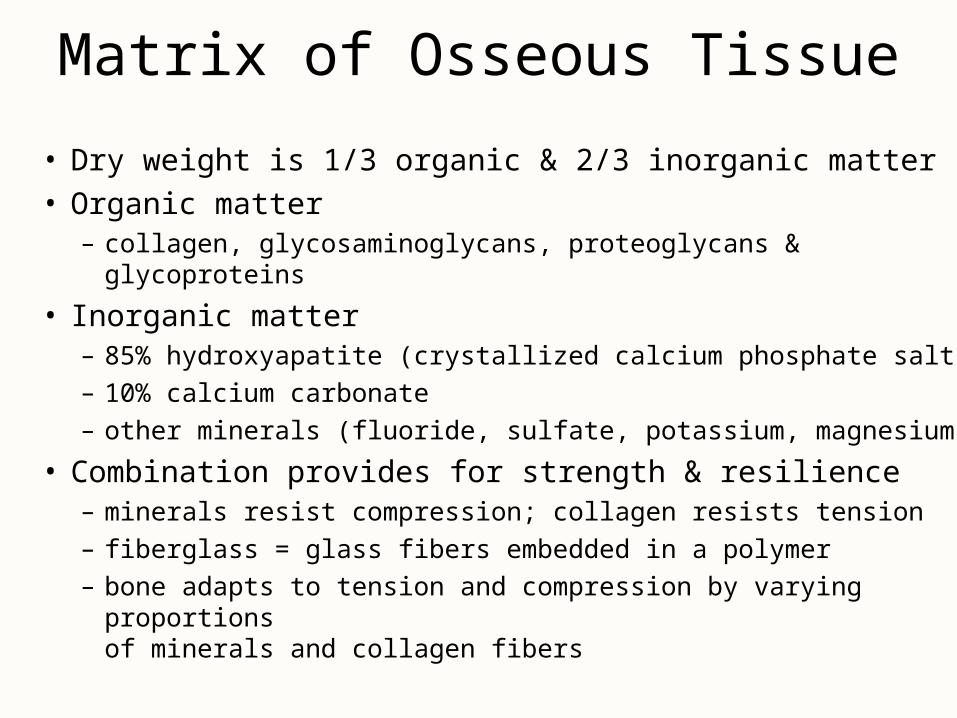
Matrix of Osseous Tissue
• Dry weight is 1/3 organic & 2/3 inorganic matter• Organic matter
– collagen, glycosaminoglycans, proteoglycans & glycoproteins
• Inorganic matter– 85% hydroxyapatite (crystallized calcium phosphate salt)
– 10% calcium carbonate
– other minerals (fluoride, sulfate, potassium, magnesium)
• Combination provides for strength & resilience– minerals resist compression; collagen resists tension
– fiberglass = glass fibers embedded in a polymer
– bone adapts to tension and compression by varying proportions of minerals and collagen fibers

Compact Bone• Osteon (haversian system) = basic structural unit
– cylinders of tissue formed from layers (lamellae) of matrix arranged around central canal holding a blood vessel
• collagen fibers alternate between right- and left-handed helices from lamella to lamella
– osteocytes connected to each other and their blood supply by tiny cell processes in canaliculi
• Perforating canals or Volkmann canals– vascular canals perpendicularly joining central canals
• Circumferential or outer lamellae

Histology of Compact Bone

Blood Vessels of Compact Bone

Spongy Bone
• Spongelike appearance formed by rods and plates of bone called trabeculae– spaces filled with red bone marrow
• Trabeculae have few osteons or central canals– no osteocyte is far from blood of bone marrow
• Provides strength with little weight– trabeculae develop along bone’s lines of stress

Spongy Bone Structure and Stress

Bone Marrow
• Soft tissue that occupies the medullary cavity of a long bone or the spaces amid the trabeculae of spongy bone
• Red marrow looks like thick blood– mesh of reticular fibers and immature cells– hemopoietic means produces blood cells– found in vertebrae, ribs, sternum, pelvic
girdle and proximal heads of femur and humerus in adults
• Yellow marrow– fatty marrow of long bones in adults
• Gelatinous marrow of old age– yellow marrow replaced with reddish jelly

Intramembranous Ossification• Produces flat bones of skull & clavicle
• Steps of the process– mesenchyme condenses into a sheet of soft tissue
• transforms into a network of soft trabeculae
– osteoblasts gather on the trabeculae to form osteoid tissue (uncalcified bone)
– calcium phosphate is deposited in the matrix transforming the osteoblasts into osteocytes
– osteoclasts remodel the center to contain marrow spaces & osteoblasts remodel the surface to form compact bone
– mesenchyme at the surface gives rise to periosteum

Intramembranous Ossification

Endochondral Ossification• Primary ossification center forms in cartilage model
– chondrocytes near the center swell to form primary ossification center
– matrix is reduced & model becomes weak at that point– cells of the perichondrium produce a bony collar– cuts off diffusion of nutrients and hastens their death
• Primary marrow space formed by periosteal bud– osteogenic cells invade & transform into osteoblasts– osteoid tissue deposited and calcified into trabeculae at
same time osteoclasts work to enlarge the primary marrow cavity

Primary Ossification Center & Marrow Space
• Both form in center of cartilage model -- same process begins again subsequently at ends of cartilage model.

The Metaphysis• Transitional zone between head and shaft of a developing
long bone
• Zone of reserve cartilage is layer of resting cartilage
• Zone of cell proliferation is layer– chondrocytes multiply forming columns of flat lacunae
• Zone of cell hypertrophy shows hypertrophy
• Zone of calcification shows mineralization between columns of lacunae
• Zone of bone deposition -- chondrocytes die and each channel is filled with osteoblasts and blood vessels to form a haversian canal & osteon

The Metaphysis

Secondary Ossification Center
• Begin to form in the epiphyses near time of birth
• Same stages occur as in primary ossification center– result is center of epiphyseal cartilage being transformed
into spongy bone
• Hyaline cartilage remains on joint surface as articular cartilage and at junction of diaphysis & epiphysis (epiphyseal plate)– each side of epiphyseal plate has a metaphysis

Metaphysis & Secondary Ossification Center
• Metaphysis is cartilagenous material that remains as growth plate between medullary cavity & secondary ossification centers in the epiphyses.

The Fetal Skeleton at 12 Weeks

Epiphyseal Plates

Bone Growth and Remodeling• Grow and remodel themselves throughout life
– growing brain or starting to walk
– athletes or history of manual labor have greater density & mass of bone
• Cartilage grows by both appositional & interstitial growth• Bones increase in length by interstitial growth of
epiphyseal plate• Bones increase in width by appositional growth
– osteoblasts lay down matrix in layers parallel to the outer surface & osteoclasts dissolve bone on inner surface
– if one process outpaces the other, bone deformities occur(osteitis deformans)

Achondroplastic Dwarfism
• Short stature but normal-sized head and trunk– long bones of the limbs
stop growing in childhood but other bones unaffected
• Result of spontaneous mutation when DNA is replicated– mutant allele is dominant
• Pituitary dwarf has lack of growth hormone– short stature with normal
proportions

Mineral Deposition• Mineralization is crystallization process in which ions
(calcium, phosphate & others) are removed from blood plasma & deposited in bone tissue
• Steps of the mineralization process– osteoblasts produce collagen fibers that spiral along the length
of the osteon in alternating directions– fibers become encrusted with minerals hardening matrix
• ion concentration must reach the solubility product for crystal formation to occur & then positive feedback forms more
• Ectopic ossification is abnormal calcification– may occur in lungs, brain, eyes, muscles, tendons or arteries
(arteriosclerosis)

Mineral Resorption
• Process of dissolving bone & releasing minerals into the blood– performed by osteoclasts “ruffled border”
• hydrogen pumps in the cell membrane secrete hydrogen ions into the space between the osteoclast & the bone
• chloride ions follow by electrical attraction• hydrochloric acid with a pH of 4 dissolves bone minerals• an enzyme (acid phosphatase) digests the collagen
• Dental braces reposition teeth, creating greater pressure on the bone on one side of the tooth and less on the other side– increased pressure stimulates osteoclasts; decreased
pressure stimulates osteoblasts to remodel jaw bone

Functions of Calcium & Phosphate
• Phosphate is a component of DNA, RNA, ATP, phospholipids, & acid-base buffers
• Calcium is needed for communication between neurons, muscle contraction, blood clotting & exocytosis
• Calcium plasma concentration is 9.2 to 10.4 mg/dL -- 45% is as Ca+2, rest is bound to plasma proteins & is not physiologically active
• Phosphate plasma concentration is 3.5 to 4.0 mg/dL & occurs in 2 forms: HPO4 -2 & H2PO4
-

Ion Imbalances
• Changes in phosphate concentration have little effect• Changes in calcium can be serious
– hypocalcemia is deficiency of blood calcium• causes excessive excitability of nervous system leading to
muscle spasms, tremors or tetany– laryngospasm may cause suffocation
• calcium normally binds to cell surface contributing to resting membrane potential
– with less calcium, sodium channels open more easily exciting neuron
– hypercalcemia • excessive calcium binding to cell surface makes sodium channels less likely
to open, depressing nervous system
• Calcium phosphate homeostasis depends on calcitriol, calcitonin & PTH

Carpopedal Spasm
• Hypocalcemia causing overexcitability of nervous system and muscle spasm of hands and feet

Calcitriol (Activated Vitamin D)• Produced by the following process
– UV radiation penetrating the epidermal keratinocytes converts a cholesterol derivative (7-dehydrocholesterol) to previtamin D3 and then (cholecalciferol) D3
– liver adds OH to convert it to calcidiol– kidney adds OH to convert calcidiol to calcitriol
• Calcitriol behaves as a hormone (blood-borne messenger)– stimulates intestine to absorb calcium, phosphate & magnesium– weakly promotes urinary reabsorption of calcium ions– promotes osteoclast activity to raise blood calcium concentration
to the level needed for bone deposition
• Abnormal softness of the bones is called rickets in children and osteomalacia in adults

Calcitriol Synthesis & Action

Hormonal Control of Calcium Balance
• Calcitriol, PTH and calcitonin maintain normal blood calcium concentration.

Calcitonin
• Secreted by C cells of the thyroid gland when calcium concentration rises too high
• Functions– reduces osteoclast activity by as much as 70% in 15
minutes– increases the number & activity of osteoblasts
• Important role in children, but little effect in adults– calcitonin deficiency is not known to cause any disease
in adults– may be useful in reducing bone loss in osteoporosis

Parathyroid Hormone
• Secreted by the parathyroid glands found on the posterior surface of the thyroid gland
• Released when calcium blood level is too low• Functions
– binds to osteoblasts causing them to release osteoclast-stimulating factor that stimulates osteoclast multiplication & activity
– promotes calcium resorption by the kidneys
– promotes calcitriol synthesis in the kidneys
– inhibits collagen synthesis and bone deposition by osteoblasts
• Injection of low levels of PTH can cause bone deposition
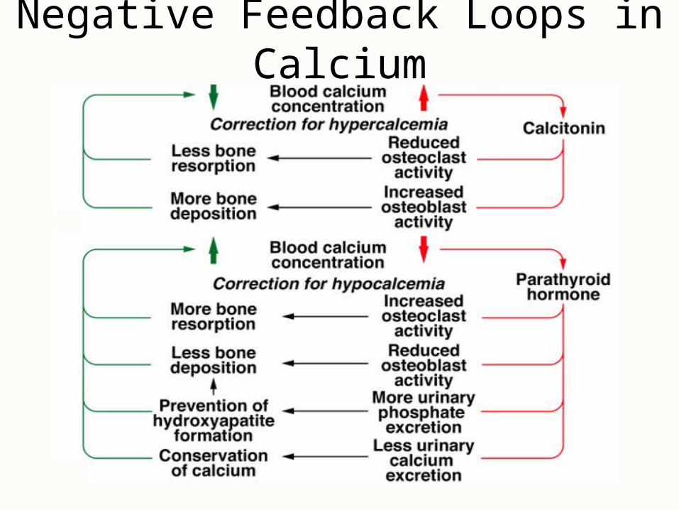
Negative Feedback Loops in Calcium

Other Factors Affecting Bone• 20 or more hormones, vitamins & growth factors
not well understood• Bone growth especially rapid at puberty
– hormones stimulate proliferation of osteogenic cells and chondrocytes in growth plate
– adolescent girls grow faster than boys & reach their full height earlier (estrogen has stronger effect)
– males grow for a longer time
• Growth ceases when epiphyseal plate “closes”– anabolic steroids may cause premature closure of growth
plate producing short adult stature

Fractures and Their Repair
• Stress fracture is a break caused by abnormal trauma to a bone– car accident, fall, athletics, etc
• Pathological fracture is a break in a bone weakened by some other disease– bone cancer or osteoporosis
• Fractures are classified by their structural characteristics -- causing a break in the skin, breaking into multiple pieces, etc– or after a physician who first described it
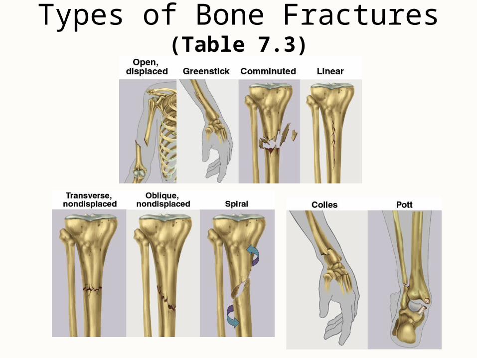
Types of Bone Fractures (Table 7.3)

Healing of Fractures• Normally healing takes 8 - 12 weeks (longer in
elderly)
• Stages of healing– fracture hematoma (1)
• broken vessels form a blood clot
– granulation tissue (2)• fibrous tissue formed by fibroblasts & infiltrated by capillaries
– callus formation (3)• soft callus of fibrocartilage replaced by hard callus of bone in
6 weeks
– remodeling (4) occurs over next 6 months as spongy bone is replaced with compact bone

Healing of Fractures
1 2
3 4

Treatment of Fractures• Closed reduction
– fragments are aligned with manipulation & casted
• Open reduction– surgical exposure & repair with plates & screws
• Traction is not used in elderly due to risks of long-term confinement to bed– hip fractures are pinned & early walking is encouraged
• Electrical stimulation is used on fractures that take longer than 2 months to heal
• Orthopedics = prevention & correction of injuries and disorders of the bones, joints & muscles

Fractures and Their Repairs

Osteoporosis
• Most common bone disease• Bones lose mass & become brittle due to loss of both
organic matrix & minerals– risk of fracture of hip, wrist & vertebral column– lead to fatal complications such as pneumonia– widow’s (dowager’s) hump is deformed spine
• Postmenopausal white women at greatest risk– by age 70, average loss is 30% of bone mass
• ERT slows bone resorption, but best treatment is prevention -- exercise & calcium intake (1000 mg/day) between ages 25 and 40
• Therapies to stimulate bone deposition are still under investigation

Effects of Osteoporosis


![8 th lecture December 10, 2015 Specialized Connective Tissue [Bone (Osseous) Tissue]](https://static.fdocuments.net/doc/165x107/5a4d1b567f8b9ab0599a95f3/8-th-lecture-december-10-2015-specialized-connective-tissue-bone-osseous.jpg)