The Skeletal System How to : identify and describe the major functions and structures; Describe...
121
The Skeletal System How to : identify and describe the major functions and structures; Describe three types of joints. Know the difference between axial and appendicular skeletons. Recognize, define, spell and pronounce terms related to
-
Upload
clinton-hancock -
Category
Documents
-
view
214 -
download
0
Transcript of The Skeletal System How to : identify and describe the major functions and structures; Describe...

The Skeletal System
How to : identify and describe the major functions and structures;
Describe three types of joints. Know the difference between axial
and appendicular skeletons. Recognize, define, spell and
pronounce terms related to the skeletal system.

Functions of the Skeletal System
Bones act as the framework of the body.
Bones support and protect the internal organs.
Joints, along with muscles, ligaments, and tendons make possible the body movements.

Structures of the Skeletal System
The structures include: bones (oste/o) cartilage (chondr/o) ligaments (ligament/o) joints (arthr/o) bursa (burs/o)

The Skeleton
There are 206 bones in the adult human body.
For descriptive purposes, the skeleton is divided into the:
axial skeleton and the appendicular skeleton

Axial Skeleton – 80 bones The term axial refers to an
imaginary line or axis that runs through the center of the body.
The axial skeleton is the skull, spinal (vertebral) column, ribs, and sternum.
It protects the major organs of the nervous, respiratory and circulatory systems.

Appendicular Skeleton – 126 bones
The term appendicular means referring to an appendage, which is anything attached to a major part of the body.
It makes body movement possible & protects the organs of digestion, excretion and reproduction.

Appendicular Skeleton
It is organized into the: upper extremities
(shoulders, arms, forearms, wrists, hands)
lower extremities (hips, thighs, legs, ankles and feet)


Learning the Bones……
Let’s look at the skeleton and start learning the names of each of the bones.
Then, we will break down each area of the body, and learn more specific information.

The Cranium
The cranium is the portion of the skull that encloses the brain.
crani means skull; -um is a noun ending.
The cranium is made of up many bones.

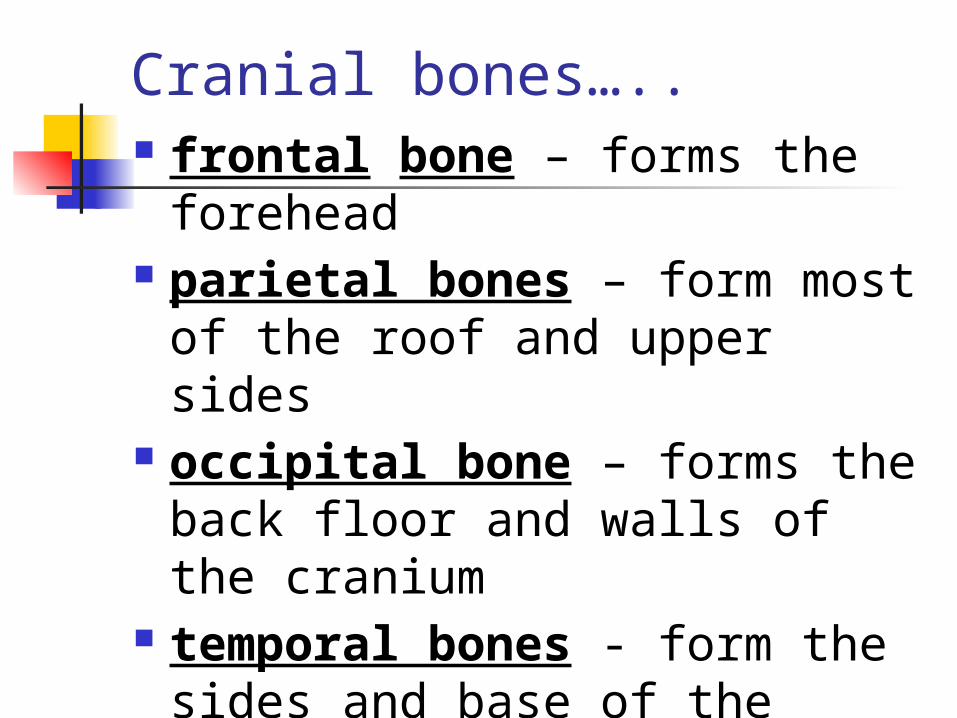
Cranial bones….. frontal bone – forms the
forehead parietal bones – form most of
the roof and upper sides occipital bone – forms the
back floor and walls of the cranium
temporal bones - form the sides and base of the cranium

sphenoid bone – forms part of the base of the skull, and parts of the floor and sides of the orbit (bony socket around eyeball).
ethmoid bone – forms part of the nose, the orbit, and the floor of the cranium.

Bones of the Face
zygomatic bones – also known as the cheekbones, join with the frontal bones.
maxilla – upper jaw mandible – lower jawbone; is the
only movable bone of the skull. It is attached to the temporo-mandibular joint (TMJ).

nasal bone – upper bridge of the nose.
Let’s review the cranial bones and the facial bones on the skeleton.

Thoracic Cavity, Ribs, and Sternum
The thoracic cavity is made up of the ribs, sternum and thoracic vertebrae.
Also known as the rib cage, this structure protects the heart and lungs.

Ribs
There are 12 pairs of ribs, called costals, which attach posteriorly to the thoracic vertebrae.
The first seven pairs of ribs, called true ribs, are attached anteriorly to the sternum.

The next three pairs of ribs, called false ribs, are attached anteriorly to cartilage that joins with the sternum.
The last two pairs of ribs, called floating ribs, are not attached anteriorly, only posteriorly.


Sternum
The sternum, also known as the breastbone, forms the middle of the front of the rib cage.
It is divided into three parts.

Sternum parts…… The manubrium, which is
bone, is the upper portion of the sternum.
The body of the sternum, which is also bone, is the middle portion of the sternum.
The xiphoid process, which is cartilage, is the lower portion of the sternum.


Shoulders
The shoulders form the pectoral girdle, which is also known as the shoulder girdle, that supports the arms and hands.
As used here, the term girdle means a structure that encircles the body.

Shoulders…… The clavicle, also known as
the collarbone, is a slender bone that connects the sternum to the scapula.
The scapula is also known as the shoulder blade.
The acromion is an extension of the scapula that forms the high point of the shoulder.


Arms
The humerus is the bone of the upper arm.
The radius is the smaller bone in the forearm. It is thumb side.
The ulna is the larger bone of the forearm. It joins with the humerus to form the elbow joint.


The olecranon process, commonly known as the “funny bone”, is a large projection on the upper end of the ulna that forms the point of the elbow that tingles when struck.


Wrists and Hands
The carpals are the wrist bones.
The metacarpals are the bones that form the back of the hand.
The phalanges are the bones of the fingers.

Each finger has three bones. These are the distal, medial,
and proximal phalanges. The thumb has two bones. These are the distal and
proximal phalanges.


Let’s play Jeopardy with what we’ve learned so far!

Spinal Column
Also known as the “vertebral column”
Consists of 26 vertebrae Functions: to support the head
and body and to protect the spinal cord.

Structures of vertebrae:
The body is the solid anterior portion of each vertebra (singular).
The lamina is the posterior portion
The vertebral foramen is the opening through the middle of the vertebra; the spinal cord passes through this opening.


Types of Vertebrae Cervical vertebrae – first 7
that form the neck. C1 through C7.
Thoracic vertebrae – second set of 12 vertebrae - form the outward curve of the spine. T1 through T12
Lumbar vertebrae - third set of 5 vertebrae; largest and strongest; form inward curve of spine. L1 through L5.


Inter/vertebr/al disks:
The intervertebral disks are made of cartilage; they separate and cushion the vertebrae from each other.
The act as “shock absorbers” and allow for movement of the spinal column.
The Sacrum It is a slightly curved,
triangular shaped bone near the base of the spine.
At birth, it is composed of five separate sacral bones; however, in the young child, they fuse together to form a single bone.


The Coccyx
Also known as the “tailbone”. It forms the end of the spine. Is made up of four small
vertebrae fused together.

PELVIC GIRDLE Also known as the hips or
pelvic bone Protects internal organs and
supports the lower extremities. Made up of three bones fused
together:

Ilium – upper, blade shaped flat / rounded part of the hip on each side of the pelvic girdle.
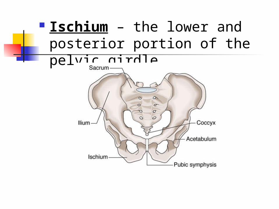
Ischium – the lower and posterior portion of the pelvic girdle.

Pubis – the anterior portion. These three bones, also known
as the pubic bones, fuse together.
The two pubic bones join at the anterior midline to form the pubis symphysis. This is a cartilaginous joint that holds the bones firmly together.

Acetabulum – large socket in the pelvic bones; forms the hip socket for the head of the femur.
Pubis, pubic symphysis, acetabulum:

Legs and Knees The femur is the upper leg bone,
known as the thigh bone, and is the largest bone in the body.
The head of the femur joins with the acetabulum.
The femoral neck is the narrow area below the head of the femur.
The trochanter is one of two bony projections on the upper end of femur.


The patella is the bony anterior portion of the kneecap.
The term popliteal refers to the posterior surface of the knee .
The anterior and posterior cruciate ligaments (ACL and PCL) make possible the movements of the knee.
Cruciate means shaped like a cross.

The lower leg is made of 2 bones:
Tibia – also known as the shinbone, is the larger weight-bearing bone of the lower leg.
Fibula – the smaller of the two bones of the lower leg.


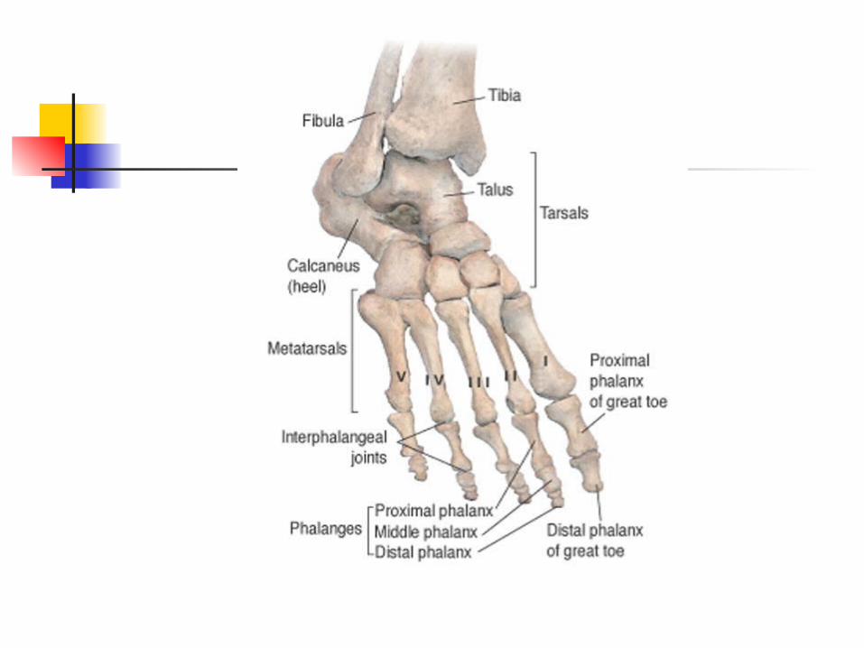
Ankles and Feet
The tarsals are the ankle bones. The metatarsals are the bones of
the foot. The phalanges are the toes. The calcaneus is the heel bone. The malleolus is the rounded
protruberance on each side of ankle.

Structure / Landmarks of Bones
Bone is a form of connective tissue and is almost the hardest tissue in the human body.
Only dental enamel is harder.

Although it is very hard and dense, bone is a living structure that changes and is capable of healing itself.
The periosteum is the tough, fibrous tissue that forms the outermost covering of the bone.
The compact bone is the hard, dense, and very strong bone that forms the outer layer of bones.

The spongy bone is lighter and not as strong as compact bone. It is commonly found in the ends and inner portions of long bones.
Red bone marrow is located within the spongy bone.
Medullary Cavity – located in the shaft of the long bone, it is line with endosteum and contains yellow bone marrow.


Bone Marrow Red bone marrow, located
within the spongy bone is hematopoietic (pertaining to the formation of blood) and manufactures red blood cells, hemoglobin, white blood cells, and cells that produce thrombocytes.

Yellow bone marrow is found in the medullary cavity. It is composed chiefly of fat cells and functions as a fat storage area.

Landmarks The diaphysis is the shaft of a
long bone. The epiphysis, which is
covered in articular cartilage, is the wide end of the long bone.
The proximal epiphysis is the end of the bone that is located nearest to the midline of the body.

The distal epiphysis is the end of the bone that is located farthest away from the midline.
The foramen is the opening in a bone through which blood vessels, nerves, and ligaments pass.
A process is a normal projection on the surface of a bone that serves as attachments for muscles and tendons.

Cartilage Cartilage is the smooth,
rubbery blue-white connective tissue that acts as a shock absorber between bones.
It is more elastic than bone. It makes up the flexible parts
of the skeleton such as the outer ear and the tip of the nose.

Articular cartilage covers the surfaces of bones that form joints to make smooth joint movement possible and to protect the bones from rubbing against each other.
The meniscus is the curved fibrous cartilage found in some joints such as the knee and the TMJ.


Joints Joints, also known as
articulations, are connections between bones.
The term articulate means to come together in a manner that allows motion between parts.
Types of joints: sutures, symphysis, and synovial.

A suture is a jagged line where bones join and form a joint that does not move. Examples: the coronal and sagittal sutures across the top of the skull in the adult.
On a baby’s head, the fontanel, or soft spot, is where the sutures between the frontal and parietal bones have not yet closed. This disappears as the child grows.


A symphysis is a cartilaginous joint, where two bones join and are held together so that they function as one bone. Example: pubis symphysis.

Synovial joints – movable joints. 2 Types:
Ball and socket joints – hips and shoulders. They allow a wide range of movement in many directions.
Hinge joints – knees and elbows. Allow movement primarily in one direction or plane.


Pathology of Joints and Bones Ankyl/osis: ankyl means
crooked, bent, or stiff. -osis is a suffix that means “abnormal condition”.
It is the loss or absence of mobility in a joint due to disease, an injury, or a surgical procedure.

Arthr/o/sclerosis: arthr/o = joint; -sclerosis is a suffix that means abnormal hardening.
Arthrosclerosis is stiffness, or hardening, of the joints, especially in the elderly.
Arthr/itis: arthr/o = joint; -itis means inflammation.
Arthritis is inflammation of a joint.

What is arthralgia? Pain in a joint. Chondr/o/malacia? Abnormal softening of
cartilage. The suffix –oma = tumor. So,
what is a chondroma? A slow-growing, usually benign,
tumor in cartilage cells.

Luxation is a term for dislocation or displacement of a bone from it’s joint.
Subluxation – is the “partial” displacement of a bone from it’s joint.
What is osteo/arthr/itis? Inflammation of bone and joint;
also known as “wear-and-tear arthritis”.

Gouty arthritis, also known as gout, is a type of arthritis caused by the formation of uric acid crystals in the joint. It causes pain, inflammation, and, if untreated, bone destruction.

Rheumatoid arthritis, also known as RA, is an autoimmune disease. The symptoms are usually more severe than osteoarthritis.
Tissues are attacked, causing the joints to become swollen, painful and immobile.
Juvenile RA affects children; symptoms are pain, swelling in joints, rash, fever, slowed growth, fatigue.

A herniated disk, or ruptured disk, is a rupture of the intervertebral disk that results in pressure on the spinal nerve.

Spondyl is a word root for “vertebrae”. What is spondylitis?
Inflammation of the vertebrae. What is spondylosis? A degenerative condition of the
vertebrae.

Spina bifida is the congenital defect that occurs during early pregnancy in which the spinal canal fails to close around the spinal cord.
Spina means pertaining to the spine; bifida means split.
Many cases are caused by a lack of folic acid (a vitamin) during the early stages of pregnancy.

What is oste/itis? Inflammation of bone. What is osteo/malacia? Abnormal softening of bones. What is osteo/myel/itis? Inflammation of the bone and
bone marrow. What is osteo/necrosis? Destruction and death of bone
tissue.

Rickets – caused by calcium and vitamin D deficiencies in early childhood, resulting in demineralized bones and related deformities.
Vitamin D was added to milk in the 1930’s.
Vitamin D also comes from sunlight.

Talipes, also known as clubfoot, is a congenital deformity in which the foot may be turned inward or outward.
Myel/oma is a malignant tumor in cells of the blood-forming tissues of the bone marrow.
It is usually progressive, may cause pathologic fractures, and is often fatal.

Osteo/chondr/oma – the most common benign bone tumor.
These tumors are growths on the surface of the bone that protrude as hard lumps, covered with a cap of cartilage.

Osteoporosis – a loss of bone density, and an increase in bone porosity associated with aging.
Oste/o = bone; -porosis = porous condition.
Osteoporosis is primarily responsible for three types of fractures:

Vertebral crush fractures, or compression fractures of the spine. The occur when one or more vertebrae become weak and collapse spontaneously or under minimal stress. It causes pain, loss of height, and spinal curvature known as dowager’s hump.
Also causes crowding of internal organs and reduced lung capacity.

The 2nd fracture (fx) caused by osteoporosis is Colle’s fracture, or fractured wrist.
It is a fracture of the lower end of the radius.
It is usually caused when a person tries to break a fall by landing on his or her hands and the trauma causes the weakened bone to break.

The third fracture caused by osteoporosis is an osteoporotic hip fracture, or broken hip.
It can occur spontaneously or as a result of a fall.
Complications from these fractures may result in death or the loss of function, mobility, and independence.

Curvatures of the Spine Kyphosis is an abnormal
increase in the outward curvature of the thoracic spine.
This condition is also known as humpback or dowager’s hump.


Lordosis is an abnormal increase in the forward curvature of the lower or lumbar spine.
This condition is also known as swayback.


Scoliosis is an abnormal lateral (sideways) curvature of the spine.
Can occur anywhere along the spine, and can be minimal or extreme.


Fracture (fx) = broken bone

In addition, there is a stress fracture – a small crack in bones that often develops from chronic, excessive impact.
Usually due to a sports injury. And a compression fracture
– occurs when the bone is pressed together (compressed) on itself, such as with osteoporosis.

As the bone heals, a callus forms a bulging deposit around the area of the break. This tissue eventually becomes bone.
A fat embolus may form when a long bone is fractured and fat cells from yellow bone marrow are released into the blood. It may become lodged and block a blood vessel.

Treatment of fractures Manipulation – or ‘closed
reduction’ – use of manual force to realign the bone before it is immobilized.
Traction – a pulling force exerted on a limb in a distal direction to return the bone or joint to it’s normal alignment or position.

Immobilization – the act of holding, suturing, or fastening the bone in a fixed position with strapping or a cast.
External fixation – pins are placed through soft tissue and bone so that an external appliance can be used to hold the pieces of bone in place. When healing is complete, the appliance is removed.

Internal fixation – also know as ORIF = open reduction internal fixation. Pins or a plate are placed directly into the bone to hold the broken pieces in place. This is usually not removed after the fracture has healed.

Ligaments A ligament is a band of fibrous
connective tissue that connects one bone to another bone.
Be careful not to confuse ligaments and tendons. Tendons attach muscles to bones.

Synovial Membranes and Fluid Synovial joints (movable joints)
are surrounded by a fibrous capsule and are lined with synovial membrane. The synovial membrane secretes synovial fluid that acts as a lubricant to make the smooth movement of the joint possible.

Synovitis is inflammation of the synovial membrane that results in swelling and pain.
It may be caused by an injury, infection, or irritation produced by damaged cartilage.

Bursa A bursa is a fibrous sac that is
lined with a synovial membrane and contains synovial fluid.
A bursa acts as a cushion to ease movement in areas that are subject to friction, such as shoulder, elbow, and knee joints where a tendon passes over a bone.


Diagnostic and Treatment Procedures
What is arthr/o/centesis? Surgical puncture of a joint
space to remove synovial fluid. What is arthr/o/scopy? Visual examination of the
internal structure of a joint, using an arthroscope.

Bone density testing – the use of radiation tests to determine bone density, to diagnosis osteoporosis, osteomalacia.
A bone scan is the use of nuclear medicine to detect bone cancer and osteomyelitis.
Radiographs, or x-rays, are used to view fractured bones.

A bone marrow biopsy is done by inserting a sharp needle into the hipbone or sternum and removing bone marrow cells. It is used as a diagnostic test to determine why blood cells are abnormal.
It is also done to find a donor match for a bone marrow transplant.

Magnetic resonance imaging (MRI) is used to image soft tissue structures such as the interior of complex joints and spinal disorders.
It is not the most effective method of imaging hard tissues such as bone.

What is arthr/o/scopic surgery?
Treatment of the interior of a joint, such as the removal of torn cartilage, with the use of an arthroscope and instruments inserted through small incisions.

What does the suffix –plasty mean?
What is chondr/o/plasty? Surgical repair of cartilage. What is arthr/o/plasty? Surgical repair of a joint. What is a crani/o/plasty? Surgical repair of the skull

What is oste/o/plasty? Surgical repair of bones. What does the suffix –ectomy
mean? What is a bursectomy? Surgical removal of a bursa What is synov/ectomy? Surgical removal of synovial
membrane.

What is disk/ectomy? Surgical removal of an
intervertebral disk. What is lamin/ectomy? Surgical removal of a lamina What is a crani/ectomy? Surgical removal of a portion of
the skull.

What is an ost/ectomy? Surgical removal of bone. What does the suffix –otomy
mean? What is peri/oste/otomy? Incision through the
periosteum. What is crani/otomy? Also known as ‘bone flap’ to
gain access to the brain

What is oste/otomy? Incision into a bone to realign a
joint damaged by arthritis. What is chondr/otomy? Incision into cartilage

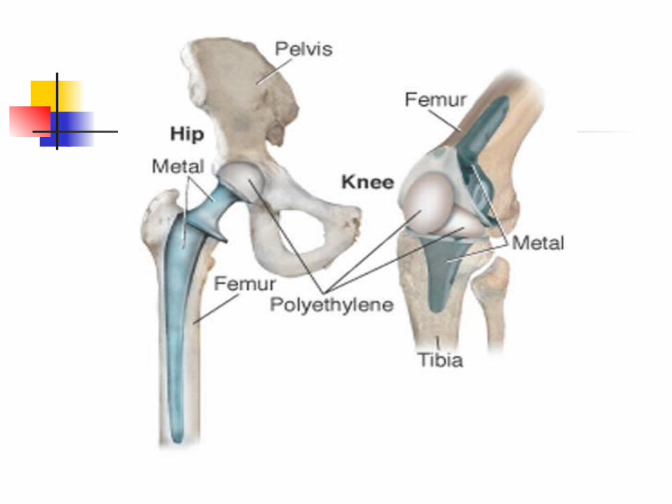
Joint Replacements
The procedures are named for the involved joint and the amount of the joint that is replaced.
The replacement part is called a prosthesis or an implant.
Total knee replacement – all of the parts of the knee are replaced.

Partial knee replacement - only part of the knee is replaced.
Total hip replacement – consists of 2 components. The thigh component is a metal shaft fitted into the femur with a metal ball at the top end. The ball fits into a plastic lined cup-shaped socket that replaces the acetabulum in the hip.

Medical Specialties A chiropractor holds of
Doctor of Chiropractic (DC) degree and specializes in manipulative treatment of disorders from mis-alignment of the spine.
An orthopedic surgeon or ‘orthopedist’ diagnoses and treats diseases and disorders of the bones, joints and muscles.

Orthotics is the field relating to the making and fitting of orthopedic appliances such as braces or splints.
Osteo/pathy is any bone disease. An Osteopathic physician holds
a Doctor of Osteopathy (DO) degree. They specialize in treating health problems by manipulation and also use traditional forms of medical treatment.