The Shaw-related Potassium Channel Gene, Kv3.1, on · PDF fileThe Shaw-related Potassium...
9
THE JOURNAL OF BIOLOGICAL CHEMISTRY 0 1992 by The American Society for Biochemistry and Molecular Biology, Inc Val. 267, No. 29, Issue of October 15, pp. 20971-20979,1992 Printed in U. S. A. The Shaw-related Potassium Channel Gene, Kv3.1, on Human Chromosome 11, Encodes the Type I K+ Channel in T Cells* (Received for publication, April 22, 1992) Stephan GrissmerS, Sanjiv GhanshaniS, Brent DethlefsS, John D. McPhersong, John J. Wasmuthg, George A. Gutmad, Michael D. CahalanS, K. George ChandySII From the Departments of $Physiology and Biophysics, §BiologicalChemistry, and TMicrobiology and Molecular Genetics, University of California, Zruine, California 92715 T lymphocytes exhibit three distinct types of voltage- gated K’channels, n, n’, and 1, that are distributed in the T cell lineage according to subset, as well as the cells’ activation and developmental status. Type Z K’ channels are found sparingly in cytotoxic T cells from normal mice and abundantly in a specific T cell subset (CD4- CDS- Thyl+)from mice with autoimmune dis- ease. Here, we show that the mouse Kv3.1 gene, when expressed in Xenopus oocytes, encodes a channel with properties remarkably similar to those of the Z-type channel. Kv3.1 transcripts were found in T cells iso- lated from the lymph nodes of MRL-Zpr micewith systemic lupus erythematosus and in a human lym- phoma cell line that also expresses the 1 channel phe- notype. By these criteria, we conclude that Kv3.1 encodes the voltage-gated type Z K’ channel in lym- phocytes. The Kv3.1 gene maps to human chromosome 11; the related Kvl.1 and Kv3.2 genes are localized on humanchromosome 12, while the IsK gene maps to human chromosome 2 1. Patch-clamp experiments have demonstrated that T lym- phocytes can express at least three distinct types of voltage- dependent K’ channels, in a pattern which is determined by their state of differentiation and mitogenic activation (Lewis and Cahalan, 1988; for review see Cahalan et al., 1991). The three channel types differ in voltage-dependent gating kinet- ics and pharmacological sensitivity. These channels appear to play essential roles in the physiology of lymphocytes, includ- ing the regulation of membrane potential, cell volume, calcium signaling, and mitogenic activation. The type n channel is the most commonly observed K’ channel in normal T lymphocytes. It activates at potentials above -40 mV, inactivates slowly during depolarizations last- ing hundreds of ms, and recovers very slowly from inactivation (30-60 s). The n channel has a single-channel conductance of 10-18 pS’ and is half-blocked by -10 mM tetraethylammo- nium (TEA) or by -3 nM charybdotoxin. A second K+channel * This study was supported by a grant from Pfizer Central Research (to K. G. C.), and by National Institute of Allergy and Infectious Diseases Grant 24783 (to K. G. C.), NS14609 (to M. D. C.), GM41514 (to M. D. C.), and GM42365 (to J. J. W.). The costs of publication of this article were defrayed in part by the payment of page charges. This articlemusttherefore be hereby marked“advertisement” in accordance with 18 U.S.C. Section 1734 solely to indicate this fact. I1 To whom correspondence should be addressed Rm. C154, Dept. of Physiology and Biophysics, Medical School, University of Califor- nia, Irvine, CA 92717. Tel.: 714-725-2133; Fax: 714-856-8540. The abbreviations used are: S, siemens; TEA, tetraethylammo- nium; bp, base pair(s);PCR, polymerase chain reaction; kb, kil- obase(s); DN, double-negative. subtype, 1, is readily distinguished from n; the 1 channel activates at more depolarized potentials (-10 mV), closes more rapidly, inactivates to a lesser extent, and has a larger single-channel conductance (25-30 pS). Pharmacologically, the 1 channel is more sensitive to TEA (half-blocked at -0.1 mM) and resistant to charybdotoxin (no block at 50 nM). The n’ channel has biophysical and pharmacological properties similar to those of the n channel but is more resistant to block by TEA (half-blocked at -100 mM TEA). Molecular cloning has revealed a superfamily of voltage- gated channels, including at least 14 distinct mammalian genes encoding K’ channels (Chandy et al., 1991). The gene nomenclature used in this report is based on the sequence relatedness of these mammalian genes to the Shaker, Shab, Shaw, and Shal genes in Drosophila (Chandy et al., 1991). The Shaker-family genes have intronless coding regions (Chandy et al., 1990a), whereas the coding sequences of the Shaw-related genes are contained in multiple exons (Ghans- hani et al., 1992) that arealternatively spliced (Luneau et al., 1991). We have shown previously that the Shaker-related mouse Kv1.3 (MK3) gene encodes the type n channel in mouse T cells (Grissmer et dl.,199Ob). The rat (RGK5) and human (HGK5, HLK3) Kv1.3 genes have also been reported to encode the type n K’ channel in T cells from those species (Douglass et al., 1990; Yun-Cai Cai et al., 1992; Attali et al., 1992). The genes encoding the types 1 and n‘ channels in T cells have not been identified. The Shaw-related Kv3.1 gene in rodents is alternatively spliced, giving rise to two distinct transcripts (Luneau et al., 1991; Yokoyama et al., 1989), Kv3.la(NgK2) and Kv3.lb (Kv4). The coding regions of the two transcripts are identical up to amino acid 501 (1,503 bp) in the C-terminal region, and they diverge completely, the last 10 amino acids of Kv3.la being replaced with another 84 amino acids in Kv3.lb (Luneau et al., 1991). Only the Kv3.la mRNA has been described in mice, whereas both transcripts arepresent in rats. When expressed in Xenopus oocytes, these two transcripts produce biophysically identical K+ channels that resemble the type 1 K’ channel in T cells (Yokoyama et al., 1989; Luneau et al., 1991). We have taken two approaches to verify experimentally that theKv3.1 gene encodes the type 1 K’ channel in T cells. First, we isolated a mouse Kv3.lb(mKv3.lb) cDNA and performed a detailed comparison of the functional properties of the cloned channel expressed in Xenopus oocytes with channel “fingerprints” of type 1 K’ channels from mouse and human lymphocytes. Our studies demonstrate that the mKv3.lbchannel has gating kinetics and pharmacological sensitivities that are so remarkably similar to those of the type 1 K’ channels in T cells that it is likely that the native 20971
Transcript of The Shaw-related Potassium Channel Gene, Kv3.1, on · PDF fileThe Shaw-related Potassium...
THE JOURNAL OF BIOLOGICAL CHEMISTRY 0 1992 by The American Society for Biochemistry and Molecular Biology, Inc
Val. 267, No. 29, Issue of October 15, pp. 20971-20979,1992 Printed in U. S. A.
The Shaw-related Potassium Channel Gene, Kv3.1, on Human Chromosome 11, Encodes the Type I K+ Channel in T Cells*
(Received for publication, April 22, 1992)
Stephan GrissmerS, Sanjiv GhanshaniS, Brent DethlefsS, John D. McPhersong, John J. Wasmuthg, George A. Gutmad, Michael D. CahalanS, K. George ChandySII From the Departments of $Physiology and Biophysics, §Biological Chemistry, and TMicrobiology and Molecular Genetics, University of California, Zruine, California 92715
T lymphocytes exhibit three distinct types of voltage- gated K’channels, n, n’, and 1, that are distributed in the T cell lineage according to subset, as well as the cells’ activation and developmental status. Type Z K’ channels are found sparingly in cytotoxic T cells from normal mice and abundantly in a specific T cell subset (CD4- CDS- Thyl+) from mice with autoimmune dis- ease. Here, we show that the mouse Kv3.1 gene, when expressed in Xenopus oocytes, encodes a channel with properties remarkably similar to those of the Z-type channel. Kv3.1 transcripts were found in T cells iso- lated from the lymph nodes of MRL-Zpr mice with systemic lupus erythematosus and in a human lym- phoma cell line that also expresses the 1 channel phe- notype. By these criteria, we conclude that Kv3.1 encodes the voltage-gated type Z K’ channel in lym- phocytes. The Kv3.1 gene maps to human chromosome 11; the related Kvl.1 and Kv3.2 genes are localized on human chromosome 12, while the IsK gene maps to human chromosome 2 1.
Patch-clamp experiments have demonstrated that T lym- phocytes can express at least three distinct types of voltage- dependent K’ channels, in a pattern which is determined by their state of differentiation and mitogenic activation (Lewis and Cahalan, 1988; for review see Cahalan et al., 1991). The three channel types differ in voltage-dependent gating kinet- ics and pharmacological sensitivity. These channels appear to play essential roles in the physiology of lymphocytes, includ- ing the regulation of membrane potential, cell volume, calcium signaling, and mitogenic activation.
The type n channel is the most commonly observed K’ channel in normal T lymphocytes. It activates at potentials above -40 mV, inactivates slowly during depolarizations last- ing hundreds of ms, and recovers very slowly from inactivation (30-60 s). The n channel has a single-channel conductance of 10-18 pS’ and is half-blocked by -10 mM tetraethylammo- nium (TEA) or by -3 nM charybdotoxin. A second K+ channel
* This study was supported by a grant from Pfizer Central Research (to K. G. C.), and by National Institute of Allergy and Infectious Diseases Grant 24783 (to K. G. C.), NS14609 (to M. D. C.), GM41514 (to M. D. C.), and GM42365 (to J. J. W.). The costs of publication of this article were defrayed in part by the payment of page charges. This article must therefore be hereby marked “advertisement” in accordance with 18 U.S.C. Section 1734 solely to indicate this fact.
I1 To whom correspondence should be addressed Rm. C154, Dept. of Physiology and Biophysics, Medical School, University of Califor- nia, Irvine, CA 92717. Tel.: 714-725-2133; Fax: 714-856-8540.
The abbreviations used are: S, siemens; TEA, tetraethylammo- nium; bp, base pair(s); PCR, polymerase chain reaction; kb, kil- obase(s); DN, double-negative.
subtype, 1, is readily distinguished from n; the 1 channel activates at more depolarized potentials (-10 mV), closes more rapidly, inactivates to a lesser extent, and has a larger single-channel conductance (25-30 pS). Pharmacologically, the 1 channel is more sensitive to TEA (half-blocked at -0.1 mM) and resistant to charybdotoxin (no block at 50 nM). The n’ channel has biophysical and pharmacological properties similar to those of the n channel but is more resistant to block by TEA (half-blocked at -100 mM TEA).
Molecular cloning has revealed a superfamily of voltage- gated channels, including at least 14 distinct mammalian genes encoding K’ channels (Chandy et al., 1991). The gene nomenclature used in this report is based on the sequence relatedness of these mammalian genes to the Shaker, Shab, Shaw, and Shal genes in Drosophila (Chandy et al., 1991). The Shaker-family genes have intronless coding regions (Chandy et al., 1990a), whereas the coding sequences of the Shaw-related genes are contained in multiple exons (Ghans- hani et al., 1992) that are alternatively spliced (Luneau et al., 1991).
We have shown previously that the Shaker-related mouse Kv1.3 (MK3) gene encodes the type n channel in mouse T cells (Grissmer et dl., 199Ob). The rat (RGK5) and human (HGK5, HLK3) Kv1.3 genes have also been reported to encode the type n K’ channel in T cells from those species (Douglass et al., 1990; Yun-Cai Cai et al., 1992; Attali et al., 1992). The genes encoding the types 1 and n‘ channels in T cells have not been identified.
The Shaw-related Kv3.1 gene in rodents is alternatively spliced, giving rise to two distinct transcripts (Luneau et al., 1991; Yokoyama et al., 1989), Kv3.la (NgK2) and Kv3.lb (Kv4). The coding regions of the two transcripts are identical up to amino acid 501 (1,503 bp) in the C-terminal region, and they diverge completely, the last 10 amino acids of Kv3.la being replaced with another 84 amino acids in Kv3.lb (Luneau et al., 1991). Only the Kv3.la mRNA has been described in mice, whereas both transcripts are present in rats. When expressed in Xenopus oocytes, these two transcripts produce biophysically identical K+ channels that resemble the type 1 K’ channel in T cells (Yokoyama et al., 1989; Luneau et al., 1991).
We have taken two approaches to verify experimentally that the Kv3.1 gene encodes the type 1 K’ channel in T cells. First, we isolated a mouse Kv3.lb (mKv3.lb) cDNA and performed a detailed comparison of the functional properties of the cloned channel expressed in Xenopus oocytes with channel “fingerprints” of type 1 K’ channels from mouse and human lymphocytes. Our studies demonstrate that the mKv3.lb channel has gating kinetics and pharmacological sensitivities that are so remarkably similar to those of the type 1 K’ channels in T cells that it is likely that the native
20971
20972 Shuw-related Gene, Kv3.1, Encodes a Lymphocyte K" Channel
channel is a homomultimeric protein. Second, we used the polymerase chain reaction (PCR) to amplify and sequence the hydrophobic core of the Kv3.1 coding region from mouse and human lymphocytes which express type 1 K' channels. Col- lectively, these data suggest that the Kv3.1 gene encodes the type 1 K+ channel in lymphocytes. In addition, we determined the chromosomal localization of four human K+ channel genes.
EXPERIMENTAL PROCEDURES
K+ Channel Nomenclature-Several vertebrate voltage-gated K+ channel genes have been isolated and have frequently been given inconsistent names. Recently, a simplified nomenclature has been proposed for this family of genes based on sequence relatedness to the Shaker, Shab, Shaw, and Shd genes in Drosophila (Chandy et al., 1991). The mKv3.lb cDNA described in this paper is the mouse homologue of the rat Kv3.lb (rKv3.lb) transcript described previ- ously by Luneau et al. (1991); the mouse Kv3.la (mKv3.la) transcript is identical to the mKv3.lb cDNA from the start codon through 1,503
Isolation of the mKu3.Ib cDNA-Approximately 7 X lo6 plaques of a mouse brain cDNA library (cDNA partially digested with EcoRI and cloned into XgtlO, a gift of Dr. Daniel Littman, University of California, San Francisco) were screened with a mixture of Kv3.3 5' (1.0-kb BamHIISacI), Kv3.3 3' (2.2-kb PstI), and Kv3.4 3 ' (1.1 kbHindIII/BglII) probes labeled with 32P to a specific activity of 1.2 X lo9 cpm/g with the random primer method (Feinberg and Vogel- stein, 1983). Hybridization was performed a t 55 "C in hybridization buffer for 16-18 h. Hybridization buffer contains 5 X SSC (1 X SSC is 0.15 M NaC1,0.15 M sodium citrate), 10 X Denhardt's (0.2% bovine serum albumin, 0.2% Ficoll, 0.2% polyvinyl pyrrolidone), and 0.1% sodium dodecyl sulfate (SDS). The blots were washed at a final stringency of 0.5 X SSC and 0.1% SDS for 60 min at 53 "C. DNA was isolated from the hybridizing X phage clone, 382, digested to comple- tion with EcoRI, and electrophoresed in a 1% agarose gel. The resultingEcoR1 cDNA fragments (1.9 kb and 0.87 kb) were subcloned into M13mp19 and pBluescript I1 KS' vectors (Stratagene, La Jolla, CA) and sequenced by the dideoxy chain termination method (Sanger et al., 1977) using a Sequenase kit (U. S. Biochemical Corp.).
Construction and Expression of mKu3.lb cRNA in Xenopus 00- cytes-The mKv3.lb coding region was inserted into the pBluescript I1 KS' cloning vector using a three-way ligation as follows. The upstream 0.87-kb EcoRI fragment of the cDNA was digested with NarI (7 bp upstream of the ATG initiation codon) and EcoRI (814 bp into the coding region) to generate an 820-bp fragment. The downstream 1.9-kb EcoRI fragment was digested with EcoRI (814 bp into the coding region) and PstI (57 bp beyond the stop codon) to generate a 995-bp fragment. The two mKv3.lb fragments were then inserted into the ClaI (compatible restriction overhang with NarI) and PstI sites of a pBluescript I1 KS' vector which had a 200-bp poly(A)+ stretch inserted between Not1 and Sac11 in the polylinker. The resulting 1,815-bp NarIIPstI insert was verified by restriction mapping, and the integrity of the splice sites was confirmed by double- stranded DNA sequencing. For transcription, the plasmid was line- arized by digestion with Sac1 in the polylinker. RNA was transcribed using the T3 promoter in pBluescript I1 KS+ and injected into oocytes as described previously (Grissmer et al., 199Ob).
Electrophysiology-Experiments were carried out on Xenopus oo- cytes with two-electrode voltage-clamp and patch-clamp techniques (Hamill et al., 1981). All experiments were done at room temperature (22-26 "C).
For the two-electrode voltage-clamp experiments, cells were bathed
mM CaCl,, 1 mM MgC12, 5 mM HEPES and adjusted to pH 7.5 with in oocyte Ringer solution containing 96 mM NaCI, 2 mM KCI, 1.8
NaOH. The pipette solution was 3 M KCI. Patch-clamp experiments were carried out under identical conditions in oocytes and in T cells, using a mammalian Ringer solution containing 160 mM NaCl, 4.5 mM KCl, 2 mM CaCI2, 1 mM MgC12, 5 mM HEPES, and adjusted to pH 7.4 with NaOH and a total osmolarity of 290-320 mosm/kg. Internal (pipette) solutions contained 140 mM KF, 2 mM MgC12, 1 mM CaCl,, 5 mM HEPES, 11 mM K,-EGTA. Charybdotoxin was purchased from Latoxan (IBF-Biotechnics, Inc., Columbia, MD).
The patch-clamp amplifier (List L/M-EPC 7, Adams and List Associates, Ltd., Great Neck, NY) was used in the voltage-clamp mode without series resistance compensation. Patch-clamp electrodes were pulled from Accu-fill 90 Micropets (Becton, Dickinson & Co.,
bP.
Parsippany, NJ) in three stages, coated with Sylgard (Dow Corning Corp., Midland, MI), and fire polished to resistances, measured in the bath, of 2-7 MR. In all patch-clamp experiments, the command input of the patch-clamp amplifier was controlled by a PDP 11/73 computer via a digital-analog converter, and membrane currents were recorded at a band width of 2 kHz. The holding potential was adjusted in all experiments to E = -80 mV. Correction for capacitative currents was achieved by analog subtraction.
Oligonucleotide Primers-Synthetic oligonucleotide primers were prepared (ChemGenes Corp., Needham, MA) from the coding regions of mKv3.la. The upstream and two downstream primers were: 5'- GAAATCGAGAAGCTTCGAAACGG-3', which lies between the S1 and S2 loop; 5'-CAGGAAGATGATCAGCTGCAGG-3', lying in the S5 transmembrane region; and 5"GTGTGGAGAGTTAACGACAG- 3', prepared from a region 112 bp downstream of S6.
PCR Amplification of mKu3.I from RNA from MRL-lpr T Cells and Human Louckes Lymphoma Cells-Total RNA isolated from the lymph nodes of diseased MRL-lpr mice and from human Louckes cells was used to generate a random-primed cDNA product as de- scribed previously (Grissmer et al., 1990b). The 20-pl reaction mixture contained 48 units of avian myeloblastosis virus reverse transcriptase (U. S . Biochemical Corp.), 20 units of RNasin (Boehringer Mann- heim), 100 pM random 14-mer nucleotide mixture (Boehringer Mann- heim), 1 pg of total RNA (5 pl), and a 1 mM concentration of each dNTP (GeneAmp kit, Perkin-Elmer/Cetus Instruments). cDNA syn- thesis was then initiated by the random primer method (Krug and Berger, 1987). The cDNA product was amplified by PCR for 33 cycles (annealing temperature 55 "C) with TAQ polymerase (GeneAmp kit) using the upstream (primer 1) and downstream (primer 3) primers. The reaction mixture contained a 1 mM concentration of each dNTP, 100 p~ of each of the primers, and 5 pl of the cDNA template. As a control for contaminating genomic DNA, 5 p1 of RNA (lpr and Louckes) without cDNA synthesis was run in parallel PCR reactions. The amplified product (65 p1) was then run on a 1.5% agarose gel in Tris-acetate/EDTA (TAE) buffer. Based on the mKv3.la sequence (Yokoyama et al., 1989), the amplified product would have a predicted size of 774 bp. The resulting 774-bp PCR product was next primed with the 5' oligonucleotide in the S 1 3 2 loop (primer 1) and the internal oligonucleotide in the S5 segment (primer 2) to amplify a second PCR product with a predicted size of 390 bp. The 774-bp PCR fragment from Louckes cells was cloned into pBluescript, and single- stranded phage was generated and sequenced by the dideoxy chain termination method. Single-stranded DNA prepared from the 774-bp fragment amplified from MRL-lpr T cells using the asymmetric PCR method (Gyllensten and Erlich, 1988) was similarly sequenced. In
other primer (5'-AAAGTGCGTGTGCTCACTGG-3') from the mid- addition to the oligonucleotide primers described above, we used one
dle of the 774-bp fragment for the asymmetric PCR experiments and for sequencing.
Chromosomal Localization-A mKv3.lb coding region probe (1.7- kb EcoRI fragment) was labeled to a specific activity of lo9 cpm/pg by the random primer method and then used for the chromosomal studies. Chromosomal assignments for Kv3.1 were determined by Southern blot analysis (genomic DNA digested with EcoRI) of a panel of 59 human-Chinese hamster cell hybrids (Ghanshani et al., 1992). We also examined HindIII-digested genomic DNA from these cell lines to resolve any ambiguities. In all experiments, the probes were also hybridized separately either to hamster or to human genomic DNA digested with EcoRI or HindIII. The Kv3.1-hybridizing hamster and human bands were readily distinguished on the basis of size. The hybrids were isolated using a variety of selectable markers (Cirullo et al., 1983; Overhauser et al., 1987). All hybrids were characterized cytogenetically by trypsin-Giemsa banding and G-11 staining to determine which human chromosomes were retained. The analyses were repeated at the time the cells were harvested for the preparation of DNA. In all cases, 20 metaphase chromosome spreads were exam- ined. Hybridization was performed at a final stringency of 65 "C and 0.1 X SSC for 30 min to avoid cross-hybridization with related genes.
A human genomic clone (clone 361) encoding Kvl.1 was isolated, and a 4-kb HindIII fragment from this clone was used to probe the DNA panel at high stringency (65 "C 0.1 X SSC). A 1.5-kb EcoRI fragment from the coding region of mouse Kv3.2 (clone MShawl2; a gift from Michael Pak and Lawrence Salkoff, Washington University, St. Louis, MO) was similarly used for chromosomal mapping. A 420- bp EcoRI/HindIII fragment from the coding region of the rat IsK (MinK) gene (gift from Richard Swanson, Merck Sharpe and Dohme, West Point, PA) was also used for chromosomal localization studies.

Shaw-related Gene, Kv3.1, Encodes a Lymphocyte K" Channel 20973
RESULTS
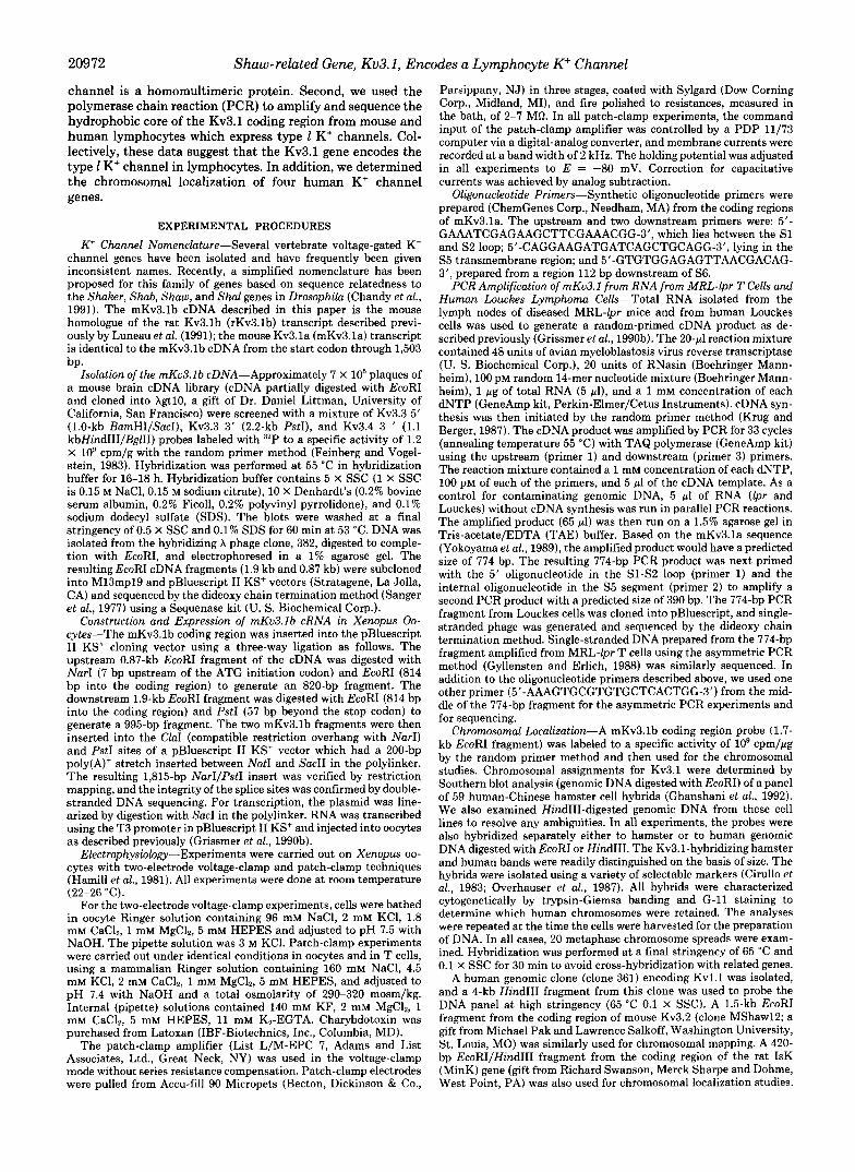
Mouse Ku3.1 b Expression in Oocytes-Xenopus oocytes ex- press a high density of voltage-activated K' channels follow- ing injection of mKv3.lb mRNA. With two-electrode voltage clamping, currents in excess of 15 pA at test potentials of +40 mV, representing on the order of lo7 channels, were routinely observed following injection of 0.5 ng of mRNA (50 nl at 0.01 pg/pl, not shown). Single-channel recordings were done on outside-out patches from these oocytes, whereas gating and inactivation kinetics and pharmacological sensitivities were determined using outside-out patches of oocyte membrane injected with a 10 times more concentrated mRNA solution (50 nl of 0.1 pg/pl mRNA). Reflecting the large currents seen in microelectrode recordings from the whole oocyte, current magnitudes in excised patches were large, sometimes repre- senting more than 1,000 channels/patch.
Voltage Dependence of the mKv3.1 b Gene Product-Oocyte mKv3.lb patch currents were very similar to whole-cell K' currents in normal mouse cytotoxic T lymphocytes (Lewis and Cahalan, 1988), in abnormal CD4- CD8- (double-nega- tive, or DN) T cells from several mouse models of autoimmune disease (Chandy et al., 1986, 1990b; DeCoursey et al., 1987; Grissmer et al., 1988, 1990a), and in the human B-lymphoma line, Louckes (Shapiro and DeCoursey, 1988; see below). Table I compares several gating properties of the mKv3.lb gene product, expressed in oocytes, with properties of type 1 K' channels in mouse T cells and in the human Louckes cells. Gating properties of the type n channels in human T cells, and those of the cloned mKv3.la and rKv3.lb gene products expressed in oocytes, are included in Table I for comparison. For these determinations, identical solutions at mammalian ionic strength (290-320 mosm/kg) were employed. Fig. 1 illustrates records and analysis of K+ currents from oocytes expressing the mKv3.lb gene product. The outward currents appear to represent a single population of K+ channels which
TABLE I Gating parameters of Ku3. I K' currents expressed i n oocytes
Type 1 and type n K' current properties in lymphocytes are included for comparison.
Channel
mV Cloned 1 channels
mKv3.lb cDNA" mKv3.la cDNA (NGK2)* 16
-1
rKv3.lb cDNA ( K v ~ ) ~ 15
Type l (human)" -4 Type l (mouse)e -9 Type n (human)x -36 Tvpe n (mouse)' -36
Native channels
mV ms ms PS
-11 630 2.5 27 -9 N D ND 26
-10 ND ND ND
-12 565 1.4 26 -9 340 2.6 21', 27/ -4.3 178 44 gh, 16h -7.3 107 49 12/. 18'
Data from this paper; numbers represent the mean of three to
Data from Yokoyama et al. (1989). ND, not determined. Data from Luneau et al. (1991). Data from DeCoursey et al. (1987).
five determinations.
'Mouse type n and type 1 channels each exhibit only one single- channel conductance state which has been variously reported to be 12 and 18 pS for type n, and 2 1 and 27 pS for type 1 (DeCoursey et al., 1987; Lewis and Cahalan, 1988).
*' Data from Cahalan et el. (1985). In human T cells two distinct single-channel events have been
described with conductances of 9 and 16 pS; these may represent two types of channels or one channel with two conductance states (Ca- halan et ul., 1985).
A
2 nA L 50 ms
+50
-80 __J
L -40 -20 0 20 40 " (mv)
FIG. 1. A, K+ currents in an outside-out patch from an oocyte injected with mKv3.lh mRNA. The membrane potential was -80 mV, and depolarizing pulses were applied every 30 s. The test poten- tial was changed from -50 to 50 mV in 10-mV increments. B, peak K' conductance-voltage relation for the K' currents shown in A. The line through the points was fitted with the Boltzmann equation, g K ( E ) = gK,,,,,/(l+exp((E, - E ) / k ) ) with parameter values, &mal = 16.1 nS; E, = 0 mV; k = -15.6 mV.
become activated at depolarizing potentials positive of about -20 mV. Upon depolarization, the channels open with a sigmoid time course, reach a peak within about 10 ms, and inactivate slightly. From the conductance-voltage relation shown in Fig. lB, the midpoint of channel activation is -0 mV. This value is characteristic of type 1 K' currents recorded in the whole-cell configuration from normal mouse cytotoxic T cells (Lewis and Cahalan, 1988), from abnormal DN T cells taken from 10 mouse models of autoimmunity (Chandy et al., 1986, 1990b; DeCoursey et al., 1987; Grissmer et al., 1988, 1990a), and from human Louckes lymphoma cells (Table I). Thus, the voltage dependence for K' channel activation of mKv3.lb currents matches that of type 1 K' currents in both human and mouse lymphocytes.
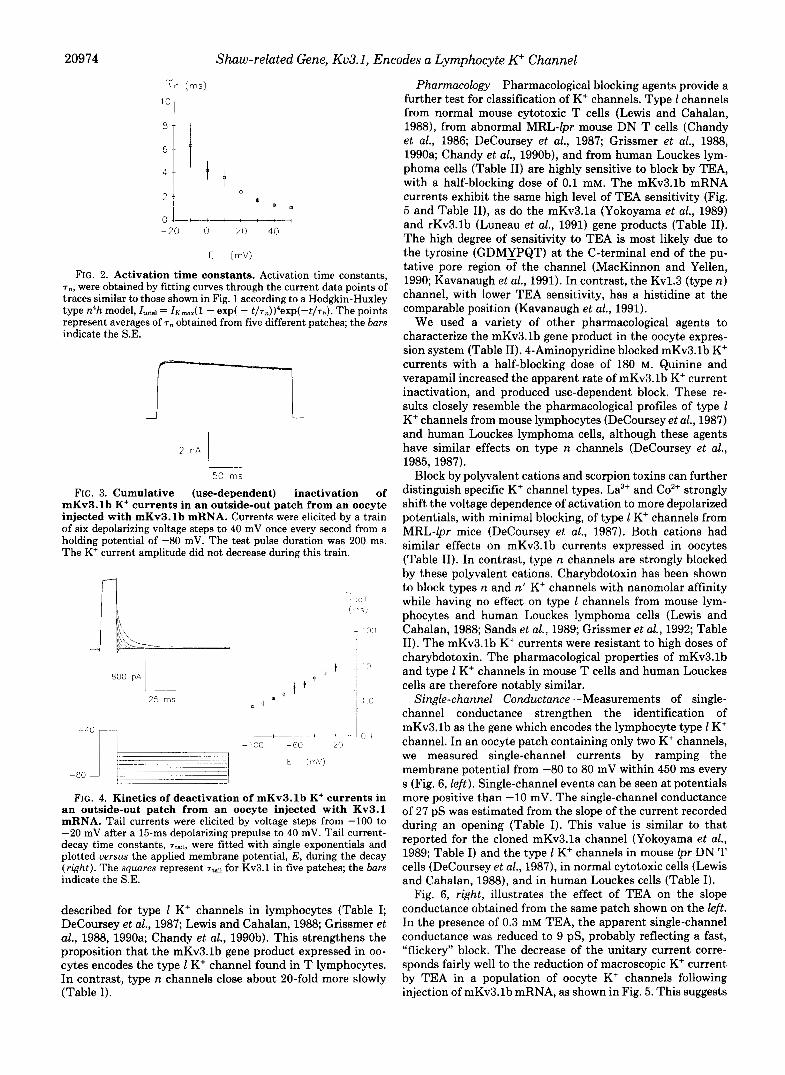
Activation and Inactivation Kinetics-Kinetic properties of activation and inactivation are also similar between mKv3.lb channels and 1 type currents from mouse and human lym- phocytes (Table I). The time course of K+ currents in five oocyte patches was analyzed using a modified Hodgkin-Hux- ley kinetic model with activation (n4) and inactivation (h); no particular microscopic mechanism for gating is implied by this quantitative description. The rates for channel activation and inactivation are similar to those reported previously for type 1 channels (Fig. 2 and Table I). Channel inactivation, in particular, provides a convenient property to distinguish be- tween diverse types of K+ channels. In contrast to "use- dependent" inactivation characteristic of type n channels, inactivation of type 1 K+ channels does not accumulate during repetitive depolarizing pulses delivered at 1 Hz. The time course of inactivation and lack of use dependence exhibited by mKv3.lb K' currents expressed in oocytes are similar to those for type 1 channels in mouse T cells and in human Louckes cells (Fig. 3 and Table I).
Closing Kinetics-The kinetics of K' channel closing can be determined by first opening the channels with a 15-ms conditioning pulse to 40 mV and then forcing the channels to close by repolarizing to different potentials (Fig. 4). The time constants, T ~ , of the resultant "tail currents" are plotted against the repolarized membrane potential. The closing rate for K' channels increases at more negative potentials. Typi- cally, a t -60 mV the tail current time constant is between 1 and 3 ms. Similar tail current time constants have been
20974 Shuw-related Gene, Ku3.1, Encodes a Lymphocyte K" Channel
' " T T n ( M S )
0- -20 0 20 40
E ( N
FIG. 2. Activation time constants. Activation time constants, T,, were obtained by fitting curves through the current data points of traces similar to those shown in Fig. 1 according to a Hodgkin-Huxley type n4h model, Itota, = I K ~ J ~ - exp( - t/Tn))4eXP(-t/Th). The points represent averages of T , obtained from five different patches; the bars indicate the S.E.
i 2 nA 1
50 rns
FIG. 3. Cumulative (use-dependent) inactivation of mKv3.lb K+ currents in an outside-out patch from an oocyte injected with mKv3.lb mRNA. Currents were elicited by a train of six depolarizing voltage steps to 40 mV once every second from a holding potential of -80 mV. The test pulse duration was 200 ms. The K' current amplitude did not decrease during this train.
FIG. 4. Kinetics of deactivation of mKv3.lb K+ currents in an outside-out patch from an oocyte injected with Kv3.1 mRNA. Tail currents were elicited by voltage steps from -100 to -20 mV after a 15-ms depolarizing prepulse to 40 mV. Tail current- decay time constants, T-:,, were fitted with single exponentials and plotted uersus the applied membrane potential, E , during the decay (right). The squares represent T-:, for Kv3.1 in five patches; the bars indicate the S.E.
described for type 1 K' channels in lymphocytes (Table I; DeCoursey et al., 1987; Lewis and Cahalan, 1988; Grissmer et al., 1988, 1990a; Chandy et al., 1990b). This strengthens the proposition that the mKv3.lb gene product expressed in oo- cytes encodes the type 1 K' channel found in T lymphocytes. In contrast, type n channels close about 20-fold more slowly (Table I).
Pharmacology-Pharmacological blocking agents provide a further test for classification of K' channels. Type 1 channels from normal mouse cytotoxic T cells (Lewis and Cahalan, 19881, from abnormal MRL-lpr mouse DN T cells (Chandy et al., 1986; DeCoursey et al., 1987; Grissmer et al., 1988, 1990a; Chandy et al., 199Ob), and from human Louckes lym- phoma cells (Table 11) are highly sensitive to block by TEA, with a half-blocking dose of 0.1 mM. The mKv3.lb mRNA currents exhibit the same high level of TEA sensitivity (Fig. 5 and Table 11), as do the mKv3.la (Yokoyama et al., 1989) and rKv3.lb (Luneau et al., 1991) gene products (Table 11). The high degree of sensitivity to TEA is most likely due to the tyrosine (GDMIPQT) at the C-terminal end of the pu- tative pore region of the channel (MacKinnon and Yellen, 1990; Kavanaugh et al., 1991). In contrast, the Kv1.3 (type n) channel, with lower TEA sensitivity, has a histidine at the comparable position (Kavanaugh et al., 1991).
We used a variety of other pharmacological agents to characterize the mKv3.lb gene product in the oocyte expres- sion system (Table 11). 4-Aminopyridine blocked mKv3.lb K' currents with a half-blocking dose of 180 M. Quinine and verapamil increased the apparent rate of mKv3.lb K' current inactivation, and produced use-dependent block. These re- sults closely resemble the pharmacological profiles of type 1 K+ channels from mouse lymphocytes (DeCoursey et al., 1987) and human Louckes lymphoma cells, although these agents have similar effects on type n channels (DeCoursey et al., 1985, 1987).
Block by polyvalent cations and scorpion toxins can further distinguish specific K+ channel types. La3+ and Co2+ strongly shift the voltage dependence of activation to more depolarized potentials, with minimal blocking, of type 1 K' channels from MRL-lpr mice (DeCoursey et al., 1987). Both cations had similar effects on mKv3.lb currents expressed in oocytes (Table 11). In contrast, type n channels are strongly blocked by these polyvalent cations. Charybdotoxin has been shown to block types n and n' K+ channels with nanomolar affinity while having no effect on type 1 channels from mouse lym- phocytes and human Louckes lymphoma cells (Lewis and Cahalan, 1988; Sands et al., 1989; Grissmer et al., 1992; Table 11). The mKv3.lb K+ currents were resistant to high doses of charybdotoxin. The pharmacological properties of mKv3.lb and type 1 K' channels in mouse T cells and human Louckes cells are therefore notably similar.
Single-channel Conductance-Measurements of single- channel conductance strengthen the identification of mKv3.lb as the gene which encodes the lymphocyte type 1 K+ channel. In an oocyte patch containing only two K+ channels, we measured single-channel currents by ramping the membrane potential from -80 to 80 mV within 450 ms every s (Fig. 6, left). Single-channel events can be seen at potentials more positive than -10 mV. The single-channel conductance of 27 pS was estimated from the slope of the current recorded during an opening (Table I). This value is similar to that reported for the cloned mKv3.la channel (Yokoyama et al., 1989; Table I) and the type 1 K' channels in mouse lpr DN T cells (DeCoursey et al., 1987), in normal cytotoxic cells (Lewis and Cahalan, 1988), and in human Louckes cells (Table I).
Fig. 6, right, illustrates the effect of TEA on the slope conductance obtained from the same patch shown on the left. In the presence of 0.3 mM TEA, the apparent single-channel conductance was reduced to 9 pS, probably reflecting a fast, "flickery" block. The decrease of the unitary current corre- sponds fairly well to the reduction of macroscopic K' current by TEA in a population of oocyte K' channels following injection of mKv3.lb mRNA, as shown in Fig. 5. This suggests
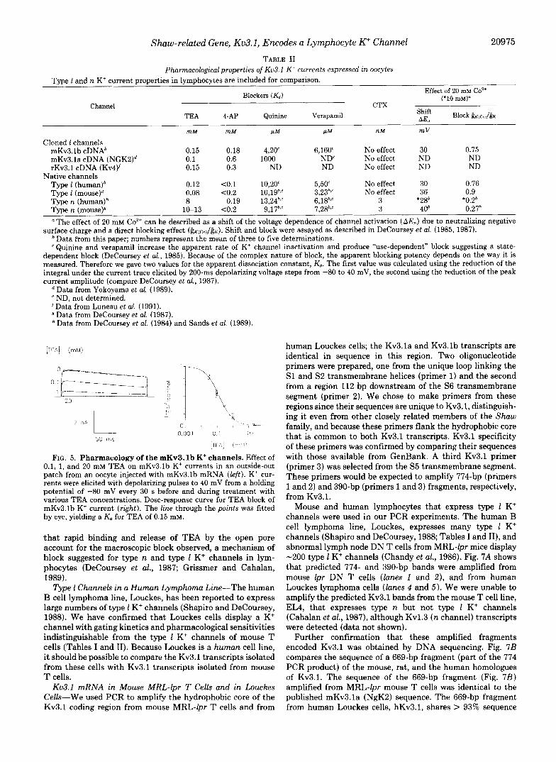
Shaw-related Gene, Kv3.1, Encodes a Lymphocyte K" Channel 20975 TABLE I1
Pharmacological properties of Ku3. I K' currents expressed in oocytes Type 1 and n K' current properties in lymphocytes are included for comparison.
Blockers ( K d ) Effect of 20 mM co2'
1+10 mM)= Channel CTX
, ~ ~~~~ I
TEA 4-AP Quinine Verapamil '2 Block & c ~ , / &
m M m M IrM IrM n M mV
Cloned 1 channels mKv3.1b cDNA' 0.15 0.18 4,20' 6,160' No effect 30 0.75 mKv3.la cDNA (NGK2)d 0.1 0.6 1000 ND' No effect ND ND rKv3.1 cDNA (Kv4)' 0.15 0.3 ND ND No effect ND ND
Type 1 (human)' 0.12 <0.1 10,20' 5,50' No effect 30 0.76 Type I (mouse)" 0.08 <0.2 10,19b,' 3,23b,' No effect 36 0.9 Type n (human)h 8 0.19 13,24',' 6,18',' 3 *28' '0.2'
Type n (mouse)p 10-13 C0.2 9,17bs' 7,28'.' 3 40' 0.27'
Native channels
The effect of 20 mM Co2+ can be described as a shift of the voltage dependence of channel activation (A&) due to neutralizing negative surface charge and a direct blocking effect (gKK(co)/gK). Shift and block were assayed as described in DeCoursey et al. (1985, 1987). ' Data from this paper; numbers represent the mean of three to five determinations. ' Quinine and verapamil increase the apparent rate of K' channel inactivation and produce "use-dependent" block suggesting a state-
dependent block (DeCoursey et al., 1985). Because of the complex nature of block, the apparent blocking potency depends on the way it is measured. Therefore we gave two values for the apparent dissociation constant, Kd. The first value was calculated using the reduction of the integral under the current trace elicited by 200-ms depolarizing voltage steps from -80 to 40 mV, the second using the reduction of the peak current amplitude (compare DeCoursey et al., 1987).
Data from Yokoyama et al. (1989). ND, not determined.
Data from DeCoursey et al. (1987). Data from DeCoursey et al. (1984) and Sands et al. (1989).
'Data from Luneau et al. (1991).
[TCA] (1m1.1)
1 ~.~ __ " 1 -\ "" """i 0
20 - " I\\ rr
\; I-
b,\
2 nA L 1 1 1
" ~, ._r~ , 1 L 7 x.
- 50 ms
i)!)01 0 I ;, ,
[ l i * j (.1,1.11
FIG. 5. Pharmacology of the mKv3.lb K+ channels. Effect of 0.1, 1, and 20 mM TEA on mKv3.lb K' currents in an outside-out patch from an oocyte injected with mKv3.lb mRNA ( le f t ) . K' cur- rents were elicited with depolarizing pulses to 40 mV from a holding potential of -80 mV every 30 s before and during treatment with various TEA concentrations. Dose-response curve for TEA block of mKv3.lb K+ current (right). The l ine through the points was fitted by eye, yielding a Kd for TEA of 0.15 mM.
that rapid binding and release of TEA by the open pore account for the macroscopic block observed, a mechanism of block suggested for type n and type 1 K' channels in lym- phocytes (DeCoursey et al., 1987; Grissmer and Cahalan, 1989).
Type 1 Channels in a Human Lymphoma Lint?-The human B cell lymphoma line, Louckes, has been reported to express large numbers of type 1 K' channels (Shapiro and DeCoursey, 1988). We have confirmed that Louckes cells display a K' channel with gating kinetics and pharmacological sensitivities indistinguishable from the type 1 K' channels of mouse T cells (Tables I and 11). Because Louckes is a human cell line, it should be possible to compare the Kv3.1 transcripts isolated from these cells with Kv3.1 transcripts isolated from mouse T cells.
Kv3.1 mRNA in Mouse MRL-lpr T Cells and in Louckes Cells-We used PCR to amplify the hydrophobic core of the Kv3.1 coding region from mouse MRL-lpr T cells and from
human Louckes cells; the Kv3.la and Kv3.lb transcripts are identical in sequence in this region. Two oligonucleotide primers were prepared, one from the unique loop linking the S1 and S2 transmembrane helices (primer 1) and the second from a region 112 bp downstream of the S6 transmembrane segment (primer 2). We chose to make primers from these regions since their sequences are unique to Kv3.1, distinguish- ing it even from other closely related members of the Shaw family, and because these primers flank the hydrophobic core that is common to both Kv3.1 transcripts. Kv3.1 specificity of these primers was confirmed by comparing their sequences with those available from GenBank. A third Kv3.1 primer (primer 3) was selected from the S5 transmembrane segment. These primers would be expected to amplify 774-bp (primers 1 and 2) and 390-bp (primers 1 and 3) fragments, respectively, from Kv3.1.
Mouse and human lymphocytes that express type 1 K' channels were used in our PCR experiments. The human B cell lymphoma line, Louckes, expresses many type 1 K+ channels (Shapiro and DeCoursey, 1988; Tables I and 11), and abnormal lymph node DN T cells from MRL-lpr mice display -200 type 1 K' channels (Chandy et al., 1986). Fig. 7A shows that predicted 774- and 390-bp bands were amplified from mouse lpr DN T cells (lanes 1 and 2) , and from human Louckes lymphoma cells (lanes 4 and 5). We were unable to amplify the predicted Kv3.1 bands from the mouse T cell line, EL4, that expresses type n but not type 1 K+ channels (Cahalan et al., 1987), although Kv1.3 (n channel) transcripts were detected (data not shown).
Further confirmation that these amplified fragments encoded Kv3.1 was obtained by DNA sequencing. Fig. 7B compares the sequence of a 669-bp fragment (part of the 774 PCR product) of the mouse, rat, and the human homologues of Kv3.1. The sequence of the 669-bp fragment (Fig. 7B) amplified from MRL-lpr mouse T cells was identical to the published mKv3.la (NgK2) sequence. The 669-bp fragment from human Louckes cells, hKv3.1, shares > 93% sequence

20976 Shaw-related Gene, Kv3.1, Encodes a Lymphocyte K” Channel
0 3 rnM TEA
A
2 PA
100 rns
4 :A L 100 ms
FIG. 6. Single-channel currents of mKv3.lb K+ currents in normal Ringer solution (left) and in the presence of 0.3 mM TEA (right) measured in an outside-out patch from an oocyte injected with mKv3.lb mRNA. The holding potential was -80 mV, and the membrane potential was continuously ramped from -80 mV to 80 mV within 450 ms every second. Single-channel events can be seen at potentials positive to -10 mV. Fitting a line through the slope of the current during openings gives an estimate of 27 pS for the single-channel conductance in Ringer; the line intercepts the voltage axis near -80 mV, close to the equilibrium potential for K+. In the presence of TEA, the single-channel conductance is 9 pS.
identity to the corresponding regions of mKv3.la and rKv3.lb (Fig. 7B).
Since the PCR primers are contained in one exon, we may have inadvertently amplified the 774- and 390-bp fragments from genomic DNA contaminating the RNA sample. To con- trol for this possibility we omitted the cDNA synthesis step (i.e. eliminated reverse transcriptase) and performed PCR directly on the RNA sample. Since Taq polymerase is a DNA- dependent polymerase, the predicted 774-bp fragment would be amplified if Kv3.1 genomic DNA was contaminating the RNA sample. The Kv3.1 band was not seen (data not shown), indicating that the fragments amplified by PCR represent legitimate Kv3.1 transcripts in mouse and human lym- phocytes.
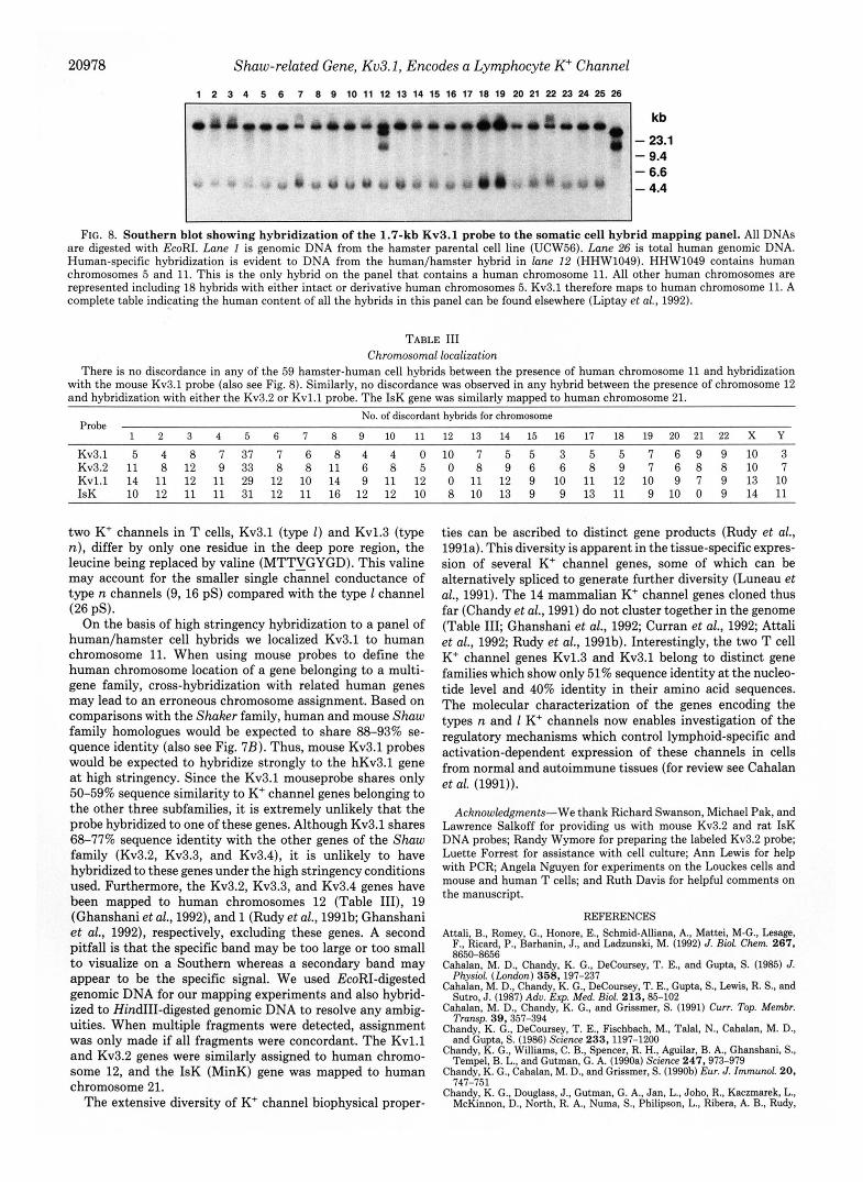
Chromosomal Localizationof Ku3.1, Kv3.2, Kul.1, and IsK- Genomic DNA from a panel of 59 human-Chinese hamster cell hybrids was hybridized to a 1.7-kb EcoRI fragment of Kv3.1 at high stringency (65 “C, 0.1 X SSC) to avoid cross- hybridization with related K’ channel genes (Fig. 8). Two Kv3.1-hybridizing bands are visible in human genomic DNA (lane 26) which is readily distinguishable from the one hy- bridizing band in hamster DNA (lane 1 ) . The Kv3.1 probe only hybridized to genomic DNA from the hamster/human hybrid (HHW1049) in lane 12. This cell line contains human chromosomes 5 and 11. All other human chromosomes are represented in the panel. Eighteen hybrids contain either intact or derivative human chromosome 5. These data indicate that hKv3.1 is located on human chromosome 11. In parallel
studies, we mapped the human chromosomal locations of three related K’ channel genes. Table I11 shows that the Shaker-related gene, Kvl.1 (Chandy et al., 1990a) and the Shaw family gene Kv3.2 (McCormack et al., 1990) are both located on human chromosome 12. The IsK gene encoding a slowly activating K’ channel (Takumi et al., 1988; Murai et al., 1989) was mapped to human chromosome 21.
DISCUSSION
Two approaches were used to identify the Kv3.1 gene product as the type 1 K’ channel in T lymphocytes. First, biophysical and pharmacological properties of the type 1 channel in mouse and human lymphocytes were compared with currents in oocytes following injection of mKv3.lb cRNA. We examined the voltage dependence and kinetics of opening and closing, conductance properties, and pharmaco- logical sensitivities of the channel encoded by the mKv3.1 gene. These properties of the mKv3.1 gene are remarkably similar to those of type 1 K’ channels in both mouse and human lymphocytes (summarized in Tables I and 11). Second, using PCR we detected Kv3.1 transcripts in MRL-lpr mouse T cells and in the human lymphoma line, Louckes; both these cell lines express type 1 K’ channels. Based upon these data, we conclude that Kv3.1 encodes type 1 K’ channels in lym- phocytes and that the channel is likely to have a homomulti- meric structure.
For quantitative comparison of channels expressed in oo- cytes with native channels, it is important that the experi- mental conditions be identical. We found several quantitative differences between two-electrode measurements and patch- clamp measurements in oocytes expressing Kv3.1 K’ channels. For example, pharmacological sensitivities to qui- nine and verapamil are lower in intact oocytes than in membrane patches2 (cf. Yokoyama et al., 1989). In addition, microelectrode measurements from oocytes do not show pro- nounced use-dependent inactivation, and the time course of inactivation is much slower than corresponding patch-clamp measurements’ (Yokoyama et al., 1989; Luneau et al., 1991; Douglass et al., 1990; Stuhmer et al., 1989). In patch-clamp measurements from oocytes, however, the inactivation time course and use-dependent inactivation of Kv1.3 and Kv3.1 channels are similar to those observed in T cells (Grissmer et al., 199Ob; this paper). One possible explanation for the dif- ference between whole oocyte voltage clamp and patch-clamp experiments could be the involvement of cytoplasmic factors within the oocyte that might contribute to channel function (cf. Ruppersberg et al., 1991).
Structure-function studies have recently identified residues that confer TEA sensitivity and determine the single-channel conductance of K’ channels. Kavanaugh et al. (1991) and MacKinnon and Yellen (1990) have provided evidence that the tyrosine at the C-terminal end of the putative pore region is the residue that confers TEA sensitivity to K’ channels. The 1-type K+ channel (Kv3.1) has a tyrosine (GDMYPKQ) at this position and so do six other mammalian K’ channel genes: Kvl.1 (Chandy et al., 1990a), Kv1.6 (Swanson et al., 1990), Kv2.1 (Frech et al., 1989), Kv3.2 (McCormack et al., 1990), Kv3.3 (Ghanshani et al., 1992), and Kv3.4 (Schroter et al., 1991; Rudy et al., 1991b; Ghanshani et al., 1992), which correlates well with the high degree of TEA sensitivity of these channels. Mutagenesis studies have shown that a leucine (MTTLGYGD) in the putative pore region is one of two residues involved in determining the single channel conduct- ance of K’ channels (Kirsch et al., 1992). Interestingly, the
S. Grissmer, unpublished observations.

Shaw-related Gene, Kv3.1, Encodes a Lymphocyte K" Channel 20977
FIG. 7. Kv3.1 is expressed in MRL-lpr mouse T cells and human Louckes cells. A , PCR amplification of 774- and 390-bp fragments from Louckes cDNA (left two lanes) and from MRL- lpr lymph node cDNA (right two lanes) with Kv3.1-specific primers. The middle lane shows the 4X molecular weight markers from top to bottom (in bp): 1,353, 1,078,872,603, 310,281,271,234, 194, 118, and 72. B, the nucleotide se- quence of the 669-bp PCR fragment am- plified from human Louckes cells (hKv3.1) is compared with the corre- sponding regions of the published mKv3.la and rKv3.lb genes. The puta- tive transmembrane senments S2-S6 are
Louckes $X L P R
1 2 3 4 5
rKv3.1 Mv3.1 hKv3.1 hKv3.1
rKv3.1 Mv3.1 hKv3.1 hKv3.1
rKv3.1 Mv3.1 hKv3.1 hKv3.1
indicated. Dashes represent nucleotides
The only difference between the m ~ n o Mv3.1 _ _ _ _ _ _ _ _ -c - -c _ _ _ _ _ _ _ __. _ _ _ . _ _ - - - _ _ _ _ _ _ - _ _ _ _ _ _ _ ~ - _ _ _ _ - _ _ ~ - _ _ ~ _ _ ~
acid sequence of the hKv3.1 and the rat hKV3.l - -T _ _ _ _ _ _ _ _ _ _ _ _ - -c _ _ _ _ _ _ _ _ _ _ _ _ _ _ _ _ _ _ _ _ _ _ _ _ _ _ _ _ _ _ _ _ _ - -A _ _ _ _ _ _ _ _ _ _ _ _ _ _ _ _ _ _ _ _ _ _ _ _ ment. a isoleucine realacine a valine
identical to those in the top sequence' rKv3.1 TTC AAG CTG ACC CGC CAT TTT GTG GGC CT: CGG GTC CTG G6C CAC ACG CTC CGT GCC AGC ACC AAC GAG TTC CTG CTG I
and homolo~es is in the s2 seg- hKv3.1 Phe Lys Leu Thr Arg His Phe Val Gly Leu Arg Val Leu Gly His Thr Leu Arg Ala Ser Thr Asn Glu Phe Leu Leu
20978 Shaw-related Gene, Kv3.1, Encodes a Lymphocyte K' Channel
1 2 3 4 5 6 7 8 9 10 11 12 13 14 15 16 17 18 19 20 21 22 23 24 25 26
kb
- 23.1 - 9.4 - 6.6 - 4.4
FIG. 8. Southern blot showing hybridization of the 1.7-kb Kv3.1 probe to the somatic cell hybrid mapping panel. All DNAs are digested with EcoRl. Lune 1 is genomic DNA from the hamster parental cell line (UCW56). Lune 26 is total human genomic DNA. Human-specific hybridization is evident to DNA from the human/hamster hybrid in lane 12 (HHW1049). HHW1049 contains human chromosomes 5 and 11. This is the only hybrid on the panel that contains a human chromosome 11. All other human chromosomes are represented including 18 hybrids with either intact or derivative human chromosomes 5. Kv3.1 therefore maps to human chromosome 11. A complete table indicating the human content of all the hybrids in this panel can be found elsewhere (Liptay et al., 1992).
TABLE 111 Chromosomal localization
There is no discordance in any of the 59 hamster-human cell hybrids between the presence of human chromosome 11 and hybridization with the mouse Kv3.1 probe (also see Fig. 8). Similarly, no discordance was observed in any hybrid between the presence of chromosome 12 and hybridization with either the Kv3.2 or Kvl.1 probe. The IsK gene was similarly mapped to human chromosome 21.
Probe
K v 3 . 1 5 4 8 7 3 7 7 6 8 4 4 0 1 0 7 5 5 3 5 5 7 6 9 9 1 0 3 Kv3.2 11 8 1 2 9 3 3 8 8 1 1 6 8 5 0 8 9 6 6 8 9 7 6 8 8 10 7 Kvl.1 14 11 12 11 29 12 10 14 9 11 12 0 11 12 9 10 11 12 10 9 7 9 13 10 IsK 10 12 11 11 31 12 11 16 12 12 10 8 10 13 9 9 13 11 9 10 0 9 14 11
No. of discordant hybrids for chromosome
1 2 3 4 5 6 7 8 9 10 11 12 13 14 15 16 17 18 19 20 21 22 x Y
two K' channels in T cells, Kv3.1 (type 1 ) and Kv1.3 (type n), differ by only one residue in the deep pore region, the leucine being replaced by valine (MTTIGYGD). This valine may account for the smaller single channel conductance of type n channels (9, 16 pS) compared with the type 1 channel
On the basis of high stringency hybridization to a panel of human/hamster cell hybrids we localized Kv3.1 to human chromosome 11. When using mouse probes to define the human chromosome location of a gene belonging to a multi- gene family, cross-hybridization with related human genes may lead to an erroneous chromosome assignment. Based on comparisons with the Shaker family, human and mouse Shaw family homologues would be expected to share 88-93% se- quence identity (also see Fig. 7 B ) . Thus, mouse Kv3.1 probes would be expected to hybridize strongly to the hKv3.1 gene at high stringency. Since the Kv3.1 mouseprobe shares only 50-59% sequence similarity to K' channel genes belonging to the other three subfamilies, it is extremely unlikely that the probe hybridized to one of these genes. Although Kv3.1 shares 68-77% sequence identity with the other genes of the Shaw family (Kv3.2, Kv3.3, and Kv3.4), it is unlikely to have hybridized to these genes under the high stringency conditions used. Furthermore, the Kv3.2, Kv3.3, and Kv3.4 genes have been mapped to human chromosomes 12 (Table 1111, 19 (Ghanshani et al., 1992), and 1 (Rudy et al., 1991b; Ghanshani et al., 1992), respectively, excluding these genes. A second pitfall is that the specific band may be too large or too small to visualize on a Southern whereas a secondary band may appear to be the specific signal. We used EcoRI-digested genomic DNA for our mapping experiments and also hybrid- ized to HindIII-digested genomic DNA to resolve any ambig- uities. When multiple fragments were detected, assignment was only made if all fragments were concordant. The Kv1.l and Kv3.2 genes were similarly assigned to human chromo- some 12, and the IsK (MinK) gene was mapped to human chromosome 21.
The extensive diversity of K+ channel biophysical proper-
(26 PSI.
ties can be ascribed to distinct gene products (Rudy et al., 1991a). This diversity is apparent in the tissue-specific expres- sion of several K+ channel genes, some of which can be alternatively spliced to generate further diversity (Luneau et dl., 1991). The 14 mammalian K' channel genes cloned thus far (Chandy et al., 1991) do not cluster together in the genome (Table 111; Ghanshani et al., 1992; Curran et al., 1992; Attali et dl., 1992; Rudy et al., 1991b). Interestingly, the two T cell K' channel genes Kv1.3 and Kv3.1 belong to distinct gene families which show only 51% sequence identity at the nucleo- tide level and 40% identity in their amino acid sequences. The molecular characterization of the genes encoding the types n and 1 K' channels now enables investigation of the regulatory mechanisms which control lymphoid-specific and activation-dependent expression of these channels in cells from normal and autoimmune tissues (for review see Cahalan et al. (1991)).
Acknowledgments-We thank Richard Swanson, Michael Pak, and Lawrence Salkoff for providing us with mouse Kv3.2 and rat IsK DNA probes; Randy Wymore for preparing the labeled Kv3.2 probe; Luette Forrest for assistance with cell culture; Ann Lewis for help with PCR Angela Nguyen for experiments on the Louckes cells and mouse and human T cells; and Ruth Davis for helpful comments on the manuscript.
REFERENCES Attali, B., Romey, G., Honore, E., Schmid-Alliana, A., Mattei, M-G., Lesage,
F., Ricard, P., Barhanin, J., and Ladzunski, M. (1992) J. Bml. Chem. 267,
Cahalan, M. D., Chandy, K. G., DeCoursey, T. E., and Gupta, S. (1985) J. Physiol. (London) 358,197-237
Cahalan, M. D., Chandy, K. G., DeCoursey, T. E., Gupta, S., Lewis, R. S., and Sutro, J. (1987) Adu. Exp. Med. Biol. 213.85-102
Cahalan, M. D., Chandy, K. G., and Grissmer, S. (1991) Curr. Top. Membr. Tramp. 39,357-394
Chandy, K. G., DeCoursey, T. E., Fischbach, M., Talal, N., Cahalan, M. D., and Gupta, S. (1986) Science 233,1197-1200
Chandy, K. G., Williams, C. B., Spencer, R. H., Aguilar, B. A., Ghanshani, S., Tempel, B. L., and Gutman, G. A. (1990a) Science 247,973-979
Chandy, K. G., Cahalan, M. D., and Grissmer, S. (1990b) Eur. J. ImmunoL 20, 747-751
Chandy, K. G., Douglass, J., Gutman, G. A., Jan, L., Joho, R., Kaczmarek, L., McKinnon, D., North, R. A., Numa, S., Philipson, L., Ribera, A. B., Rudy,
8650-8656

Shaw-related Gene, Ku3.1, Encodes a Lymphocyte K” Channel 20979 B., Salkoff, L., Swanson, R. A,, Steiner, D., Tanouye, M., and Tempel, B. L. (1991) Nature 352 ,26
Cirullo. R. E.. Arrendo-Vega. F. X., Smith, M., and Wasmuth, J. J. (1983)
Curran, M. E., Landes, G. M., and Keating, M. T. (1992) Gemmics 12 , 729- Somatic Celt Genet. 9,215-233
l a 1
DeCoursey, T. E., Chandy, K. G., Gupta, S., and Cahalan, M. D. (1984) Nature
DeCoursev. T. E.. Chandv. K. G.. Guuta, S., and Cahalan, M. D. (1985) J.
, “ I
307,465-468
Neuroimmunol.’lO, 71-65
Physiol. 89,379-404
Adelman, J. P. (1990) J. Immunol. 144,4841-4850
- .
DeCoursey, T. E., Chandy, K. G., Gupta, S., and Cahalan, M. D. (1987) J. Gen.
Douglass, J., Osborne, P. B., Cai, Y. C., Wilkinson, M., Christie, M. J., and
Feinberg, A. P., and Vogelstein, B. (1983) Anal. Biochem. 132,6-13 Frech, G. C., VanDongen, A. M. J., Schuster, G., Brown, A. M., and Joho, R.
Ghanshani, S., Pak, M., McPherson, J. D., Strong, M., Dethlefs, B., Wasmuth, (1989) Nature 340,642-645
J. J., Salkoff, L., Gutman, G. A,, and Chandy, K. G. (1992) Genomics 12 , 190-196
Grissmer, S., and Cahalan, M. D. (1989) Biophys. J. 5 5 , 203-206 Grissmer, S., Cahalan, M. D., and Chandy, K. G. (1988) J . Immunol. 141 ,
Grissmer, S., Hanson, D., Natoli, E. J., Cahalan, M. D., and Chandy, K. G. 1137-1142
Grissmer, S., Dethlefs, B., Wasmuth, J., Goldin, A. L., Gutman, G. A,, Cahalan, (1990a) J. Immunol. 145,2105-2109
M. D., and Chandy, K. G. (1990b) Proc. Natl. Acad. Sci. U. S. A . 87, 9411- 9415
Grissmer, S., Lewis, R. S., and Cahalan, M. D. (1992) J. Gen. Physiol. 9 9 , 1- 23
Gyllensten, U. B., and Erlich, H. A. (1988) Proc. Natl. Acad. Sci. U. S. A . 8 5 , 7652-7656
Hamill, 0. P., Marty, A,, Neher, E., Sakmann, B., and Sigworth, F. J. (1981) Pfligers Arch. Eur. J. Physiol. 391 , 85-100
Kavanaugh, M. P., Varnum, M. D., Osborne, P. B., Christie, M. J., Buscb, A. E., Adelman, J. P., and North, R. A. (1991) J. Biol. Chem. 2 6 6 , 7583-7587
Kirsch, G. E., Drewe, J. A,, Hartmann, H. A,, Tagliatela, M., De Biasi, M., Brown, A. M., and Joho, R. (1992) Neuron 8,499-505
Krug, M. S., and Berger, S. L. (1987) Methods Enzymol. 152 , 316-325 Lewis, R. S., and Cahalan, M. D. (1988) Science 239 , 771-775 Liptay, S., Schmid, R. M., Perkins, N. D., Meltzer, P., Altherr, M. R., Mc-
Pherson, J. D., Wasmuth, J. J., and Nabel, G. J. (1992) Genomics 13 , 287-
Luneau, C. J., Williams, J. B., Levitan E. S., Oliva, C., Smith, J. S., Antanavage, 292
J., Folander, K., Stein, R. B., Swanson, R., Kaczmarek, L., and Buhrow, S. A. (1991) Proc. Natl. Acad. Sci. U. S. A. 88,3932-3936
MacKinnon, R., and Yellen, G. (1990) Science 260,276-279 McCormack, K., Vega-Saenz de Miera, E. C., and Rudy, B. (1990) Proc. Natl.
Murai, T., Kakizuka, A,, Takumi, T , Ohkubo, H., and Nakanishi, S. (1989)
Overhauser, J., McMahon, J , and Wasmuth, J. J. (1987) Nucleic Acids Res. 15 ,
Rudy, B., Kentros, C., and Vega-Saenz de Miera, E. (1991a) Mol. Cell. Neurosci.
Rudy, B., Sen, K., Vega-Saenz de Miera, Lau, D., Ried, T., and Ward, D. C.
Ruppersberg, J. P., Stocker, M., Pongs, O., Heinemann, S. H., Frank, R., and
Sands, S. B., Lewis, R. S., and Cahalan, M. D. (1989) J. Gen. Physiol. 9 3 ,
Sanger, F., Nicklen, S., and Coulson, A. R. (1977) Proc. Natl. Acad. Sci. U. S. A .
Scbroter, K. H., Ruppersberg, J. P., Wunder, F., Rettig, J., Stocker, M., and
Shapiro, M., and DeCoursey, T. E. (1988) Biophys. J. 53,550a (ahstr.) Stuhmer, W. J., Ruppersberg, J. P., Schroter, K. H., Sakmann, B., Stocker, M.,
Giese, K. P., Perschke, A., Baumann, A,, and Pongs, 0. (1989) EMBO J. 8 ,
Swanson, R., Marshall, J., Smith, J. S., Williams, J. B., Boyle, M. B., Folander, 3235-3244
K., Luneau, C. J., Antanavage, J., Oliva, C., Buhrow, S. A,, Bennet, C., Stein, R. B., and Kaczmarek, L. K. (1990) Neuron 4,929:939
Acad. Sci. U. S. A. 8 7 , 5227-5231
Biochem. Biophys. Res. Commun. 161,176-181
4617-4627
2,89-102
(1991b) J. Neurosci. Res. 2 9 , 401-412
Koenen, M. (1991) Nature 352 , 711-714
1061-1074
74,5463-5467
Pongs, 0. (1991) FEES Lett. 278,211-216
Takumi, T., Ohkubo, H., and Nakanishi, S. (1988) Sctence 242,1042-1045 Yokovama. S.. Imoto. K.. Kawamura. T.. Hisashida. H.. Iwabe. N.. Mivpt-
and Numa, 8. (1989) PEES Lett. 259; 37y42
DNA Cell Biol. 11 , 163-172
, , I I I I”) I . ,
Yun-Cai Cai, Osborne, P. B., North, R. A,, Dooley, D. C., Douglass, J. (1992)