Advanced glycation end-products activate the renin-angiotensin

The Role of the Receptor for Advanced
Glycation End Products (RAGE) in
idiopathic Pulmonary Fibrosis
Inaugural Dissertation
Zur Erlangung des Doktorgrades der Naturwissenschaften
-Dr. rer. nat.- vorgelegt von
Markus Alexander Queisser aus Berlin, Deutschland
angefertigt am Institut für Biochemie Fachbereich Medizin und dem Fachbereich für Biologie
und Chemie Justus-Liebig-Universität Giessen
From the Department of Medicine
Institute of Biochemistry
Director: Prof. Dr. Klaus T. Preissner
at the Justus Liebig University Giessen
First Supervisor and Committee Member Prof. Dr. A. Pingoud
Second Supervisor and Committee Member Prof. Dr. K.T. Preissner
Committee members: Prof. Dr. W. Clauss and Prof. Dr. R. Dammann
Date of Doctoral Defense ···················································15.05.2009

“Forschung ist nie zu Ende, sie lebt von der Kritik und der intelligenten Skepsis, die nicht die Arroganz des Ignoranten ist.“
Prof. Dr. Lothar Jaenicke

Table of contents
I
Table of contents································································· I List of figures····································································· IIIList of abbreviations ························································· IVSummary ··········································································· VIIZusammenfassung·························································· VIII1. Introduction····································································· 11.2 Ligands of the recetor for advanced glycation end products ···········11.2.1 Advanced glycation end products (AGE) ·····································21.2.2 Amyloid �-peptides·······································································31.2.3 High mobility group box-protein B1 (HMGB1) ······························31.2.4 S100/Calgranulins········································································41.2.5 Mac-1 (CD11b/CD18) ··································································41.3 Physiological function of RAGE ······················································51.4 RAGE expression and its involvement in pathogeneses·················51.4.1 RAGE in vascular and renal complications of diabetes mellitus···51.4.2 RAGE in tumor progression and metastasis ································71.4.3 RAGE in innate and adapted immunity ········································81.5 Physiology and pathophysiology of the lung ···································91.5.1 Anatomy of the pulmonary system·············································101.5.2 Interstitial lung diseases·····························································121.5.3 Idiopathic pulmonary fibrosis······················································121.5.4 Pathogenesis of IPF···································································121.5.4.2 Chronic injury hypothesis ························································131.5.4.3 Sequential injury hypothesis ···················································131.5.4.4 Circulating fibrocyte-hypothesis ··············································141.5.4.5 Epithelial-mesenchymal transition (EMT) hypothesis··············151.5.5 Genetic factors···········································································161.6 Animal models of pulmonary fibrosis·············································171.6.1 Bleomycin model········································································181.6.2 Asbestos, silicia model·······························································181.6.3 Fluorescein isothiocyanate-model··············································181.6.4 Irradiation model ········································································191.6.5 Transgenic model·······································································191.7 Hypothesis ····················································································201.8 Aims ······························································································202. Materials ········································································ 212.1. Chemicals ····················································································212.1.2 Enzymes ····················································································232.1.3 Cytokines ···················································································232.1.4 Antibodies ··················································································232.1.5 DNA-Primers ··············································································242.1.6 Small interfering RNA (siRNA) ···················································242.1.7 General consumable ··································································242.1.8 Cell culture ·················································································252.1.9 Machines and systems·······························································252.2 Patient Population ·········································································263 Methods·········································································· 263.1 Animal Treatment··········································································26

Table of contents
II
3.2 Isolation and Culture of Human Alveolar Epithelial Cells type II····263.3 Isolation and Culture of Human Pulmonary Fibroblasts ················273.4 Cytokine Stimulation ·····································································273.5 Immunohistochemistry ··································································283.6 Immunofluorescence·····································································283.7 siRNA knock down········································································283.8 Reverse Transcriptase (RT)-PCR ·················································293.9 Real-time PCR ··············································································293.10 Western Blot ···············································································303.11 Extracellular Matrix Preparation ··················································313.12 Adhesion Assay ··········································································313.13 Proliferation Assay ······································································313.14 Migration (chemotaxis) Assay ·····················································313.15 Wound Healing Assay·································································323.16 Basolateral membrane isolation··················································324. Statistics··························································································335. Results··········································································· 345.1 Differential expression of RAGE in mouse tissue··························345.2 Distribution of RAGE in donor and IPF lung tissue ·······················345.3 RAGE expression in donor, IPF lungs,alveolar type II cells and fibroblasts ····················································355.4 RAGE Expression in the bleomycin mouse model of lung fibrosis 385.5 Influence of Cytokines on RAGE Expression ································385.6 Relation between RAGE and Cell Adhesion, Migration and Proliferation ·································································396. Discussion ···································································· 456.1 The role of RAGE in pulmonary fibrosis ········································456.2 RAGE as a biomarker for lung injury·············································476.3 RAGE-ligand signaling in the lung·················································486.4 potential mechanism of RAGE downregulation·····························496.4.1 RAGE downregulation by micro-RNA ········································506.4.2 RAGE downregulation by proteases ··········································506.4.3 Downregulation of RAGE in relation to caveolae ·······················516.4 Involvement of RAGE in epithelial-mesenchymal transition ··········516.5 RAGE as an adhesion molecule ···················································527. Declaration ···································································· 538. Curriculum vitae ··························································· 549. Acknowledgements······················································ 5710. References ·································································· 59
List of figures
III
List of figures
Figures Introduction
Figure 1: RAGE isoforms and signaling cascade···································· 2 Figure 2: Endothelial dysfunction by AGE-RAGE interaction ·················· 6 Figure 3: RAGE dependent regulation of cellular invasion.····················· 8Figure 4: Schematic diagram of lung anatomy········································ 10Figure 5: Air-blood barrier ······································································· 11 Figure 6 Hypothetical scheme of the abnormal wound healing model for idiopathic pulmonary fibrosis.····························································· 14 Figure 7: Alveolar epithelial transdifferentiation pathways. ····················· 16
Figures Results
Figure 8: Abundant RAGE Expression in the Lung. ································ 34Figure 9: RAGE distribution in IPF and Donor lungs. ······························ 35Figure 10: RAGE Downregulation in IPF lung homogenate. ··················· 36Figure 11: RAGE Downregulation in alveolar epithelial cells type II from IPF patients.···················································································· 37Figure 12: Cytokine-Dependent RAGE Downregulation in A549 Cells. ·· 39Figure 13:Cytokine-Dependent RAGE Downregulationin Pulmonary Fibroblasts········································································· 40Figure 14: Impairement of Cell Adhesion on Collagen andExtracellular Matrix by RAGE Blocking. ·················································· 41Figure 15 Increased Cell Proliferation and Migration due to siRNAmediated RAGE knockdown ·············································· 42 Figure 16: Increased Cell Migration in Wound Scratch Assay ···············. 43Figure 17: RAGE is associated with the Cytoskeleton ···························· 44
List of abbreviations
IV
List of abbreviations
A� = Amyloid-beta
ADAM = A Disintegrin And Metalloproteinase protein
AEC = alveolar epithelial cells
AGE = Advanced Glycation End Products
ALI = Acute lung injury
ARDS = Acute respiratory distress syndrome
AT = Alveolar type
ATP = Adenosin triphosphate
BAL = Bronchoalveolar lavage fluid
BCA = Bicinchoninic acid
Bcl-2 = B-cell lymphoma 2
CD = Cluster of differentiation
CML = Carboxymethyl lysine
CMPC = Circulating mesenchymal progenitor cells
Col = Collagen
CT = Cycle of threshold
CXCR = Chemokine-CXC-motif Receptor
DMEM = Dulbecco’s modified Eagle medium
DNA = Deoxyribonucleic acid
dNTP = Desoxy nucleotide triphosphate
DTT = DL-Dithiothreitol
EC = Endothelial Cell
ECL = Enhanced Chemiluminescence
ECM = Extracellular matrix
EDTA = Eythelene diamino tetra acetic acid
Egr-1 = Early growth factor-1
EF = Elongation factor
EMT = Epithelial-mesenchymal transition
EN-RAGE = Extracellular newly identified RAGE binding protein
(S100A12)
List of Aberrations
V
ERK = Extracellular signal-regulated kinase
esRAGE = Endogenous soluble RAGE
FBS = Fetal bovine serum
FGF-2 = Fibroblast growth factor 2
FITC = Fluorescein isothiocyanate
FSP-1 = Fibroblast specific protein
GTPase = GTP hydrolase
GTP = Guanosine triphosphate
HBSS = Hank's Buffered Salt Solution
HEPES = 4-(2-hydroxyethyl)-1-piperazineethanesulfonic acid
HGF = Hepatocyte growth factor
Hmbs = Hydroxymethylbilane synthase
HMGB1 = High mobility group binding-protein B1
HRP = Horse-radish peroxidase
ICAM-1 = Inter-Cellular Adhesion Molecule 1
IgG = Immune globuline
IL-1� = Interleukin-1�
IL-6 = Interleukin-6
ILD = Interstitial lung diseases
IFN-� = Interferon-�
IPF = Idiopathic Pulmonary Fibrosis
JNK = C-Jun-N-terminal kinase
KGF = Keratinocyte growth factor
MAPK = Mitogen-activated protein kinase
MCP-1 = Monocyte chemotactic protein-1
MMP = Matrix Metalloprotease
mRNA = messenger RNA
miRNA = micro RNA
NADPH = Nicotinamide adenine dinucleotide phosphate
NF-�B = Nuclear factor -�B
pDC = Plasmacytoid dendritic cells
PAI-1 = Plasminogen-activator inhibitor 1
PCR = Polymerase chain reaction

List of Aberrations
VI
PBS = Phosphate buffered saline
PDGF = Platelet derived growth factor
PI3K = Phosphoinositide kinase 3
PMVEC = Pulmonary microvascular endothelial cells
PRP = Pattern recognition receptor
PDVF = Polyvinylidene difluoride
RAGE = Receptor for Advanced Glycation End Products
RBC = Red blood cells
RNA = Ribonucleic acid
RNP = Ribonucleoprotein
SDS = Dodecyl sodium salt
siRNA = Small interfering RNA
SMA = Smooth muscle actin
SP-C = Surfactant protein C
sRAGE = Soluble RAGE
ROS = Reactive oxygen species
RT = Reverse Transcriptase
TBS = Tris buffered saline
TBST = Tris buffered saline tween
TEMED = N,N,N',N'-Tetramethylethylenediamine
TERT = Telomerase reverse transcriptase
TF = Tissue factor
TIMP = Tissue inhibitors of MMP
TGF-� = Tumor growth factor-�
TLR = Toll-like receptor
TNF-� = Tumor-necrosis factor-�
TR = Telomerase RNA
TRIS = Tris(hydroxymethyl)aminomethane
UIP = Usual interstitial pneumonia
UTR = Untranslated region
VCAM-1 = Vascular cellular adhesion molecule 1
ZO-1 = Zonula occludens-1

Summary
VII
Summary
The Receptor for Advanced Glycation End Products (RAGE) is a
transmembrane receptor of the immunoglobulin superfamily. While vascular
RAGE expression is associated with kidney and liver fibrosis, under
physiological conditions high expression level of RAGE is found in the lung.
In this work, RAGE expression in idiopathic pulmonary fibrosis (IPF) was
assessed, and the relation of the receptor to functional changes of epithelial
cells and pulmonary fibroblasts in the pathogenesis of the disease was
investigated. Significant downregulation of RAGE was observed in lung
homogenate and alveolar epithelial cells (AEC) type II from IPF patients as
well as in bleomycin-treated mice, demonstrated by RT-PCR, western
blotting and immunohistochemistry. RAGE downregulation was provoked by
stimulation of primary human lung fibroblasts and A549 epithelial cells with
the pro-inflammatory cytokines, transforming growth factor-�1 or tumor
necrosis factor-� in vitro. Blockade of RAGE resulted in impaired cell
adhesion, and siRNA induced knock down of RAGE increased cell
proliferation and migration of A549 cells and human primary fibroblasts in
vitro. These results indicate that RAGE serves a protective role in the lung
and that loss of the receptor is related with functional changes of pulmonary
cell types with the consequences of fibrotic disease. The study provides
evidence that the expression and regulation of RAGE in the pulmonary
system differs from that in the vascular system. Here, a possible functional
mechanism of RAGE in pulmonary fibrosis is described for the first time.

Zusammenfassung
VIII
Zusammenfassung
Der Rezeptor für “Advanced glycation end products” (RAGE) ist ein
Transmembranrezeptor aus der Superfamilie der Immunglobuline. Die
vaskuläre RAGE Expression ist mit Nieren- und Leberfibrose assoziert,
während eine hohe Expression von RAGE in der Lunge unter normalen
physiologischen Bedingungen gefunden wurde. In dieser Studie wurde die
Expression von RAGE in Patienten der idiopathischen Lungenfibrose (IPF)
gemessen, und die Beziehung zwischen RAGE und die funktionellen
Änderungen von Epithelzellen und pulmonalen Fibroblasten wurde
untersucht. Signifikante Absenkung der Expression von RAGE wurde in
Lungenhomogenaten und isolierten alveolaren Epithelzellen type II von IPF
Patienten sowie auch in Bleomycin-behandelten Mäusen, nachgewiesen
mittels RT-PCR, Western-blot und Immohistochemie. In vitro wurde die
Repression von RAGE durch die pro-inflammatorischen Zytokine TGF-� und
TNF-� in primären Fibroblasten und A549 Epithelzellen erreicht. Desweiteren
führte die Blockade von RAGE mittels anti-RAGE Antikörpern zu reduzierter
Zelladhäsion. siRNA-induzierte Inhibierung der Expression von RAGE in
A549 und Fibroblasten führte zur vermehrten Zellproliferation und -Migration
in vitro.
Diese Ergebnisse deuten auf eine Schutzfunktion der RAGE Expression in
der Lunge hin, hingegen trägt der Verlust an RAGE zu zellulären Änderungen
und fibrotischen Erkrankungen bei. Diese Studie deckt molekulare
Zusammenhänge auf, die zur Erklärung der Unterschiede in der Expression
und Regulation von RAGE zwischen dem pulmonalen und vaskulären
System führen können. Ein möglicher, funktioneller Mechanismus von RAGE
in der pulmonalen Fibrose wurde hier zum ersten Mal beschrieben.
Introduction
1
1. Introduction
1.1 The receptor for advanced glycation end products (RAGE) The receptor for advanced glycation end products (RAGE) is a type I
transmembrane receptor of the immunoglobulin superfamily composed of
three extracellular immunoglobulin-like domains V, C1, C2, a transmembrane
helix and a short, highly negatively charged, cytoplasmatic tail with no known
binding motif at the intracellular C-terminus.
Several shorter isoforms exist beside the RAGE full-length receptor. An N-
truncated receptor lacking the V-domain and two soluble RAGE isoforms
composed of the extracellular domains which can be derived by alternative
splicing, called endogenous soluble RAGE (esRAGE) or which arise from
cleavage by the matrix metallo-proteases ADAM10 or MMP-9, called soluble
RAGE (sRAGE) (Raucci, Cugusi et al. 2008; Zhang, Bukulin et al. 2008). It
was shown that calcium is a critical regulator of the intramembrane-
proteolysis of RAGE, catalyzed by ADAM10 and the �-secretase (Galichet,
Weibel et al. 2008). The function and possible benefit of the processing of
RAGE is broadly unknown and not well understood. However, it is widely
accepted that the soluble isoforms of RAGE can intercept and prevent certain
ligand interactions with RAGE.
The human gene for RAGE, ager (advanced glycation end products receptor),
is localized on chromosome 6 in the histocompatibility complex between
class II and class III. The ager gene is composed of 10 introns and 11 exons
which can undergo alternative splicing to derive splice variants. The RAGE-
promoter contains nuclear factor (NF)-�B sites, interferon-� response element
and an interleukin-6 (IL-6) DNA binding motif, whereby the NF-�B sites
control the expression and connect the expression to inflammation (Bierhaus,
Humpert et al. 2005)
1.2 Ligands of the recetor for advanced glycation end products (RAGE)
RAGE was originally identified as a binding receptor for advanced glycation
end products (AGE) by Neeper et al. (Neeper, Schmidt et al. 1992).

Introduction
2
Nowadays, it is known that RAGE is a multi-ligand receptor, interacting with a
wide variety of different molecules such as AGE, �-amyloid peptide, high
mobility group binding-protein B1 (HMGB1), S100/Calgranulins and the
leukocyte adhesion molecule Mac-1 (CD11b/CD18) (Chavakis, Bierhaus et al.
2003).
Figure 1: RAGE isoforms and signaling cascade There are multiple isoforms of the RAGE receptor. The major isoforms are known as full-length RAGE, N-truncated RAGE and soluble RAGE (sRAGE) or endogenous soluble RAGE (esRAGE), The (e)sRAGE receptor is released from the cell and allowed to interact with RAGE ligands prior to their interacting at the plasma membrane. The N-truncated RAGE lacks the intracellular signaling domain, and therefore binds RAGE ligands without directly transducing a signal.
1.2.1 Advanced glycation end products (AGE)
AGE are glycated proteins derived by a non-enzymatic reaction, called
Maillard-reaction, between a primary amine (preferably lysine and arginine)
and a reducing sugar or an aldehyde, leading to the formation of an initial
Schiff-base association product, followed by oxidation, reduction and cross-
linking with other amines to Amadori products finally leading to the formation
Introduction
3
of carboxymethyllysine (CML), pentosidine or arginine-pyramidine. These
AGE are highly heterogenous in their degree of modification and their
structural/functional characteristics. Despite this diversity, AGE binds only to
the V-domain of RAGE. The binding affinity of AGE to RAGE depends on the
degree of glycation of the ligand (10 �M – 100 nM) (Dattilo, Fritz et al. 2007),
and the AGE-RAGE interaction can activate p21(ras), MAP Kinase (ERK1/2),
MAPK p38 or cdc 42 (Rac) and NF-�B action (Yeh, Sturgis et al. 2001). In
addition, AGE can induce NADPH activation and ROS production via RAGE
(Yan, Schmidt et al. 1994; Wautier, Chappey et al. 2001).
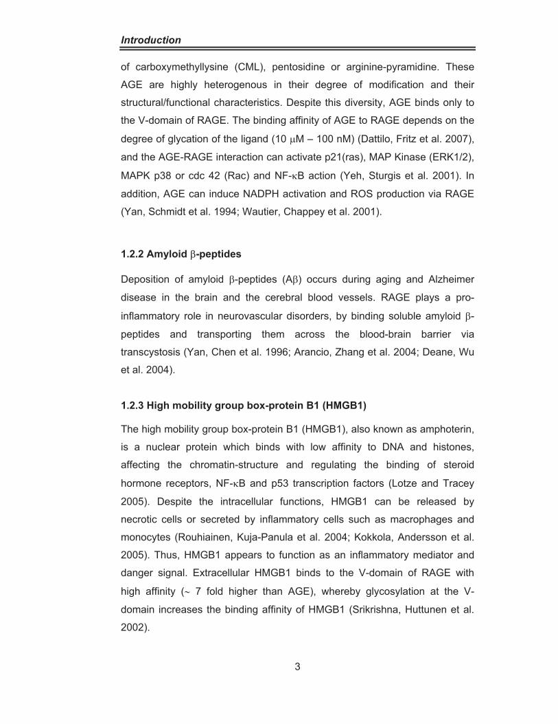
1.2.2 Amyloid �-peptides
Deposition of amyloid �-peptides (A�) occurs during aging and Alzheimer
disease in the brain and the cerebral blood vessels. RAGE plays a pro-
inflammatory role in neurovascular disorders, by binding soluble amyloid �-
peptides and transporting them across the blood-brain barrier via
transcystosis (Yan, Chen et al. 1996; Arancio, Zhang et al. 2004; Deane, Wu
et al. 2004).
1.2.3 High mobility group box-protein B1 (HMGB1)
The high mobility group box-protein B1 (HMGB1), also known as amphoterin,
is a nuclear protein which binds with low affinity to DNA and histones,
affecting the chromatin-structure and regulating the binding of steroid
hormone receptors, NF-�B and p53 transcription factors (Lotze and Tracey
2005). Despite the intracellular functions, HMGB1 can be released by
necrotic cells or secreted by inflammatory cells such as macrophages and
monocytes (Rouhiainen, Kuja-Panula et al. 2004; Kokkola, Andersson et al.
2005). Thus, HMGB1 appears to function as an inflammatory mediator and
danger signal. Extracellular HMGB1 binds to the V-domain of RAGE with
high affinity (� 7 fold higher than AGE), whereby glycosylation at the V-
domain increases the binding affinity of HMGB1 (Srikrishna, Huttunen et al.
2002).

Introduction
4
1.2.4 S100/Calgranulins
S100/Calgranulins are calcium binding proteins characterized by two calcium
binding elongation factor (EF)-motifs, found in granulocytes, monocytes,
macrophages well as induced in epithelial cells under inflammatory
conditions. S100/Granulin proteins have a broad spectrum of intracellular
functions in cell homeostasis but under cell damage, infection or
inflammatory conditions, they convert into cytokine-like mediators which are
secreted in a non-classical, Golgi-independent manner, and function as
danger signals after release in the extracellular space similar to HMGB1. It
was shown that some of S100/Calgranulin proteins such as S100A6
(calcyclin), and S100A12 (EN-RAGE) bind specifically to all three
extracellular domains of RAGE (Hofmann, Drury et al. 1999; Xie, Burz et al.
2007).
1.2.5 Mac-1 (CD11b/CD18)
Mac-1 (CD11b/CD18) is a member of the �2-integrin family which is
exclusively expressed on the surface of leukocytes. Under inflammatory
conditions and in concert with �1-integrins, �2-integrins recognize their
counterligands such as ICAM-1, VCAM-1 or surface associated fibrinogen
(FBG) on the endothelium, required for integrin-mediated adhesion and
diapedesis of activated leukocytes into the inflamed tissue. Recent studies
have shown that RAGE mediates leukocyte recruitment in vivo based on the
RAGE-Mac-1 interaction (Chavakis, Bierhaus et al. 2003) . For the first time,
these results shed light on the cell-adhesive functions of RAGE.
The reason for the different binding abilities to interact with such a diversity of
ligands may be explained with the concept that RAGE is a pattern recognition
receptor (PRP) which recognizes a conserved molecular structure such as
the �-sheet fibrilliar structure on diverse ligands. The characteristics of a
typical PRP are a multidomain structure, consiting of several similar structural
subunits; recognition of diverse types of ligands is brought about by their
comman recognition motifs (Gordon 2002). Although RAGE has no similar
structural subunits, it clearly fulfills characteristics of a PRP.
Introduction
5
1.3 Physiological function of RAGE
RAGE displays high expression during embryogenesis and organ
development in the nervous system and the lung (Hori, Brett et al. 1995;
Reynolds, Kasteler et al. 2008). After birth, RAGE is downregulated in almost
all organs which indicates RAGE’s physiological function of RAGE in
developmental processes. However, the RAGE-/- mice develops normal with
no obvious pathological phenotype (Liliensiek, Weigand et al. 2004). Further
studies in neuronal cells showed that activation of RAGE by HMGB1 or
S100B can facilitate cell survival by increased expression of the anti-
apoptotic protein Bcl-2 (Huttunen, Kuja-Panula et al. 2000). However, the
RAGE-/- mice demonstrated neither neuronal deficits nor behavior
abnormalities (Wendt, Tanji et al. 2003; Bierhaus, Haslbeck et al. 2004).
Further experiments have to be performed to challenge RAGE-/- mice with
various stimuli to explore the contribution of RAGE in diverse functions of the
organism.
1.4 RAGE expression and its involvement in pathogeneses
The expression pattern of RAGE and its splice-isoforms is tissue- and cell-
type specific. Basically, under physiological conditions, the RAGE expression
is on a low level and appears to be upregulated under inflammatory
conditions via the activation of the NF-�B-promoter or direct ligand-RAGE
interaction leads to an amplification of RAGE expression in different cell
types (Bierhaus, Humpert et al. 2005).
1.4.1 RAGE in vascular and renal complications of diabetes mellitus
Diabetes mellitus (type I and II) is a multi-phenotypic disease which is
characterized by hyperglycemia with subsequent macro- and microvascular
late complications, in particular increased atherosclerosis, retinopathy,
nephropathy and retinopathy. Under hyperglycemic conditions, the
progressive formation of modified proteins, termed advanced glycation end
products (AGE), is associated with vascular complications and cellular
senescence in diabetic patientens (Brownlee 1995; Hammes, Alt et al. 1999).

Introduction
6
Hyperglycemia has direct effects on the vessel wall by promoting glycation
and cross-linking of long-living extracellular matrix proteins such as collagen,
laminin and vitronectin, involving basement membrane thickening, decrease
in proteoglycans density, charge and permeability changes (Hammes, Weiss
et al. 1996). Finally, formed AGE induce production of reactive oxygen
species (ROS) by activation of NADPH oxidase at least partly through the
inflammatory RAGE-signaling in the endothelium as well as in macrophages
(Wautier, Chappey et al. 2001; Ding, Kantarci et al. 2007; Gao, Zhang et al.
2008). The AGE-RAGE interaction results in amplification of inflammatory
responses by activation of NF-�B (Bierhaus, Schiekofer et al. 2001),
production of cytokines such as monocyte chemotactic protein-1 (MCP-1),
tumor-necrosis factor-� (TNF-�) (Csiszar and Ungvari 2008), tumor growth
factor-� (TGF-�) (Li, Huang et al. 2004) interleukin-1� (IL-1�), tissue factor,
endothelin-1 and furthermore to the upregulation of RAGE, the vascular cell
adhesion molecules-1 (VCAM-1) and the inter-cellular adhesion molecule-1
(ICAM-1) (Boulanger, Wautier et al. 2002). Under inflammatory conditions,
high expression level of endothelial RAGE provides the molecular basis for
elevated leukocyte infiltration where leukocyte MAC-1 interacts with its
counter-receptor RAGE and facilitates leukocyte recruitment (Chavakis,
Bierhaus et al. 2003). In summary, the anti-coagulant endothelium turns into
a pro-coagulant cellular surface required for inflammatory signaling.
Figure 2: Endothelial dysfunction by AGE-RAGE interaction AGE-RAGE interaction on endothelial cells induces expression of tissue factor (TF), upregulation of adhesion molecules such as ICAM and VCAM and cytokine release such as MCP-1 and IL-6, followed by leukocyte recruitment and increased permeability of the endothelial monolayer (Wautier and Schmidt 2004).

Introduction
7
Diabetic retinopathy and renal fibrosis demonstrates another example where
RAGE acts as a pathogenic factor. Despite the fact that AGE and RAGE are
co-localized in diabetic kidney (Abel, Ritthaler et al. 1995; Heidland,
Sebekova et al. 2001; Hou, Ren et al. 2004), Yamamoto et al. demonstrated
in a transgeneic model that diabetic mice over-expressing RAGE developed
characteristics of diabetic nephropathy such as kidney enlargement,
albuminurea, glomerulosclerosis and tubulointerstitial fibrosis (Yamamoto,
Kato et al. 2001). Based on the inflammatory response of RAGE signaling,
namely the induction of the main fibrotic cytokine TGF-� as well as
inflammatory cell recruitment, several studies indicated a pro-fibrotic role for
RAGE due to its involvement in kidney and liver fibrosis (Oldfield, Bach et al.
2001; Forbes, Thallas et al. 2003; Hyogo and Yamagishi 2008).
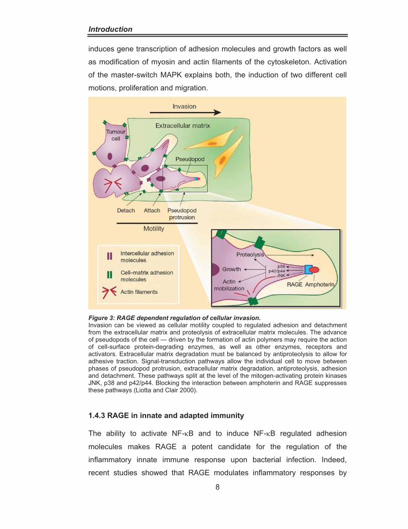
1.4.2 RAGE in tumor progression and metastasis
Tumor tissue (malignant neoplasm) is characterized by transformed cells
which display uncontrolled cell proliferation and impaired cell apoptosis
caused by changes on the genetic and epigenetic level. Beside the
uncontrolled growth, malignant neoplasm exhibits cellular invasion and often
metastasis. Interestingly, HMGB1 is expressed in a wide range of
transformed cells indicating a general role of HMGB1 in cell motility and
invasive migration of tumor cells. Invasion comprises spatial and temporal
coordination. Motility included regulated adhesion to the extracellular matrix
and degradation of matrix proteins, resulting in the migration of the cell
through the matrix. Receptor-ligand and proteolysis-antiproteolysis reactions
regulate the sensing and traction of the moving cell. Here, HMGB1 can
function as a generation site for the proteolytic enzyme plasmin. The complex
activates pro-matrixmetalloproteases (MMP), resulting in the degradation of
extracellular matrix. Forthermore, HMGB1-RAGE interaction leads to
proliferation and migration of cells (Taguchi, Blood et al. 2000). The mitogen-
activated protein kinase (MAPK) signals p42/p44, p38 and the c-Jun-N-
terminal kinase (JNK) are involved in this signal transduction between cell
membrane, cytoskeleton and nucleus. The MAPK can be directly activated or
indirectly by the GTPases Ras, Rac, Cdc42 and Rho. Activated MAPK
Introduction
8
induces gene transcription of adhesion molecules and growth factors as well
as modification of myosin and actin filaments of the cytoskeleton. Activation
of the master-switch MAPK explains both, the induction of two different cell
motions, proliferation and migration.
Figure 3: RAGE dependent regulation of cellular invasion. Invasion can be viewed as cellular motility coupled to regulated adhesion and detachment from the extracellular matrix and proteolysis of extracellular matrix molecules. The advance of pseudopods of the cell — driven by the formation of actin polymers may require the action of cell-surface protein-degrading enzymes, as well as other enzymes, receptors and activators. Extracellular matrix degradation must be balanced by antiproteolysis to allow for adhesive traction. Signal-transduction pathways allow the individual cell to move between phases of pseudopod protrusion, extracellular matrix degradation, antiproteolysis, adhesion and detachment. These pathways split at the level of the mitogen-activating protein kinases JNK, p38 and p42/p44. Blocking the interaction between amphoterin and RAGE suppresses these pathways (Liotta and Clair 2000).
1.4.3 RAGE in innate and adapted immunity
The ability to activate NF-�B and to induce NF-�B regulated adhesion
molecules makes RAGE a potent candidate for the regulation of the
inflammatory innate immune response upon bacterial infection. Indeed,
recent studies showed that RAGE modulates inflammatory responses by
Introduction
9
induction of the expression of adhesion molecules such as ICAM-1 and
VCAM-1 which enhances the recruitment of inflammatory cells (Fiuza, Bustin
et al. 2003; Treutiger, Mullins et al. 2003). Furthermore, RAGE itself functions
as a counter-receptor for leukocyte by binding to the �2-integrin Mac-1 and
amplifying the leukocyte infiltration. Induced systemic inflammation in the
RAGE-/- demonstrated decreased inflammatory cell recruitment (Chavakis,
Bierhaus et al. 2003).
In addition, it was shown that RAGE signaling can interact with the toll-like
receptor 9 (TLR-9)-pathway, to detect invading pathogens and to
distinguished infection-mediated from tissue damage by normal cell necrosis
(Tian, Avalos et al. 2007). Under non-infectious cell death, the necrotic cells
release HMGB1 which binds to RAGE on plasmacytoid dendritic cells (pDC)
or B-cells with no further cell activation. However, under infectious cell death,
HMGB1 forms a complex with CpG-containing pathogen DNA, whereby
activated RAGE and pathogenic DNA co-interact with TLR9 resulting in
interferon-� (IFN-�) secretion or B-cell proliferation. Besides IFN-�
production, maturing DC secrete HMGB1 in an autocrine/paracrine manner,
leading to RAGE activation and migration of the DC to the draininig lymph
nodes, they interact with naive T-cells to establish the T-cell dependent
immune-response, indicating that RAGE is involved in DC homing to lymph
nodes as well (Dumitriu, Baruah et al. 2005).
1.5 Physiology and pathophysiology of the lung
Oxygen is essential for multicellular aerobic organisms, cellular respiration
and ATP synthesis serves as electron acceptor in the respiratory chain. The
main function of the lungs is to provide continuous gas exchange between
inhaled air and the blood in the pulmonary circulation, supplying oxygen to
the organism and removing carbon dioxide, which is then removed from the
lungs by expiration. Survival is dependent upon this physiological process
being sustained and efficient, whereby this system responses to pathological
challenged in different ways to maintain optimal gas exchange.

Introduction
10
Figure 4: Schematic diagram of lung anatomy a) Anatomy and localisation of the respiratory tract including Larynx, Trachea and Bronchus. b) cross-section of a bronchus with lining ciliated epithelium and mucin-secreting goblet cells, surrounded by cartilage and smooth muscle cells. c) cross-section of an aloveolar duct at the end of the respiratory bronchiole. The alveolar duct is characterized by an interrupted wall with smooth muscle knobs. d) cross-section of the terminal part of the airway, the alveolus, composed of alveolar epithelia cells capillaries. The outer side is outlined with a surfactant layer (Effros 2006). 1.5.1 Anatomy of the pulmonary system
The respiratory tract extends from the mouth and nose cavities through the
bronchial tract down to the distant alveoli. The upper airway serves to filter
airborne particles, humidify and warm the inspired gases. The air is passing
the larynx, trachea, bronchi, bronchioles (terminal bronchiole) and alveolar
duct before reaching the alveolus where the gas exchange takes place
between the red blood cells (RBC) in the pulmonary capillaries and the
alveolar septae. In the septae three layers (endothelial cells, basallamina and
epithelial cells) function as the so called “air-blood barrier” which is very thin
(0.1-1.5 �m) and facilitates the diffusion of the gas. The air-blood barrier
functions as a barrier which enables the selective exchange between
molecules.

Introduction
11
Figure 5: Air-blood barrier The lung is a gas-exchanging organ for the provision of O2 to the blood and removel of CO2 from the blood. Alveolar capillaries are closely apposed to the alveolar lumen. Gas exchange by passive diffusion occurs across the air blood barrier consisting of type I alveolar cells, dual basal lamina, endothelial cells and the plasma membrane of red blood cells. Type II alveolar cells contribute indirectly to the gas-exchange process by secreting surfactant, a lipid-protein complex that reduces the surface tension of the alveolus and prevents alveolar collapsing (Kierszenbaum 2007). The pulmonary microvascular endothelial cells (PMVEC) form a tight barrier,
connected by tight-junctions and desmosomal structures between cells. The
endothelial cells are placed on their basolateral side on the basallamina
which separates vascular endothelium and pulmonary epithelium. On the
alveolar side, the epithelia cells form a tight cellular layer, connected by tight-
junctions, facing with their apical side towards the alveolar space. The
alveolar epithelium is composed of two different types of alveolar epithelial
cells (AEC), whereby the type I cells represent about 40% of the epithelial
cells, yet lining 90% of the alveolar surface. Type II cells only cover 10% of
the alveolar surface but represent 60% of total cells and are primarily located
at the branching of the alveolar septae. Type II cells produce and secrete
surfactant (surfactant protein-C positive cells), composed of hydrophobic
phospholipid-proteins which maintain alveolar expansion by lowering the
surface tension. As putative progenitors, the type II cells are considered to
differentiate into type I cells (aquaporin-5 positive cells) (Adamson and
Bowden 1974). The interstitium contributes tissue fibroblasts between both
layers of alveolar epithelial cells on the alveolar septum, embeded capillaries,
and elastic and collagen fibers produced by interstitial fibroblasts.
Introduction
12
1.5.2 Interstitial lung diseases
Interstitial lung diseases (ILD), caused by infections or other noxes, is a term
for over 200 different lung diseases which are characterized by damage to
the lining of the alveoli, increase of the interstitial and/ or vascular spaces,
leading to inflammation and fibrosis of the interstitium. A comman symptom in
ILD is progressive shortness of breath at rest and more dramatically during
physical exercise. The most common ILD include sarcoidosis and usual
interstitial pneumonia (UIP).
1.5.3 Idiopathic pulmonary fibrosis
Idiopathic Pulmonary Fibrosis (IPF) is classified as “a specific form of chronic
fibrosing interstitial pneumonia of unknown etiology, limited to the lung and
associated with the histological entity of usual interstitial pneumonia”
(Demedts and Costabel 2002). IPF is a progressive degenerative disease of
unknown etiology, for which no effective treatment exists. IPF is
characterized histologically by unrestricted interstitial fibroblast proliferation
and excessive deposition of extracellular matrix (Maher, Wells et al. 2007).
1.5.4 Pathogenesis of IPF
Although the cause of IPF is still elucidate, it is broadly accepted that the
pathogenesis starts with multiple damages to alveolar epithelial cells,
resulting in activated epithelial cells which release cellular agonists such as
TGF-�, TNF-�, platelet derived growth factor (PDGF), tissue factor (TF) and
plasminogen-activator inhibitor 1 (PAI-1) by activated epithelial cells (Selman,
King et al. 2001). TGF-� and PDGF induce proliferation and migration of sub-
epithelial fibroblasts as well as differentiation to myofibroblasts (Raghu,
Masta et al. 1989; Zhang and Phan 1999; Evans, Tian et al. 2003; Khalil, Xu
et al. 2005). The primary sites of injury become areas of fibroblast
proliferation, forming fibroblast-foci which are sites of active collagen
synthesis (Kuhn and McDonald 1991; Tzortzaki, Koutsopoulos et al. 2006).
Thus, foci formation is a hallmark of fibrosis. At these sites, epithelial cells
and myofibroblasts are producing gelatinases (MMP 9 and 2) which induce
Introduction
13
basement membrane disruption to enable the fibroblast/myofibroblast
migration to the injured surface. Intra- and interstitial fibroblast/myofibroblast
secrete extracellular matrix proteins, mainly collagen. An imbalance between
MMP and tissue inhibitors of MMP (TIMP) leads to deposition and
accumulation of extracellular matrix proteins (Pardo and Selman 2002). The
release of angiogenic factors from fibroblasts such as fibroblast growth factor
2 (FGF-2) and vascular endothelia growth factor (VEGF) leading to
angiogenesis to some extent. In parallel, Myofibroblasts show an increased
cell survival and delayed apoptosis provoking impaired reepithelialization and
tissue fibrosis (Zhang and Phan 1999). The whole sequence of events can be
seen as a process of abnormal wound repair where the response to injury is
overwhelmed by fibroblasts/myofibroblast proliferation and excessive matrix
deposition. Several hypotheses aim to provide the basis for the pathogenesis
(Thannickal, Toews et al. 2004; Maher, Wells et al. 2007).
1.5.4.2 Chronic injury hypothesis
Following the original hypothesis, IPF is caused by unknown stimuli which
lead to chronic inflammation inducing epithelial injury and subsequent fibrosis.
The inflammation theory might represent a major mechanism of ILD such as
sarcoidosis or hypersensivity pneumonitis. However, IPF patients display
mild or non-inflammatory cell recruitment to fibrotic lesions. In addition, anti-
inflammatory drugs such as steroids provide no significant improvement of
the pathogenesis (Nadrous, Ryu et al. 2004). These observations lead to the
assumption that inflammation is probably not necessary for the development
of pulmonary fibrosis (Gross and Hunninghake 2001).
1.5.4.3 Sequential injury hypothesis
The sequential injury hypothesis postulates that IPF is derived from
sequential acute lung injury where the repetitive wound repair results in
fibrosis by proliferation of fibroblasts, differentiation to myofibroblasts with an
contractile phenotype by expression of stress fibers such as �-smooth
muscle actin (�-SMA) and the production of collagen. Factors such as
Introduction
14
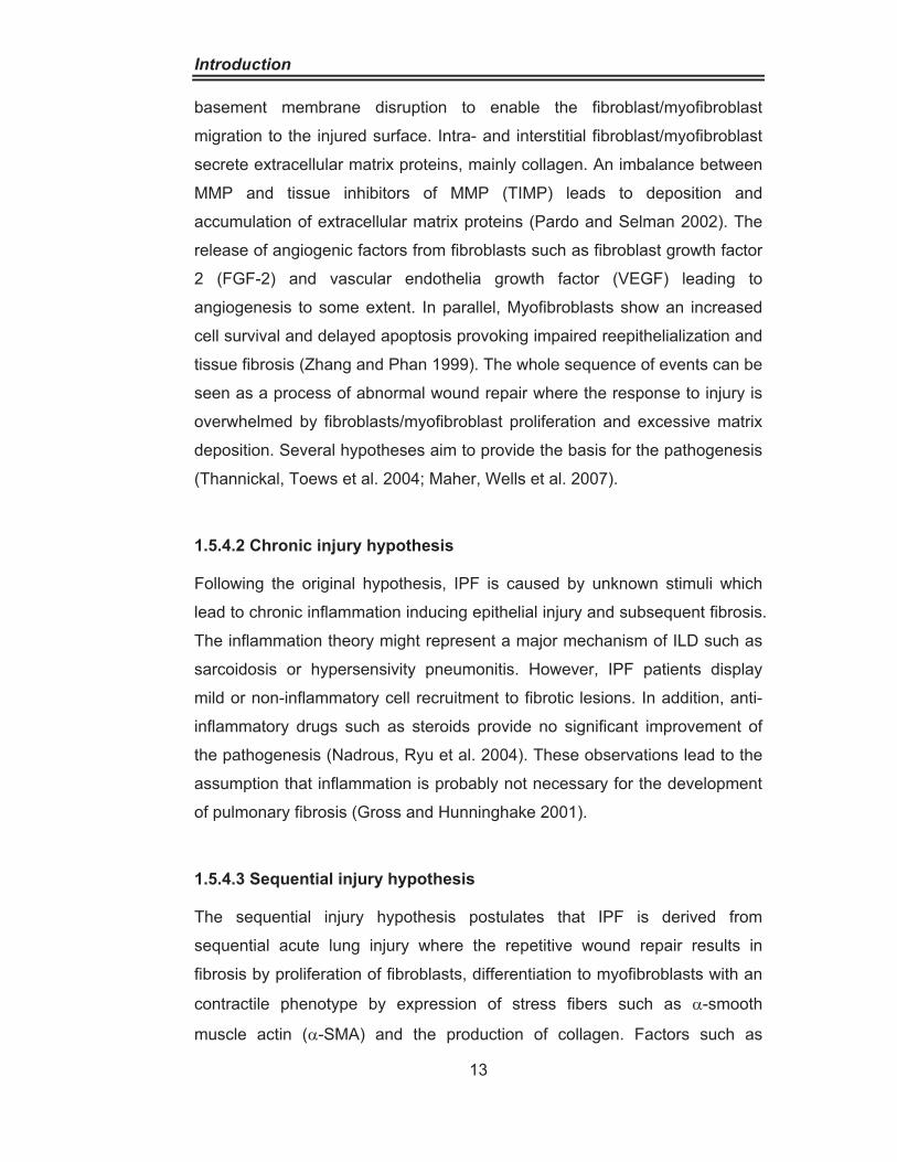
cigarette smoking, viral infection, environmental toxins or genetic background
can regulate and modify the fibrotic response (Gross and Hunninghake 2001).
Figure 6: Hypothetical scheme of the abnormal wound healing model for idiopathic pulmonary fibrosis. Multiple damage and activate alveolar epithelial cells, which in turn induce an antifibrinolytic environment in the alveolar spaces, enhancing wound clot formation. Alveolar epithelial cells secrete growth factors and induce migration and proliferation of fibroblasts and differentiation into myofibroblasts. Subepithelial myofibroblasts may increase basement membrane disruption and allow fibroblast–myofibroblast migration. Interstitial myofibroblasts secrete extracellular matrix proteins, mainly collagens. An imbalance between interstitial collagenases and tissue inhibitors of metalloproteinases provokes the progressive deposit of extracellular matrix and further impairing reepithelialization (Selman, King et al. 2001).
1.5.4.4 Circulating fibrocyte-hypothesis
Philips and colleges discovered a fibroblast-like cell population, sharing
leukocyte markers (CD34+ CD45+, CXCR4+, Col I+ and Vimentin+), called
fibrocytes. Fibrocytes are circulating mesenchymal progenitor cells (CMPC)
which can differentiate into multiple mesenchymal cell types depending on
the tissue environment (Phillips, Burdick et al. 2004). Fibrocytes are believed
to be involved in adipogenesis, pulmonary hypertension with associated
vascular wall remodeling, wound healing and pulmonary fibrosis. Leukocytes
and CMPC are generated in the bone marrow and extravasate to specific
region within tissues by trafficking, involving adhesion molecules,
chemoattractants and chemoattractant receptors. Lung injury results in high
level of the chemokine CXCL12, creating a chemokine gradient for CXCR4+
Introduction
15
positive fibrocytes to be released from the bone marrow and recruited from
the circulation to the lung (Strieter, Gomperts et al. 2007). In the lung,
fibrocytes can proliferate; they differentiate into myofibroblast-like cells with
the expression of �-SMA+ and the loss of CD45 and CD34 after the
stimulation with TGF-� or endothelin and synthesis to extracellular matrix and
thus contribute to pulmonary fibrosis (Gomperts and Strieter 2007; Mehrad,
Burdick et al. 2007).
1.5.4.5 Epithelial-mesenchymal transition (EMT) hypothesis
EMT is a well-known process during development where epiblasts undergo a
cell phenotype changes early in morphogenesis to form primary
mesenchyme. EMT is defined as a process by which differentiated epithelial
cells undergo a phenotypic conversion to mesenchymal cells such as
fibroblasts and myofibroblasts (Petersen, Nielsen et al. 2003; Radisky, Kenny
et al. 2007). The main aspect of EMT is the ability of epithelial cells to lose
polarity, disassemble from intracellular arrangements, acquire cell-motility,
and move from one location to another. So called secondary EMT which
occurs in fully differentiated epithelial cells is an accepted concept in cancer
metastasis and kidney fibrosis (Dasari, Gallup et al. 2006; Peinado, Olmeda
et al. 2007; Wynn 2008).
The transdifferention of AEC type II to type I cells reflects a normal process of
re-epithelialisation after epithelial cell injury where the epithelial cells undergo
apoptosis or necrosis. It was proposed that epithelial cells can alternatively
undergo transition to a mesenchymal phenotype. This transition is
characterized by the loss of epithelial cell markers such as E-cadherin and
zonula occludens-1 (ZO-1) and the expression of fibroblast and myofibroblast
markers such as fibroblast specific protein (FSP-1), a member of the S100
family, and �-SMA. Thus, cells which are in the process of EMT, express
both, epithelial and myofibroblast markers at the same time. Interestingly, the
fibrotic cytokine TGF-� has the ability to induce EMT by loss of E-cadherin
via Smad-dependent target genes which are mainly controlled by Smad3
(Masszi, Di Ciano et al. 2003). In concert with Smad-independent signaling
Introduction
16
such as Rho kinase, Ras, ERK, p38 MAPK, Notch and Wnt proteins, NF-�B
or phosphoinositide kinase 3 (PI3K) affect the EMT process as well (Zavadil
and Bottinger 2005). The EMT hypothesis provides another explanation for
epithelial cell loss and increasing myofibroblast population with excessive
extracellular matrix production in pulmonary fibrosis (Willis, duBois et al.
2006).
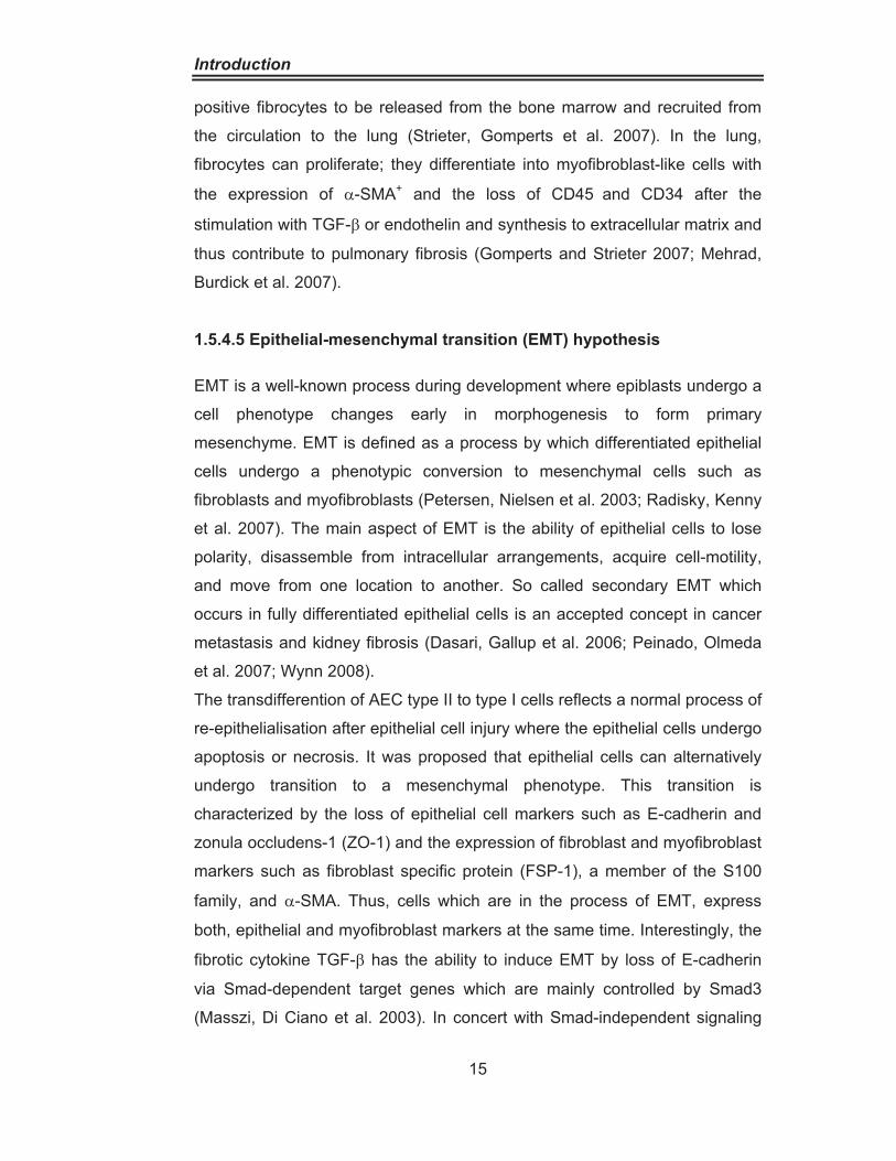
Figure 7: Alveolar epithelial transdifferentiation pathways. AECs demonstrate pluripotency; under normal conditions, alveolar type II (AT2) cells transdifferentiate into alveolar type I (AT1) cells. Depending on the cellular environment and stimuli, AECs respond to injury by traveling down one of a number of pathways: apoptosis/necrosis (1); proliferation, transdifferentiation, and re-epithelialization (2); or EMT (3) to amyofibroblast phenotype, resulting in extracellular matrix (ECM) deposition, destruction of lung architecture, and fibrosis (Willis, duBois et al. 2006).
1.5.5 Genetic factors
The dominant prevalence of IPF in some families raised the question
concerning the genetic background of the disease. Familiar IPF is identified
when two or more member of the same family are affected. The precise
prevalence is not known but is estimated to be at 7-11 in 100.000 of the
population. The familial form of IPF is probably transmitted via an autosomal
dominant trait with reduced penetrance (Allam and Limper 2006).
Genetic analysis verified some mutations in the surfactant protein C (SP-C)
molecule (Nogee, Dunbar et al. 2001). SP-C is probably the most
Introduction
17
hydrophobic protein in the human body, containing a valine, leucine and
isoleucine rich domain which forms a stable a-helical structure resulting in
insoluble random structures in the aqueous environment. SP-C is secreted by
AEC type II cells and facilitates to multify the surface tension in the alveolar
space by lining up the alveolar epithelium with a thin lipoproteinlayer. The
mutation causes a deletion of 37 amino acids, lacking a cystein residue which
is important for protein-disulphide mediated protein folding. In patients with
these mutations, the absence of mature SP-C in lung tissue and
bronchoalveolar lavage fluid (BAL) was observed, indicating that the
precursor protein has not been processed and secreted normally (Nogee,
Dunbar et al. 2001).
Recently, in some cases of familiar IPF, germ-line mutations in the genes
htert and htr, encoding telomerase reverse transcriptase and telomerase
RNA, was found in familiar cases of IPF (Armanios, Chen et al. 2007).
Telomerase reverse transcriptase (hTERT) is a polymerase that conjugates
telomere repeats (TTAGGG) to the ends of chromosomes during DNA
replication, whereas the telomerase RNA (hTR) provides the template for
nucleotide addition. The addition of telomeric repeats to the ends of the
chromosome partly re-do the shortening that occurs during DNA replication.
Telomeres shorten with each cell division and ultimately activate a DNA
damage response that leads to apoptosis. Mutations in htert and htr affect the
telomerase activity and shorten the telomers.
The limited number of familiar IPF patients makes it difficult to perform
genetic studies. However, mutations in familiar IPF could be detected in only
1-8% cases, indicating that IPF is a multi-cause disease.
1.6 Animal models of pulmonary fibrosis
Animal models for pulmonary fibrosis are restricted in several ways which
has to be considered when results from animal studies are transferred into
patient situation (Moore and Hogaboam 2008). Due to the unknown cause of
pulmonary fibrosis, several agents are used to induce lung injury with the
development of a fibrotic response (Gauldie and Kolb 2008). In addition, the
development of symptomatic in patients occurs between 10-20 years of age

Introduction
18
(IPF � 6 years) whereas the animal model takes only 21-28 days. The most
common rodent fibrosis models are here discussed.
1.6.1 Bleomycin model
The bleomycin model is the most commonly used model for lung fibrosis in
rodents because of its well characterized feature and the fast development
(Adamson and Bowden 1974). Bleomycin is a glycopeptide antibiotic with
anti-tumor activity that causes cytotoxic and mutagenic effects by mediating
single-strand and double-strand DNA damage in many cell types. Bleomycin
can be delivered equelly efficiant to the lung intratracheally, intraperitoneally
or intranasally. Bleomycin causes epithelial cell apoptosis and necrosis,
followed by an acute inflammation phase (1-7 days) which results in an
fibrotic response with increased collagen deposition (day 14) and the
establishment of severe fibrotic leasions (day 21-28). However, in this model,
fibrosis is self-limiting and starts to resolve after 28 days. Furthermore, this
model shows fibrosis untytipical acute inflammatory phase between day one
and seven. In addition, the mouse strain Balb/c is rather insensitive towards
developing pulmonary fibrosis in this model.
1.6.2 Asbestos, silicia model
Asbestos and silicia such as siliciumoxide can be used to induce pulmonary
fibrosis by a persistent fibrotic stimulus which is similary to that observed in
humans exposed to occupational dusts and particulates (Bozelka, Sestini et
al. 1983). The prolonged presence of the particles in the lung protract
cytotoxicity, induce inflammation (to a lower level as compared to the
bleomycin-model) and induce the release of cytokines and growth factors.
The asbestos model has clinical relevance due to long term exposure in
working environment. The clear disadvantage of this fibrosis model takes 12-
16 weeks to develop.
1.6.3 Fluorescein isothiocyanate-model
The Fluorescein isothiocyanate (FITC)-model demonstrates a fast fibrotic

Introduction
19
response within 14-28 days which varies considerably depending on the
amount of FITC (Christensen, Goodman et al. 1999). Although a fast
response, the fibrotic response persists at least for six months in Balb/c and
C57Bl/6 mice. The big advantage of this approach is the ability to visualize
areas of lung injury by the characteristic fluorescence of FITC. However, this
model is not of clinical relevance.
1.6.4 Irradiation model
The irradiation model is probably the only model which shows fibrosis
development without significant inflammation and therefore the closest model
to IPF with clinical relevance (Franko and Sharplin 1994). However, the
development of fibrosis takes over 30 weeks and is very cost intensive.
Therefore, this model is not commonly used.
1.6.5 Transgenic model
The transgenic model allows studying the effect of a single molecule
overexpression in a cell-specific manner. Frequently, the gene of interest is
cloned into an adenoviral vector which is used to infect the animal. The
adenovirus is transient overexpressing the gene of interest for limited period
of time (Bonniaud, Margetts et al. 2003). Recently, the use of transgenetic
mice which contain additional DNA or deleted parts of DNA into the genome
in every cell, became popular. The gene expression can be controlled by cell-
specific promoters, such as SP-C for specific epithelial cell type II expression.
However, the amount of expressed protein does not necessarily correspond
with physiological levels.
A more accurate way is to control the protein expression by using a
transcriptionally-regulated promoter using the tetracycline-resistance operon
(Tet-system). Gene expression from this promoter is tightly controlled by the
presence or absence of tetracycline or tetracycline derivatives such as
doxycycline. In the Tet-On system, the Tet-On activator requires doxycycline
for binding the chimeric transcriptional activator. In contrast, in the Tet-Off
system doxycycline prevents DNA binding and subsequent gene expression
(Gossen and Bujard 1992).
Aims
20
1.7 Hypothesis
Due to its involvement in inflammatory reactions, tissue fibrosis, myoblast
and tumor formation, it was hypothesized that RAGE expression in the lung,
in contrast to blood vessels, has a protective role in the pulmonary system
against degenerative processes, such as IPF pathogenesis.
1.8 Aims
The aim of the study was to investigate the role of RAGE in IPF by
addressing the following approches:
First, the expression of RAGE and the cell-specific distribution in lungs from
IPF patients and donors was characterized by immunohistology, real-time
PCR and western-blot analysis. Furthermore, the expression level of RAGE
was investigated in the bleomycin model and compared to the situation in IPF
patients.
Second, the regulation of RAGE expression during pulmonary fibrosis
development, as well as the effect of pro-fibrotic cytokines on RAGE
expression was demonstrated in alveolar epithelial cells and pulmonary
fibroblasts.
Third, since cell-proliferation and –migration are key events in pulmonary
fibrosis, a potential pathomechanistic role of RAGE was investigated by
blocking RAGE with anti-RAGE antibody and siRNA-mediated knock down in
epithelial cells and fibroblast and analyzed for cell-proliferation and –
migration.

Materials and Methods
21
2. Materials
2.1. Chemicals
Acetone Roth, Karlsruhe, GermanyAcrylamide-Bisacrylamide Roth, Karlsruhe, GermanyAmmoniumchlorid Roth, Karlsruhe, GermanyAmmonium persulphate Roth, Karlsruhe, GermanyBovine serum albumin Fraction V Sigma AldrichBromophenol blue Roth, Karlsruhe, GermanyBCA Protein assay kit Pierce, Rockford, USABleomycin sulphate Almirall Prodesfarma, Barcelona,
SpainCalciumchlorid Roth, Karlsruhe, GermanyChrystal blue violett Roth, Karlsruhe, Germanycitrate monohydrate Roth, Karlsruhe, Germany(trisodium)-citrate dehydrate Roth, Karlsruhe, GermanyCollagen I BD Biosciences, Franklin Lakes,
USADeoxyribonucleotide triphosphates Finnzymes, Espoo, FinlandDimethylsulfoxide Roth, Karlsruhe, GermanyDL-Dithiothreitol (DTT) Roth, Karlsruhe, GermanyDodecyl sodium salt (SDS) Roth, Karlsruhe, GermanyDynal magnet Dynal Biotech, Oslo, NorwayEnhanced Chemiluminescence (ECL) Plus reagents™
GE Healthcare (Amersham), Buckinghamshire, UK
Ethanol Roth, Karlsruhe, GermanyEythelene diamino tetra acetic acid (EDTA)
Roth, Karlsruhe, Germany
Formaldehyde alcohol free � 37% Roth, Karlsruhe, GermanyGenElute mammalian total RNA kit Sigma Aldrich,St. Louis, USAGlycerol Roth, Karlsruhe, GermanyGlycine Roth, Karlsruhe, GermanyHaematoxylin Roth, Karlsruhe, GermanyHBSS Invitrogen (Gibco), Carlsbad, USAHistostain Plus kit Zymed Laboratories, San Francisco,

Materials and Methods
22
USAHydrochloric acid Roth, Karlsruhe, GermanyHydrogen peroxide Roth, Karlsruhe, GermanyIsofluorane Forene® Abbott, Wiesbach, Germany�-mercaptoethanol Sigma Aldrich,St. Louis, USAMethanol Roth, Karlsruhe, GermanyNon-fat dry milk powder Roth, Karlsruhe, GermanyMounting medium, Vectashild (with dapi)
Vector Laboratories, Peterborough, UK
Paraffin Roth, Karlsruhe, GermanyParaformaldehyde Roth, Karlsruhe, GermanyPercoll Sigma Aldrich,St. Louis, USAProtease Inhibitor cocktail complete™ Roche, Mannheim, GermanyPotassium chloride Roth, Karlsruhe, GermanyPotassium dihydrogen phosphate Roth, Karlsruhe, GermanyRotiphorese® gel 30 Roth, Karlsruhe, GermanyRNasin RNase inhibitor Promega, Madison, USASaline solution, physiological Baxter, München, GermanySodium Chloride Roth, Karlsruhe, GermanySodium dihydrogen phospahate Roth, Karlsruhe, GermanySodium hydrogen carbonate Roth, Karlsruhe, GermanySYBR Green PCR master mix Invitrogen, Carlsbad, USAN,N,N',N'-Tetramethylethylenediamine (TEMED)
Roth, Karlsruhe, Germany
TransPass R1 transfection Reagent New England Biolabs, Ipswich, USAtris(hydroxymethyl)aminomethane (TRIS) base
Roth, Karlsruhe, Germany
Triton-X-100 Roth, Karlsruhe, GermanyTween 20 Roth, Karlsruhe, GermanyXylol Roth, Karlsruhe, Germany
Materials and Methods
23
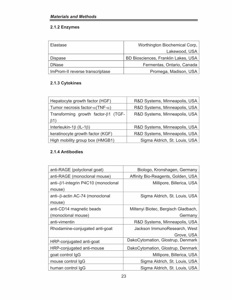
2.1.2 Enzymes
Elastase Worthington Biochemical Corp, Lakewood, USA
Dispase BD Biosciences, Franklin Lakes, USADNase Fermentas, Ontario, CanadaImProm-II reverse transcriptase Promega, Madison, USA
2.1.3 Cytokines
Hepatocyte growth factor (HGF) R&D Systems, Minneapolis, USATumor necrosis factor-�(TNF-�) R&D Systems, Minneapolis, USATransforming growth factor-�1 (TGF-�1)
R&D Systems, Minneapolis, USA
Interleukin-1� (IL-1�) R&D Systems, Minneapolis, USAkeratinocyte growth factor (KGF) R&D Systems, Minneapolis, USAHigh mobility group box (HMGB1) Sigma Aldrich, St. Louis, USA
2.1.4 Antibodies
anti-RAGE (polyclonal goat) Biologo, Kronshagen, Germanyanti-RAGE (monoclonal mouse) Affinity Bio-Reagents, Golden, USAanti–�1-integrin P4C10 (monoclonal mouse)
Millipore, Billerica, USA
anti–�-actin AC-74 (monoclonal mouse)
Sigma Aldrich, St. Louis, USA
anti-CD14 magnetic beads (monoclonal mouse)
Miltenyi Biotec, Bergisch Gladbach, Germany
anti-vimentin R&D Systems, Minneapolis, USARhodamine-conjugated anti-goat Jackson ImmunoResearch, West
Grove, USAHRP-conjugated anti-goat DakoCytomation, Glostrup, Denmark
HRP-conjugated anti-mouse DakoCytomation, Glostrup, Denmarkgoat control IgG Millipore, Billerica, USAmouse control IgG Sigma Aldrich, St. Louis, USAhuman control IgG Sigma Aldrich, St. Louis, USA

Materials and Methods
24
2.1.5 DNA-Primers
Gene Primer Sequence (5’–3’) Forward Reverse RAGE (ager) (Homo sapiens)
caggaccagggaacctacag catgtgttgggggctatctt
RAGE (ager) (Mus musculus)
gggtgctggttcttgctcta tggagaaggaagtgcctcaa
hprt-1 (H. sapiens) aaggaccccacgaagtgttg gctttgtattttgcttttccapbgd (Mus musculus) atgtccggtaacggcggc ggtacaaggctttcagcatcgc
2.1.6 Small interfering RNA (siRNA)
Gene Antisense sequence pool (5’-3’) RAGE (ager) (Homo sapiens)
1.ttccattcctgttcattgctt 2.tactgctccaccttctggctt 3.tgttccttcacagatactctt 4.tttgaggagagggctgggctt
2.1.7 General consumable
Eppendorf tubes (0.5 ml, 1.5 ml, 2.0 ml)
Eppendorf, Hamburg, Germany
Falcon tubes (15 ml, 50 ml) BD Biosciences, Franklin Lakes, USADisposable pipettes ( 2 ml, 5 ml, 10 ml, 25 ml, 50 ml)
BD Biosciences, Franklin Lakes, USA
3MM Whatman paper GE Healthcare (Amersham), Buckinghamshire, UK
Hybond-C polyvinylidene difluoride (PDVF) membrane
GE Healthcare (Amersham), Buckinghamshire, UK
Pipettes tips (2�l, 20�l, 200 �l, 1000 �l)
Gilson, Middelton, USA
Materials and Methods
25
2.1.8 Cell culture
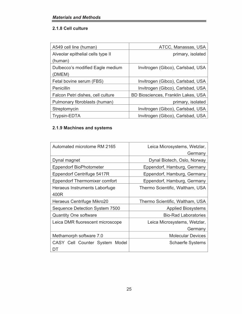
A549 cell line (human) ATCC, Manassas, USAAlveolar epithelial cells type II (human)
primary, isolated
Dulbecco’s modified Eagle medium (DMEM)
Invitrogen (Gibco), Carlsbad, USA
Fetal bovine serum (FBS) Invitrogen (Gibco), Carlsbad, USAPenicillin Invitrogen (Gibco), Carlsbad, USAFalcon Petri dishes, cell culture BD Biosciences, Franklin Lakes, USAPulmonary fibroblasts (human) primary, isolatedStreptomycin Invitrogen (Gibco), Carlsbad, USATrypsin-EDTA Invitrogen (Gibco), Carlsbad, USA
2.1.9 Machines and systems
Automated microtome RM 2165 Leica Microsystems, Wetzlar, Germany
Dynal magnet Dynal Biotech, Oslo, NorwayEppendorf BioPhotometer Eppendorf, Hamburg, GermanyEppendorf Centrifuge 5417R Eppendorf, Hamburg, GermanyEppendorf Thermomixer comfort Eppendorf, Hamburg, GermanyHeraeus Instruments Laborfuge 400R
Thermo Scientific, Waltham, USA
Heraeus Centrifuge Mikro20 Thermo Scientific, Waltham, USASequence Detection System 7500 Applied BiosystemsQuantity One software Bio-Rad LaboratoriesLeica DMR fluorescent microscope Leica Microsystems, Wetzlar,
GermanyMethamorph software 7.0 Molecular DevicesCASY Cell Counter System Model DT
Schaerfe Systems

Materials and Methods
26
2.2 Patient Population
Lung tissue was obtained from six subjects with IPF and six donor lungs
rejected for transplantation (mean age 45.6 ± 15.7 years; 3 females, 3 males).
The diagnosis of IPF was made in accordance with American Thoracic
Society-European Respiratory Society criteria (2002). All patients exhibited
the typical usual interstitial pneumonia (UIP) pattern (mean age 52.4 ± 11.8
years; 2 females, 4 males). The study protocol was approved by the Ethics
Committee of the Justus-Liebig-University School of Medicine (AZ 31/93).
Informed consent was obtained from each subject for the study protocol.
3 Methods
3.1 Animal Treatment
C57BL/6J mice were purchased from the Jackson Laboratory (Bar Habor,
ME) and used for bleomycin challenge to induce pulmonary fibrosis.
Bleomycin sulphate (Almirall Prodesfarma, S.A., Barcelona, Spain) was
dissolved in sterile saline and applied by microspray as a single dose of 0.08
mg/mouse in a total volume of 200 μl. Control mice received 200 μl of saline.
Mice were sacrificed at days 7, 14 and 21 after bleomycin exposure. The
lungs were perfused via vasculature and shock frozen or paraffin-immersed
for 24 h at room temperature. All experiments were performed in accordance
with the guidelines of the Ethics Committee of the University of Giessen,
School of Medicine, and approved by local and national authorities.
3.2 Isolation and Culture of Human Alveolar Epithelial Cells type II
Human AEC II cells were isolated, as previously described (Fang X 2006).
Cells were isolated after the lungs had been preserved for 4–8 h at 4°C. The
pulmonary artery was perfused with a 37°C PBS solution, and the distal air
spaces were lavaged with warmed Ca2- and Mg2-free PBS solution (0.5 mM
EDTA) few times. Afterwards, 13 U/ml elastase in Ca2-and Mg2-free HBSS
were instilled into the distal air spaces through segmental bronchial intubation.

Materials and Methods
27
After digestion for 45 min, the lung was minced finely in the presence of fetal
bovine serum (FBS) and DNase (500 �g/ml). The solution was then layered
onto a discontinuous Percoll density gradient 1.04 –1.09 g/ml solution and
centrifuged at 400 g for 20 min. The upper band containing a mixture of type
II cells and alveolar macrophages was collected and centrifuged at 150 g for
10 min. The cell pellet was washed and resuspended in Ca2- and Mg2-free
PBS containing 5% FBS. The cells were then incubated with magnetic beads
coated with anti-CD-14 antibodies at 4°C for 40 min. Then the beads were
depleted with a Dynal magnet. The remaining cell suspension was incubated
in human IgG-coated tissue culture-treated Petri dishes in a humidified
incubator (5% CO2, 37°C) for 90 min. Unattached cells were collected and
counted. The purity of isolated human AEC type II cells was examined by
Papanicolaou staining. The purity and viability of AEC preparations was
consistently between 90% and 95%.
3.3 Isolation and Culture of Human Pulmonary Fibroblasts
Fibroblasts were isolated from human donor lungs, as described previously
(Wang, Zhang et al. 2006). The lungs were perfused via pulmonary artery
and lavaged. Lung tissue was dissected from the airways, minced into 2-mm3
pieces and placed in tissue culture flasks in a humidified incubator at 37�C
under 5% CO2 atmosphere with a minimal volume of DMEM supplemented
with 10% FBS, 100 units/ml penicillin, 100 μg/ml streptomycin. An
appropriate volume of DMEM medium was then added to the cell culture
dishes and the cells were maintained until fibroblasts began to migrate out
from the tissue. Identification of fibroblasts was based on the morphology and
presence of vimentin staining. Passages 2 to 5 were used for experiments.
3.4 Cytokine Stimulation
Cells were cultured in DMEM containing 0.5% (v/v) FBS for 24 h and 48 h in
the absence or presence of TNF-� (10 ng/ml) or TGF-�1 (5 ng/ml) in a
humidified incubator at 37�C under 5% CO2 atmosphere.
Materials and Methods
28
3.5 Immunohistochemistry
IPF and donor lung sections were cut in 3-�m thick paraffin sections and
transferred onto glass slides and incubated overnight at 37°C. Lung sections
were dewaxed by immersion in xylol (3x 10 min) and dehydrated though a
series of graded ethanol (2x 100%, 2x 95%, and 2x 70% v/v) 5 min each,
followed by PBS washing. Then, the sections were cooked for 20 min in 10
mM citrate buffer (citrate monohydrate and trisodium citrate dehydrate) for
antigen retrieval. Potential endogenous peroxidase activity was blocked with
3% (v/v) H2O2 for 20 min. The blocking reagent (donkey serum) was applied
for 10 min to prevent nonspecific binding. Sections were incubated with the
primary anti-RAGE antibody overnight at 4°C. On the following day, tissue
slides were incubated with a biotinylated secondary goat antibody for 10 min,
followed by streptavidin-conjugated enzyme for another 10 min, and
chromogen substrate incubation for 10 min. All the steps described were
intermitted by washing 2x 5 min with PBS. Finally, sections were
counterstained with haematoxylin for 5 min and washed under running tap
water for 10 min. Sections were mounted by mounting medium and sealed
with nail polish. The sections were analyzed under a bright field microscope.
3.6 Immunofluorescence
Cells were washed with PBS, fixed with ice-cold methanol for 5 min at -20�C,
blocked with 5% (v/v) FBS in PBS for 2 h at room temperature and stained
with goat anti-RAGE antibody over night at 4�C, washed four times with PBS
and incubated with a rhodamine-conjugated anti-goat secondary antibody in
2.5% (v/v) FBS in PBS for 1 h at room temperature. After intensive washing
with PBS (4x 5 min), Sections were mounted by dapi-containing mounting
medium and sealed nail polish. The sections were analyzed under a
fluorescence microscope.
3.7 siRNA knock down
Cells were seeded and cultured in starvation medium (FBS free DMEM) for 4
h prior to transfection. Cells were transfected using the transfection reagent

Materials and Methods
29
TransPass R1 with scrambled siRNA from Santa Cruz or siRNA SMARTpool
with the following antisenses: 5’-ttccattcctgttcattgctt-3’; 5’-
tactgctccaccttctggctt-3’; 5’-tgttccttcacagatactctt-3’ and 5’-
tttgaggagagggctgggctt-3’. Transfection reagent was mixed with DMEM
without serum, vortexed and incubated for 5 min at room temperature, and
the siRNA were added, gently mixed and incubated at room temperature for
20 min. The siRNA-transfection reagent complex was added to the cells. The
cells were transfected with 150 nM siRNA and incubated for 24 or 48 h.
Afterwards, the cells were harvested and used for downstream applications.
3.8 Reverse Transcriptase (RT)-PCR
Total RNA was extracted from lung tissue and cells using the GenElute
mammalian total RNA kit, following the manufacturer’s instructions. Briefly, 1x
106 cells were resuspend in 500 �l lysisbuffer containing �-Mercaptoethanol,
pipetted the lysed cells into a filtration column and centrifuged for 2 min. the
eluate was diluted with equal volume of 70% ethanol and mixed thoroughly.
The mixture was loaded onto a binding column and centrifuged for 15 sec.
The flow-through liquid was discard and the binding column washed with
washing solution1 (first column wash), centrifuged and washed with washing
solution 2 (2x) (second, third column wash). Finally the binding column was
transferred to a new collection tube and 20 �l of the elution solution was
pipetted onto the binding tube and centrifuged for one additional minute. The
RNA concentration of the eluate was determined by measuring the
absorbance at 260 nm. All described centrifugation steps were carried out at
14 000 g. 1 μg of total RNA was used for each reverse transcription (RT)
reaction. ImProm-II reverse transcriptase, random primers, RNasin
ribonuclease inhibitor and dNTPs were used as recommended by
manufacture’s instructions.
3.9 Real-time PCR
Expression levels of RAGE-mRNA transcripts from human lungs were
quantified by real-time PCR. cDNAs were mixed with SYBR Green PCR
master mix and primers, and real-time PCR was performed using the
Materials and Methods
30
Sequence Detection System 7500. In addition to profiling all samples for the
target sequence, samples were profiled for hydroxymethylbilane synthase
(hmbs) expression as reference. For each single well amplification reaction, a
threshold cycle (CT) was observed in the exponential phase of amplification,
and the quantification of relative expression levels was achieved using
standard curves for both the target and endogenous controls. Relative
transcript abundance of a gene is expressed in Ct values (Ct = Ctreference –
Cttarget).
3.10 Western Blot
Protein extraction from lung tissue samples was performed with minor
changes as described before (Xu, Mora et al. 2006). Frozen lung tissue was
homogenized under liquid nitrogen with a mortal and suspended in lysis-
buffer (50 mM HEPES pH 7.0, 250 mM, NaCl, 5 mM EDTA, 1 mM DTT, and
0.1 % triton-x100). The protein concentration was determined by the BCA
Protein Assay Reagent Kit. For western blotting, 20 μg of total lysate was
resuspended in Laemmli sample buffer [10% (w/v) SDS, 10 mM �-
mercaptoethanol, 20% (v/v) glycerol, 200 mM TRIS-HCl pH 6.8, 0.05% (w/v)
bromephenol blue] and resolved on a 10% SDS-PAGE gel for 1.5 h with 80 V
and blotted onto a PVDF membrane in a tank blotting system containing
transfer buffer [24 mM Tris base, 193 mM glycine, 10% (v/v) methanol] for 1
h and 100 V at 4°C. Afterwards, the membrane was blocked in blocking
solution [5% dry-milk (m/v), 1x TBS, 0.01% tween-20 (v/v)] for 2h at room
temperature. The membrane was incubated overnight with a primary anti-
RAGE antibody in blocking solution at 4°C. Next, the membrane was washed
with TBST for 4 x 15 min. A HRP-conjugated secondary antibody was
incubated for 1 h in blocking solution at room temperature and washed again
4 x 15 min in TBST afterwards. The membrane was incubated for 5 min with
ECL detection reagent to detect the RAGE antibody. Finally, the membrane
was stripped with stripping buffer (0.1 M glycine, pH 2.9) washed, blocked
and reprobed with an anti-�-actin antibody for loading control.
Materials and Methods
31
3.11 Extracellular Matrix Preparation
Adherent fibroblast cells were washed 3x with PBS containing 2% (m/v) BSA
and 0.1 mM CaCl2, followed by incubation with 0.5% (v/v) Triton-X-100 in
PBS for 15 min at 37�C. Plates were then washed with PBS containing 0.1 M
NH4Cl to remove the cells. Cell-free extracellular matrix (ECM) was blocked
with PBS containing 3% (m/v) BSA for 30 min at room temperature.
3.12 Adhesion Assay
Cell adhesion to ECM, collagen (2 �g/ml) or BSA (as control) was tested, as
described previously (Chavakis, Kanse et al. 2000). Multiwell plates were
coated with collagen (2 �g/ml) or BSA (as control) dissolved in
bicarbonatebuffer, (pH 9.6), respectively, and blocked with 3% (w/v) BSA. 1
x104 cells were plated onto precoated wells as described above in the
absence or presence of an anti-RAGE antibody (5 μg/ml), control IgG, anti-
�1-integrin antibody (10 μg/ml) or sRAGE (10 μg/ml). After 30 min of
incubation in serum-free DMEM, the wells were washed with PBS and
Adherent cells were fixed with methanol/acetone (1:1) and stained with
crystal violet blue and quantified by absorbance at 590 nm.
3.13 Proliferation Assay
Cell proliferation was determined by cell counting using the CASY Cell
Counter System. Cells were transfected with 150 nM siRNA under starvation
conditions for 4 h and cultured for further 48 h prior to assess proliferation.
KGF (10 ng/ml) and TGF-�1 (10 ng/ml) were used as positive controls for
A549 and fibroblast cell proliferation, respectively.
3.14 Migration (chemotaxis) Assay
The migration of cells was analyzed using a Boyden chamber as previously
described. Cells were allowed to migrate towards different chemotactic
stimuli, including HGF (10 ng/ml) and TGF-�1 (10 ng/ml) or 5% FBS, and the
extent of migration was measured by densitometric image analysis with
Quantity One software (Bio-Rad Laboratories) and expressed as optical
Materials and Methods
32
density/mm².
3.15 Wound Healing Assay
Wound healing assay was performed as previously described (Katsuhiko
Asanuma and Mundel 2006). Briefly, cells were seeded overnight in Lab-Tek
chamber wells and transfected 48 h prior to scratch. Each coverslip was then
scratched with a sterile 200 μl pipette tip, washed with PBS and placed into
fresh medium with 5% FBS. After 24 h, cells were fixed with 4%
paraformaldehyde and cell nuclei were stained with DAPI. Pictures were
captured by fluorescent microscopy under a ×10 objective on a Leica DMR
microscope at 0 and 24 h after scratching, and the number of cells that had
migrated into the same-sized square fields (marked in fig. 7) were counted
with Methamorph software 7.0 (Molecular Devices).
3.16 Basolateral membrane isolation
The basolateral membrane can be easily isolated from a total membrane
fraction using a percoll gradient. The used method is based on Hammond et
al. (Hammond, Verroust et al. 1994). First, the cell pellets were homogenized
in 0.5 ml homogenization buffer (300 mM Mannitol in12 mM Hepes, pH: 7.4
with a pestle device 2x 1 min. the homogenate was centrifuged in a 1.5 ml
tube at 2000 g for 8 min to obtain the nuclear pellet. Afterwards, the pellet
was rehomogenized in 0.5 ml of homogenization buffer and centrifuged as
indicated above. The supernatants were transferred in a prechilled
ultracentrifuge tube (polycarbonate 11 x 34 mm) and centrifuged at 14 0000 g
for 20 min to obtain a mitochondrial pellet. The supernatant was recovered to
another prechilled ultracentrifuge tube and centrifuged at 48.000 g for 30 min
in a TLA 100.2 rotor. Next, the pellet was resuspended in 0.3 ml of
homogenization buffer, first with the pipette and second with the
homogeneizer for 30 seconds. The volume was adjusted to 1 ml with
homogenization buffer and 0.2 ml of percoll (16 % gradient) was added and
mixed by pipetting. Finally, the samples were centrifuged at 48.000 g for 30
min in a TLS 55 rotor. The basolateral membrane fraction (blurry phase) was
Materials and Methods
33
recovered and the quantity of proteins was measured by BCA assay. All
steps were performed on ice or 4°C.
4. Statistics
All data were expressed as mean � SD (n � 3) unless otherwise indicated.
Experimental conditions were compared by using Student’s t-test for single
measurments or containing multiple comparisons were analyzed using
analysis of variance (ANOVA). Differences were considered significant at
p<0.05. All Ct values obtained from qRT-PCR were analyzed for normal
distribution using the Shapiro-Wilk-test. Data were assumed to be normally
distributed, when p > 0.05.

Results
34
5. Results
5.1 Differential expression of RAGE in mouse tissue
The expression and distribution of RAGE was analyzed in different mouse
organs. RAGE was abundantly expressed in the lung, in comparison to other
organs, such as the brain or heart, where substantially lower levels of the
protein were detected (Fig. 8). The anti-RAGE antibody detected multiple
bands of different molecular mass in the lung, which resulted from post-
translational modifications of RAGE (Hanford, Enghild et al. 2004).
Figure 8: Abundant RAGE expression in the lung.RAGE expression was appreciably high in the lung and localized to the epithelium. Mouse organ homogenates were prepared and analyzed by western-blot analysis. RAGE exhibited a tissue-specific expression pattern, and was highly expressed in the lung. Three variants were detected (55 kDa, 50 kDa and 45 kDa).
5.2 Distribution of RAGE in donor and IPF lung tissue
In donor lung tissue, RAGE was localized to bronchial epithelial cells,
alveolar epithelial cells and pulmonary fibroblasts. In IPF lung sections,
RAGE staining was clearly weaker in the bronchial- and alveolar epithelium,
as well as in pulmonary fibroblasts (Fig. 9).

Results
35
Figure 9: RAGE distribution in IPF and donor lungs. Human lung sections were stained for RAGE (red) and counterstained with H&E (blue). RAGE was localized to the alveolar and bronchial epithelium as well as to fibroblasts. In comparison to donor lungs, IPF lungs exhibited a weak RAGE staining in alveolar epithelium as well in fibroblasts. Magnification: top panel 20x, middle 40x and bottom 63x.
5.3 RAGE expression in donor, IPF lungs, alveolar type II cells and
fibroblasts
RAGE expression at the mRNA and protein level was investigated in IPF
(n=6) and donor lung samples (n=6). While the quantitative PCR amplified
RAGE transcript in a high amount in all donor samples, it was highly
downregulated in the IPF lung homogenates (Fig. 10A). Furthermore,
downregulation of RAGE was detected at the protein level in IPF lung
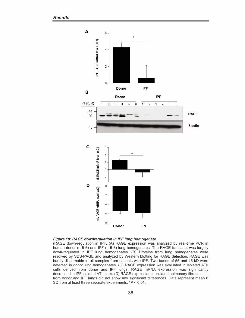
Results
36
Figure 10: RAGE downregulation in IPF lung homogenate. (RAGE down-regulation in IPF. (A) RAGE expression was analyzed by real-time PCR in human donor (n 5 6) and IPF (n 5 6) lung homogenates. The RAGE transcript was largely down-regulated in IPF lung homogenates. (B) Proteins from lung homogenates were resolved by SDS-PAGE and analyzed by Western blotting for RAGE detection. RAGE was hardly discernable in all samples from patients with IPF. Two bands of 55 and 45 kD were detected in donor lung homogenates. (C) RAGE expression was evaluated in isolated ATII cells derived from donor and IPF lungs. RAGE mRNA expression was significantly decreased in IPF isolated ATII cells. (D) RAGE expression in isolated pulmonary fibroblasts from donor and IPF lungs did not show any significant differences. Data represent mean 6 SD from at least three separate experiments; *P < 0.01.

Results
37
homogenates as shown by western blotting. Two major isoforms (55 kDa and
45 kDa) were identified in donor lung homogenates. In contrast, little RAGE
was discernable in IPF lung extracts (Fig. 10B). Moreover, there was a
significant downregulation of RAGE mRNA levels in IPF derived ATII cells
(n=4) as compared to donor ATII cells (n=2) (Fig. 10C). No significant
changes were observed between IPF (n=3) and donor (n=3) isolated
fibroblasts (Fig. 10D).
Figure 11 RAGE downregulation in the bleomycin model. Lung homogenates from saline- and bleomycin-treated mice were analyzed for RAGE expression at the mRNA and protein levels. (A) RNA samples from saline- and bleomycin-treated mice (n 5 3) were subjected to RT-PCR. The RAGE mRNA was amplified in all samples, independent of the time period of bleomycin exposure. No significant changes at the mRNA level were observed after bleomycin treatment. (B) RAGE was significantly down-regulated at the protein level in the bleomycin-treated mice (n 5 3) in comparison with saline-treated mice, as demonstrated by Western blotting.

Results
38
5.4 RAGE Expression in the bleomycin mouse model of lung fibrosis
To study possible mechanistic relationships between pulmonary fibrosis and
the downregulation or loss of RAGE, an established mouse model was
employed in which pulmonary fibrosis was provoked by bleomycin inhalation.
In bleomycin-treated mice, no significant decrease in RAGE expression was
noted at the mRNA level (Fig. 11A). In contrast, at the protein level, RAGE
was significantly downregulated in bleomycin treated mice, exemplified by the
appearance of very weak protein bands upon western blotting (Fig. 11B).
These data are consistent with the observations made on RAGE protein
expression in IPF patient lungs. In contrast to the human studies, an
additional 50 kDa RAGE variant was detected.
5.5 Influence of Cytokines on RAGE Expression
Since RAGE expression is controlled by cytokines (Tanaka, Yonekura et al.
2000), the influence of TNF-� and TGF-�1 at RAGE mRNA and protein levels
was tested in an alveolar epithelial cell line, A549, and primary human
pulmonary fibroblasts after 24 and 48 hours stimulation. RAGE expression
was significantly altered at the mRNA and protein level by both TNF-� and
TGF-�1 in A549 cells after 48 hours as analyzed by quantitative PCR and
western blotting (Fig. 12A, B).
In pulmonary fibroblasts, RAGE downregulation at the mRNA level was
provoked by TNF-�, and to a lesser extent by TGF-�1 after 24 hour
stimulation (Fig. 13A, B). RAGE expression was decreased at the protein
level after 48 hours by both cytokines full length RAGE was mainly affected.
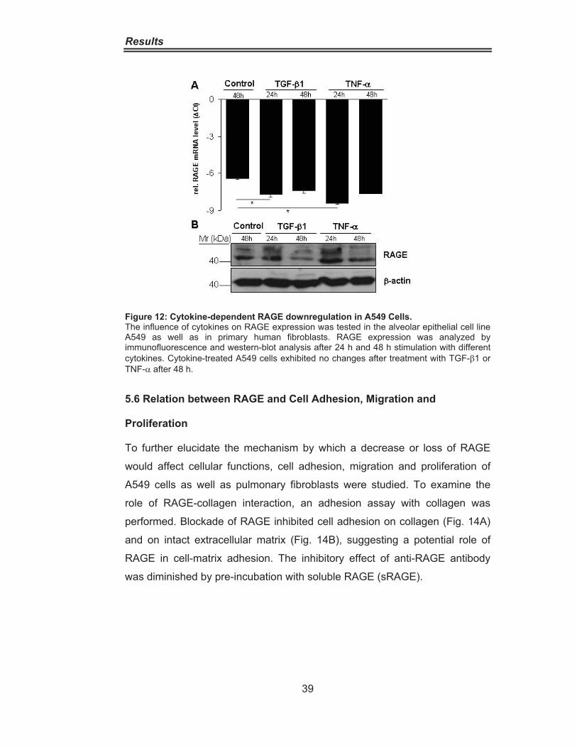
Results
39
Figure 12: Cytokine-dependent RAGE downregulation in A549 Cells. The influence of cytokines on RAGE expression was tested in the alveolar epithelial cell line A549 as well as in primary human fibroblasts. RAGE expression was analyzed by immunofluorescence and western-blot analysis after 24 h and 48 h stimulation with different cytokines. Cytokine-treated A549 cells exhibited no changes after treatment with TGF-�1 or TNF-� after 48 h.
5.6 Relation between RAGE and Cell Adhesion, Migration and
Proliferation
To further elucidate the mechanism by which a decrease or loss of RAGE
would affect cellular functions, cell adhesion, migration and proliferation of
A549 cells as well as pulmonary fibroblasts were studied. To examine the
role of RAGE-collagen interaction, an adhesion assay with collagen was
performed. Blockade of RAGE inhibited cell adhesion on collagen (Fig. 14A)
and on intact extracellular matrix (Fig. 14B), suggesting a potential role of
RAGE in cell-matrix adhesion. The inhibitory effect of anti-RAGE antibody
was diminished by pre-incubation with soluble RAGE (sRAGE).

Results
40
Figure 13:Cytokine-dependent RAGE downregulation in pulmonary fibroblasts. The influence of cytokines on RAGE expression was tested in primary human fibroblasts. RAGE expression was analyzed by immunofluorescence and western-blot analysis after 24 h and 48 h stimulation with different cytokines. Cytokine-treated fibroblasts were analyzed after 24 h. After stimulation with TNF-� and TGF-�1, the expression of RAGE was decreased. TGF-�1 and TNF-� exposure significantly decreased RAGE expression after 48h. Data represent mean ± SD. from at least three separate experiments. * p 0.05.
In contrast, control IgG and sRAGE had no influence on cell adhesion,
indicating that the full length RAGE is essential for its adhesive function. In
addition, knock down of RAGE by specific siRNA led to increased
proliferation of epithelial cells (A549) and fibroblasts as demonstrated by cell
counting (Fig. 15 B,C).
Keratinocyte growth factor (KGF) and TGF-�1 served as positive controls for
cell proliferation of A549 and pulmonary fibroblasts, respectively. RAGE
knock down resulted in increased migration of fibroblasts and epithelial cells
(A549) as shown by chemotaxis migration assay (Fig. 15 D). Hepatocyte
growth factor (for epithelial cells) and TGF-�1 (for fibroblast) served as
positive controls. Undirected chemokinesis was tested by a scratch wound
healing assay with siRNA transfected A549 cells and pulmonary fibroblasts.
Cells transfected with RAGE specific siRNA migrated faster inducing closure
of the scratch, in comparison to scrambled siRNA transfected cells after 24
hours (Fig. 16). siRNA knock down of RAGE in A549 cells affected mainly
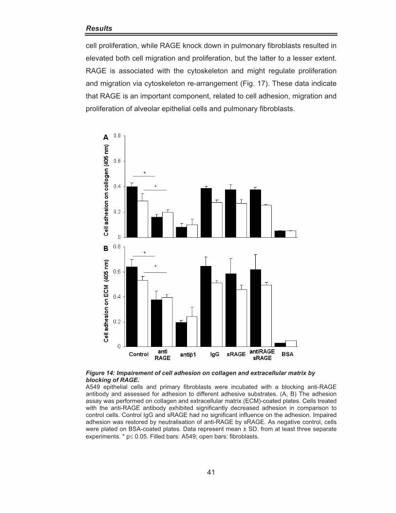
Results
41
cell proliferation, while RAGE knock down in pulmonary fibroblasts resulted in
elevated both cell migration and proliferation, but the latter to a lesser extent.
RAGE is associated with the cytoskeleton and might regulate proliferation
and migration via cytoskeleton re-arrangement (Fig. 17). These data indicate
that RAGE is an important component, related to cell adhesion, migration and
proliferation of alveolar epithelial cells and pulmonary fibroblasts.
Figure 14: Impairement of cell adhesion on collagen and extracellular matrix by blocking of RAGE. A549 epithelial cells and primary fibroblasts were incubated with a blocking anti-RAGE antibody and assessed for adhesion to different adhesive substrates. (A, B) The adhesion assay was performed on collagen and extracellular matrix (ECM)-coated plates. Cells treated with the anti-RAGE antibody exhibited significantly decreased adhesion in comparison to control cells. Control IgG and sRAGE had no significant influence on the adhesion. Impaired adhesion was restored by neutralisation of anti-RAGE by sRAGE. As negative control, cells were plated on BSA-coated plates. Data represent mean ± SD. from at least three separate experiments. * p 0.05. Filled bars: A549; open bars: fibroblasts.
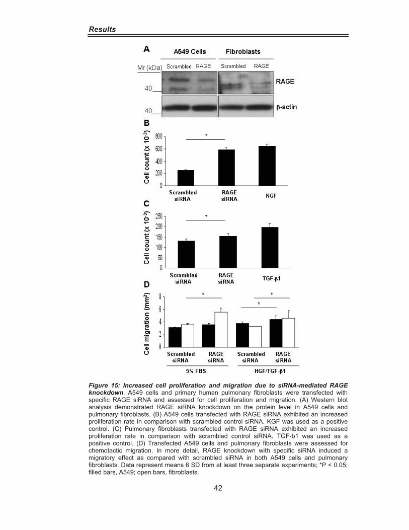
Results
42
Figure 15: Increased cell proliferation and migration due to siRNA-mediated RAGE knockdown. A549 cells and primary human pulmonary fibroblasts were transfected with specific RAGE siRNA and assessed for cell proliferation and migration. (A) Western blot analysis demonstrated RAGE siRNA knockdown on the protein level in A549 cells and pulmonary fibroblasts. (B) A549 cells transfected with RAGE siRNA exhibited an increased proliferation rate in comparison with scrambled control siRNA. KGF was used as a positive control. (C) Pulmonary fibroblasts transfected with RAGE siRNA exhibited an increased proliferation rate in comparison with scrambled control siRNA. TGF-b1 was used as a positive control. (D) Transfected A549 cells and pulmonary fibroblasts were assessed for chemotactic migration. In more detail, RAGE knockdown with specific siRNA induced a migratory effect as compared with scrambled siRNA in both A549 cells and pulmonary fibroblasts. Data represent means 6 SD from at least three separate experiments; *P < 0.05; filled bars, A549; open bars, fibroblasts.

Results
43
Figure 16: Increased cell migration due to siRNA-mediated RAGE knock down. A549 cells and primary human pulmonary fibroblasts were transfected with specific RAGE siRNA and assessed for cell migration. (A) A549 cells transfected with RAGE siRNA exhibited an increased migration rate, in comparison to scrambled control siRNA in a Boyden chamber. (B,C) RAGE knockdown induced wound closure. A549 cells and primary human pulmonary fibroblasts were transfected with RAGE-specific siRNA and assessed for wound healing assay. (B) A549 cells transfected with RAGE siRNA exhibited increased migration and wound closure in comparison with scrambled siRNA-transfected cells. (C) Pulmonary fibroblasts exhibited increased migration and wound closure in comparison with scrambled siRNA-transfected cells. Data represent means 6 SD from at least three separate experiments. Scale bar 5 100 mm; *P < 0.05.

Results
44
Figure 17: RAGE is associated with the cytoskeleton. A549 cells were separated by subcellular fractionation and analyzed by western-blot analysis. RAGE was detected in the cytoplasm, the membrane/organelle and predominantly in the cytoskeleton fraction.
Discussion
45
6. Discussion
The physiological and pathophysiological role of RAGE in the pulmonary
system remains elusive. The following section will reflect the results of the
current thesis and will discuss them in the context with the existing literature.
6.1 The role of RAGE in pulmonary fibrosis
In the present work, the relation between RAGE downregulation in fibrotic
lungs and the loss of control of cell adhesion, migration and proliferation,
alterations believed to be involved in fibrosis development, was described.
These data provide new mechanistic insight into the regulatory role of RAGE
in cell communication in the lung and are supported by recent findings of
other investigators (Hanford LE 2003; Englert, Hanford et al. 2008).
Decreased RAGE expression in lungs of IPF patients and in aveolar type II
cells (AT II) in vitro are in accordance with the observed alterations in the
animal model of bleomycin-induced lung fibrosis (Hanford LE 2003).
As demonstrated in this study, downregulation and/or loss of RAGE
expression by TNF-� and TGF-�1 in primary human pulmonary fibroblasts
and A549 cells as well as its involvement in cell adhesion appear to be
processes linked to the onset and/or progression of fibrosis pathogenesis.
Here, the profibrotic cytokine TNF-� plays a critical role, possibly driving the
inflammatory phase into fibrosis (Miyazaki, Araki et al. 1995; Oikonomou N
2006). Another hallmark of the pathogenesis of pulmonary fibrosis are the
alterations in cellular phenotype and functions, accompanied by changes in
cell adhesion and communication of lung epithelial cells. The results
demonstrate RAGE protein localization mainly on AT I cells, but also on AT II
cells, which contain high amounts of RAGE mRNA. It remains to be further
established whether RAGE is a specific marker for AT I or AT II epithelial
cells (Katsuoka F 1997; Fehrenbach H 1998). Whether these differences are
related in any way to the transition from AT II to AT I cells remains to be
elucidated. However, it was recently shown the RAGE is upregulated during
the transition from AT I to AT II cells (Wang, Edeen et al. 2007). Based on the

Discussion
46
these findings, the following function is proposed for RAGE in alveolar
epithelial cells.
Under normal physiological conditions AT II cells contain low level of RAGE
protein, although there is appreciable RAGE mRNA detactable, allowing the
cells to proliferate, and to reepithelialzate. However, during
transdifferentiation of AT II cells to AT I cellls, there is increase on RAGE
protein levels (Dobbs ref.), which by utelizing with collagen in the ECM,
immobilieses AT I cells and thus contributing to there non-proliferative
phenotype. RAGE knock down in AT II-like A549 cells as well as pulmonary
fibroblasts resulted in elevated proliferative responsiveness to serum and
increased cellular motility. Interestingly, RAGE knock down had a more
proliferative effect on A549 cells and a higher migratory effect on pulmonary
fibroblasts, indicative for a cell type-specific role of RAGE as well. Moreover,
the cell phenotype changes provoked by RAGE downregulation in lung
fibroblasts are reminiscent of morphological alterations of these cells in IPF,
demonstrating increased proliferation and migration in comparison to
fibroblasts derived from donors (Suganuma, Sato et al. 1995; Ramos,
Montano et al. 2001; Moodley, Scaffidi et al. 2003). Together, RAGE
downregulation appears to be associated with both, cytokine and adhesion-
related cellular changes and may thus be mechanistically linked to the switch
of chronic inflammation to fibrosis in fibrotic lung disease.
The adhesive properties of RAGE in mediating cell-matrix contacts described
here and in previous reports (Fehrenbach H 1998; Demling N 2006) appear
to be similar and comparable to integrin-mediated adhesion of lung epithelial
cells, and loss of RAGE is associated with disturbed cellular contacts.
Moreover, preliminary data from our laboratory indicate a tight linkage of
RAGE to cytoskeletal elements in lung epithelial cells, suggesting that RAGE
provides a regulatory adhesion function linked to cytoskeleton-related
signaling systems, also characteristic for integrin functions. It may thus be
hypothesized that signaling pathways that would lead to inside-out signaling
to affect RAGE function may not be unlikely. Since RAGE was described by
our group as the major inflammation-related counter-receptor on endothelial
cells for recognition of ß2-integrins on leukocytes (Chavakis, Bierhaus et al.
Discussion
47
2003), it remains to be investigated whether loss of RAGE in the aleolar
epithelium may lead to disturbances in inflammatory cell interactions in the
lung as well. Although the clarification of the cell-stabilizing role of RAGE in
the lung requires further work, RAGE appears to serve an "opposite" role in
the vasculature, where it becomes upregulated upon inflammatory processes
and promotes e.g. leukocyte recruitment into diseased tissue (Chavakis,
Bierhaus et al. 2003).
Although the bleomycin model of lung fibrosis used in this study may not
necessarily reflect all alterations of fibrosis pathogenesis as observed in
humans, data from this in vivo model are in accordance with our in vitro and
ex vivo data: A major loss of RAGE expression was seen in these mice that
is supported by recent findings from Englert et al. (Englert, Hanford et al.
2008) who indicated that RAGE-/- mice developed more severe asbestos-
induced lung fibrosis than wild-type controls, and underlined our data using a
different animal model for lung fibrosis. In contrast, He et al. (He, Kubo et al.
2007) reported that RAGE-/- mice were protected from bleomycin-induced
lung fibrosis. Furthermore, it has been shown that RAGE levels are
decreased in the alveolar epithelium after in vitro treatment of rat lung
sections with CdCl2 and TGF-�1 (Kasper, Seidel et al. 2004).
Our observations agree with and further complement the findings that RAGE
is downregulated in non-small cell lung carcinoma, and its expression impairs
the proliferative stimulus of lung fibroblasts on lung cancer cells (Bartling,
Hofmann et al. 2005; Bartling, Demling et al. 2006). Thus, loss of RAGE
leads to increased cellular proliferation and migration of pulmonary cells in
association with different pathologies, and (therapeutic) prevention of RAGE
downregulation may serve as a potential antagonizing mechanism in the
diseased lung
6.2 RAGE as a biomarker for lung injury
The finding of biomarkers for certain lung diseases is essential for a
diagnostic tool and therapy development. Due to its high expression in lung
homogenate as well as the appearance of its soluble form in bronchial
alveolar fluid (BAL), RAGE was studied as a marker for acute lung injury

Discussion
48
(ALI). ALI is often seen as part of a systemic inflammatory process, such as
sepsis, with lung manifestations such as widespread destruction of the
capillary endothelium, extravascation of protein rich fluid and interstitial
edema. Indeed, in an animal model of acute lung injury (ALI), evaluated
levels of sRAGE were associated with the severity of the experimentally
induced lung injury (Uchida, Shirasawa et al. 2006). in addition, sRAGE was
increased in serum samples from patients with acute respiratory distress
syndrome (ARDS), a more severe form of ALI (Calfee, Ware et al. 2008). The
underlying mechanism for this phenomenon is not clear. It could be noted
that under inflammatory conditions metalloproteases such as MMP-9 and
ADAM10 proteolytically dissociate RAGE from the epithelial membrane or
that epithelial injury-induced apoptosis or necrosis leads to sRAGE release
into the alveolar space. The present results show that RAGE is dramatically
downregulated in lung homogenate and in alveolar type II cells from IPF
patients as well as in the bleomycin model. Furthermore, sRAGE level in BAL
from IPF patients is decreased as well (Bargagli, Penza et al. 2008),
indicating that RAGE expression is different in pulmonary fibrosis than in
acute lung injury. However, RAGE might not be useful as a biomarker for IPF
since downregulation of RAGE was observed as well in lung adenocarcinoma
(Hofmann, Hansen et al. 2004) (Stav, Bar et al. 2007).
In respect to cell-type specific markers, RAGE was proposed to be an AT I
marker (Fehrenbach, Kasper et al. 1998; Shirasawa, Fujiwara et al. 2004).
While it remains to be further established whether RAGE is a specific marker
for AT I or AT II epithelial cells (Katsuoka F 1997; Fehrenbach H 1998), the
present results demonstrate RAGE protein localization mainly in AT I cells,
but also in AT II cells, which contain high amounts of RAGE mRNA. Whether
these differences are related in any way to the transition from AT II to AT I
cells remains to be elucidated.
6.3 RAGE-ligand signaling in the lung
The role of RAGE and its ligands in maintaining and amplifying inflammation
has been investigated in several tissues. RAGE-mediated tissue damage is
being involved in chronic inflammatory pathology of multiple organs. In the

Discussion
49
lung, several RAGE ligands were indentified under inflammatory conditions.
AGE accumulation was demonstrated in lung tissue after smoke exposure,
and cigarette smoke extract induced RAGE expression via early growth
factor-1 (Egr-1) in vitro (Morbini, Villa et al. 2006; Reynolds, Kasteler et al.
2008). However, the situation in vivo is by far more complex. AGE in the
alveolar space have to face several surfactant proteins with AGE-binding
properties such as lysozyme and lactoferrin which both contains AGE-binding
domains and might contribute to AGE clearence (Li, Tan et al. 1995). In
addition, high sRAGE levels are present in BAL and might prevent AGE-
RAGE interaction on the alveolar epithelium.
The situation with the S100 proteins is similar, S100A12 (EN-RAGE) and
S100B are expressed under inflammatory conditions and in acute lung injury
(Morbini, Villa et al. 2006; Wittkowski, Sturrock et al. 2007). However,
whether this association indicates a potential pro-inflammatory signaling
pathway remain to be unsolved. The most likely interaction could occur
between epithelial cell-expressed RAGE and HMGB1, which has the highest
affinity for RAGE. HMGB1 is released from injured and necrotic cells. In
addition, the concentration of HMGB1 in sepsis, which is associated with
acute lung injury, as well as in bleomycin-induced pulmonary fibrosis (He,
Kubo et al. 2007), can reach high levels which are unlikely to be blocked
completely by endogenous sRAGE. Indeed, applied HMGB1 leads to
cytokine release and inflammatory cell recruitment to the lung (Abraham,
Arcaroli et al. 2000). However, since RAGE downregulation is taking place
early after bleomycin administration as well the fact the RAGE is
downregulated in IPF patients (present results), it is more likely that the
observed effects are mediated via TLR2 and TLR4 receptors as described
before (Tsung, Klune et al. 2007).
6.4 Potential mechanism of RAGE downregulation
The RAGE promoter which is controlled by three NF-�B binding sites links
the RAGE expression to inflammation. Thus, it is recognized that RAGE
controls inflammation in several diseases (Bierhaus, Humpert et al. 2005).
Surprisingly enough, in IPF patients as well as in the bleomycin mouse model,

Discussion
50
with its acute inflammatory phase, RAGE is largely downregulated. This
phenomenon might be explained by non-classical signaling events.
6.4.1 RAGE downregulation by micro-RNA
In lung homogenate from IPF patients and AT II cells, the RAGE transcripts
were downregulted. One possible explanation is that micro-RNA (miRNA)
might interact with RAGE mRNA and could regulate its expression. miRNA
gene regulation was recently discovered as an additional regulatory
mechanism caused by endogenous, 21-nucleotide-long derived miRNA.
Similar to siRNA, miRNA can regulated gene transcription as well as
translation by degradation or blocking transcripts of translation (Pillai,
Bhattacharyya et al. 2007; Flynt and Lai 2008).
The excat pairing between miRNA and target mRNA trigges degradation
through a mechanism similar to RNA interferance. However, mostly miRNAs
regulate gene expression by imperfect base pairing to the 3’ UTR of the
target mRNA, causing translational repression or exonucleolytic degradation
of target mRNA. The miRNA is incooperated in a ribonucleoprotein complex
(miRNP) which is composed of Argonaute proteins which exhibit nucleolytic
activity. The RAGE encoding ager mRNA transcripts from mouse and human
have several potential binding sites for miRNA, indicating a potential gene
regulatory mechanism. the functions of these miRNAs are not yet described
and need to be further analyzed. However, miRNA-mediated mechanisms
might explain the effect of stable RAGE mRNA expression in mice with
absence of RAGE on the protein level.
6.4.2 RAGE downregulation by proteases
RAGE mRNA transcripts are not significantly downregulated regulated in the
bleomycin model, indicating that other mechanisms are taking place on the
post-transcriptional level. ADAM10 and MMP9 are known proteases which
mediate RAGE shedding and production/processing of sRAGE (Raucci,
Cugusi et al. 2008; Zhang, Bukulin et al. 2008). However, it was recently
reported though that sRAGE is downregulated as well in BAL from IPF
patients (Bargagli, Penza et al. 2008).

Discussion
51
6.4.3 Downregulation of RAGE in relation to caveolae
Another explanation for RAGE downregulation on the protein level could be
due to an indirect mechanism where by membrane proteins which are
associated with RAGE would be targeted by TGF-� or TNF-� leading to their
downregulation together with RAGE. Indeed, it was reported that RAGE is
associated with caveolin-rich membrane fractions (Lisanti, Scherer et al.
1994). Caveolin-1, the main component of caveolae, is a critical regulator of
lung fibrosis because Caveolin-1 limits TGF-� -induced production of
extracellular matrix and restores alveolar epithelial-repair processes.
Caveolin-1 expression was markedly reduced in lung tissue from patients
with idiopathic pulmonary fibrosis and that this reduction was predominant in
alveolar epithelial cells. In addition, fibroblasts had low levels of caveolin-1
expression in patients with idiopathic pulmonary fibrosis (Wang, Zhang et al.
2006; Le Saux, Teeters et al. 2008). Therefore, further investigations should
focus on a possible connection between RAGE with caveolae.
6.4 Involvement of RAGE in epithelial-mesenchymal transition
Beside the involvement of RAGE in cell proliferation, RAGE might be
involved as well in cell transdifferentiation, especially under conditions of
epithelial to mesenchymal transition (EMT). It was reported that AGE can
induce EMT by RAGE activation, inducing TGF-� expression in
tubulointerstitial fibrosis (Oldfield, Bach et al. 2001). In the lung, induction of
EMT by HMGB1 via RAGE in pulmonary fibrosis was suggested (He, Kubo et
al. 2007). However, the early downregulation of RAGE in fibrosis can act, if at
all, only for a very limited period of time in the onset of the fibrogenesis.
Another way for RAGE involvement in EMT can be associated with the
present results, suggesting that beside AT II cells also AT I can undergo EMT.
Following TGF-� stimulation this process would lead to RAGE
downregulation and enables the transformed AT I cells (myofibroblasts) to
proliferate excessively and contributes to fibrosis.
Discussion
52
6.5 RAGE as an adhesion molecule
Chavakis et al. showed a cell-adhesive function for RAGE, based on the
RAGE-Mac-1 interaction, for the first time (Chavakis, Bierhaus et al. 2003).
Furthermore, a role for RAGE was proposed in cytoskeleton reorganization
and involvement in ���3 integrin signaling in osteoclasts (Zhou, Immel et al.
2006). The present results indicate a direct interaction between RAGE and
the cytoskeleton. In addition, the results show that RAGE can interact with
collagen and extracellular matrix. The binding interaction between RAGE and
collagen suggests that RAGE might fullfill mainly adhesion function in the
lung. RAGE is localised at the basolateral membrane of type I cells, indicative
for interaction with collagen of the extracellular membrane (Fehrenbach,
Kasper et al. 1998). Surprisingly enough, RAGE-/- mice shows accumulation
of macrophages in the lung as opposed to wild-type mice after injury,
indicating that RAGE can influence the circulation and adhesiveness of
leukocytes (He, Kubo et al. 2007).
The present results supporting a cell-adhesive function for RAGE and show
an its physiological relevance in the pulmonary system.
Declaration
53
7. Declaration
Herewith, I declare that I have completed this dissertation single-handedly
without the unauthorized help of a second party and only with the assistance
acknowledged therein. I have appropriately acknowledged and referenced all
text passages that are derived literally from or are based on the content of
published or unpublished work of others, and all information that relates to
verbal communications. I have abided by the principles of good scientific
conduct laid down in the charter of the Justus Liebig University of Giessen in
carrying out the investigations described in this dissertation.

Der Lebenslauf wurde aus der elektronischen Version der Arbeit entfernt.
The curriculum vitae was removed from the electronic version of the paper.

Acknowledgements
57
9. Acknowledgements
I would like to express my deep gratitude to my supervisor Prof. Dr. Klaus T.
Preissner who gave me the opportunity to perform my PhD in his laboratory
and selected me for the International Training Research group “Signaling
Mechanism in Lung Physiology and Disease (SMLPD)”. During this time, he
guided me in new directions and let me start to think “outside” the cell. Most
of all, he gave me the freedom to perform research in a Humboldt’sche way
which I highly appreciated and very much enjoyed.
My sincere gratefulness applies to Prof. Dr. Oliver Eickelberg who introduced
me to the pulmonary medicine and put great effort in teaching me how
science works. Prof. Dr. Oliver Eickelberg encourages the spirit of science to
me and the importance of professional ethics. Especially, I would like to thank
him for the annual retreat where I could present my research to invited
international experts in the field. These scientific workshops were the ideal
platform for an intense international exchange with scientists on an informal
basis.
Furthermore, I thank the Molecular Biology and Medicine of the Lung (MBML)
graduate program and my mentors Prof. Dr. Oliver Eickelberg, Dr. Rory Morty
and Dr. Melanie Königshoff for scientific education as good as it gets.
I would like to sincerely thank Prof. Dr. Werner Seeger who established a
creative and recognized community in lung science and who demonstrates
that the faith in God and science has not to be contradictory.
I thank Prof. Dr. Michael Brownlee from the Albert Einstein College of
Medicine, Yeshiva University, who integrated me in his group and treated me
as a coequal scientist from the first day. I admire his scientific integrity and
his craft to master life.
I specially thank Prof. Dr. Pingoud for his efforts to enable me, as a
biochemist, to achieve the doctor rerum naturalium from the Justus-Liebig
University.
I thank the Deutsche Forschungsgemeinschaft (DFG) for generous financial
support in Giessen and New York.

Acknowledgements
58
I am deeply indebted to my honorable parents for their enormous support in
good and bad times throughout my entire life and to my grandparents for their
tremendous support and education in many ways.
Special thanks to Heinz Wummel, my mentor in statistics, for his enduring
patience.
I am deeply grateful for the help and support of Fotini Kouri who always
believed in me and never let me down.
Finally, I thank all my friends around the world, thanks for having you here
and there.

References
59
10. References
(2002). "American Thoracic Society/European Respiratory Society International Multidisciplinary Consensus Classification of the Idiopathic Interstitial Pneumonias . This Joint Statement of the American Thoracic Society (ATS), and the European Respiratory Society (ERS) was adopted by the ATS Board of Directors, June 2001 and by The ERS Executive Committee, June 2001." Am. J. Respir. Crit. Care Med. 165: 277-304.
Abel, M., U. Ritthaler, et al. (1995). "Expression of receptors for advanced glycosylated end-products in renal disease." Nephrol Dial Transplant 10: 1662-7.
Abraham, E., J. Arcaroli, et al. (2000). "HMG-1 as a mediator of acute lung inflammation." J Immunol 165: 2950-4.
Adamson, I. Y. and D. H. Bowden (1974). "The pathogenesis of bloemycin-induced pulmonary fibrosis in mice." Am J Pathol 77: 185-97.
Adamson, I. Y. and D. H. Bowden (1974). "The type 2 cell as progenitor of alveolar epithelial regeneration. A cytodynamic study in mice after exposure to oxygen." Lab Invest 30: 35-42.
Allam, J. S. and A. H. Limper (2006). "Idiopathic pulmonary fibrosis: is it a familial disease?" Curr Opin Pulm Med 12: 312-7.
Arancio, O., H. P. Zhang, et al. (2004). "RAGE potentiates Abeta-induced perturbation of neuronal function in transgenic mice." EMBO J 23: 4096-105.
Armanios, M. Y., J. J. Chen, et al. (2007). "Telomerase mutations in families with idiopathic pulmonary fibrosis." N Engl J Med 356: 1317-26.
Bargagli, E., F. Penza, et al. (2008). "Controversial role of RAGE in the pathogenesis of idiopathic pulmonary fibrosis." Respir Physiol Neurobiol.
Bartling, B., N. Demling, et al. (2006). "Proliferative Stimulus of Lung Fibroblasts on Lung Cancer Cells Is Impaired by the Receptor for Advanced Glycation End-Products." Am. J. Respir. Cell Mol. Biol. 34: 83-91.
Bartling, B., H.-S. Hofmann, et al. (2005). "Down-regulation of the receptor for advanced glycation end-products (RAGE) supports non-small cell lung carcinoma." Carcinogenesis 26: 293-301.
Bierhaus, A., K. M. Haslbeck, et al. (2004). "Loss of pain perception in diabetes is dependent on a receptor of the immunoglobulin superfamily." J Clin Invest 114: 1741-51.
Bierhaus, A., P. M. Humpert, et al. (2005). "Understanding RAGE, the receptor for advanced glycation end products." J Mol Med 83: 876-86.
Bierhaus, A., S. Schiekofer, et al. (2001). "Diabetes-associated sustained activation of the transcription factor nuclear factor-kappaB." Diabetes 50: 2792-808.
Bonniaud, P., P. J. Margetts, et al. (2003). "Adenoviral gene transfer of connective tissue growth factor in the lung induces transient fibrosis." Am J Respir Crit Care Med 168: 770-8.

References
60
Boulanger, E., M. P. Wautier, et al. (2002). "AGEs bind to mesothelial cells via RAGE and stimulate VCAM-1 expression." Kidney Int 61: 148-56.
Bozelka, B. E., P. Sestini, et al. (1983). "A murine model of asbestosis." Am J Pathol 112: 326-37.
Brownlee, M. (1995). "Advanced protein glycosylation in diabetes and aging." Annu Rev Med 46: 223-34.
Calfee, C. S., L. B. Ware, et al. (2008). "Plasma receptor for advanced glycation end products and clinical outcomes in acute lung injury." Thorax 63: 1083-9.
Chavakis, T., A. Bierhaus, et al. (2003). "The pattern recognition receptor (RAGE) is a counterreceptor for leukocyte integrins: a novel pathway for inflammatory cell recruitment." J Exp Med 198: 1507-15.
Chavakis, T., S. M. Kanse, et al. (2000). "Different mechanisms define the antiadhesive function of high molecular weight kininogen in integrin- and urokinase receptor-dependent interactions." Blood 96: 514-522.
Christensen, P. J., R. E. Goodman, et al. (1999). "Induction of lung fibrosis in the mouse by intratracheal instillation of fluorescein isothiocyanate is not T-cell-dependent." Am J Pathol 155: 1773-9.
Csiszar, A. and Z. Ungvari (2008). "Endothelial dysfunction and vascular inflammation in type 2 diabetes: interaction of AGE/RAGE and TNF-alpha signaling." Am J Physiol Heart Circ Physiol 295: H475-6.
Dasari, V., M. Gallup, et al. (2006). "Epithelial-mesenchymal transition in lung cancer: is tobacco the "smoking gun"?" Am J Respir Cell Mol Biol 35: 3-9.
Dattilo, B. M., G. Fritz, et al. (2007). "The extracellular region of the receptor for advanced glycation end products is composed of two independent structural units." Biochemistry 46: 6957-70.
Deane, R., Z. Wu, et al. (2004). "RAGE (yin) versus LRP (yang) balance regulates alzheimer amyloid beta-peptide clearance through transport across the blood-brain barrier." Stroke 35: 2628-31.
Demedts, M. and U. Costabel (2002). "ATS/ERS international multidisciplinary consensus classification of the idiopathic interstitial pneumonias." Eur Respir J 19: 794-6.
Demling N, E. C., Kasper M, Laue M, Knels L, Rieber EP (2006). "Promotion of cell adherence and spreading: a novel function of RAGE, the highly selective differentiation marker of human alveolar epithelial type I cells." Cell Tissue Res 323: 475-88.
Ding, Y., A. Kantarci, et al. (2007). "Activation of RAGE induces elevated O2- generation by mononuclear phagocytes in diabetes." J Leukoc Biol 81: 520-7.
Dumitriu, I. E., P. Baruah, et al. (2005). "Requirement of HMGB1 and RAGE for the maturation of human plasmacytoid dendritic cells." Eur J Immunol 35: 2184-90.
Effros, R. (2006). Anatomy, development, and physiology of the lungs, GI Motility online
Englert, J. M., L. E. Hanford, et al. (2008). "A Role for the Receptor for Advanced Glycation End Products in Idiopathic Pulmonary Fibrosis." Am J Pathol.
Evans, R. A., Y. C. Tian, et al. (2003). "TGF-beta1-mediated fibroblast-myofibroblast terminal differentiation-the role of Smad proteins." Exp

References
61
Cell Res 282: 90-100. Fang X, S. Y., Hirsch J, Galietta LJ, Pedemonte N, Zemans RL, Dolganov G,
Verkman AS, Matthay MA. (2006). "Contribution of CFTR to apical-basolateral fluid transport in cultured human alveolar epithelial type II cells." Am J Physiol Lung Cell Mol Physiol 290: L242-249.
Fehrenbach, H., M. Kasper, et al. (1998). "Receptor for advanced glycation endproducts (RAGE) exhibits highly differential cellular and subcellular localisation in rat and human lung." Cell Mol Biol (Noisy-le-grand) 44: 1147-57.
Fehrenbach H, K. M., Tschernig T, Shearman MS, Schuh D, Muller M. (1998). "Receptor for advanced glycation endproducts (RAGE) exhibits highly differential cellular and subcellular localisation in rat and human lung." Cell Mol Biol (Noisy-le-grand). 44: 1147-57.
Fiuza, C., M. Bustin, et al. (2003). "Inflammation-promoting activity of HMGB1 on human microvascular endothelial cells." Blood 101: 2652-60.
Flynt, A. S. and E. C. Lai (2008). "Biological principles of microRNA-mediated regulation: shared themes amid diversity." Nat Rev Genet 9: 831-42.
Forbes, J. M., V. Thallas, et al. (2003). "The breakdown of preexisting advanced glycation end products is associated with reduced renal fibrosis in experimental diabetes." FASEB J 17: 1762-4.
Franko, A. J. and J. Sharplin (1994). "Development of fibrosis after lung irradiation in relation to inflammation and lung function in a mouse strain prone to fibrosis." Radiat Res 140: 347-55.
Galichet, A., M. Weibel, et al. (2008). "Calcium-regulated intramembrane proteolysis of the RAGE receptor." Biochem Biophys Res Commun 370: 1-5.
Gao, X., H. Zhang, et al. (2008). "AGE/RAGE produces endothelial dysfunction in coronary arterioles in type 2 diabetic mice." Am J Physiol Heart Circ Physiol 295: H491-8.
Gauldie, J. and M. Kolb (2008). "Animal models of pulmonary fibrosis: how far from effective reality?" Am J Physiol Lung Cell Mol Physiol 294: L151.
Gomperts, B. N. and R. M. Strieter (2007). "Fibrocytes in lung disease." J Leukoc Biol 82: 449-56.
Gordon, S. (2002). "Pattern recognition receptors: doubling up for the innate immune response." Cell 111: 927-30.
Gossen, M. and H. Bujard (1992). "Tight control of gene expression in mammalian cells by tetracycline-responsive promoters." Proc Natl Acad Sci U S A 89: 5547-51.
Gross, T. J. and G. W. Hunninghake (2001). "Idiopathic pulmonary fibrosis." N Engl J Med 345: 517-25.
Hammes, H. P., A. Alt, et al. (1999). "Differential accumulation of advanced glycation end products in the course of diabetic retinopathy." Diabetologia 42: 728-36.
Hammes, H. P., A. Weiss, et al. (1996). "Modification of vitronectin by advanced glycation alters functional properties in vitro and in the diabetic retina." Lab Invest 75: 325-38.
Hammond, T. G., P. J. Verroust, et al. (1994). "Heavy endosomes isolated from the rat renal cortex show attributes of intermicrovillar clefts." Am J

References
62
Physiol 267: F516-27. Hanford, L. E., J. J. Enghild, et al. (2004). "Purification and Characterization
of Mouse Soluble Receptor for Advanced Glycation End Products (sRAGE)." J. Biol. Chem. 279: 50019-50024.
Hanford LE, F. C., Shaefer LM, Enghild JJ, Valnickova Z, Oury TD. (2003). "Regulation of receptor for advanced glycation end products during bleomycin-induced lung injury." Am J Respir Cell Mol Biol. 29: S77-81.
He, M., H. Kubo, et al. (2007). "The role of the receptor for advanced glycation end-products in lung fibrosis." Am J Physiol Lung Cell Mol Physiol 293: L1427-36.
Heidland, A., K. Sebekova, et al. (2001). "Advanced glycation end products and the progressive course of renal disease." Am J Kidney Dis 38: S100-6.
Hofmann, H. S., G. Hansen, et al. (2004). "Discrimination of human lung neoplasm from normal lung by two target genes." Am J Respir Crit Care Med 170: 516-9.
Hofmann, M. A., S. Drury, et al. (1999). "RAGE mediates a novel proinflammatory axis: a central cell surface receptor for S100/calgranulin polypeptides." Cell 97: 889-901.
Hori, O., J. Brett, et al. (1995). "The receptor for advanced glycation end products (RAGE) is a cellular binding site for amphoterin. Mediation of neurite outgrowth and co-expression of rage and amphoterin in the developing nervous system." J Biol Chem 270: 25752-61.
Hou, F. F., H. Ren, et al. (2004). "Enhanced expression of receptor for advanced glycation end products in chronic kidney disease." J Am Soc Nephrol 15: 1889-96.
Huttunen, H. J., J. Kuja-Panula, et al. (2000). "Coregulation of neurite outgrowth and cell survival by amphoterin and S100 proteins through receptor for advanced glycation end products (RAGE) activation." J Biol Chem 275: 40096-105.
Hyogo, H. and S. Yamagishi (2008). "Advanced glycation end products (AGEs) and their involvement in liver disease." Curr Pharm Des 14: 969-72.
Kasper, M., D. Seidel, et al. (2004). "Early signs of lung fibrosis after in vitro treatment of rat lung slices with CdCl2 and TGF-beta1." Histochem Cell Biol 121: 131-40.
Katsuhiko Asanuma, E. Y.-A., Christian Faul, Yasuhiko Tomino, Kwanghee Kim and a. P. Mundel (2006). "Synaptopodin orchestrates actin organization and cell motility via regulation of RhoA signalling." Nat Cell Biol. 8: 485-91.
Katsuoka F, K. Y., Arai T, Imuta H, Fujiwara M, Kanma H, Yamashita K. (1997). "Type II alveolar epithelial cells in lung express receptor for advanced glycation end products (RAGE) gene." Biochem Biophys Res Commun. 238: 512-6.
Khalil, N., Y. D. Xu, et al. (2005). "Proliferation of pulmonary interstitial fibroblasts is mediated by transforming growth factor-beta1-induced release of extracellular fibroblast growth factor-2 and phosphorylation of p38 MAPK and JNK." J Biol Chem 280: 43000-9.
Kierszenbaum, A. (2007). Histology and Cell Biology. Philadelphia, Mosby Elsevier.
References
63
Kokkola, R., A. Andersson, et al. (2005). "RAGE is the major receptor for the proinflammatory activity of HMGB1 in rodent macrophages." Scand J Immunol 61: 1-9.
Kuhn, C. and J. A. McDonald (1991). "The roles of the myofibroblast in idiopathic pulmonary fibrosis. Ultrastructural and immunohistochemical features of sites of active extracellular matrix synthesis." Am J Pathol 138: 1257-65.
Le Saux, O., K. Teeters, et al. (2008). "The role of caveolin-1 in pulmonary matrix remodeling and mechanical properties." Am J Physiol Lung Cell Mol Physiol 295: L1007-17.
Li, J. H., X. R. Huang, et al. (2004). "Advanced glycation end products activate Smad signaling via TGF-beta-dependent and independent mechanisms: implications for diabetic renal and vascular disease." FASEB J 18: 176-8.
Li, Y. M., A. X. Tan, et al. (1995). "Antibacterial activity of lysozyme and lactoferrin is inhibited by binding of advanced glycation-modified proteins to a conserved motif." Nat Med 1: 1057-61.
Liliensiek, B., M. A. Weigand, et al. (2004). "Receptor for advanced glycation end products (RAGE) regulates sepsis but not the adaptive immune response." J Clin Invest 113: 1641-50.
Liotta, L. A. and T. Clair (2000). "Cancer. Checkpoint for invasion." Nature 405: 287-8.
Lisanti, M. P., P. E. Scherer, et al. (1994). "Characterization of caveolin-rich membrane domains isolated from an endothelial-rich source: implications for human disease." J Cell Biol 126: 111-26.
Lotze, M. T. and K. J. Tracey (2005). "High-mobility group box 1 protein (HMGB1): nuclear weapon in the immune arsenal." Nat Rev Immunol 5: 331-42.
Maher, T. M., A. U. Wells, et al. (2007). "Idiopathic pulmonary fibrosis: multiple causes and multiple mechanisms?" Eur Respir J 30: 835-9.
Masszi, A., C. Di Ciano, et al. (2003). "Central role for Rho in TGF-beta1-induced alpha-smooth muscle actin expression during epithelial-mesenchymal transition." Am J Physiol Renal Physiol 284: F911-24.
Mehrad, B., M. D. Burdick, et al. (2007). "Circulating peripheral blood fibrocytes in human fibrotic interstitial lung disease." Biochem Biophys Res Commun 353: 104-8.
Miyazaki, Y., K. Araki, et al. (1995). "Expression of a tumor necrosis factor-alpha transgene in murine lung causes lymphocytic and fibrosing alveolitis. A mouse model of progressive pulmonary fibrosis." J Clin Invest 96: 250-9.
Moodley, Y. P., A. K. Scaffidi, et al. (2003). "Fibroblasts isolated from normal lungs and those with idiopathic pulmonary fibrosis differ in interleukin-6/gp130-mediated cell signaling and proliferation." Am J Pathol 163: 345-54.
Moore, B. B. and C. M. Hogaboam (2008). "Murine models of pulmonary fibrosis." Am J Physiol Lung Cell Mol Physiol 294: L152-60.
Morbini, P., C. Villa, et al. (2006). "The receptor for advanced glycation end products and its ligands: a new inflammatory pathway in lung disease?" Mod Pathol 19: 1437-45.
Nadrous, H. F., J. H. Ryu, et al. (2004). "Impact of angiotensin-converting
References
64
enzyme inhibitors and statins on survival in idiopathic pulmonary fibrosis." Chest 126: 438-46.
Neeper, M., A. M. Schmidt, et al. (1992). "Cloning and expression of a cell surface receptor for advanced glycosylation end products of proteins." J Biol Chem 267: 14998-5004.
Nogee, L. M., A. E. Dunbar, 3rd, et al. (2001). "A mutation in the surfactant protein C gene associated with familial interstitial lung disease." N Engl J Med 344: 573-9.
Oikonomou N, H. V., Zalevsky J, Valavanis C, Kotanidou A, Szymkowski DE, Kollias G, Aidinis V. (2006). "Soluble TNF mediates the transition from pulmonary inflammation to fibrosis." PLoS ONE 1(e108).
Oldfield, M. D., L. A. Bach, et al. (2001). "Advanced glycation end products cause epithelial-myofibroblast transdifferentiation via the receptor for advanced glycation end products (RAGE)." J Clin Invest 108: 1853-63.
Pardo, A. and M. Selman (2002). "Molecular mechanisms of pulmonary fibrosis." Front Biosci 7: d1743-61.
Peinado, H., D. Olmeda, et al. (2007). "Snail, Zeb and bHLH factors in tumour progression: an alliance against the epithelial phenotype?" Nat Rev Cancer 7: 415-28.
Petersen, O. W., H. L. Nielsen, et al. (2003). "Epithelial to mesenchymal transition in human breast cancer can provide a nonmalignant stroma." Am J Pathol 162: 391-402.
Phillips, R. J., M. D. Burdick, et al. (2004). "Circulating fibrocytes traffic to the lungs in response to CXCL12 and mediate fibrosis." J Clin Invest 114: 438-46.
Pillai, R. S., S. N. Bhattacharyya, et al. (2007). "Repression of protein synthesis by miRNAs: how many mechanisms?" Trends Cell Biol 17: 118-26.
Radisky, D. C., P. A. Kenny, et al. (2007). "Fibrosis and cancer: do myofibroblasts come also from epithelial cells via EMT?" J Cell Biochem 101: 830-9.
Raghu, G., S. Masta, et al. (1989). "Collagen synthesis by normal and fibrotic human lung fibroblasts and the effect of transforming growth factor-beta." Am Rev Respir Dis 140: 95-100.
Ramos, C., M. Montano, et al. (2001). "Fibroblasts from idiopathic pulmonary fibrosis and normal lungs differ in growth rate, apoptosis, and tissue inhibitor of metalloproteinases expression." Am J Respir Cell Mol Biol 24: 591-8.
Raucci, A., S. Cugusi, et al. (2008). "A soluble form of the receptor for advanced glycation endproducts (RAGE) is produced by proteolytic cleavage of the membrane-bound form by the sheddase a disintegrin and metalloprotease 10 (ADAM10)." FASEB J 22: 3716-27.
Reynolds, P. R., S. D. Kasteler, et al. (2008). "RAGE: developmental expression and positive feedback regulation by Egr-1 during cigarette smoke exposure in pulmonary epithelial cells." Am J Physiol Lung Cell Mol Physiol 294: L1094-101.
Rouhiainen, A., J. Kuja-Panula, et al. (2004). "Regulation of monocyte migration by amphoterin (HMGB1)." Blood 104: 1174-82.
Selman, M., T. E. King, et al. (2001). "Idiopathic pulmonary fibrosis: prevailing and evolving hypotheses about its pathogenesis and
References
65
implications for therapy." Ann Intern Med 134: 136-51. Shirasawa, M., N. Fujiwara, et al. (2004). "Receptor for advanced glycation
end-products is a marker of type I lung alveolar cells." Genes Cells 9: 165-74.
Srikrishna, G., H. J. Huttunen, et al. (2002). "N -Glycans on the receptor for advanced glycation end products influence amphoterin binding and neurite outgrowth." J Neurochem 80: 998-1008.
Stav, D., I. Bar, et al. (2007). "Usefulness of CDK5RAP3, CCNB2, and RAGE genes for the diagnosis of lung adenocarcinoma." Int J Biol Markers 22: 108-13.
Strieter, R. M., B. N. Gomperts, et al. (2007). "The role of CXC chemokines in pulmonary fibrosis." J Clin Invest 117: 549-56.
Suganuma, H., A. Sato, et al. (1995). "Enhanced migration of fibroblasts derived from lungs with fibrotic lesions." Thorax 50: 984-9.
Taguchi, A., D. C. Blood, et al. (2000). "Blockade of RAGE-amphoterin signalling suppresses tumour growth and metastases." Nature 405: 354-60.
Tanaka, N., H. Yonekura, et al. (2000). "The Receptor for Advanced Glycation End Products Is Induced by the Glycation Products Themselves and Tumor Necrosis Factor-alpha through Nuclear Factor-kappa B, and by 17beta -Estradiol through Sp-1 in Human Vascular Endothelial Cells." J. Biol. Chem. 275: 25781-25790.
Thannickal, V. J., G. B. Toews, et al. (2004). "Mechanisms of pulmonary fibrosis." Annu Rev Med 55: 395-417.
Tian, J., A. M. Avalos, et al. (2007). "Toll-like receptor 9-dependent activation by DNA-containing immune complexes is mediated by HMGB1 and RAGE." Nat Immunol 8: 487-96.
Treutiger, C. J., G. E. Mullins, et al. (2003). "High mobility group 1 B-box mediates activation of human endothelium." J Intern Med 254: 375-85.
Tsung, A., J. R. Klune, et al. (2007). "HMGB1 release induced by liver ischemia involves Toll-like receptor 4 dependent reactive oxygen species production and calcium-mediated signaling." J Exp Med 204: 2913-23.
Tzortzaki, E. G., A. V. Koutsopoulos, et al. (2006). "Active remodeling in idiopathic interstitial pneumonias: evaluation of collagen types XII and XIV." J Histochem Cytochem 54: 693-700.
Uchida, T., M. Shirasawa, et al. (2006). "Receptor for advanced glycation end-products is a marker of type I cell injury in acute lung injury." Am J Respir Crit Care Med 173: 1008-15.
Wang, J., K. Edeen, et al. (2007). "Differentiated human alveolar epithelial cells and reversibility of their phenotype in vitro." Am J Respir Cell Mol Biol 36: 661-8.
Wang, X. M., Y. Zhang, et al. (2006). "Caveolin-1: a critical regulator of lung fibrosis in idiopathic pulmonary fibrosis." J Exp Med 203: 2895-906.
Wautier, J. L. and A. M. Schmidt (2004). "Protein glycation: a firm link to endothelial cell dysfunction." Circ Res 95: 233-8.
Wautier, M. P., O. Chappey, et al. (2001). "Activation of NADPH oxidase by AGE links oxidant stress to altered gene expression via RAGE." Am J Physiol Endocrinol Metab 280: E685-94.
Wendt, T. M., N. Tanji, et al. (2003). "RAGE drives the development of
References
66
glomerulosclerosis and implicates podocyte activation in the pathogenesis of diabetic nephropathy." Am J Pathol 162: 1123-37.
Willis, B. C., R. M. duBois, et al. (2006). "Epithelial origin of myofibroblasts during fibrosis in the lung." Proc Am Thorac Soc 3: 377-82.
Wittkowski, H., A. Sturrock, et al. (2007). "Neutrophil-derived S100A12 in acute lung injury and respiratory distress syndrome." Crit Care Med 35: 1369-75.
Wynn, T. A. (2008). "Cellular and molecular mechanisms of fibrosis." J Pathol 214: 199-210.
Xie, J., D. S. Burz, et al. (2007). "Hexameric calgranulin C (S100A12) binds to the receptor for advanced glycated end products (RAGE) using symmetric hydrophobic target-binding patches." J Biol Chem 282: 4218-31.
Xu, J., A. L. Mora, et al. (2006). "Increased bleomycin-induced lung injury in mice deficient in the transcription factor T-bet
10.1152/ajplung.00006.2006." Am J Physiol Lung Cell Mol Physiol 291: L658-667.
Yamamoto, Y., I. Kato, et al. (2001). "Development and prevention of advanced diabetic nephropathy in RAGE-overexpressing mice." J Clin Invest 108: 261-8.
Yan, S. D., X. Chen, et al. (1996). "RAGE and amyloid-beta peptide neurotoxicity in Alzheimer's disease." Nature 382: 685-91.
Yan, S. D., A. M. Schmidt, et al. (1994). "Enhanced cellular oxidant stress by the interaction of advanced glycation end products with their receptors/binding proteins." J Biol Chem 269: 9889-97.
Yeh, C. H., L. Sturgis, et al. (2001). "Requirement for p38 and p44/p42 mitogen-activated protein kinases in RAGE-mediated nuclear factor-kappaB transcriptional activation and cytokine secretion." Diabetes 50: 1495-504.
Zavadil, J. and E. P. Bottinger (2005). "TGF-beta and epithelial-to-mesenchymal transitions." Oncogene 24: 5764-74.
Zhang, H. Y. and S. H. Phan (1999). "Inhibition of myofibroblast apoptosis by transforming growth factor beta(1)." Am J Respir Cell Mol Biol 21: 658-65.
Zhang, L., M. Bukulin, et al. (2008). "Receptor for advanced glycation end products is subjected to protein ectodomain shedding by metalloproteinases." J Biol Chem.
Zhou, Z., D. Immel, et al. (2006). "Regulation of osteoclast function and bone mass by RAGE." J Exp Med 203: 1067-80.