The Role of Physical Therapy in Tinnitus Management
43
The Role of Physical Therapy in Tinnitus Management October 6, 2017 Kay Cherian PT, DPT, Cert MDT [email protected]
Transcript of The Role of Physical Therapy in Tinnitus Management
The Role of Physical Therapy in Tinnitus Management
October 6, 2017
Kay Cherian PT, DPT, Cert MDT

1. Describe the physical therapy component of the
Tinnitus Management Clinic at Cleveland
Clinic.
2. Present case examples / common orthopedic
findings.
3. Review our past reviews/research on tinnitus.
4. Identify who needs physical therapy and which
physical therapist should treat tinnitus patients.
Goals

Tinnitus Management Clinic
Physical Therapy
Physical Therapy
• Has anyone had PT before?
– For what body part?
• Consider your onset of Tinnitus
–Was it insidious? after MVA/fall? etc..
• Do you have any additional symptoms?
–Neck pain/tightness/stiffness
–Headache
–Jaw pain/popping/clicking/locking
–Shoulder or mid back pain

Cervical Spine
• Derangement vs
dysfunction
• Joint vs muscle
• C0-1, C1-2, C2-3
• upper cervical region
• 50% of rotation at C1-2
• C5-6-7-8-T1
–More strain with
protrusion
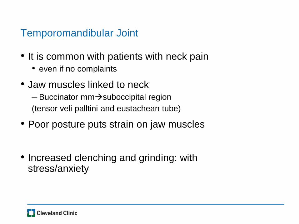
Temporomandibular Joint
• It is common with patients with neck pain
• even if no complaints
• Jaw muscles linked to neck
– Buccinator mmsuboccipital region
(tensor veli palltini and eustachean tube)
• Poor posture puts strain on jaw muscles
• Increased clenching and grinding: with stress/anxiety
Physical Therapy
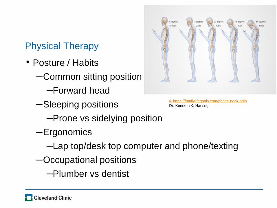
• Posture / Habits
–Common sitting position
–Forward head
–Sleeping positions
–Prone vs sidelying position
–Ergonomics
–Lap top/desk top computer and phone/texting
–Occupational positions
–Plumber vs dentist
© https://familylifegoals.com/phone-neck-pain
Dr. Kenneth K. Hansraj
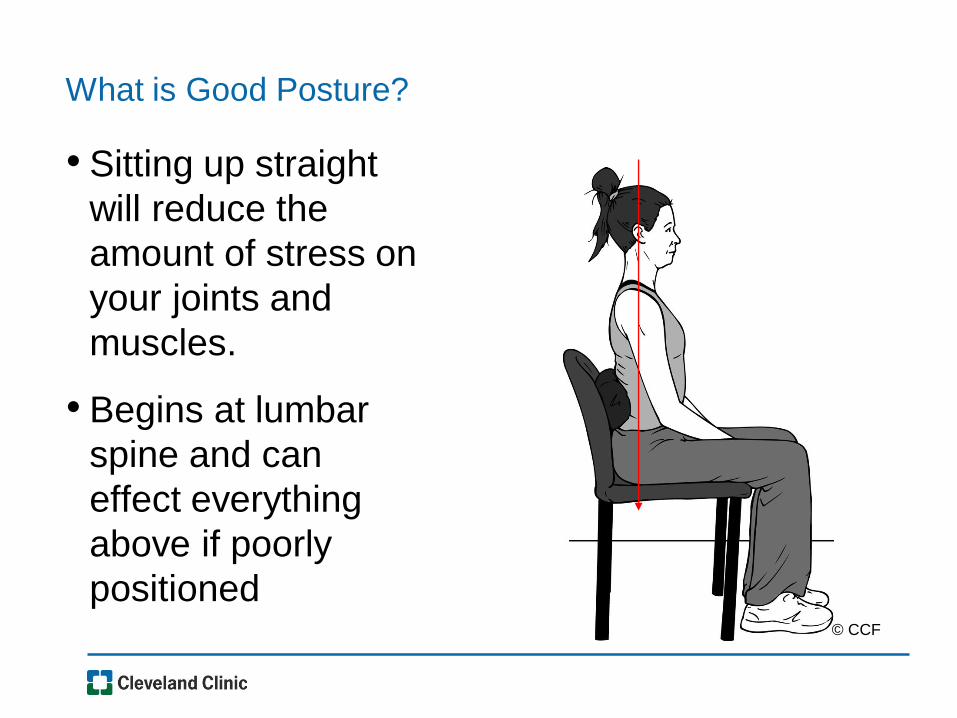
What is Good Posture?
• Sitting up straight
will reduce the
amount of stress on
your joints and
muscles.
• Begins at lumbar
spine and can
effect everything
above if poorly
positioned© CCF
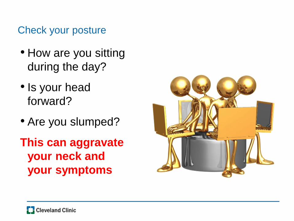
Check your posture
• How are you sitting
during the day?
• Is your head
forward?
• Are you slumped?
This can aggravate
your neck and
your symptoms
Tinnitus Screen
©CCF
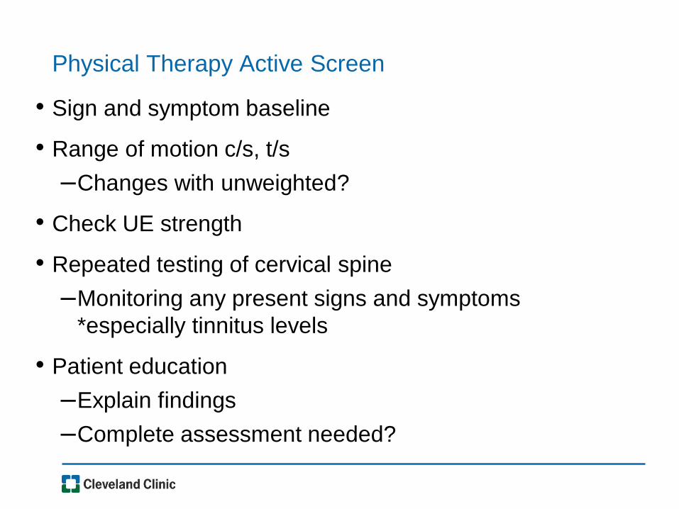
Physical Therapy Active Screen
• Sign and symptom baseline
• Range of motion c/s, t/s
–Changes with unweighted?
• Check UE strength
• Repeated testing of cervical spine
–Monitoring any present signs and symptoms
*especially tinnitus levels
• Patient education
–Explain findings
–Complete assessment needed?
Why should physical therapy be included in
assessment of tinnitus?
Why look at the neck and jaw?
Literature is suggesting we should consider these
areas for tinnitus management:
Sanchez TG, Rocha CA, Latifpour DH, Michiels S, Buergers R
Montazem A.
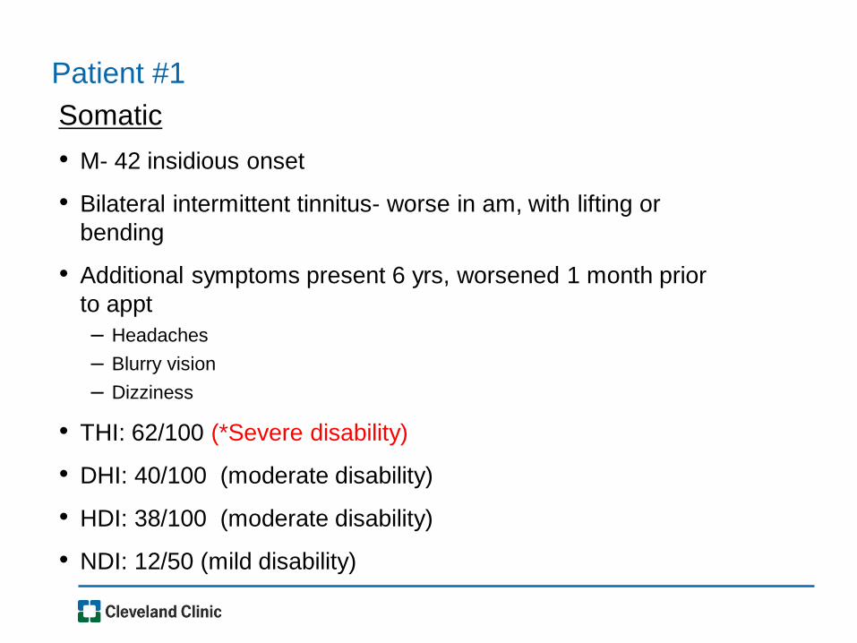
Patient #1
Somatic
• M- 42 insidious onset
• Bilateral intermittent tinnitus- worse in am, with lifting or
bending
• Additional symptoms present 6 yrs, worsened 1 month prior
to appt
– Headaches
– Blurry vision
– Dizziness
• THI: 62/100 (*Severe disability)
• DHI: 40/100 (moderate disability)
• HDI: 38/100 (moderate disability)
• NDI: 12/50 (mild disability)
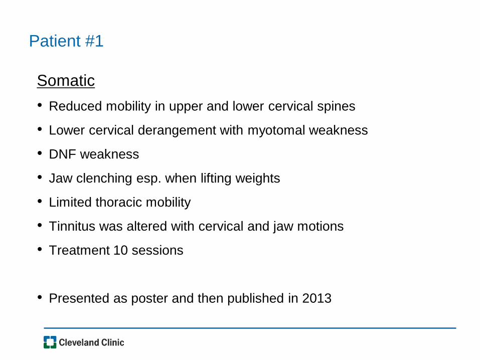
Patient #1
Somatic
• Reduced mobility in upper and lower cervical spines
• Lower cervical derangement with myotomal weakness
• DNF weakness
• Jaw clenching esp. when lifting weights
• Limited thoracic mobility
• Tinnitus was altered with cervical and jaw motions
• Treatment 10 sessions
• Presented as poster and then published in 2013
Tinnitus patients would benefit from a physical therapy evaluation for the following reasons:
1. To identify any biomechanical abnormalities in the cervical spine and / or jaw
2. To educate patients on proper posture, ergonomics, and e xercise techniques
Studies are needed to cri tically evaluate the role of mechanical interventions of the cervical spine in
treatment of tinnitus. This is crucial because all of the other available treatments have conflicting and
inconclusive evidence regarding eff icacy. Given this, any safe approaches with a potential for benefit
should be further investigated.
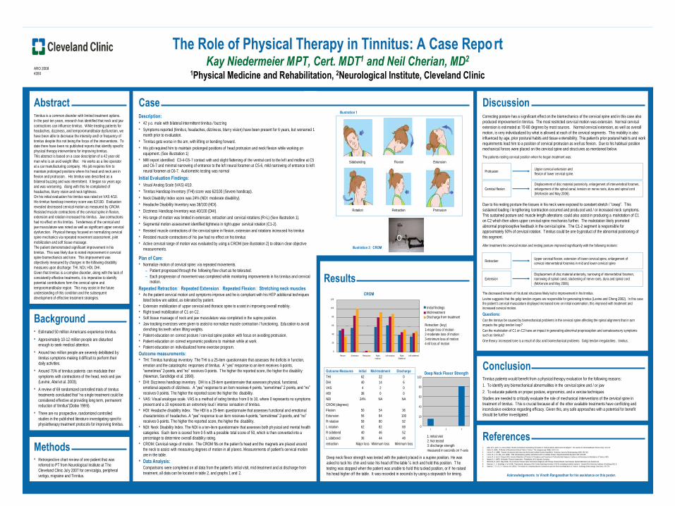
The Role of Physical Therapy in Tinnitus: A Case ReportKay Niedermeier MPT, Cert. MDT1 and Neil Cherian, MD2
1Physical Medicine and Rehabilitation, 2Neurological Institute, Cleveland Clinic
Abstract Tinnitus is a common disorder with limited treatment options.
In the past ten years, research has identified that neck and jaw
contractions can influence tinnitus. While treating patients for
headaches, dizziness, and temporomandibular dysfunction, we
have been able to decrease the intensity and/ or frequency of
tinnitus despite this not being the focus of the interventions. To
date there have been no published reports that identify specific
physical therapy interventions for improving tinnitus.
This abstract is based on a case description of a 42 year old
man who is an avid weight lifter. He works as a line operator
at a car manufacturing company. His job requires him to
maintain prolonged positions where his head and neck are in
flexion and protrusion. His tinnitus was described as a
bilateral buzzing and was intermittent. It began six years ago
and was worsening. Along with this he complained of
headaches, blurry vision and neck tightness.
On his initial evaluation his tinnitus was rated on VAS 4/10.
His tinnitus handicap inventory score was 62/100. Evaluation
revealed decreased cervical motion as measured by CROM.
Resisted muscle contractions of the cervical spine in flexion,
extension and rotation increased his tinnitus. Jaw contractions
had no effect on his tinnitus. Tenderness of the cervical and
jaw musculature was noted as well as significant upper cervical
dysfunction. Physical therapy focused on normalizing cervical
spine mechanics via repeated movement assessment, joint
mobilization and soft tissue massage.
The patient demonstrated significant improvement in his
tinnitus. This was likely due to noted improvement in cervical
spine biomechanics and tone. This improvement was
objectively measured by changes in the following disability
measures upon discharge: THI, NDI, HDI, DHI.
Given that tinnitus is a complex disorder, along with the lack of
consistently effective treatments, it is imperative to identify
potential contributions form the cervical spine and
temporomandibular region. This may assist in the future
understanding of this condition and the subsequent
development of effective treatment strategies.
Background • Estimated 50 million Americans experience tinnitus.
• Approximately 10-12 million people are disturbed
enough to seek medical attention.
• Around two million people are severely debilitated by
tinnitus symptoms making it dif ficult to perform their
daily activities.
• Around 75% of tinnitus patients can modulate their
symptoms with contractions of the head, neck and jaw
(Levine, Abel et al. 2003).
• A review of 69 randomized controlled trials of tinnitus
treatments concluded that “no s ingle treatment could be
considered effective at providing long term, permanent
reduction of tinnitus”(Dobie 1999).
• There are no prospective, randomized controlled
studies in the publi shed literature investigating specif ic
physiotherapy treatment protocols for improving tinnitus.
Methods • Retrospective chart review of one patient that was
referred to PT from Neurological Institute at T he
Cleveland Clinic July 2007 for cervicalgia, peripheral
vertigo, migraine and Tinnitus.
Case Description:
• 42 y.o. male with bilateral intermittent t innitus / buzz ing
• Symptoms reported (tinnitus, headaches, dizziness, blurry vision) have been present for 6 years, but worsened 1
month prior to evaluation.
• Tinnitus gets worse in the am, with lifting or bending forward.
• His job required him to maintain prolonged positions of head protrusion and neck f lexion while working on
equipment. (See illustration 1)
• MRI report identified: C3-4-C6-7 contact with and slight flattening of the ventral cord to the lef t and midline at C5
and C6-7 and minimal narrowing of entrance to the lef t neural foramen at C5-6, mild narrowing of entrance to left
neural foramen at C6-7. Audiometric testing was normal
Initial Evaluation Findings:
• Visual Analog Scale (VAS) 4/10.
• Tinnitus Handicap Inventory (THI) score was 62/100 (Severe handicap).
• Neck Disability Index score was 24% (NDI: moderate disability).
• Headache Disability Inventory was 38/100 (HDI).
• Dizziness Handicap Inventory was 40/100 (DHI).
• His range of motion was limited in extension, retraction and cervical rotations (R>L) (Se e illustration 1).
• Segmental motion assessment identified tightness in right upper cervical rotation (C1-2).
• Resisted muscle contractions of the cervical spine in f lexion, extension and rotations increa sed his tinnitus
• Resisted muscle contractions of his jaw had no effect on his tinnitus
• Active cervical range of motion was evaluated by using a CROM (see illustration 2) to obtai n clear objective
measurements.
Illustration 1
Sidebending Flexion Extension
Rotation Retraction
Illustration 2: CROM
Plan of Care:
• Normalize motion of cervical spine: v ia repeated movements.
– Patient progressed through the following flow chart as he tolera ted.
– Each progression of movement was completed while monitoring improvements in his t innitus and cervical
motion.
Repeated Retraction→ Repeated Extension→ Repeated Flexion→ Stretching neck muscles• As the patient cerv ical motion and symptoms improve and he is compliant with his HEP additional techniques
listed below are added, as tolerated by patient.
• Extension mobilization of upper cervical and thoracic spine to a ssist in improving overall mobility.
• Right towel mobilization of C1 on C2.
• Soft tissue massage of neck and jaw musculature was completed in the supine posit ion.
• Jaw tracking exercises were given to assist to nor malize muscle contraction / functioning. Education to avoid
clenching his teeth when lifting weights.
• Patient education on correct posture / cervical spine position with focus on avoiding protrusion.
• Patient education on correct ergonomic positions to maintain while at work.
• Patient education on individualized home exercise program.
Outcome measurements:• THI: Tinnitus handicap inventory. The THI is a 25-item questionnaire that assesses the d eficits in function,
emotion and the catastrophic responses of tinnitus. A “yes” response to an item receives 4-points,
“sometimes” 2-points, and “no” receives 0-points. The higher the reported score, the higher th e disability
(Newman, Sandridge et al. 1998).
• DHI: Dizziness handicap inventory. DHI is a 25-item questionnaire that assesses physical, functional,
emotional aspects of dizziness. A “yes” response to an item receives 4 points, “sometimes” 2 points, and “no”
receives 0 points. The higher the reported score the higher the disability.
• VAS: Visual analogue scale. VAS is a method of rating tinnitus f rom 0 to 10, where 0 represents no symptoms
present and a 10 represents an extremely loud / intense sensation of tinnitus.
• HDI: Headache disability index. The HDI is a 25-item questionnaire that assesses f unctional and emotional
characteristics of headaches. A “yes” response to an item receives 4-points, “sometimes” 2-points, and “no”
receives 0-points. The higher the reported score, the higher the disability.
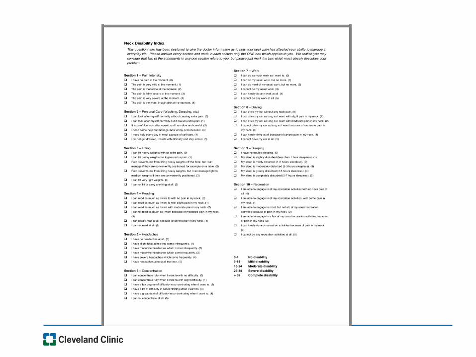
• NDI: Neck Disability Index. The NDI is a ten-ite m questionnaire that assesses both ph ysical and mental health
categories. Each item is scored from 0-5 with a possible total score of 50, which is then converted into a
percentage to determine overall disabilit y rating.
• CROM: Cervical range of motion. The CROM fits on the patien t’s head and the magnets are placed around
the neck to assist with measuring degrees of motion in all p lanes. Measurements of patient’s ce rvical motion
are in the table.
• Data Analysis:
Comparisons were completed on all data from the patient’s initial visit, mid-treatment and at discharge from
treatment, all data can be located in table 2, and graphs 1 and 2.
Results
Initial findings
Mid-treatment
Discharge from treatment
Retraction: (key)
1-major loss of motion
2-moderate loss of motion
3-minimum loss of motion
4-nil loss of motion
Outcome Measures Initial Mid-treatment Discharge
THI 62 22 0
DHI 40 14 6
VAS 4 2 0
HDI 38 0 0
NDI 24% NA NA
CROM (degrees)
Flexion 50 54 36
Extension 56 84 100
R rotation 58 80 92
L rotation 62 82 88
R sidebend 40 46 52
L sidebend 36 44 48
retraction Major loss Minimum loss Minimum loss
1: initial visit
2: Not tested
3: discharge strength
measured in seconds on Y-axis
Deep neck flexor strength was tested with the patient p laced in a supine position. He was
asked to tuck his chin and raise his head off the table ¼ inch and hold this position. T he
testing was stopped when the patient was unable to hold this tucked position, or if he raised
his head higher off the table. It was recorded in seconds by using a stopwatch for timing.
Discussion Correcting posture has a s ignificant effect on the biomechanics of the cervical spine and in this case also
produced improvement in tinnitus. The most restricted cerv ical motion was extension. Normal cervical
extension is estimated at 70-80 degrees by most sources. Normal cervical extension, as well as overall
motion, is very individualized by what is allowed at each of the cervical segments. This mobility is also
influenced by age, prior postural habits and tissue e xtensibility. This patient’s prior postural habi ts and work
requirements lead him to a position of cervical protrusion as well as flexion. Due to his habitua l position
mechanical forces were placed on the cervical spine and struct ures as mentioned below.
The patients resting cervical position when he began treatment was:
References 1. Abel, M. D. and R. A. Levine (2004). "Muscle Contractions and Auditory Perception in Tinnitus Patients and Nonclinical Subjects." The Journal of Craniomandibular Practice 22(3): 181-1 91.
2. Dobie, R. (1999 ). "A Review of Randomized Clinical Trials in Tinnitus." The Laryngoscope 109(8): 1202-1211.
3. Levine, R. A. (1999). "Somatic (Craniocervical) tinnitus and the Dorsal Cochlear Nucleus Hypothesis." American Journal of Otolaryngology 20(6): 351-362.
4. Levine, R. A., M. Abel, et al. (2003). "CNS somatosensory-auditory interactions elicit or modulate tinnitus." Experimental Brain Researc h 153: 643-648.
5. Levine, R. A. and H. Cheng (2002). Somatic Modulation of Tinnitus III: Prevalence and Properties in Profoundly Deaf Subjects, Cochlea is not Necessary for Modulation of Tinnitus. ARO.
6. Magee, D. J. (1997). Orthopedic Physical Assessment. Philadelphia, W.B. Saunders Company.
7. McKenzie, R. and S. May (2006). The Cervical & Thoracic Spine: Mechanical Diagnosis and Therapy. Raum ati Beach, New Zealand, Spinal Publications new Zealand Ltd.
8. Newman, C., S. Sandridge, et al. (1998). "Psychometric adequacy of the Tinnitus Handicap Inventory ( THI) for evaluating treatment outcom e." Journal of the American Academy of Audi ology 9(2): 8.
9. Sanchez, T. G., G. C. Y. Guerra, et al. (2002). "The Influence of Voluntary Muscle Contractions upon the Onset and Modul ation of Tinnitus." Audiology & Neur otology 7(Nov/Dec): 370-375.
Protrusion
Cervical flexion
Upper cervical extension and
flexion of lower cervical spine.
Due to his resting posture the tissues in his neck were exposed to constant stretch / “creep”. This
sustained loading / lengthening /contraction occurred and produ ced and / or increased neck symptoms.
This sustained posture and muscle length alterations could also assist in producing a malrotation of C1
on C2 which then alters upper cervical spine mechanics further. The malrotation likely promoted
abnormal proprioceptive feedback in the cervical spine. The C1-2 segment is responsible for
approximately 50% of cervical rotation. T innitus could be one byproduct of the abnormal positioning of
this segment.
After treatment his cervical motion and resting posture improved significantly with the following motions:
Upper cervical flexion, extension of lower cervical spine, enlargement of
cervical intervertebral foramina in mid and lower cervical spine
Displacement of disc material anteriorly, narrowing of intervertebral foramen,
narrowing of spinal canal, slackening of nerve roots, dura and spinal cord
(McKenzie and May 2006).
The decreased tension of his dural structures likely led to improvement in his tinnitus.
Levine suggests that the golgi tendon organs are responsible for generating tinnitus (Levine and Cheng 2002). In this case
the patient’s cervical musculature displayed increased tone on initial examination, this improved with treatment and
increased cervical motion.
Questions:
Can the tinnitus be caused by biomechanical problems in the cervical spine affecting the spinal alignment that in turn
impacts the golgi tendon loop?
Can the malrotation of C1 on C2 have an impact in generating abnormal proprioception and somatosensory symptoms
such as tinnitus?
One theory: increased tone is a result of disc and biomechanical problems Golgi tendon irregularities→ tinnitus.
ConclusionDeep Neck Flexor Strength
0
20
40
60
80
1 2 3
100
CROM
0
20
40
60
80
100
120
Flexion Extension Retract ion Right
rotation
Left rotation Right
Sidebe nd
Left s idebend
Protrusion
Displacement of disc material posteriorly, enlargement of intervertebral foramen,
enlargement of the spinal canal, tension on nerve roots, dura and spinal cord
(McKenzie and May 2006).
Retraction
Extension
Acknowledgements to Vinoth Ranganathan for his assistance on this poster.
ARO 2008
#260
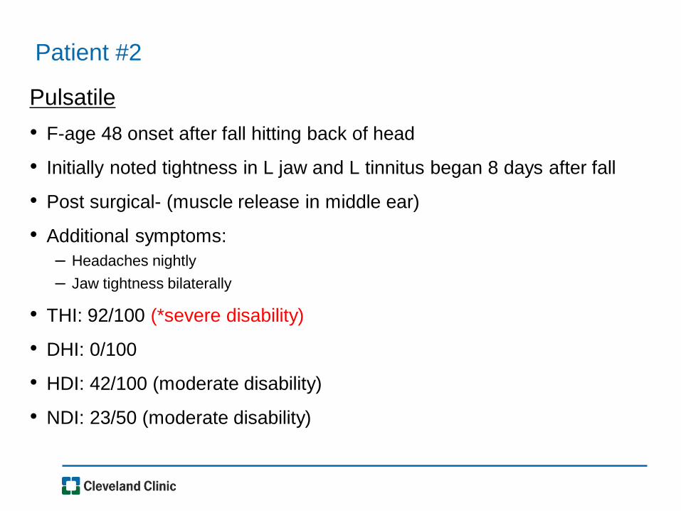
Patient #2
Pulsatile
• F-age 48 onset after fall hitting back of head
• Initially noted tightness in L jaw and L tinnitus began 8 days after fall
• Post surgical- (muscle release in middle ear)
• Additional symptoms:
– Headaches nightly
– Jaw tightness bilaterally
• THI: 92/100 (*severe disability)
• DHI: 0/100
• HDI: 42/100 (moderate disability)
• NDI: 23/50 (moderate disability)
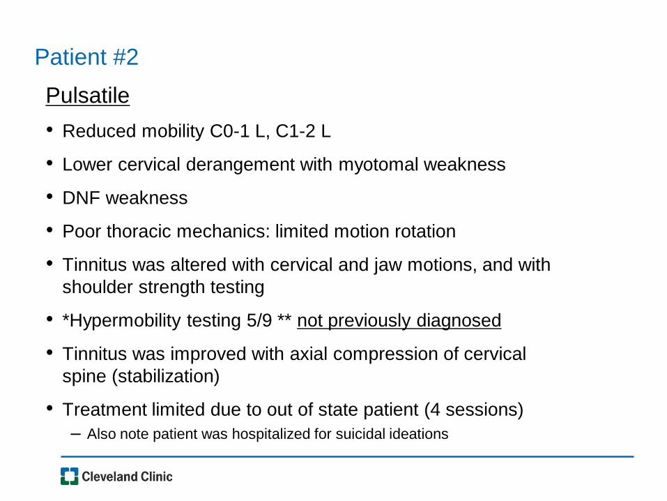
Patient #2
Pulsatile
• Reduced mobility C0-1 L, C1-2 L
• Lower cervical derangement with myotomal weakness
• DNF weakness
• Poor thoracic mechanics: limited motion rotation
• Tinnitus was altered with cervical and jaw motions, and with
shoulder strength testing
• *Hypermobility testing 5/9 ** not previously diagnosed
• Tinnitus was improved with axial compression of cervical
spine (stabilization)
• Treatment limited due to out of state patient (4 sessions)
– Also note patient was hospitalized for suicidal ideations
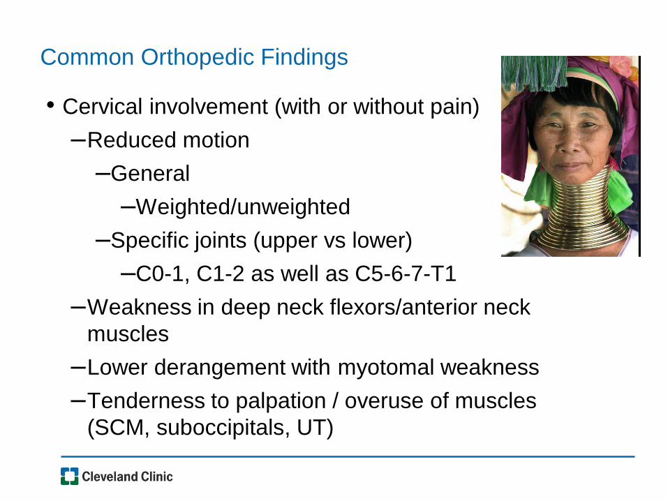
Common Orthopedic Findings
• Cervical involvement (with or without pain)
–Reduced motion
–General
–Weighted/unweighted
–Specific joints (upper vs lower)
–C0-1, C1-2 as well as C5-6-7-T1
–Weakness in deep neck flexors/anterior neck
muscles
–Lower derangement with myotomal weakness
–Tenderness to palpation / overuse of muscles
(SCM, suboccipitals, UT)
Common Orthopedic Findings cont.
• Jaw involvement
–Pain with jaw motion
–Parafunction:
–Clenching/grinding, biting lips etc.
–Abnormal mechanics:
–Popping/clicking, limited motion, hypermobility
–Tenderness to palpation of TMJ or muscles of
mastication
–Poor posture
–Leaning on hand
–Sleeping on side

Trigger points in these neck and jaw
muscles have been known to
contribute to tinnitus.
Travell and Simons

Common Orthopedic Findings cont.
• General findings
–Posture
–Forward head/posterior cranial rotation,
protruded jaw
–Alters mechanics of neck and jaw
–Weakness of anterior neck, tightness of
posterior mm
–Rounded shoulders
–Can aggravate shoulders and
thoracic/lumbar spines
–Poor ergonomic awareness/endurance

Our findings
CCF Pilot Study
• 2008 study of 10 patients
– Limited demographic information
• Monitored THI, CROM, DHI, NDI, neck strength at
initial visit, mid, and at discharge
• 10 PT sessions: manual and exercise
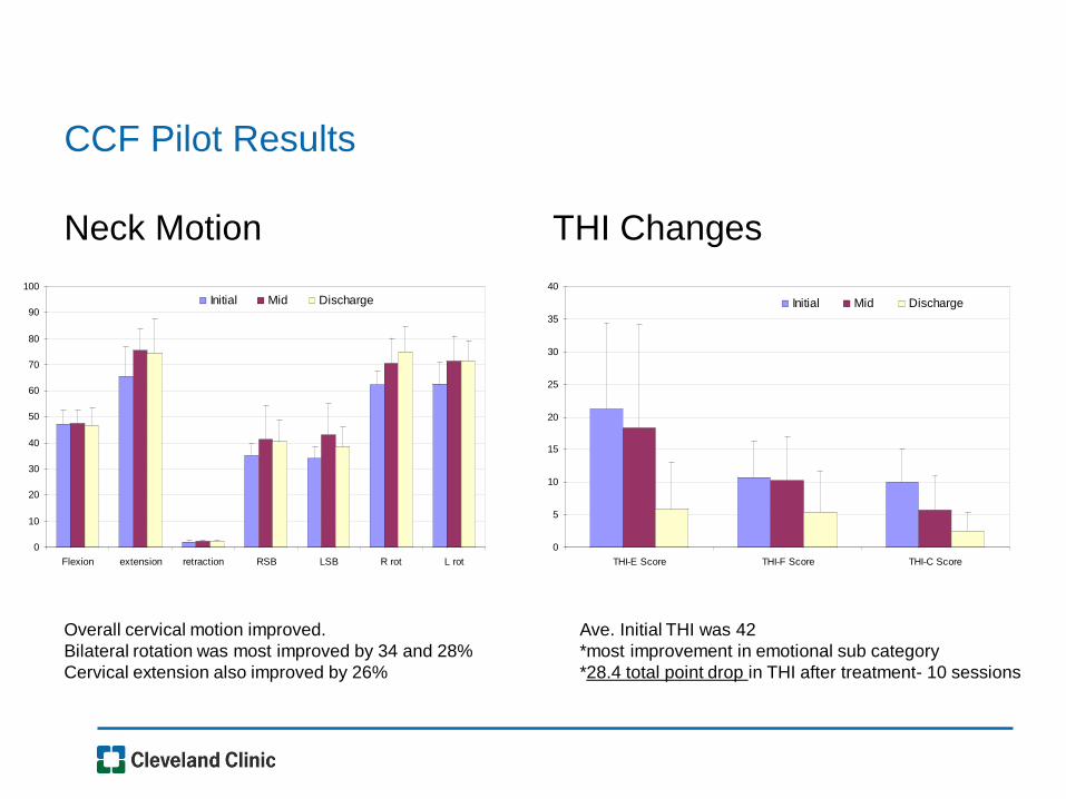
CCF Pilot Results
Neck Motion THI Changes
0
5
10
15
20
25
30
35
40
THI-E Score THI-F Score THI-C Score
Initial Mid Discharge
0
10
20
30
40
50
60
70
80
90
100
Flexion extension retraction RSB LSB R rot L rot
Initial Mid Discharge
Ave. Initial THI was 42
*most improvement in emotional sub category
*28.4 total point drop in THI after treatment- 10 sessions
Overall cervical motion improved.
Bilateral rotation was most improved by 34 and 28%
Cervical extension also improved by 26%
2014 TMC Review
Introduction
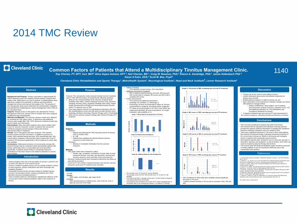
Common Factors of Patients that Attend a Multidisciplinary Tinnitus Management Clinic. Kay Cherian, PT, DPT, Cert. MDT1 Alma Gojani Axhemi, DPT 2, Neil Cherian, MD 3, Craig W. Newman, PhD,4 Sharon A. Sandridge, PhD,4 James Kaltenbach PhD 5
Karyn A Kahn, DDS,4 Scott M. Bea, PsyD3
Cleveland Clinic Rehabilitation and Sports Therapy1, MetroHealth System2, Neurological Institute3, Head and Neck Institute4, Lerner Research Institute5
Abstract Case Description
Discussion
Pre-Intervention Post-Intervention
Introduction and Objectives
Case Description
Methods
Results
Results
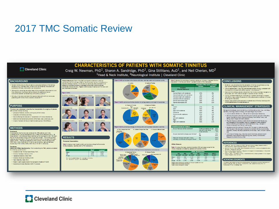
Background and Purpose: Tinnitus is prevalent in approximately 50 million Americans and in the majority of these patients it impacts their
quality of life. While there is no cure for tinnitus, a multidisciplinary team approach is helpful and worthwhile to address and help patients
manage their tinnitus and improve their quality of life. The purpose of this retrospective study was to determine the characteristics of patients
that attended the multidisciplinary Tinnitus Management Clinic (TMC) at
the Cleveland Clinic. Subjects: Medical charts of 108 patients who attended the Tinnitus
Management Clinic from January 2010 to October 2013 were analyzed
to determine their characteristics. Methods and Materials: The outcome measure results were obtained
by reviewing the TMC paper charts. To determine what additional services were sought after TMC, EPIC (electronic medical records) was
used to obtain such information.
Data Analysis: Basic descriptive statistics were used to obtain mean scores for outcome measures, number of patients that returned for
individual evaluation and percentage of those that showed improvement after PT treatment.
Results: Out of 108 patients that were analyzed, 75% reported
constant tinnitus and 25% intermittent. In addition to presence of tinnitus, 60% reported having neck pain, 40% reported jaw pain, 29%
scored mild to complete in HDI and 26% mild to severe DHI. Post PT treatment THI decreased significantly in 45% of patients for which data
was available.
Conclusions: Addressing mechanics of cervical spine and jaw with physical therapy may be a treatment option that benefits patients with
tinnitus, particularly when it is associated with neck discomfort. Specifically, in patients whose tinnitus is somatically-induced or
modulated as it has been shown to be related to disorders in cervical spine and jaw.
References
Methods
Conclusions
1140
• Tinnitus (ringing of the ear) is the perception of sound in a person’s ear or head in the absence of an external sound.
• Tinnitus is related to auditory factors such as otologic problems, history of noise exposure, ototoxic medications and other neurologic and
metabolic disorders.1 • Somatically-induced tinnitus has been related to whiplash injuries,
recurrent cervical spine injuries, temporo-mandibular joint (TMJ)
disorders and poor prolonged postures.2,3 • There is limited evidence in the literature regarding the influence of the
cervical spine and/or temporomandibular region in the treatment of
tinnitus.
Purpose: This retrospective study reviewed medical records of patients seen in TMC with the purpose of answering the following questions:
1. What are the common findings of the outcome measures Neck Disability Index (NDI), Tinnitus Handicap Inventory (THI), Dizziness
Handicap Inventory (DHI), Headache Disability Index (HDI), Patient Health Questionnaire-9 (PHQ9) and Generalized Anxiety Disorder
(GAD-7) in patients seen at TMC
2. How many patients returned for individualized evaluations after the first multidisciplinary screen and with which provider did they return:
Audiology, Dentistry, Neurology, Physical Therapy and/ or Psychology
3. What are the outcome measure findings after Physical Therapy (PT) treatment
Subjects: Inclusion:
• Patients that attended the TMC during the period of January 2012 to October 2013
• N= 108 • Patients that completed the aforementioned outcome
measures on their initial visit
Exclusion: • Missing or incomplete information from the outcome
measures
Methods:
• TMC paper charts were reviewed to collect: • Gender, age, occupation, mechanism of onset, date of onset,
symptoms (tinnitus, neck pain, jaw discomfort, hearing loss),
outcome measure scores and date of first screening visit • Electronic medical records were reviewed to determine any follow up
care with the providers • If seen by PT, the number of physical therapy visits and the outcome
measures scores post PT treatment were recorded
• Pattern of tinnitus: • 75% reported constant tinnitus, 25% intermittent
• Additional mechanical complaints: • 60% of patients reported having neck pain, 40% jaw pain,
29% scored mild to complete HDI scores and 26% mild to complete DHI scores
• Further assessments:
• 57% returned for additional assessment • Audiology: 20, Dentistry: 11, Neurology: 9,
• Psychology: 56 were recommended to follow up, 58 were
recommended a workbook, 48 nothing further suggested • PT: follow up recommended in 107 due to neck and/or jaw
impairments identified, 89 were local patients, 22 sought evaluation, data available on 11.
• The average score of THI was 52: severe disability
• The average scores for DHI, HDI and NDI were 12, 13 and 15: mild
disability.
• The PHQ-9 and GAD-7 average scores were 7.2 and 6 which correspond
to moderate depression and anxiety.
• 11/22 patients were included in the review of PT, others eliminated due to
incomplete data (2 still undergoing treatment, 2 at different PT/facility)
• TMC has been beneficial for patients that suffer from tinnitus • We surmised that the education and individual screening sessions
provided the patients sufficient information to manage their symptoms, therefore individual evaluations were not needed in 43%
• There was a significant decrease in THI score in 45% of the patients that returned for physical therapy, indicating mechanical treatment of
the cervical spine and jaw are helpful in decreasing and managing
tinnitus in patients with somatically-induced tinnitus • Most patients reported other symptoms such as neck pain, jaw pain or
discomfort, dizziness and headache, as well as tinnitus, all of which can
easily be treated by physical therapy interventions • The cervical spine and temporomandibular region should be assessed
in tinnitus patients to rule out mechanical influences due to the connectivity of the systems
• Tinnitus can be the result of many different factors • Limited information about active and effective treatments for
tinnitus exist currently • Multidisciplinary programs to address tinnitus are rare
• Basic information on the importance of lifestyle changes are shared with patients to manage tinnitus
• Dietary modifications, sleep hygiene, use of auditory
devices/sounds, postural correction, relaxation music / CD and additional education via “The Mindfulness and
Acceptance Workbook for Anxiety”.
• A majority of patients that were seen by PT had improvement in the outcome measures, indicating mechanical influences on tinnitus
1. Crummer RW, Hassan GA (2004). "Diagnostic approach to tinnitus". Am Fam Physician. 69 (1): 120–6.
2. Sanchez, T. G., Guerra, G.C.Y et al The Influence of Voluntary Muscle Contractions upon
the Onset and Modulation of Tinnitus. Audiology & Neurotology.2002;7:370-375.
3. Bjorne, A. (2007). Assessment of temporomandibular and cervical spine disorders in
tinnitus patients, Elsevier. 4. Levine, R. A. (1999). "Somatic (Craniocervical) tinnitus and the Dorsal Cochlear Nucleus
Hypothesis." American Journal of Otolaryngology 20(6): 351-362.
5. Coad ML, Lockwood A, Salvi R, Burkard R. Characteristics of patients with gaze-evoked
tinnitus. Otol Neurotol;2001 Sep;22(5):650-4.
6. Wright DD, Ryugo DK. Mossy fiber projection from the cuneate nucleus to the cochlear in the rat. J Comp Neurol.1996;365(1):159-172
7. Cherian K, Cherian N, Cook Ch, Kaltenbach JA. Improving Tinnitus with Mechanical
Treatment of the Cervical Spine and Jaw. J Am Acad Audiol; 2012:24:1-11
The authors have no disclosures*
Purpose
• N=108 • Gender:
• 59% males, 41% females, age range 30-84 • Location:
• 59% reported tinnitus in bilateral ears, 14% in left ear, 12% in right ear, and 15% in the head
Graph 2a: Baseline Outcome Measure
!
Graph 2b: Additional Baseline Outcome Measures
!
• 45% of patients for which data were available showed significant decrease in THI score.
• 1 patient showed increase in THI as well as increase in DHI, HDI and NDI.
Nu
mb
er
of
pa
tie
nts
Graph 1: Patient Report of Inciting Factor of Tinnitus
2014 TMC Review
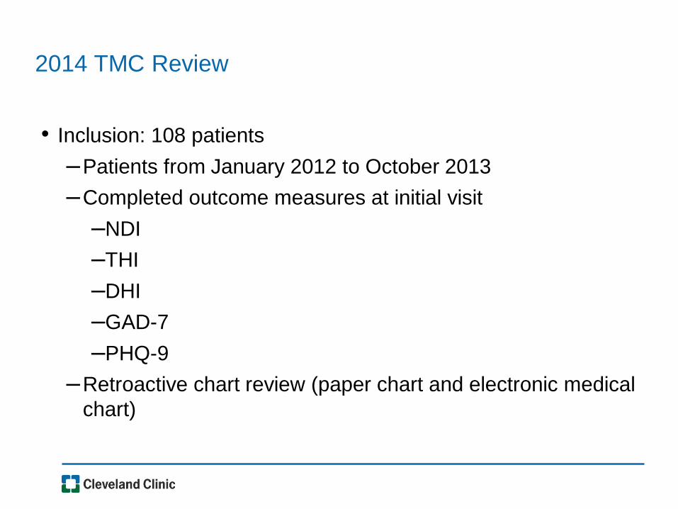
• Inclusion: 108 patients
–Patients from January 2012 to October 2013
–Completed outcome measures at initial visit
–NDI
–THI
–DHI
–GAD-7
–PHQ-9
–Retroactive chart review (paper chart and electronic medical
chart)
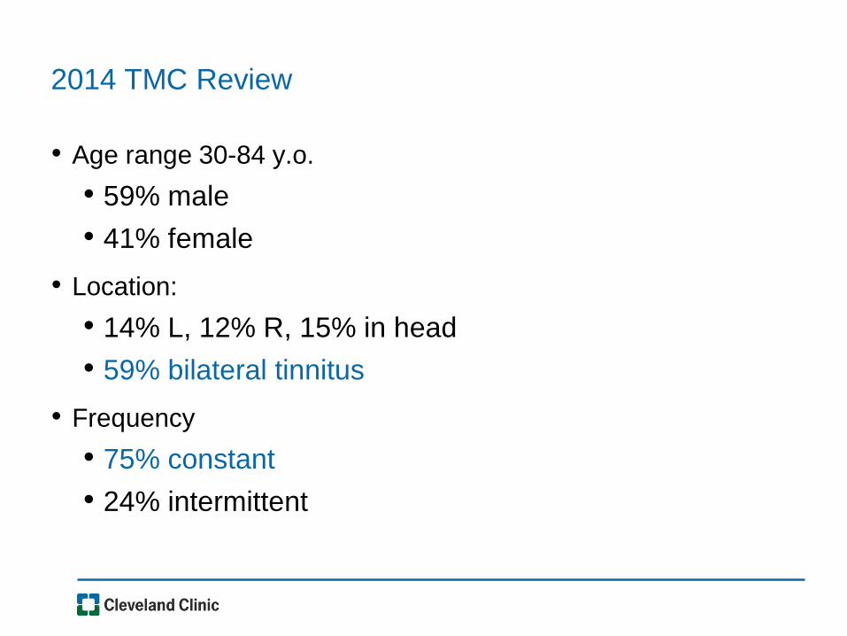
2014 TMC Review
• Age range 30-84 y.o.
• 59% male
• 41% female
• Location:
• 14% L, 12% R, 15% in head
• 59% bilateral tinnitus
• Frequency
• 75% constant
• 24% intermittent
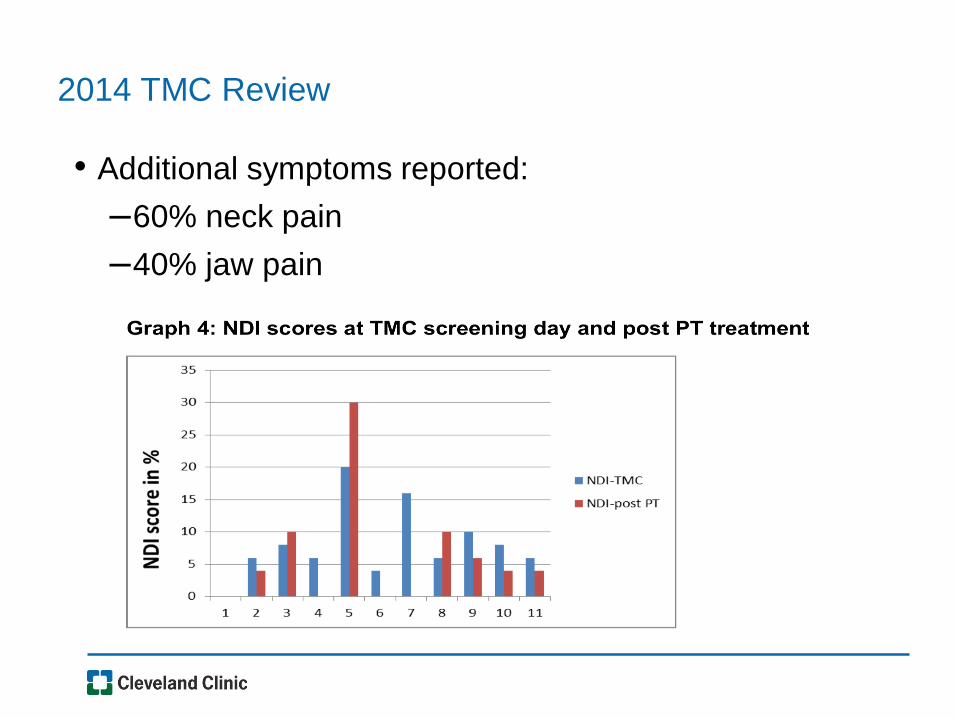
2014 TMC Review
• Additional symptoms reported:
–60% neck pain
–40% jaw pain

2017 TMC Somatic Review

2017 TMC Somatic Review
• 69/138 > 5 on NDI (> mild disability)
• Breakdown:
– 0-4: 56/138 (no disability)
– 5-14: 49/138 (mild disability)
– 15-24: 16/138 (moderate disability)
– 25-34:2/138 ** (severe disability)
– 35-50: 0/138 (complete disability)
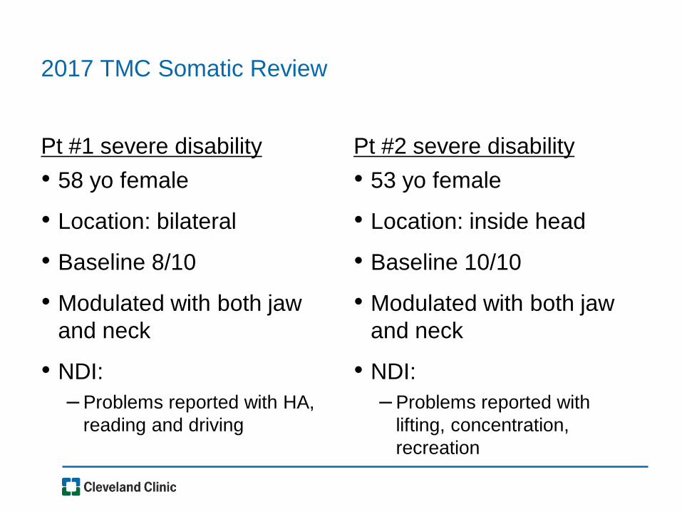
2017 TMC Somatic Review
Pt #1 severe disability
• 58 yo female
• Location: bilateral
• Baseline 8/10
• Modulated with both jaw
and neck
• NDI:
– Problems reported with HA,
reading and driving
Pt #2 severe disability
• 53 yo female
• Location: inside head
• Baseline 10/10
• Modulated with both jaw
and neck
• NDI:
– Problems reported with
lifting, concentration,
recreation
2017 Somatic Review
• We are not using a jaw outcome measure at this time
• Consider this in the future for additional information
• Patients do have dental exam regardless of additional
symptoms
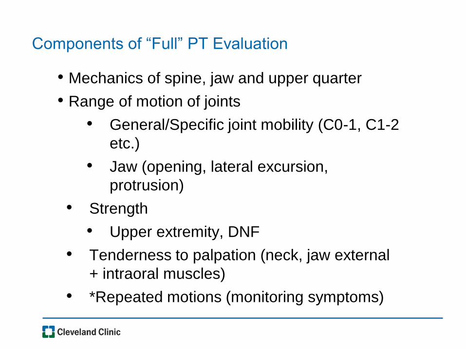
Components of “Full” PT Evaluation
• Mechanics of spine, jaw and upper quarter
• Range of motion of joints
• General/Specific joint mobility (C0-1, C1-2
etc.)
• Jaw (opening, lateral excursion,
protrusion)
• Strength
• Upper extremity, DNF
• Tenderness to palpation (neck, jaw external
+ intraoral muscles)
• *Repeated motions (monitoring symptoms)
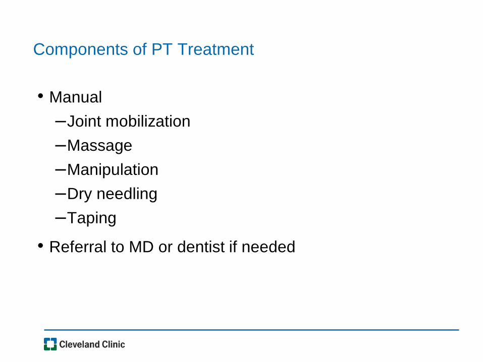
Components of PT Treatment
• Manual
–Joint mobilization
–Massage
–Manipulation
–Dry needling
–Taping
• Referral to MD or dentist if needed
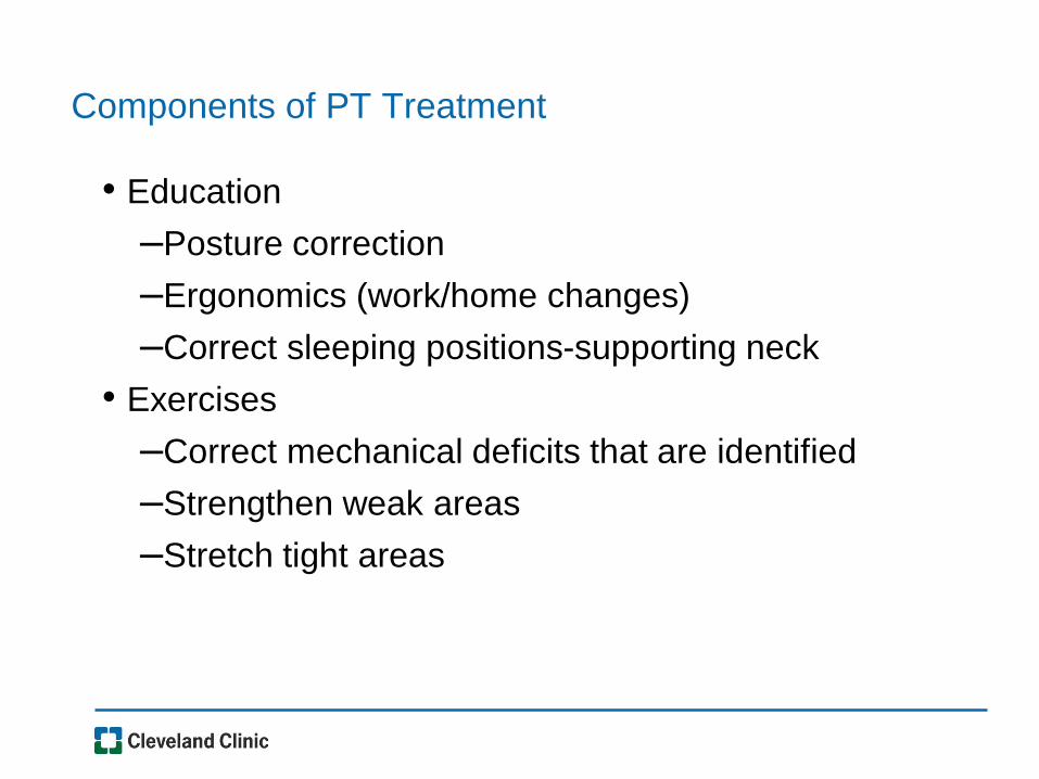
Components of PT Treatment
• Education
–Posture correction
–Ergonomics (work/home changes)
–Correct sleeping positions-supporting neck
• Exercises
–Correct mechanical deficits that are identified
–Strengthen weak areas
–Stretch tight areas

Take home messages…
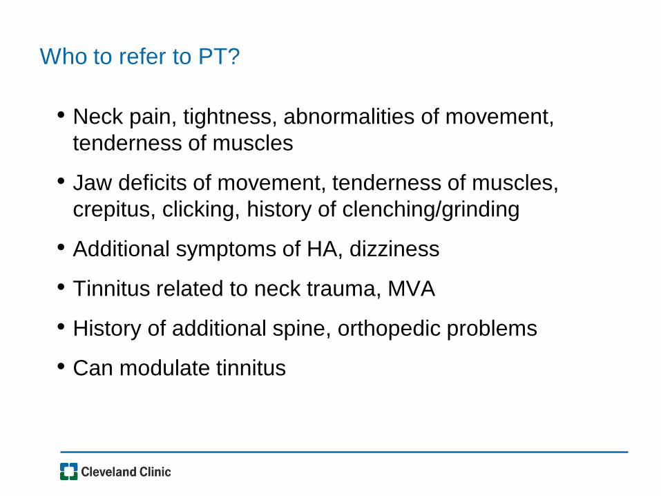
Who to refer to PT?
• Neck pain, tightness, abnormalities of movement,
tenderness of muscles
• Jaw deficits of movement, tenderness of muscles,
crepitus, clicking, history of clenching/grinding
• Additional symptoms of HA, dizziness
• Tinnitus related to neck trauma, MVA
• History of additional spine, orthopedic problems
• Can modulate tinnitus

How to identify an appropriate PT
• What to look for:
–Active PT
–Patient is involved in their care, progress
–Home exercises are a must
–Manual therapy: massage, mobilizations, “hands on approach”
–Passive PT
–Patient is not as involved in care
–No home exercises
–Electric stimulation, hot pack, cold packs, general gym exercises

Locating a PT
• www.apta.org
– Find a PT- helps to ID PT local to patient
– Look for OCS (orthopedic specialist certification)
– Look for manual certifications (COMT, OMT etc)
• www.mckenziemdt.org
– Specialized training in cervical and lumbar spine mechanics
– Find a certified or diplomaed therapist on the list
*May need to ask if they are comfortable treating neck, headache
and dizzy patients to get with appropriate PT. This PT can handle
tinnitus patients even though they may not have treated them in
the past.
Now
we welcome questions