The Role Of miRNAs In CD8 T Cell Differentation · In CD8+ T Cell Differentation ... A citocina com...
87
The Role Of miRNAs In CD8 + T Cell Differentation Ana de Oliveira Rodrigues Amorim Mestrado em Biologia Celular e Molecular Departamento de Biologia 2013-2014 Orientador Bruno Silva-Santos, Ph.D, Professor Associado, FMUL
Transcript of The Role Of miRNAs In CD8 T Cell Differentation · In CD8+ T Cell Differentation ... A citocina com...

The Role Of miRNAs In CD8+ T Cell Differentation
Ana de Oliveira Rodrigues Amorim Mestrado em Biologia Celular e Molecular Departamento de Biologia 2013-2014 Orientador Bruno Silva-Santos, Ph.D, Professor Associado, FMUL

Todas as correções determinadas pelo júri, e só essas, foram efetuadas.
O Presidente do Júri, Porto, ______/______/_________

Dissertação de candidatura ao grau de Mestre em Biologia Celular e Molecular submetida à Faculdade de Ciências da Universidade do Porto. O presente trabalho foi desenvolvido sob a orientação científica do Professor Doutor Bruno Silva-Santos, no Institituto de Medicina Molecular, Faculdade de Medicina da Universidade de Lisboa. Dissertation for applying to a Master’s Degree in Cell and Molecular Biology, submitted to the Faculty of Sciences of the University of Porto. The present work was developed under the scientific supervision of Professor Bruno Silva-Santos and was done at the Institute of Molecular Medicine, Faculty of Medicine, University of Lisbon.

!

FCUP The Role Of miRNAs In CD8+ T Cell Differentiation
III
!
Acknowledgements I couldn’t miss the chance to thank to everyone who contributed for another step in
my education and in my personal development.
First of all I would like to thank Professor Bruno Silva-Santos for the opportunity
given to me, for integrated me in his team and in this project. And especially for
always pushing me into giving my best and perfect myself, I have learned a lot.
I thank Anita Gomes for all her help, teaching and her caring!
To the person that contributed the most to my growth, not only scientifically, but
personal as well, Nina Schmolka. Since the first day you received me with a wide-
open smile! It was a joy to have you with me in the lab, I couldn’t have asked for
better! You made my stay in Lisbon and final year really especial; I thank you for all
the knowledge you passed on to me, for your support and for your friendship!
To Miguel, for your help with the FlowJo, for the coffee breaks, and our tours with
Joana at the weekends through the museums! You also contributed for a special
year in Lisbon.
Julie, for being available and helping me when I needed and Natacha, for helping me
to loose my fear with the 384 well plates and for all your advices!
I thank the rest of the Silva-Santos group, Ana Pamplona, Margarida, Sofia, Sergio,
Haakan, Joana, Tiago, Francisco and Daniel for the company at the diner time in the
hospital.
Many thanks to all the team of the flow cytometry facility, especially Ana Vieira; you
made flow cytometry easier for me!
I thank my best friend Maria, we survived, you helped me going for all this process
and you never rejected any of my long, long phone calls. Thank you for the boost of
confidence, wise words, and the nights in Lisbon.
To my parents and sister, without you this journey would not have been possible.
You supported me in all the possible ways, were always present for me, and always
rooted for me. You are the best parents and sister and I will always have you close to
my heart.
Finally to you, Diogo, you have been my best friend, my support. Always made me
go after what I wanted and helped me in the darkest hours. Even with the Atlantic
Ocean between us you were presented everyday, to support me and give me love.
You are an amazing person, and I am glad to have you in my life.

!

FCUP The Role Of miRNAs In CD8+ T Cell Differentiation
V
!
Resumo Os linfócitos T CD8+ desempenham um papel fundamental na defesa do hospedeiro
contra patogénios intracelulares e tumores. A citocina com maior relevância
produzida pelas células T CD8+ é o interferão-gama (IFN-γ), caracterizada por
possuir efeitos pleiotrópicos sobre uma vasta gama de células do sistema
imunológico e por ser essencial tanto na imunidade inata como na adaptativa. Em
2003 foi identificada a Eomesodermina, considerada como um factor de transcrição
principal, necessário e suficiente para regular a transcrição e a produção de IFN-γ
em células T CD8+.
Para além da regulação a nível transcricional, a diferenciação de sub-populações de
células T efetoras encontra-se também sujeita a mecanismos de regulação pós-
transcrição, mediados por microRNAs (miRNAs). Apesar desta função estar
claramente demonstrada para células T CD4+ helper, os miRNAs que controlam a
diferenciação de células CD8+ T produtoras de IFN-γ são ainda em grande parte
desconhecidos.
Os miRNAs são moléculas de RNA não-codificantes de pequenas dimensões, que
inibem pós-transcricionalmente a expressão genética através da diminuição da
estabilidade e/ou bloqueio da tradução de um dado mRNA, o que lhes permite
desempenhar um papel relevante na diferenciação e proliferação celular.
A análise de ratinhos deficientes para a produção de miRNAs especificamente em
linfócitos T (ratinhos LckCre Dicer), demonstrou que os miRNAs possuem um papel
global na diferenciação de células T CD8+ e na produção de IFN-γ. Verificou-se um
aumento da frequência de células T CD8+ produtoras de IFN-γ, tanto na periferia
(nódulos linfáticos e baço) como no timo, em ratinhos deficientes para a produção de
miRNAs quando comparados com ratinhos controlo.
Com o objectivo de identificar os miRNAs implicados na diferenciação das células T
CD8+, realizaram-se microarrays que permitiram a identificação de 22 miRNAs
diferencialmente expressos entre timócitos CD8+ YFP+ e CD8+YFP- de ratinhos
repórter para o IFN-γ (IFN-γ-YFP). Focamos a nossa análise sobre os 3 miRNAs
mais expressos nas células T CD8+ YFP+, miR-139, miR-200a, miR-451a, e os 3
miRNAs mais expressos em células T CD8+ YFP-γ-, miR-132, miR-181a e miR-322.
Curiosamente, a expressão destes miRNAs não se encontra restrita às células T
CD8+, sendo os mesmos também expressos noutras populações de células T,

FCUP The Role Of miRNAs In CD8+ T Cell Differentiation
VI
!sugerindo que possam ter funções pleiotrópicas nas células T. A sua expressão foi
influenciada pela ativação do receptor das células T e pela presença de citocinas. É
importante salientar que a expressão de dois dos nossos candidatos - miR-132 e
miR-451 - foi mais elevada em condições indutoras da produção de IFN-γ, sugerindo
que estes miRNAs poderão ser induzidos no decorrer da diferenciação de células T
CD8+ em células efetoras produtoras de IFN-γ. Por último, efetuaram-se ensaios
funcionais dos nossos candidatos com recurso a vectores retrovirais e verificou-se
para um miRNA em particular, o miR-132, uma redução significativa da produção de
IFN-γ em células T CD8+ que sobre-expressem este miRNA quando comparadas
com as células controlo. No seu conjunto,, estes dados sugerem que o miR-132 é
um possível regulador da expressão de IFN-γ em células T CD8+. Como tal e para
compreender os mecanismos moleculares pelos quais o miR-132 regula a produção
de IFN-γ nas células T CD8+, recorreu-se a ferramentas bioinformáticas e pesquisa
bibliográfica para encontrar possíveis mRNAs alvo. Foram encontrados vários
candidatos promissores envolvidos na regulação do IFN-γ, incluindo Stat4, TWIST1
e RUNX3. A expressão destes candidatos está atualmente a ser analisada em
estudos funcionais realizados em células T CD8+. Estes e outros candidatos serão
futuramente caracterizados em experiências que elucidarão as redes moleculares de
mRNAs controladas pelo miR-132 em células T CD8+ produtoras de IFN-γ. No seu
conjunto, Os resultados obtidos neste estudo abrem perspectivas de novos
mecanismos de regulação da diferenciação de células T CD8+ produtoras de IFN-γ.
A sua consolidação em estudos subsequentes dará uma contribuição essencial para
a compreensão de respostas imunes contra infecções e tumores mediadas por
células T CD8+.
Palavras-chave: Diferenciação de células T, células T CD8+, interferão-gama,
microRNAs, regulação pos-transcricional.

FCUP The Role Of miRNAs In CD8+ T Cell Differentiation
VII
!
Abstract CD8+ T lymphocytes play a crucial role in host defense against intracellular
pathogens and tumors. The key effector cytokine produced by CD8+ T cells is
Interferon-gamma (IFN-γ) which has pleiotropic effects on a wide range of immune
cells and is essential for both innate and adaptive immunity. In 2003 the “master”
regulatory transcription factor Eomesodermin (Eomes) was identified, which is
sufficient and necessary to drive IFN-γ production in CD8+ T cells.
Besides transcriptional regulation, also post-transcriptional mechanisms mediated by
microRNAs (miRNA) impact on the differentiation of effector T cell subsets, as clearly
demonstrated for CD4+ T helper cells. However, the miRNAs controlling the
differentiation of IFN-γ-producing CD8+ T cells are largely unknown.
miRNAs are small non-coding RNA molecules that regulate gene expression at the
post-transcriptional level, repressing gene expression by targeting mRNA stability
and/or blocking translation, which enables them to play key roles in cell differentiation
and proliferation. Our analysis of T cell-specific miRNA-deficient mice (LckCre Dicer -
/- mice) revealed a global role of the miRNA network in the differentiation of IFN-γ
producing CD8+ T cells. We observed an increase frequency of IFN-γ-producing
CD8+ T cells in both the thymus and periphery (lymph nodes and spleen) of Dicer-
deficient mice compared to control mice. To identify individual miRNAs implicated in
CD8+ T cell differentiation, we undertook a transcriptome-wide analysis of miRNA
expression in YFP+ versus YFP- CD8+ thymocytes from an Ifng-YFP reporter mouse.
We identified 22 miRNA differentially expressed between the two cell populations
and focused our analysis on the top 3 miRNAs up-regulated in CD8+ IFN-γ+ T cells,
i.e. miR-139, miR-200a, miR-451a, and the top 3 miRNAs up-regulated in CD8+ IFN-
γ- T cells, miR-132, miR-181a and miR-322.
Interestingly, our candidate miRNAs were expressed in T cell populations other than
CD8+ T cells, suggesting that they might have pleiotropic functions in T cells, and
their expression was influenced by T cell receptor and cytokine activation.
Importantly, the expression levels of two of our candidates – miR-132 and miR-451 -
were up-regulated in the presence of IFN-γ driving conditions suggesting that these
miRNAs are induced in the course of CD8+ T cell differentiation towards IFN-γ
producing effector cells. Finally, when employing a retroviral mediated over-
expression strategy to investigate the impact of the candidate miRNAs on IFN-γ
production by naïve CD8+ T cells, we detected a significant reduction of IFN-γ

FCUP The Role Of miRNAs In CD8+ T Cell Differentiation
VIII
!production in CD8+ T cells over-expressing miR-132 compared to control cells.
Collectively, our data suggest that miR-132 is a possible negative regulator of IFN-γ
expression in CD8+ T cells. To address the molecular mechanisms by which miR-132
regulates IFN-γ production by CD8+ T cells we have initiated a miR-132 target search
based on published evidence and bioinformatics analysis. Several promising mRNA
candidates involved in IFN-γ regulation, including Stat4, Twist1 and Runx3, are
currently being analyzed at the expression level in functional studies on CD8+ T cells.
These and other candidates will be further characterized in future experiments that
will elucidate the molecular mRNA networks controlled by miR-132 regulating the
differentiation of IFN-γ-producing CD8+ T cells. Ultimately the results will contribute to
a better understanding of CD8+ T cell differentiation into IFN-γ producing effector
cells that make key contributions to immune responses against infections and
tumors.
Key-words: T cell differentiation, CD8+ T cells, interferon-gamma, microRNAs, post-
transcriptional regulation.

!
FCUP The Role Of miRNAs In CD8+ T Cell Differentiation
XI
!
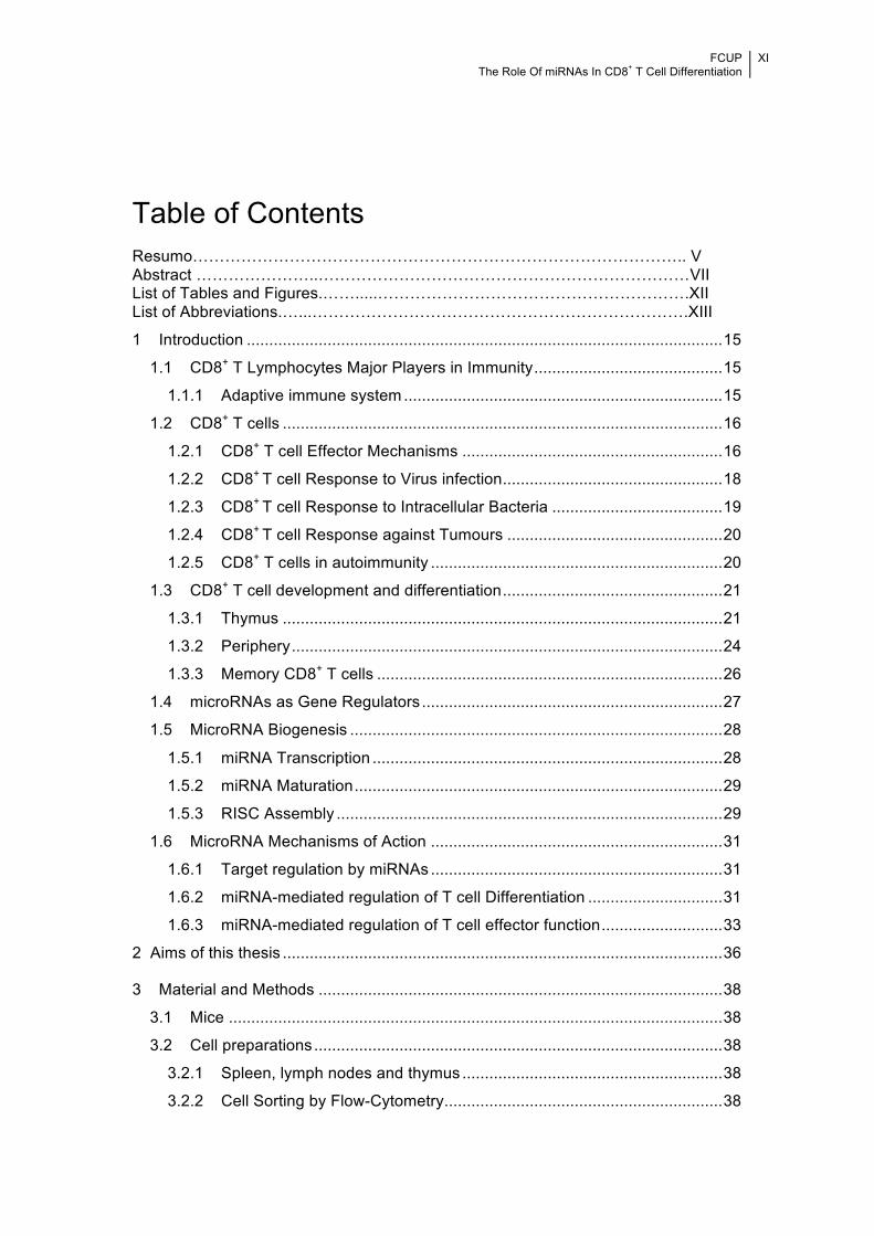
Table of Contents Resumo……………………………………………………………………………….. V Abstract …………………..……………………………………………………………VII List of Tables and Figures.…….....………………………………………………….XII List of Abbreviations.…...…………………………………………………………….XIII
1 Introduction .......................................................................................................... 15
1.1 CD8+ T Lymphocytes Major Players in Immunity .......................................... 15
1.1.1 Adaptive immune system ....................................................................... 15
1.2 CD8+ T cells .................................................................................................. 16
1.2.1 CD8+ T cell Effector Mechanisms .......................................................... 16
1.2.2 CD8+ T cell Response to Virus infection ................................................. 18
1.2.3 CD8+ T cell Response to Intracellular Bacteria ...................................... 19
1.2.4 CD8+ T cell Response against Tumours ................................................ 20
1.2.5 CD8+ T cells in autoimmunity ................................................................. 20
1.3 CD8+ T cell development and differentiation ................................................. 21
1.3.1 Thymus .................................................................................................. 21
1.3.2 Periphery ................................................................................................ 24
1.3.3 Memory CD8+ T cells ............................................................................. 26
1.4 microRNAs as Gene Regulators ................................................................... 27
1.5 MicroRNA Biogenesis ................................................................................... 28
1.5.1 miRNA Transcription .............................................................................. 28
1.5.2 miRNA Maturation .................................................................................. 29
1.5.3 RISC Assembly ...................................................................................... 29
1.6 MicroRNA Mechanisms of Action ................................................................. 31
1.6.1 Target regulation by miRNAs ................................................................. 31
1.6.2 miRNA-mediated regulation of T cell Differentiation .............................. 31
1.6.3 miRNA-mediated regulation of T cell effector function ........................... 33
2 Aims of this thesis .................................................................................................. 36
3 Material and Methods .......................................................................................... 38
3.1 Mice .............................................................................................................. 38
3.2 Cell preparations ........................................................................................... 38
3.2.1 Spleen, lymph nodes and thymus .......................................................... 38
3.2.2 Cell Sorting by Flow-Cytometry .............................................................. 38

FCUP The Role Of miRNAs In CD8+ T Cell Differentiation
XI
!3.3 In vitro cell stimulation and polarisation ........................................................ 39
3.4 Restimulation and FACS staining ................................................................. 40
3.5 Quantitaive RT-PCR ..................................................................................... 40
3.6 Retroviral transduction for miRNA overexpression ....................................... 41
3.7 Redirected cytotoxicity assay ........................................................................ 43
3.8 Statistical analysis ......................................................................................... 43
4 Results ................................................................................................................. 45
4.1 Increased differentiation of IFN-γ producing CD8+ T cells in pLck-Cre
DICERfl/fl mice ........................................................................................................ 45
4.2 YFP expression encompasses intracellular IFN-γ production in Yeti mice ... 46
4.3 YFP+ (IFN-γ+) CD8+ T cells display increased cytotoxicity ............................ 47
4.4 YFP+ versus YFP- CD8+ T cells from Ifng-YFP mice have different miRNA
repertoires .............................................................................................................. 49
4.5 RT-qPCR validation of miRNA expression in thymic YFP+ versus YFP- CD8+
T cells from Ifng-YFP mice ..................................................................................... 49
4.6 RT-qPCR analysis of peripheral YFP+ versus YFP- CD8+ T cells from Ifng-
YFP mice ............................................................................................................... 51
4.7 Candidate miRNAs expression in T cells subsets ........................................ 52
4.8 Candidate miRNAs expression under IFN-γ promoting conditions in vitro .... 53
4.9 Overexpression of candidate miRNAs in the 3T3 cell line ............................ 55
4.10 miRNA-132-3p over-expression down-regulates IFN-γ production in
peripheral CD8+ T cells .......................................................................................... 56
4.11 mRNA targets of miR-132 .......................................................................... 58
5 Discussion ............................................................................................................ 61
6 Bibliography ......................................................................................................... 69
!
FCUP The Role Of miRNAs In CD8+ T Cell Differentiation
XII
!
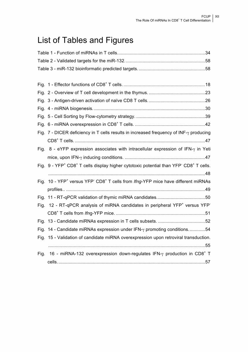
List of Tables and Figures Table 1 - Function of miRNAs in T cells. .................................................................... 34
Table 2 - Validated targets for the miR-132. .............................................................. 58
Table 3 - miR-132 bioinformatic predicted targets. .................................................... 58
Fig. 1 - Effector functions of CD8+ T cells. ................................................................ 18
Fig. 2 - Overview of T cell development in the thymus. ............................................ 23
Fig. 3 - Antigen-driven activation of naïve CD8 T cells. ............................................ 26
Fig. 4 - miRNA biogenesis. ....................................................................................... 30
Fig. 5 - Cell Sorting by Flow-cytometry strategy. ...................................................... 39
Fig. 6 - miRNA overexpression in CD8+ T cells. ....................................................... 42
Fig. 7 - DICER deficiency in T cells results in increased frequency of INF-γ producing
CD8+ T cells. ...................................................................................................... 47
Fig. 8 - eYFP expression associates with intracellular expression of IFN-γ in Yeti
mice, upon IFN-γ inducing conditions. ............................................................... 47
Fig. 9 - YFP+ CD8+ T cells display higher cytotoxic potential than YFP- CD8+ T cells.
........................................................................................................................... 48
Fig. 10 - YFP+ versus YFP- CD8+ T cells from Ifng-YFP mice have different miRNAs
profiles.. ............................................................................................................. 49
Fig. 11 - RT-qPCR validation of thymic miRNA candidates. ..................................... 50
Fig. 12 - RT-qPCR analysis of miRNA candidates in peripheral YFP+ versus YFP-
CD8+ T cells from Ifng-YFP mice. ...................................................................... 51
Fig. 13 - Candidate miRNAs expression in T cells subsets. ..................................... 52
Fig. 14 - Candidate miRNAs expression under IFN-γ promoting conditions. ............ 54
Fig. 15 - Validation of candidate miRNA overexpression upon retroviral transduction.
........................................................................................................................... 55
Fig. 16 - miRNA-132 overexpression down-regulates IFN-γ production in CD8+ T
cells. ................................................................................................................... 57
FCUP The Role Of miRNAs In CD8+ T Cell Differentiation
XIII
!
List of Abbreviations
APC Antigen presenting cell
CTL Cytotoxic T lymphocyte
DN Double negative
DP Double positive
Eomes Eomesodermin
FACS Fluorescence activated cell sorting
IFN Interferon
IL Interleukin
LN Lymph node
mAb Monoclonal antibody
MHC Major histocompatibility complex
miR microRNA
miRNA microRNA
NK Natural Killer
PCR Polymerase chain reaction
qPCR Quantitative Polymerase chain reaction
RNA Ribonucleic acid
RT Reverse transcriptase
SP Single positive
Spl Spleen
TCR T cell receptor
Th T helper cell
TNF Tumor necrosis factor
Treg Regulatory T cell
YFP Yellow Fluorescent Protein
WT Wild type
Note: List of all abbreviations used at least two times on different paragraphs
throughout the manuscript. Additional abbreviations are defined in the text once they
are first introduced to the reader.

!
!!!!!!!!
Introduction !

FCUP The Role Of miRNAs In CD8+ T Cells Differentiation
15
!
1 Introduction !
1.1 CD8+ T Lymphocytes Major Players in Immunity !
1.1.1 Adaptive immune system !The adaptive immune system provides specific protection against pathogens, has the
ability to form ‘memory’ (the basis of vaccination), that enables a rapid response to
previously encountered pathogens, fights nascent cancers and mediates tumor
destruction 1,2.
The major mediators of the adaptive immunity, responsible for providing efficient,
specific and long-lasting immunity, are lymphocytes 3. Lymphocytes can be
subdivided into two separate lineages: thymic-derived (T) lymphocytes (T cells) and
bone marrow-derived (B) lymphocytes (B cells) that further differentiate into plasma
cells to secrete antibodies 4.
T cells are involved in cell-mediated immunity, provide help for B cells to produce
antibodies (humoral immunity) and regulate immune responses 4. T cells are further
classified according to the co-receptor (either CD4 or CD8) that they express at the
cell surface, thus marking CD4+ T cells or CD8+ T cells. These markers are important
for T cell function, because they help to determine the interactions between the T cell
and its T cell receptor (TCR) and other cells expressing major histocompatibility
complex (MHC) molecules. The TCR complex of conventional T cells is a
transmembrane heterodimer composed of two polypeptide chains α and β chains,
which associate with co-receptor CD3 molecules. Each TCR chain consists of a
constant (C) and a variable (V) region, and is formed by a process termed somatic
recombination which joins variable (V), joining (J), and diversity (D) gene segments
to generate combinatorial diversity 4. Additionally, the addition or removal of
nucleotides at the joining sites increases the repertoire of TCRs.
The TCR recognizes specific peptides presented by MHC molecules: CD4+ T cells
recognize MHC class II and CD8+ T cells recognize MHC class I 5. After activation
upon its first encounter with an antigen, T cells proliferate and differentiate into
functional effector T lymphocytes. These include CD8+ cytotoxic T cells, which
secrete pro-inflammatory cytokines and kill cells that are infected with viruses or
other intracellular pathogens; CD4+ helper T cells, which provide essential additional
FCUP The Role Of miRNAs In CD8+ T Cells Differentiation
16
!signals that influence the behavior and activity of B cells and innate immune cells;
and CD4+ regulatory T cells that suppress the activity of other lymphocytes and help
to control immune responses 4.
1.2 CD8+ T cells !Activated CD8+ T cells differentiate into cytotoxic T cells and secrete high amount of
pro-inflammatory cytokines. They are crucial to fight against intracellular pathogens
but also to eliminate tumor cells. On the other hand, they are also involved in the
rejection of transplants and in the pathogenesis of a number of autoimmune diseases 6–11. CD8+ T cells recognize peptides derived from proteins present in infected or
transformed cells, bound to MHC class I molecules (pMHC). The direct lysis of target
cells mediated by CD8+ T cells is one of the most powerful actions of T cells and is
therefore tightly regulated, for example CD8+ T cells require more co-stimulation for
their activation compared to CD4+ T cells 12. Additionally CD8+ T cells can form
memory T cells, contributing to a long-lived immunological protection 13. As CD8+ T
cells are the focus of this thesis, different aspects of CD8+ T cell biology will be
discussed in detail in the following sections.
1.2.1 CD8+ T cell Effector Mechanisms !Naïve CD8+ T cells acquire two critical effector functions after antigen activation:
secretion of cytokines and direct contact-mediated cytotoxicity 14. Concurrent with the
initiation of proliferation is the establishment of gene expression that arms the CD8+
T cell with effector mechanisms to combat infection, such as increased expression of
the master transcription factors Eomesodermin and T-bet, encoded by Tbx21 15,16
(that promotes IFN-γ expression), elevated levels of mTOR and CD25 17, the high
affinity IL-2Rα chain, thereby potentiating IL-2 signals which further support effector
differentiation 18.
The CD8+ T cell effector mechanisms include: a) contact-mediated cytotoxicity which
proceeds through the release of preformed cytolytic molecules into the synaptic cleft
between the CD8+ T cell and its target cell; b) triggering of the TNFR family member
CD95 (Fas) and c) secretion of effector cytokines which contribute to a broad range
of immunological effects and contribute to local inflammatory responses 19.
CD8+ T cells are able to induce cytolysis of infected or abnormal cells by two distinct
molecular pathways 20: the granule exocytosis pathway, dependent on the pore-
forming molecules, or the upregulation of FasL (CD95L), which can initiate
programmed cell death by binding to Fas receptors (CD95) on target cells. Both

FCUP The Role Of miRNAs In CD8+ T Cells Differentiation
17
!pathways, activated in response to signals from the TCR, stimulate the caspase
cascade in the target cell, leading to apoptotic death 21. Efficient lysis by the granule
exocytosis pathway requires the coordinated delivery of perforin and granule
enzymes, such as granzymes A and B, into the target cell 22,23. CD8+ T cells also
release the cytokines interferon-gamma (IFN-γ), tumor necrosis factor-a (TNF-α) and
lymphotoxin-α (LT-α), as well as chemokines that function to recruit and/or activate
the microbicidal activities of effector cells such as macrophages and neutrophils 24
which contribute to host defense. Of note, CD8+ T cells rapidly produce IFN-γ and
TNF-α when their TCR is engaged by the pMHC complex of the target cell but will
immediately cease IFN-γ production when antigenic contact is broken, presumably
until they encounter the next target cell 25,26. TNF-α production is even more strictly
regulated and stops after a short period even when antigen contact is sustained 25.
Effector cytokines produced by antigen-specific CD8+ T cells are likely to be strictly
regulated to minimize the damage to the host 19. Cytokines may also directly interfere
with pathogen attachment or pathogen gene expression, or they may restrict
intracellular replication 27.
In contrast with the on/off cycling of cytokines, expression of the pore-forming
cytotoxic protein perforin is constitutively maintained 26. Another important capacity
that effector and subsets of memory CD8+ T cells acquire is the ability to migrate to
virtually any extra-lymphoid tissues after both localized and systemic infections 28.
1.2.1.1 The function of IFN-γ !Activated CD8+ T cells are crucial providers of the pro-inflammatory cytokine IFN-γ.
IFN-γ was discovered around five decades ago 29 and is critical for the regulation of
the host immune response against viral and intracellular bacterial pathogens.
Based on the type of receptor through which they signal, interferons have been
classified into three major types, and IFN-γ is the sole type II IFN. It is structurally
unrelated to type I IFNs, binds to a different receptor, is encoded by a separate
chromosomal locus and it is produced by T cells, Natural killer (NK) cells,
macrophages and macrophage-derived dendritic cells (mDCs).
The fundamental role of IFN-γ is clearly demonstrated by the study of IFN-γ or IFN-γ
receptor 1- (IFNGR1) deficient mice, which failed to appropriately clear mycobacterial
and other bacterial, parasitic, and viral infections 30–34. Furthermore, IFN-γ is
implicated in tumour surveillance 35,36 and is an important anti-tumoral mediator 37.

FCUP The Role Of miRNAs In CD8+ T Cells Differentiation
18
!Already in 1986, early clinical trials on IFN-γ began to evaluate the therapeutic
potential of its anti-infectious and anti-tumoral functions and until today it has been
used in a wide variety of clinical indications (reviewed in 38).
This notwithstanding, the excessive release of IFN-γ has been associated with the
pathogenesis of chronic inflammatory and autoimmune sclerosis, and plays a pivotal
role in the development and severity of autoimmune diseases such as hashimoto
thyroiditis, type I diabetes, lupus, arthritis and colitis 39–42.
The mechanism whereby IFN-γ leads to systemic autoimmunity remains unclear;
however, the importance of IFN-γ to T cell differentiation and immunoglobulin class
switching in B cells underlines a substantial contribution to adaptive immune
responses in autoimmunity.
!Fig. 1 - Effector functions of CD8+ T cells. Naïve CD8+ T cells acquired their critical effector functions after antigen
activation, and produce cytokines, chemokines and activate the direct contact-mediated cytotoxicity program14.
1.2.2 CD8+ T cell Response to Virus infection !Cytotoxic T cells (CTL) are the main effector T cells that act against cells infected
with viruses. Antigens derived from the virus multiplying inside the infected cell are
displayed on the cells surface, where they are recognized by the antigen receptors of
cytotoxic T cells. Hence, MHC class I surface expression is essential for antiviral
immunity. During virus infection, viral gene products expressed in the cytosol may be
targeted for degradation and presented by class I molecules 43. In this manner, CTL
can act early to eliminate the infected cell before viral replication is complete and
new viruses are released 44.
After antigenic stimulation, CTL up-regulate the expression of cytotoxic granule

FCUP The Role Of miRNAs In CD8+ T Cells Differentiation
19
!proteins, such as granzymes and perforin, and become cytolytic, and gain the ability
to enter non-lymphoid tissues 45–51. They also acquire antiviral effector functions,
including the ability to rapidly produce cytokines, such as IFN-γ, which inhibits viral
replication and is an important inducer of MHC class I molecule expression,
macrophage activation, and drives TNF-α expression. Also CD8+ T cell Fas-
dependent–mediated cytolysis, is critical for resistance against some non-lytic, such
as lymphocytic choriomeningitis virus (LCMV)52 and at least some lytic viruses such
as vesicular stomatitis virus (VSV), influenza virus, Herpes Simplex Virus Type
1(HSV-1) 53–55. In addition to changes in the expression of these effector molecules,
the overall pattern of gene expression is dramatically altered during this activation
phase, and a complex pattern of genetic regulation accompanies T-cell activation
and expansion 48.
Cytotoxic T cells kill infected targets with great precision, sparing adjacent normal
cells. This precision is crucial in minimizing tissue damage while allowing the
eradication of infected cells.
1.2.3 CD8+ T cell Response to Intracellular Bacteria !Whereas CD8+ T cells are principally associated with defence against viral infections,
they also combat intracellular bacterial infections 56.
All intracellular bacteria enter eukaryotic cells in a membrane-bound structure.
Organisms such as Mycobacteria, Salmonella, and Chlamydia survive within a
membrane-bound structure, whereas Listeria and Shigella escape from the vesicle
into the cytosol of the infected cell. As with most pathogens, the immune response to
bacterial infection is complex, and CD8+ T cells are frequently but not always major
effectors in this process 57.
While bacterial entry into the cytosol provides direct access to the MHC class I
antigen-processing pathway, allowing direct priming of CD8+T cells, vacuolar
pathogens such as Mycobacteria and Salmonella, although not directly, also induce
CD8+ T cell protective responses 27.
CD8+ T cells contribute to resistance against intracellular infections with bacterial
pathogens through perforin dependent cytolysis in the case of L. monocytogenes
infection, and its action appears to be most potent in the spleen and dispensable in
the liver. In contrast, no evidence for perforin dependent immunity against Chlamydia
or M. tuberculosis has been reported. In fact, CD8+ T cell-derived production of IFN-γ
is an important mediator of resistance to Chlamydia infection, and this issue remains
to be addressed in MTB infection 58–60.

FCUP The Role Of miRNAs In CD8+ T Cells Differentiation
20
!Although limited, there is evidence suggesting the possibility that the subcellular
location of the bacterial pathogen may impact on the relevance of specific CD8+ T
cell effector mechanisms 27.
1.2.4 CD8+ T cell Response against Tumours !The immune system has three primary roles in the prevention of tumors. First, it
protects the host from virus-induced tumors through elimination or suppression of
viral infections. Second, the timely elimination of pathogens and rapid resolution of
inflammation prevents the establishment of an inflammatory environment conducive
to tumorigenesis. Third, the immune system can specifically identify and eliminate
tumor cells in certain tissues on the basis of their expression of tumor-specific
antigens (TSAs). This third process, referred to as cancer immunosurveillance,
occurs when immune cells like CD8+ CTLs identify (upon recognition of pMHC class I
complexes) transformed cells that have escaped cell-intrinsic tumor-suppressor
mechanisms 61. These transformed cells are directly lysed by CTLs before they can
establish malignancy. Much attention has been given to the role of CD8+ CTLs
because most tumors are MHC class I positive, but negative for MHC class II.
1.2.5 CD8+ T cells in autoimmunity !Although the principal purpose of CD8+ T cells is to protect the host from “non-self”
(i.e. pathogens) and “altered self” (i.e. tumours), there has been an growing evidence
implicating CD8+ T cells in the pathogenesis of several autoimmune disorders, such
as type 1 diabetes, systemic lupus erythematosus, multiple sclerosis (MS),
rheumatoid arthritis (RA), inflammatory bowel disease (IBD) and Psoriasis vulgaris 7,11,62–65.
Much of what is currently known has been provided by employing animal models of
human type 1 diabetes (T1D), such as non-obese diabetic (NOD) mice. In these
mice T1D results from selective destruction of the insulin-producing pancreatic β
cells by autoreactive CD4+ and CD8+ T cells 62. This happens due to the cross-
presentation of autoantigens by dendritic cells to naïve autoreactive CD8+ T cells in
the pancreatic lymph nodes. The quality and quantity of this cross-presentation event
is determinant for whether the cognate CD8+ T cells undergo productive (and
potentially pathogenic) or non-productive activation (leading to antigenic
unresponsiveness or cell death) 66.
Other factors that also influence the outcome of cross-presentation includes the
activation state of the DCs, the cytokines present during the cross-presentation, the

FCUP The Role Of miRNAs In CD8+ T Cells Differentiation
21
!total number of autoantigenic peptide–MHC complexes that are presented on the DC
surface 8, and the affinity of the TCR for peptide–MHC complexes 67,68. In transgenic
models of spontaneous 67 and virus-induced diabetes 68, for example, the incidence
of diabetes clearly correlates with the avidity of the T cell–DC interaction.
CD8+ T cell-mediated killing of target cells might also foster autoimmune disease
progression, since it might facilitate the access of autoantigens to the cross-
presentation pathway.
Upon activation, CD8+ T cells secrete TNF-α and IFN-γ, among other cytokines, and
these cytokines have a role in autoimmune disease. TNF-α and IFN-γ can contribute
to disease progression by ligating TNF receptor 1 on DCs and promoting the
presentation of autoantigens,62. TNF-α also plays important roles in EAE/MS,
inflammatory bowel disease (IBD), experimental myasthenia gravis and rheumatoid
arthritis (RA) 69. CTL-mediated lysis of chondrocytes has been reported d to require
the upregulation of MHC class I molecules by IFN-γ 70.
1.3 CD8+ T cell development and differentiation
1.3.1 Thymus !T cell development is unique relative to other hematopoietic lineages, as T cells
complete the majority of their development in the thymus instead of the bone marrow
(BM) 71. The differentiation in the thymus is a complex and tightly controlled process
that begins with the immigration of bone marrow-derived progenitor cells, and ends
with the generation of self-tolerant, lineage committed T cells capable of performing
an array of immune functions upon recognition of their antigen 72.
Progenitor T cells begin to migrate to the thymus from the early sites of
hematopoiesis at about day 11 of gestation in mice and ninth week of gestation in
humans. Upon thymic settling, progenitors undergo a series of differentiation events
accompanied by migration through the thymic microenvironment where they receive
various inductive signals 73. Progenitors settle the thymus with the potential to
generate multiple blood lineages 74,75. However, as they differentiate and mature in
the thymus, their potential for alternative lineages is constrained, and then
irreversibly lost as the cells ultimately become restricted to the T lineage 71.
The thymus can be divided anatomically into an outer cortex, where most of the
differentiation takes place, and an inner medulla, where the newly formed cells
undergo final maturation before exiting and seeding peripheral lymphoid organs 76.

FCUP The Role Of miRNAs In CD8+ T Cells Differentiation
22
!Early intrathymic progenitor cells lack CD4 and CD8 expression and are referred to
as double negative (DN) cells 77. The precursors that first enter the thymus do not
express the antigen recognition machinery, lacking both the co-receptors CD4 and
CD8 that direct MHC recognition by T cells and the T cell receptors for antigen
recognition, TCRαβ or TCRγδ 72. DN progenitors enter the thymus at the cortico-
medullary junction (CMJ) and subsequently migrate to the subcapsular zone (SCZ) 78–80. This migration is accompanied by a progressive differentiation of these
progenitors indicating that differentiation-inducing signals locate to distinct cortical
regions 81.
Fig. 2 - Overview of T cell development in the thymus. The thymus is organized into cortical and medullary areas,
each of which is characterized by the presence of particular stromal cell types, as well as thymocyte precursors at
different maturation steps. Thymocyte differentiation is characterized by the expression of well-defined cell-surface
markers, including CD4, CD8, CD44 (or CD117) and CD25, as well as the status of the T-cell receptor (TCR).
Interactions between Notch receptor-expressing thymocytes and thymic stromal cells that express Notch ligands are
responsible for the induction of T cell maturation in the thymus, which results in the generation CD4+ T cells and CD8+
T cells, which emigrate from the thymus to establish the peripheral T-cell pool. Image adapted from Zúñiga-Pflücker,
J. et al. 200491.
Based on the expression of the surface molecules CD25 and CD44 on lineage-
negative cells, four early differentiation stages have been defined, double negative 1
to 4 (DN1-4), 82. Differentiation to the DN1 stage (CD25- CD44 high) proceeds in
proximity to the site of thymic entry 83, whereas the consecutive differentiation of

FCUP The Role Of miRNAs In CD8+ T Cells Differentiation
23
!stages DN2 (CD25+ CD44 high) and DN3 (CD25+ CD44 low) occur while cells
migrate outwards of this region into the mid and outer cortex, respectively. DN3 cells
accumulate in the SCZ where they differentiate to DN4 (CD25- CD44-), the pre–
double positive (DP; CD4+ CD8+) stage of development. Transition from the DN3 to
the DN4 stage is accompanied by a reversion of the migration polarity, which finally
guides the DP thymocytes across the cortex toward the medulla, although only
positive selected cells will actually enter the medulla, where the functional maturation
is completed 81 (Fig. 2).
The genes encoding the highly diverse TCRs undergo a carefully programmed series
of DNA rearrangements triggered within the thymus in a stepwise fashion, beginning
in DN stage thymocytes 72. For conventional TCRαβ T cells that recognize peptide
antigens presented by classical MHC molecules, commitment to the T cell lineage is
sealed by rearrangement of the TCRβ gene. The process of β selection tests the
accuracy of this rearrangement event, and drives the proliferation and CD4 and CD8
co-receptor expression by those cells expressing a functional TCRβ chain defined as
one that pairs with the product of the unrearranged pre-Tα gene 84. The result is a
large population of CD4+CD8+ double positive (DP) thymocytes that initiate TCRα
rearrangement. Accurate TCRα rearrangement, along with successful TCRαβ chain
pairing and surface expression is required for positive selection, the second critical
checkpoint for maturing T cells. Positive selection is driven by the successful, low
affinity interaction between the expressed TCRαβ receptor on a DP thymocyte and
self-peptide in the context of self-MHC 84,85. Positive selection rescues DP
thymocytes from the alternative destiny of programmed cell “death by neglect”, and
drives the accurate alignment between co-receptor expression and lineage
commitment 84–86. This process results in a population of CD4+CD8− single positive
(SP) thymocytes that can differentiate into helper T cells upon further recognition of
peptide presented by MHC class II molecules, and CD4−CD8+ SP thymocytes that
can differentiate into cytotoxic T cells upon encounter with antigen presenting cells
whose MHC class I molecules carry the appropriate peptides. At the DP or SP
stages, thymocytes are subjected to negative selection, the third checkpoint that
regulates T cell development. During this process, central to the establishment of
self-tolerance among developing T cells, TCRαβ+ thymocytes that react with high
avidity to self-peptide/MHC complexes are deleted 87–89. The remaining 1% of
thymocytes that successfully transit β selection, positive selection, and negative
selection undergo additional maturation that promotes their regulated exit from the
thymus 72. The naïve T cell population that exits the thymus after this selection
expresses a broad array of unique TCRs that are able to detect a wide range of

FCUP The Role Of miRNAs In CD8+ T Cells Differentiation
24
!foreign antigens. In steady state, the survival of naïve peripheral CD8+ T-cell pools
depends on interleukin-7 (IL-7) and interaction with MHC class I molecules 90.
1.3.2 Periphery !Upon exiting the thymus, mature naïve CD8+ T cells circulate in the blood and
lymphoid organs and signals from the interleukin-7 (IL-7) and IL-15 receptors
promote their survival. These cells lack most of the effector functions characteristic of
activated cytotoxic T lymphocytes (CTLs) 92.
Once in the periphery, naïve T cells constantly survey and sample antigen presenting
cells (APCs) in secondary lymphoid tissues in search of cognate pMHC molecules 90.
The professional APCs, in particular dendritic cells (DC), collect antigen in the
periphery and undergo a maturation process, then travel to secondary lymphoid
organs, including the spleen, lymph nodes (LN), Peyer’s patches (PP), tonsils and
appendix. Naïve T cells, which have yet to encounter their cognate antigen, are
programmed to recirculate continuously between the blood and these organs. If
naïve T cells encounter a cognate antigen presented by MHC molecules the
response is initiated in the immunologic synapse (IS). The outcome is antigenic
stimulation upon engagement of the TCR and CD8, as a co-receptor, that binds to
cognate pMHC complexes presented by APCs 90,93. TCR-mediated signalling induces
phosphorylation of several residues in the CD3 coreceptor chain and activation of ζ-
associated protein of 70 kDa (ZAP-70) and the src-family kinases Lck and Fyn,
thereby initiating downstream signalling pathways that lead to proliferation and
differentiation (35). Costimulatory signals augment TCR signals and prevent
induction of anergy or apoptosis by TCR signalling alone 94,95. The main
costimulatory receptor for T cells is the immunoglobulin (Ig) superfamily member
CD28, which is constitutively expressed on all naive T cells. If the appropriate signals
are present, naïve T cells are programmed to undergo clonal expansion, develop
effector functions, and establish a long-lived memory population following clearance
of antigen 92,96,97. There is now considerable evidence demonstrating that naïve cells
can be stimulated by antigen, referred to as signal 1, and CD28-dependent co-
stimulation, signal 2, to undergo several rounds of cell division. However,
programming for survival, effector function, and memory requires a third signal that
can be provided by either interleukin 12 (IL-12) or type I interferons (IFNs). IL-12
helps to develop a strong clonal expansion and cytolytic activity by the naive cells 90,92. In the absence of this signal, in the case of steady-state presentation of antigen
by immature DCs, the antigen-stimulated cells fail to develop effector functions, and
those that survive long term are tolerant by default 92. Other cytokines (such as TNF

FCUP The Role Of miRNAs In CD8+ T Cells Differentiation
25
!and IL-4) are not secreted in a directional preference and thus can potentially also
act on bystander cells 90.
For both CD4+ and CD8+ T cells, transient exposure to antigen is sufficient to induce
an antigen-dependent program of proliferation and differentiation (2–5), although the
kinetics and efficiency of CD8+T cell proliferation differ substantially from those of
CD4+T cell proliferation. The time of antigen exposure required to launch the
proliferative program for naive CD8+ T cells seems to be less than that required for
naive CD4+ T cells (3,4,8,9). CD8+ T cells also divide sooner and have a faster rate
of cell division than do CD4+ T cells 98–101.
Antigen-driven activation of naïve CD8 T cells is a crucial first step in the
differentiation process which generates heterogeneous subsets of cells, that vary in
their phenotypic attributes, functional capacity, anatomical location, and ability to
persist over time 13 (Fig. 3). Following exposure to antigens in an appropriate
inflammatory environment, these cells undergo a period of massive expansion,
dividing as many as 15–20 times and increasing up to 50,000-fold in number 52,102,103.
After receiving all of the signals necessary to program a response, the resulting CTL
population is limited in its capacity to continue to expand, due to the development of
an anergic state. This anergy can be rapidly reversed by IL-2, and possibly by other
proliferative signals, to allow continued expansion of the CTL population 96. IL-2 was
initially characterized as a potent T-cell growth factor in vitro, but the function during
primary expansion of CD8+ T cells in vivo is dispensable in lymphoid organs and to
some extent required in non-lymphoid tissues 104–106. IL-2 signals during priming,
nonetheless, do contribute to secondary clonal expansion of memory CD8+ T cells,
but it is thus far unknown whether CD4 or DC-derived IL-2 are essential for this
phenomenon or if autocrine IL-2 production is sufficient 105,106. When a CD8+ T cell
response has occurred and antigen is cleared from the system, either because the
initial CTL expansion was sufficient or because IL-2-dependent help was available to
maintain and expand the CTL population until clearance was achieved, the effector
CTLs decline in number as the cells undergo apoptosis, leaving behind a long-lived
population of memory cells 96.

FCUP The Role Of miRNAs In CD8+ T Cells Differentiation
26
!
!Fig. 3 - Antigen-driven activation of naïve CD8 T cells. After the encounter with an antigen presenting cell, CD8+ T
cells go into the differentiation process which generates heterogeneous subsets of cells, that vary in their phenotypic
attributes, functional capacity, anatomical location, and ability to persist over time 13.
!
1.3.3 Memory CD8+ T cells !The cytokines required for programming and maintaining a CD8+ T cell response that
leads to memory are provided, either directly or indirectly, by CD4+ T cells, or by
alternative ways. The CD4+ T cell-mediated help during the primary response or
thereafter and during recall is essential for the generation and maintenance of
functional memory CD8+ T cells to both non-inflammatory and inflammatory agents 107–110.
The maintenance of the memory population is a dynamic process that requires slow
proliferation of the cells in response to endogenous IL-15 and/or IL-7. In terms of the
cell-intrinsic factors required for CD8+ memory T cell generation, it is reported that T-
bet deficiency, particularly coupled with Eomesodermin deficiency impairs memory T
cells development 96.
Memory CD8+ T cells have higher frequencies than naïve T cells and can be
maintained for long periods of time without antigenic stimulation. This increment in
size, the ability to rapidly reactivate and kill upon antigenic stimulation and the varied
tissue distribution makes the memory CD8+ T-cell compartment able to protect its
host in a better and faster way to recurrent infections when compared to naïve T-cell
pools. Depending on the subtype of memory T cell, most but not all cell-surface
markers are gradually reversing to baseline during the ensuing development of
effector cells into memory T cells. Commonly, memory T cells are subdivided into

FCUP The Role Of miRNAs In CD8+ T Cells Differentiation
27
!two main subsets: first, effector memory cells (TEM) are found in non-lymphoid
tissues, and are CD62Llo, CCR7−; and second central memory cells (TCM) that
reside in lymphoid organs, and are CD62Lhi, CCR7+. Additionally, other memory
subsets defined by markers like CD27, CD28, CD43 exist as well as memory CD8+ T
cells with mixed phenotypes, such as CD62Llo, CCR7+ 111. TEM are preferentially
localized in non-lymphoid tissues and mucosal sites and have more rapid cytotoxic
potential, whereas TCM are mainly present in secondary lymphoid organs and
possess superior expansion potential. Thus, protection is essentially linked to the
anatomical location of memory T cell subsets and to the route of infection.
Memory CD8+ T cells in tissues such as the liver and lung are not a sessile, tissue-
resident population, and studies have shown that memory cells continuously enter
non-lymphoid organs from the bloodstream. In contrast, memory CD8+ T cells
present in the brain and lamina propria equilibrated very slowly with blood-borne
memory cells 112.
Interestingly, recent studies correlate the protection of vaccines with multifunctional T
cells (i.e. simultaneous production of the cytokines IFN-γ, TNF, and IL-2) 113, implying
that not only location and quantity of memory CD8+ T cells but also ‘fitness’ should be
considered important for protection. The three phases towards memory cell formation
(expansion, contraction, and memory development) are found in response to many
different types of acute infection and for different epitopes within the same pathogen,
indicating a common pathway for memory T cell formation. At the moment, it is still
controversial how naïve CD8+ T cells differentiate into effector and central memory T
cells and several models have been suggested that regulate this differentiation
process 114. Contrary to the dominant linear model of differentiation, a very recent
study based on single-cell PCR suggested pre-commitment of CD8+ T cells to either
the effector or memory lineages as early as in the first division after activation 115.
1.4 microRNAs as Gene Regulators
MicroRNAs (miRNAs) are critical post-transcriptional regulators of gene expression.
miRNAs are an abundant class of evolutionarily conserved small non-coding RNAs
of approximately ~21-25 nucleotides in length that can regulate gene expression by
binding to the 3´UTR of specific mRNAs leading to the degradation or translational
block of one or more target mRNAs 116.
In mammals, miRNAs are predicted to control the activity of ~50% of all protein-
coding genes 117 setting this post-transcriptional control pathway within nearly every
major gene cascade 118,119. Functional studies indicate that miRNAs participate in the

FCUP The Role Of miRNAs In CD8+ T Cells Differentiation
28
!regulation of diverse aspects of biology, including developmental timing,
differentiation, proliferation, cell death and metabolism 120–122. Changes in their
expression levels are associated with several human pathologies, including cancer,
heart ailments and neurological dysfunctions 123.
Since their discovery in Caenorhabditis elegans in 1993, thousands of miRNAs have
been identified in plants, animals, and viruses by molecular cloning and
bioinformatics approaches 121. Furthermore miRNAs and their accessory proteins
have been shown to be conserved throughout phylogeny 124.
Interestingly miRNAs are now being used as both targets and therapeutics for a
growing industry hoping to tackle the power of RNA-guided gene regulation to
combat disease and infection 119, highlighting the importance of microRNAs in the
gene regulation and therapeutic field.
1.5 MicroRNA Biogenesis
1.5.1 miRNA Transcription
miRNAs are encoded in regions of the genome including both protein coding and
non-coding transcription units. It is estimate that 50% of miRNAs are derived from
non-coding RNA transcripts, while approximately ~40% are located within the introns
of protein coding genes 125,126.
miRNAs are transcribed by RNA polymerase II and bear a 7-methyl guanylate cap at
the 5' end and a poly (A) tail at the 3' end, similar to mRNAs 124,127,128. The nascent
transcripts are referred to as primary (pri-) miRNAs. The pri-miRNAs can be long,
typically over 1kb and contain one or more secondary structures primarily consisting
of extended stem-loop structures 124,129. The pri-miRNA is then processed within the
nucleus by a multiprotein complex called the microprocessor, of which the core
components are the RNase III enzyme Drosha and the double-stranded RNA-binding
domain (dsRBD) protein DGCR8/Pasha 121,130–132.
A special subset of miRNA, mirtrons, bypass the Drosha cleavage step 122, as the
spliced intron itself corresponds exactly to a single, processed miRNA precursor 133 .
After being processed by Drosha or the being excise as a mirtron, the resulting ~70
nucleotide long RNAs, now precursor (pre-)miRNAs, folds into mini-helical structures,
allowing for recognition by Exportin 5 (Exp5), the nuclear export factor responsible
for trafficking pre-miRNAs from the nucleus to the cytoplasm 124,134,135 (Fig. 4).
FCUP The Role Of miRNAs In CD8+ T Cells Differentiation
29
!1.5.2 miRNA Maturation
After the translocation to the cytoplasm, the pre-miRNA is cleaved near the terminal
loop by the RNaseIII enzyme Dicer and generates a ~22-nt double- stranded miRNA 122,136,137 (Fig. 4).
Dicer is highly conserved throughout evolution and it is present in nearly all-
eukaryotic organisms; 122. The cleavage by Dicer takes place in a complex, that
includes the human immunodeficiency virus trans-activating response RNA-binding
protein (TRBP or TARBP2, known as loquacious in Drosophila), which contains three
dsRNA-binding domains and stabilizes the interaction of Dicer with the pre-miRNA 138–140. The resulting 19-24mers double-stranded RNA duplexes contain the mature
miRNAs, also known as guide strand, and its antisense strand, also known as the
passenger strand or miRNA* strand 141.
The antisense miRNA strand can also be found in libraries of cloned miRNAs
although in a much lower frequency than the guide strands 142,143.
1.5.3 RISC Assembly
The final step in miRNA biogenesis is the subsequent incorporation of the miRNA
duplex into the RNA-induced silencing complex (RISC), the effector complex whose
diverse functions can include mRNA cleavage, translation suppression,
transcriptional silencing and heterochromatin formation 124,144 (Fig.4).
The primary component of the RISC complex and the effectors of miRNA-mediated
repression are the Argonaute (Ago) proteins 145. While all of the Ago proteins have
the ability to interact with small RNAs, Ago2 is the only one with RNA cleavage
activity and is thought to play a prominent role in miRNA-mediated silencing.
In vivo, Ago2 associates with Dicer and the double-stranded RNA binding proteins
(TRBP) and also with protein kinase R-activating protein (PACT) to form the RISC
Loading Complex (RLC). This allows the tight coupling of Dicer cleavage to the
incorporation of miRNA into the RISC complex 140. In vitro reconstituted RLC
composed of recombinant Dicer, TRBP, and Ago2 efficiently catalyses pre-miRNA
cleavage 146,147. An important role of the RLC is the unwinding of the double-stranded
miRNA, which is followed by incorporation of the guide strand into the miRNA-
containing ribonucleoprotein (miRNP) complex and degradation of the passenger
strand 148.

FCUP The Role Of miRNAs In CD8+ T Cells Differentiation
30
!
!Fig. 4 - miRNA biogenesis. miRNAs are processed by RNA polymerase II as precursors (pri-miRNA) from intronic,
intergenic or polycistronic gene regions. In the canonical pathway the primary precursor (pri-miRNA) processing
occurs in a two steps cleavage by Drosha together with DGCR8 into 70-nucleotide stem loop known as pre-miRNA,
and then by Dicer. In contrast in the non-canonical miRNA pathway mirtrons are processed by the spliceosome.
Exportin 5 transports the pre-miRNAs to the cytosol, where they are further processed by Dicer together with TRBP
to mature miRNA. The mature miRNA, indicated in red, is incorporated into the RNA-induced silencing complex
(RISC) whose core components are the Argonaute family proteins (Ago1-4). The RISC complex either mediates
mRNA degradation or translational repression (from154)
Usually, the strand with the 5’ terminus located at the thermodynamically less-stable
end of the duplex is the one selected to function as a mature miRNA, and the other
strand is degraded 149–152.
The assembly of RNA into the RISC complex is driven by thermodynamic properties,
though may be also subject to additional regulation as the ratio of miRNA:miRNA*
can vary dramatically, depending on the specific properties of the miRNA duplex, on
the tissue itself and on the developmental stages 142,153. These findings suggest that
FCUP The Role Of miRNAs In CD8+ T Cells Differentiation
31
!differential strand selection could represent a yet unappreciated mechanism of
miRNA regulation. After the assembly of the miRNAs into the miRNPs or miRNA-
induced silencing complexes (miRISCs) the effector complex is ready for targeting
the mRNAs.
1.6 MicroRNA Mechanisms of Action
1.6.1 Target regulation by miRNAs
miRNAs interact with their mRNA targets via sequence-specific base-pairing. With
few exceptions, miRNAs base pair with their targets imperfectly, following a
combination of rules, that have been formulated, based on experimental and
bioinformatics analyses 118. It is known that an individual miRNA is able to control the
expression of more than one target mRNA and that each mRNA may be regulated by
multiple miRNAs.
The 5’ region, termed the “seed” sequence, of the miRNA is used to recognize
complementary regions mainly in the 3’ UTRs of mRNAs leading to deadenylation or
inhibition of translation, ultimately resulting in mRNA decapping and decay 123,155,156.
Recent studies based on ribosome profiling have shown that, although there are
some contribution of translational inhibition, for most of the proteins (>84 %)
regulated by miRNAs 157, the inhibition was accounted by destabilization of the target
mRNA158,159 .
Perfect pairing of a miRNA with its target sites supports direct endonucleolitic
cleavage of the mRNA by Argonaute, both in plants and animals. This is a common
mechanism in plants but is very rare in animals.
1.6.2 miRNA-mediated regulation of T cell Differentiation
The initial studies that established the role of miRNAs in T cells were based on the
generation of mice in which global miRNA maturation was blocked. Several T cell-
specific miRNA-deficient mice were generated through targeting of essential
components of the miRNA biogenesis, namely Dicer, Drosha or Dgcr8. Interestingly,
early elimination of Dicer in the T cell lineage, mediated by lckCre expression,
caused a dramatic (~10-fold) reduction of total thymocyte numbers, most probably
due to increased cell death 160. The relative numbers at the various developmental
stages remained intact and only a 4-fold reduction of peripheral CD8+ T cells was
detected 160. In general it was shown, by using a conditional gene ablation approach,
that Dicer-sufficient cells outgrow Dicer-deficient cells, implicating miRNA in the

FCUP The Role Of miRNAs In CD8+ T Cells Differentiation
32
!general T cell fitness 161. Furthermore, Dicer and Dgcr8-deficient T cells have
decreased T cell proliferation capacity after activation 162,163. Importantly, a striking
effect on T cell differentiation was observed in all T cell-specific miRNA (entire
miRnome) knockout lines. This data clearly demonstrated that miRNAs are involved
in the establishment and/or maintenance of specific T cell identities 163–165. Dicer-
deficient CD4+ T cells were strongly biased towards IFN-γ-producing (Th1) cells,
suggesting a specific role of miRNAs to repress the Th1 program 161,162,165. Other T
cell subsets were also affected, as miRNAs are essential for the homeostasis and
suppressive function of FoxP3+ regulatory T cells. Dicer and Drosha-deficient mice
displayed a scurfy-like disease and their fatal autoimmunity could not be
distinguished from FoxP3-deficient mice 165–167. Consistent with this, Dicer-deficient
Tregs lose the expression of FoxP3 167. In another mouse model using depletion of
AGO2, which is the key AGO protein in haematopoietic cells and crucial for the
maintenance of physiological miRNA levels, an increased proportion of IFN-γ and IL-
4 double producing CD4+ T cells were detected 168,169.
In summary, even if miRNA-deficient T cells are still able to function, the approaches
using T cell-specific miRNA-deficient mice, clearly demonstrated that miRNAs are
implicated in various aspects of T cell biology, namely proliferation, survival and
differentiation. However, the interpretation of the resulting phenotypes is complex, as
the question remains which miRNAs are important for the detected dysfunctions.
Deletion of two counteracting miRNAs can mask their role and prevent them from
establishing a phenotype. Therefore the “second” generation of miRNA research is
focusing on the role of individual miRNAs. The starting point is usually the profiling of
miRNAs in multiple T cell types. Unlike miR-122 or miR-1 which are exclusively
expressed in the liver and the heart, respectively, no individual miRNAs were found,
that are uniquely expressed in lymphocytes 170,171.
miR-181a was identified as an important regulator of positive selection, which may
constitute up to half of the total microRNA content of DP cells 172. It is specifically
enriched at the CD4+ CD8+ DP stage of thymocyte development and suppresses the
expression of Bcl-2, CD69, and T cell receptor (TCR), all of which are important in
positive selection 173. miR-181a has also been shown to increase sensitivity to
peptide antigens by down-regulating multiple phosphatases 174. These findings have
indicated that miR-181a functions as an intrinsic “rheostat” in TCR signalling, which
is very important in T cell development 175. In addition to miR-181a, miR-150 is
important in T cell development as its up-regulation inhibits the expression of the
target gene NOTCH 3 176.

FCUP The Role Of miRNAs In CD8+ T Cells Differentiation
33
!Interestingly, the expression of miRNAs changes during activation and differentiation
of individual T cell subsets. Most miRNAs expressed in resting T cells are down-
regulated after T cell activation, with a few exceptions, such as miR-155 and miR-17-
92, that are selectively up-regulated, suggesting a role for these miRNAs in the
transition of naïve T cells to specialized effector cells 168,177–181.
1.6.3 miRNA-mediated regulation of T cell effector function
Until now several miRNAs have been identified for regulating the differentiation of the
T cells effector function. These are summarized in Table 1.
miRNAs expression rapidly change after activation in CD8+ T cells as well in CD4+ T
cells 170,181, and miR-155 is an example of those miRNAs. It is induced in CD8+ T
cells during activation and rapidly declines to regulate CD8+ memory T cell
differentiation 187. It is also known that miR-155 is required for normal CD8+ T cell
responses to lymphocytic choriomeningitis virus (LCMV) and Listeria monocytes
infections 188.
The miR-17-92 cluster, is also induced in viral infections 189 and promotes
proliferation. The down-regulation of this cluster after the initial expansion phases is
needed for the normal memory CD8+ T cell formation 189. Therefore miR-17-92
cluster appears to be also involved in T cell differentiation.
Another important role of this cluster, is that in its absence the production of many
cytokines including IFN-γ, IL-2, IL-4, IL-5 and TNF-α, was impaired, suggesting that
multiple Th subsets and possibly CD8+ T cells were affected 190.
A microRNA with an important role in cytokine production, especially in the
production of IFN-γ is the miR-29. miR-29 suppresses IFN-γ production by indirectly
targeting two mRNAs coding for transcription factors that promote Th1 differentiation,
Tbx21 (T-bet), and Eomes 162,190 or by directly targeting IFN-γ 190. Interestingly mice
infected with Listeria monocytogenes or Mycobacterium bovis bacillus Calmette-
Guérin (BCG) exhibit a down-regulated miR-29 expression in IFN-γ-producing natural
killer cells, CD4+ T cells, and CD8+ T cells 191.
Also miR-146a, one of the best studied miRNAs in immune cells, mediates immune
suppression in all cell types analyzed so far, namely by inhibiting the production of
IFN-γ and IL-17 in CD4+ and CD8+ T cells 192–195.
Additionally, numerous miRNAs, including miR-150, miR-155, and the let-7 family
were shown to be associated with the development of effector and central memory
CD8+T cells using an in vitro system where CD8+ T cells activity was driven by IL-2 or

FCUP The Role Of miRNAs In CD8+ T Cells Differentiation
34
!IL-15 cytokines. In particular, miR-150 regulates the protein expression of Kv channel
interacting protein 1 (KChiP1) in mouse central memory T cells 196.
In sum, until today several miRNAs were identified as critical regulators of T cell
differentiation. In this thesis we will address the role of miRNAs in the differentiation
of IFN-γ-producing CD8 T cells (see section: 2), as the role of miRNAs in this
differentiation process is poorly understood.
Table 1 - Function of miRNAs in T cells. Adapted from Kroesen et al. 2014 197.

!
Aims of The Thesis

FCUP The Role Of miRNAs In CD8+ T Cells Differentiation
36
!
2 Aims of the thesis !The published observations that CD8+ T cell survival, activation and migration are
compromised in the genetic absence of Dicer, clearly implicate miRNAs in CD8+ T
cell physiology. This notwithstanding, it remains unknown which specific miRNAs
control the development and activation of CD8+ T cells, and how they may regulate
the production of IFN-γ and the cytotoxic function of CD8+ T cells. Such
understanding may pave the way to novel clinical interventions in settings of infection
and cancer where CD8+ T cells are pivotal effectors in immune responses.
In this thesis we set out to investigate the role of miRNAs in the post-transcriptional
regulation of IFN-γ producing CD8+ T cell differentiation. Preliminary data from the
host lab detected increased IFN-γ production in CD8+ T cells from miRNA-deficient
mice, both in the thymus and in the periphery compared to control mice. Building on
these interesting findings we proposed to identify, in this study, specific miRNAs that
might control IFN-γ expression in CD8+ T cells. We used Ifng-YFP reporter mice to
isolate YFP+ and YFP- thymic CD8+ T cell populations for miRnome profiling.
Additionally we proposed to analyze candidate miRNA regulation under TCR and
cytokine stimulation, and finally to conduct functional over-expression experiments.
The final goal would be to identify and characterize specific miRNAs implicated in
IFN-γ−producing CD8+ T cell differentiation.
!!

!
Material and Methods

FCUP The Role Of miRNAs In CD8+ T Cells Differentiation
38
!
3 Material and Methods !
3.1 Mice !For all experiments adult mice (6 – 12 weeks old) were used. C57BL/6J mice (6 – 12
weeks old) were from Jackson Laboratories (Bar Harbor, ME). lckCre-DicerΔ/Δ mice
were kindly provided by Dr. M. Merkenschlager (London, UK). IFNγ-IRES-YFP-
BGHpolyA knockin (YETI) mice were from Jackson Laboratories (Bar Harbor, ME).
Both female and male mice were used, however, individual experiments were
conducted with either females or males. Mice were maintained within the specific-
pathogen-free animal facilities at the Instituto de Medicina Molecular (Lisbon,
Portugal). All experiments involving animals were done in compliance with the
relevant laws and institutional guidelines and were approved by local and European
ethic committees.
!
3.2 Cell preparations
For all in vitro analyses, cells were obtained from spleen, thymus and lymph nodes
(axillary, brachial, inguinal, mesenteric and lumbar).
3.2.1 Spleen, lymph nodes and thymus
The lymph nodes and spleen, or thymus, were strained using BD Falcon Cell strainer
70 µM. Red blood cells (RBC) were lysed with RBC Lysis Buffer from Biolegend
(#420301) and spun for 5 minutes (min) at 1500 rotations per minute (rpm). The
supernatant was discarded and the pellet resuspended in approximately 3 ml of
complete RPMI medium (RPMI media 1640, containing 1mM sodium pyruvate
#11360-039, 1x non-essential acids amines #11140-050, 10 mM hepes #15630-056,
penicillin-streptomycin #15140-122, 50µg/ml gentamycin, 50 µM βmercaptoethanol
and 10% fetal calf serum (FCS) al from Gibco®).
3.2.2 Cell Sorting by Flow-Cytometry
Respective staining antibodies were added to the cells and incubated for at least 15
min at 4ºC in complete RPMI medium. For sorting of T cells subsets the following

FCUP The Role Of miRNAs In CD8+ T Cells Differentiation
39
!antibodies were used: anti-CD3e PerCP-Cy5.5 (145-2C11 e-Bioscience), anti-CD8
APC-eFluor 780 (53-6.7 e-Bioscience), anti-TCRγδ PE (GL3 e-Bioscience), anti-
CD27 PE-Cy7 (LG.7F9 e-Bioscience), anti-CD4 eFluor-450 (RM4-5 e-Bioscience)
and anti-CD25 APC (PC61 BD Pharmingen) (Fig. 5).
For sorting of CD8+ T cells the following antibodies were used: anti-CD3e PerCP-
Cy5.5 (145-2C11 e-Bioscience), and anti-CD4 eFluor-450 (RM4-5 e-Bioscience) and
anti-CD8 APC-eFluor 780 (53-6.7 e-Bioscience). Cells were washed by adding an
excess of medium to the suspension followed by centrifugation for 5 min at 1500
rpm, after which pellets were resuspended in complete RPMI medium and
transferred to 96 well U bottom plates from TPP (#92097) with 200,000 cells in 100µl
per well and kept in an incubator at 37°C and 5% CO2. Cell sort was performed with
either BD FACSAria I or BD FACSAria III cell sorter.
!Fig. 5 - Cell Sorting by Flow-cytometry strategy. CD4+ T cells, CD8+ T cells and gd T cells were sorted within the
gate of CD3+ cells. Within CD4+ gate, Tregs were sorted in CD25+ gate, and within the γδ TCR, CD27+ and CD27-
cells were sorted.
3.3 In vitro cell stimulation and polarisation
CD8+ T cells were sorted by flow cytometry and subjected to various stimulation
conditions for 48h for functional studies or 12h for miRNA expression analysis. Cells
were incubated with various cytokines including IL-12 (5 ng/ml PeproTech), IL-4 (10
ng/ml eBiosciences), IL-7 (10 ng/ml; eBiosciences), IL-18 (10 ng/m PeproTech), IL-2
(10 ng/m PeproTech), IL-15 (10 ng/m PeproTech) and or activated with plate-bound
monoclonal antibody (mAb) anti-CD3 (5 µg/ml; 145.2C11; eBiosciences) and mAb
FCUP The Role Of miRNAs In CD8+ T Cells Differentiation
40
!anti-CD28 (5 mµg/ml; 37.51; eBiosciences).
!
3.4 Restimulation and FACS staining
To measure cytokine secretion, cells were restimulated with PMA (50 ng/ml, Sigma;
P-8139) and Ionomycin (1 µg/ml, Sigma; I-0634) for 4h at 37°C, with the addition of
Brefeldin A (10 µg/ml, Sigma; B-7651). Cells were transferred into Nunc® 96 well V
bottom plates and washed once in FACS buffer (PBS, 0.5% FCS, 2 mM EDTA) (5
min, 1500 rpm). For extracellular cell staining, cells were resuspended in 50µl FACS
buffer with the respective antibodies and incubated for 30 min at 4ºC. For intracellular
cell staining the cells were resuspended in 100µl of fixation/permeabilization (BD
Cytofix/CytopermTM Fixation/Permeabilization kit; # 554714) and incubated for 30
min at 4°C. Cells were washed twice in Perm/wash (BD Cytofix/CytopermTM
Fixation/Permeabilization kit; # 554714) (5 min, 2000 rpm). Cells were resuspended
in 40µl of Perm/wash containing Anti-mouse anti-FcR (2.4G2; BD Pharmingen) and
incubated for 15 min at RT. Cells were then stained without washing with respective
antibodies in an additional 10 µl of the same Perm/wash buffer and incubated for 30
min at RT. For cytokine expression analysis with FACS the following antibodies were
used: anti-IL-17A Alexa Fluor 488 (17B7; eBiosciences), anti-IFN-γ APC (XMG1.2
BD Pharmingen) and anti-TNF-α PE (MP6-XT22 BD Pharmingen).
Cells were washed once in Perm/wash and once in FACS buffer (5 min, 20000 rpm).
Cells were finally resuspended in FACS buffer and FACS acquisition was performed
on BD LSRFortessa cell analyser.
All the data were analysed using FlowJo v.9.3.3 software.
3.5 Quantitaive RT-PCR
All quantitative RT-PCRs were performed in MicroAmp® Optical 384-Well
Reaction Plate (Applied Biosystems® #4343370) using Applied Biosystems
ViiATM 7 Real-Time PCR system. Data was analysed using ViiATM 7
software v1.2.1.
For miRNA expression analysis: RNA was isolated from sorted cell
populations by flow cytometry using miRNeasy Mini Kit (Qiagen). For cDNA
synthesis and real-time PCR amplification the miRCURY LNATM Universal RT
microRNA PCR protocol (Exiqon) was performed. LNATM PCR primer sets
(Exiqon) were used and relative quantification of specific miRNAs to small

FCUP The Role Of miRNAs In CD8+ T Cells Differentiation
41
!RNA reference miR-423-3p was carried out using SYBR on ABI ViiA7 cycler
(Applied Biosystems).
3.6 Retroviral transduction for miRNA overexpression
The native precursor stem loop of our miRNAs candidates were cloned into the
retroviral vector MSCV-IRES-GFP (pMIG) (Fig. 6) using genomic DNA as a template
and the following primers:
miR-451a-F– CGTTTCTGCCTGTAACTCTGG
miR-451a-R – CTCACAAAGGTCCTCCCATC
miR-132-F– GCCGCCTTCAGTAACAGTCT
miR-132-R – AGGACTCCTGATCCCATCG
miR-200a-F – CCTAGTGGGGCTACTCAAGC
miR-200a-R – GCATCCTCACTAACCCTCACA
miR-322-F – CCGGGGACAATAAATGAGAC
miR-322-R –TGCCACCTTGCTATTCACAC
miR-181a-F – CCCAGCATGTGTTATGGTCTT
miR-181a-R – CCGCAGTAACTGAAACTGGA
miR-139-F – AGAGGACTAACAACCCCTGC
miR-139-R – GGAGAGGAGGCATAAGGGTG
Viral supernatant was produced by transfecting the plasmids pMIG, pCMV-VSV-G
and pCL-Eco with Opti-MEM® (Life Technologies) and X-tremeGENE DNA
Transfection Reagent (Roche) in 293T/17 [HEK 293T/17] (ATCC® CRL-11268™)
packaging cell line cultured in TPP 100mm cell culture dishes. The efficiency of the
viral particles was tested by transducing 200.000 NIH/3T3 (ATCC® CRL-1658™)
cells per well, in a Nunc™ 6 well plate, with 8µg/ml of polybrene during a 60 min
centrifugation at 37ºC, 2200 rpm. After 24h the media was changed for fresh media.
And after 120h the cells were washed with FACS buffer (1500 rpm 5 min), and
resuspended in FACS buffer for FACS acquisition.
For viral transduction of peripheral and thymic CD8+ T cells, the cells were cultured in
a TPP 96 well U bottom plate ,with 200.000 sorted CD8+ T cells per well. The cells
were activated for 48h with plate-bound anti-CD3ε mAb (1µg/ml; 145.2C11;
eBiosciences) and anti-CD28 mAb (1µg/ml; 37.51; eBiosciences) in presence with IL-
2 (10 ng/m PeproTech) for peripheral CD8+ T cells and IL-7 (10 ng/ml; eBiosciences)
for thymic CD8+ T cells. Per well 200.000 CD8+ T cells were transduced with miR-

FCUP The Role Of miRNAs In CD8+ T Cells Differentiation
42
!181a-pMIG, miR-200a-pMIG, miR-132-pMIG, miR-139-pMIG or empty-pMIG vector
using 50µl of concentrated viral supernatant. The transduction was performed with
8µg/ml of polybrene during a 60 min centrifugation at 37ºC, 2200 rpm. After 120h
GFP+ (transduced) CD8+ T cells were sorted and restimulated for intracellular
cytokine staining for IFN-γ and TNF-α.
!Fig. 6 - miRNA overexpression in CD8+ T cells. Workflow of the miRNA overexpression in CD8+ T cells using
retroviral transduction. miRNAs were cloned into the retroviral plasmid pMIG, using the precursor stem loop and
genomic DNA as template. Viral particles were produced in 293T/17 [HEK 293T/17] (ATCC® CRL-11268™)
packaging cell line and their efficiency tested in the NIH/3T3 (ATCC® CRL-1658™) cell line. CD8+ T cells were then
transduced with the viral particles for 120h and afterwards sorted for GFP+. Later GFP+ (transduced) CD8+ T cells
were restimulated and analyzed for intracellular cytokine staining for IFN-γ and TNF-α.
!

FCUP The Role Of miRNAs In CD8+ T Cells Differentiation
43
!3.7 Redirected cytotoxicity assay
YFP+ and YFP- CD8+ T cells were sorted and were either cultured in complete RPMI
for 72h in a TPP 96 well U bottom plate, with plate-bound anti-CD3ε mAb (1µg/ml;
145.2C11; eBiosciences) and anti-CD28 mAb (1µg/ml; 37.51; eBiosciences), or the
cells were incubated directly after sorting in a 96 well U bottom plate for 4h at 37ºC
with P815 (ATCC® TIB-64™) mouse mastocytoma cell line, label with DDAOse
(1µM), with soluble anti-CD3e mAb (1µg/ml; 145.2C11; eBiosciences).
After 4h of incubation with P815 (ATCC® TIB-64™) mouse mastocytoma cell line,
cells were transferred to a 96 well V bottom plate, and washed with FACS buffer
(1500 rpm 5 min).
For Annexin V/Dead Cell Apoptosis staining Alexa Fluor® 488 Annexin V/Dead Cell
Apoptosis Kit (V13241; Life Technologies) was used. Cells were ressuspended in
100ul per well in 5x annexin-binding buffer (Component C), and stained with Alexa
Fluor® 488 annexin V (Component A) for 15 min at room temperature.
Cells were resuspended in 5x annexin-binding buffer (Component C) for FACS
acquisition on BD LSRFortessa cell analyser.
3.8 Statistical analysis
A two-tailed non-parametric Mann-Whitney test was used for statistical analysis. P
values of <0.05 were considered significant and are indicated on the figures.

!
Result

FCUP The Role Of miRNAs In CD8+ T Cells Differentiation
45
!
4 Results !
4.1 Increased differentiation of IFN-γ producing CD8+ T cells in pLck-Cre DICERfl/fl mice
To address the overall role of miRNAs in the regulation of IFN-γ producing CD8+ cells
we analyzed T cell-specific miRNA-deficient mice (kindly provided by Dr. M.
Merkenschlager, Imperial Colege, London). Dicer, a crucial enzyme in the miRNA
biogenesis pathway, is essential for the generation of mature miRNAs. As Dicer
mutation in mice or mouse embryonic stem (ES) cells results in developmental failure 198,199, we used T-cell specific conditional Dicer deletion in mice using the Cre-lox
system. The Cre recombinase was expressed under the lck proximal promotor, which
is active from the earliest stages of T cell development 200.Therefore in lckCre-Dicer∆/∆
mice, Dicer is deleted from the CD44-CD25+ (DN3) stage onwards, while -importantly
for our work- does not prevent CD8+ T cell development in the thymus 164.
Fig. 7 - DICER deficiency in T cells results in increased frequency of INF-γ producing CD8+ T cells. Frequency of IFN-
γ+ CD8+ T cells isolated from lckCre-Dicerlox/lox (wild-type control) and lckCre-Dicer∆/∆ mice (left). Representative
intracellular staining of IFN-γ and IL-17 in (A) thymic and (B) peripheral CD8+ T cells isolated from those mice (right).
Each symbol (in A, B, left panels) represents an individual mouse. * P ≤ 0.05 (Mann-Whitney two-tailed test).

FCUP The Role Of miRNAs In CD8+ T Cells Differentiation
46
!
Interestingly, we observed a ~8-fold higher frequency of IFN-γ producing CD8+ T cells
in the thymus (Fig.7 A) of Dicer∆/∆ compared to Dicerlox/lox mice and a ~3-fold higher
frequency of IFN-γ producing CD8+ T cells in the lymph nodes. We additionally
analyzed the frequency of another pro-inflammatory cytokine, IL-17A, that is
expressed in activated CD8+ T cells after differentiation in a specific cytokine milieu
including TGF-β and IL-6. We did not detect any expression of IL-17A in ex vivo
isolated CD8+ T cells in Dicer∆/∆ mice in the thymus (Fig. 7 A) and in the periphery
(Fig. 7 B). These data suggested an important role of miRNAs in selectively regulating
the differentiation of IFN-γ producing CD8+ T cells.
4.2 YFP expression encompasses intracellular IFN-γ production in Yeti mice
!On the basis of the previous data we hypothesized that mature miRNAs, play a role in
the differentiation of IFN-γ producing CD8+ T cells in the thymus and periphery. To
examine the role of specific miRNAs in the differentiation of IFN-γ producing CD8+ T
cells we relied on a bicistronic reporter mouse strain Ifng-YFP (termed ‘‘Yeti’’ mice), in
which transcription of the Ifng gene also results in enhanced yellow fluorescent protein
(eYFP) reporter expression 201. Therefore, immune cells producing IFN-γ mRNA will
also be YFP+ 202 .
To ensure that the expression of YFP fluorescence associated with intracellular IFN-γ
production we stimulated CD8+ T cells of the Ifng-YFP reporter mice with diverse
cytokines cocktails capable of triggering IFN-γ production.
Thymic CD8+ T cells were cultured for four days with plate bound anti-CD3 and anti-
CD28 mAb in the presence of IL-7; IL-7 plus IL-2; IL-7 plus IL-12; IL-7 plus IL-18; and
IL-7, IL-12 plus IL-18. The combination of IL-7 plus IL-12 was the most potent cytokine
cocktail to induce IFN-γ production in thymic CD8+ T cells and resulted in ~ 40% of
IFN-γ producing CD8+ T cells and ~ 90% YFP positive cells (Fig. 8 B).
The peripheral CD8+ T cells were cultured for four days with plate bound anti-CD3 and
anti-CD28 in the presence of IL-2; IL-2 plusIL-4, IL-2 plus IL-18; and IL2, IL-12 plus IL-
18. The combination of IL-2, IL-12 plus IL-18 was the most potent cytokine cocktail to
induce IFN-γ production in peripheral CD8+ T cells and resulted in ~ 60% of IFN-γ
producing CD8+ T cells and ~ 90% YFP positive cells (Fig. 8 B).
YFP expression encompassed intracellular IFN-γ production (Fig. 8 A and B). There
are at least two possible reasons with YFP expression is more frequent than the

FCUP The Role Of miRNAs In CD8+ T Cells Differentiation
47
!actual cytokine: faster translation rates; or increased mRNA or protein stability.
Importantly, they follow similar tendency in the different conditions tested in thymic
and in peripheral CD8+ T cells, as increased frequency of IFN-γ+ CD8+ T cells
associated with increased expression of YFP.
These data suggests that Yeti mice can be a reliable tool to study and characterize
IFN-γ producing CD8+ T cells, without the need of intracellular staining for IFN-γ, since
the expression of YFP encompasses IFN-γ production.
Fig. 8 - eYFP expression associates with intracellular expression of IFN-γ in Yeti mice, upon IFN-γ inducing
conditions. Representative intracellular staining for IFN-γ and IL-17A in (A) thymic and (B) peripheral CD8+ T cells of
Ifng-YFP mice stimulated in vitro for four days with plate bound anti-CD3 and anti-CD28 and in the presence of
different cytokines (IL-7, IL-2, IL-4, IL-12 and IL-18) (left, top). Representative histograms indicating the eYFP
expression in the same conditions (left, bottom). Frequency of IFN-γ+ and YFP+ (A) thymic and (B) peripheral CD8+ T
cells (right).
4.3 YFP+ (IFN-γ+) CD8+ T cells display increased cytotoxicity !CD8+ T cells are known to mediate protection against infection through the secretion
of cytokines, such as IFN-γ and tumor necrosis factor (TNF), and through CTL activity

FCUP The Role Of miRNAs In CD8+ T Cells Differentiation
48
!via the release of cytotoxic granules containing granzymes, granulysins and perforin
(Pfn)203. Interestingly, previous studies have shown a strong relationship of IFN-γ or
perforin expression and the cytotoxic ability of virus-specific CD8+ T cells and CD8-
mediated cytotoxicity against tumors 204–207.
!!
!!Fig. 9 - YFP+ CD8+ T cells display higher cytotoxic potential than YFP- CD8+ T cells. Representative FACS plots of
Annexin-V staining (right) and % of specific lysis (left) of the redirected lysis assay with (DDAO-SE-labelled)
mastocytoma P815 target cell line by CD8+ YFP+ cells and CD8+ YFP- at (A) day 0 and (B) day three.
We confirmed the association of the cytotoxic potential and IFN-γ production in CD8+
T cells isolated from Ifng-YFP reporter mice. We performed a redirected lysis assay
against P815 mastocytoma cell line comparing the cytotoxic responses of YFP+
versus YFP- CD8+ T cells. Interestingly, we observed an increased killing capacity of
freshly isolated YFP+ CD8+ T cells (IFN-γ+) cells, when compared with the YFP- CD8+
T (IFN-γ-) cells at an 1:10 target:effector ratio (40% of lysed cells versus 20% of lysed
cells) (Fig. 9 A). This enhanced cyotoxicity of YFP+ CD8+ T cells diminished in long
term cultures, (60% of lysed cells versus 50% of lysed cells) (Fig. 9 B). This is most
possibly due to the induction of differentiation in YFP- CD8+ T cells. Of note, we
observed the highest percentage of lysis by both YFP+ and YFP- CD8+ T cells at day 3
(Fig. 9 B).

FCUP The Role Of miRNAs In CD8+ T Cells Differentiation
49
!These data confirmed an increased CTL activity of IFN-γ producing CD8+ T cells
compared to IFN-γ non-producing CD8+ T cells in our redirected lysis assay, pointing
to a polyfunctionality of CD8+ T cells.
4.4 YFP+ versus YFP- CD8+ T cells from Ifng-YFP mice have different miRNA repertoires
!We next aimed at identifying individual miRNAs that regulate the differentiation of IFN-
γ+ CD8+ T cells and could mediate the increased frequency of IFN-γ+ CD8+ T cells in
Dicer∆/∆ mice. Following the observations made in sections 2 and 3, we used Yeti
reporter mice to profile the individual miRNAs present in YFP+ versus YFP- CD8+ T
cells. For this purpose we stimulated CD8+ thymocytes from Yeti mice, for two hours
with PMA and Ionomycin, and sorted YFP+ and YFP- cells (Fig.10 A). We then profiled
the microRNAs using ready-to-use PCR panels from Exiqon which analyzes the
expression of 372 miRNAs. We identified 22 microRNAs differentially expressed in
YFP+ versus YFP- CD8+ T cells (Fig.10 B), 12 of which were higher expressed in YFP+
cells, and 10 were higher expressed in YFP- CD8+ cells.
Fig. 10 - YFP+ versus YFP- CD8+ T cells from Ifng-YFP mice have different miRNAs profiles. Representative FACS
staining of YFP expression in thymic CD8+ T cells stimulated for two hours with PMA and Ionomycin (A). Differential
miRNA expression level in thymic YFP+ versus YFP- CD8+ T cells. The expression levels are represented relative to
YFP+CD8+ T cells and converted to LOG2 fold changes (B). !
4.5 RT-qPCR validation of miRNA expression in thymic YFP+
versus YFP- CD8+ T cells from Ifng-YFP mice !We chose to concentrate on the six miRNA candidates based on the highest
differential expression in thymic YFP+ versus YFP- CD8+ T cells. miR-139, miR-451a

FCUP The Role Of miRNAs In CD8+ T Cells Differentiation
50
!and miR-200a were higher expressed and miR-132, miR-322 and miR-181a were
lower expressed in YFP+ compared to YFP- CD8+ T cells. We validated their
differential expression by independent miRNA expression analysis using specific
primers for each miRNA (Exiqon).
Fig. 11 - RT-qPCR validation of thymic miRNA candidates. Quantitaive RT-PCR analysis of (A) miR-139-5p, (B) miR-
451, (C), miR-200a, (D) miR-132, (E) miR-181a-5p, (F) miR-322-5p expression in thymic CD8+ YFP+ (IFN-γ+) and
CD8+ YFP- (IFN-γ-) T cells. Expression levels are relative to the reference miRNA, miR-423-3p. The graphs show the
geometric mean of the miRNA expression from four independent experiments. –The differences were statistically not
significant (n=3-4).
!The RT-qPCR results confirmed the array data for miR-139, miR-451a, and miR-322
(Fig. 11 A, B, E, F). Although the differences are not significant between samples
(due to low sample numbers), the trend observed in the qPCR array is maintained.
miR-139 and miR-451a are higher expressed in YFP+ (Fig. 11 A and B) and miR-322
is higher expressed in YFP- CD8+ T cells (Fig. 11 E and F).
The differentially expression of miR-181a and miR-200a was not confirmed (Fig. 11 C
and E). While miR-132 seemed to be more expressed in YFP+ cells (Fig. 11 D), in
contradiction to the initial screening data (Fig.10 B).
In sum, we could validate the expression pattern of 3 out of our 6 miRNA candidates,
and miR-132 showed an interesting differential expression in YFP+ compared to YFP-
CD8+ T cells which was not detected in the initial screening.

FCUP The Role Of miRNAs In CD8+ T Cells Differentiation
51
!!
4.6 RT-qPCR analysis of peripheral YFP+ versus YFP- CD8+ T cells from Ifng-YFP mice
!We further analyzed the expression of the six miRNA candidates in peripheral eYFP+
versus YFP- CD8+ T cells isolated from pooled LNs and spleen and investigated if the
differential expression detected in the thymus of the candidate miRNAs was
maintained in peripheral CD8+ T cell subsets.
!
Fig. 12 - RT-qPCR analysis of miRNA candidates in peripheral YFP+ versus YFP- CD8+ T cells from Ifng-YFP mice.
Quantitative RT-PCR analysis of (A) miR-139-5p, (B) miR-451, (C), miR-200a, (D) miR-132, (E) miR-181a-5p, (F) miR-
322-5p expression in peripheral CD8+ YFP+ and CD8+ YFP- T cells isolated from Ifng-YFP mice. Expression levels are
relative to the reference miRNA, miR-423-3p. The graphs show the geometric mean of the miRNA expression from
four independent experiments. *P < 0.05 and **P < 0.0021. ns – not significantly different.
!miR-139 was significantly higher expressed in YFP+ CD8+ T cells, P < 0.05, (Fig. 12 A)
when compared with their YFP- counterparts, in accordance with the thymic
expression pattern. By contrast, miR-451a and miR-200a were similarly expressed in
the two subsets (Fig.12 B and C).
miR-132 was significantly higher expressed in YFP+ CD8+ T cells (Fig.12 D), P <
0.0021, in accordance with the previous RT-qPCR profiling in thymic CD8+ T cells.
miR-181a showed a non-significant trend to be higher expressed in YFP- CD8+ T cells
(Fig.12 E). miR-322 was significantly higher expressed in the YFP- CD8+ T cells, P <
0.05 (Fig. 12 F), in accordance with the previous results in thymic CD8+ T cells. In
FCUP The Role Of miRNAs In CD8+ T Cells Differentiation
52
!conclusion, our expression analysis showed various expression patterns in our
miRNA candidates. miR-139 and miR-132 are consistently higher expressed in CD8+
YFP+ CD8+ T cells, and miR-322 is consistently higher expressed in YFP- CD8+ T cells
in both thymus and periphery, whereas another miRNA, e.g miR-451, only showed a
differential expression in the thymus. Additionally we could not confirm a significant
differential expression in miR-200a and miR-181a in neither the thymus nor the
periphery. !
4.7 Candidate miRNAs expression in T cells subsets !We further analysed the expression of our miRNAs candidates in other T cell subsets
such as CD4+ T cells, Tregs (CD4+ CD25+), γδ CD27+ and γδ CD27- T cells.
All of the miRNAs candidates were expressed in the different T cell subsets (Fig.13).
miR-139 was the most abundant miRNA in all subsets (Fig. 13 A), on the other hand
the least expressed miRNA in the majority of the subsets was miR-451a, being only
slightly more abundant in γδ CD27- T cells (Fig. 13 B).
miR-200a was highest expressed in CD8+ T cells and in γδ CD27+ T cells (Fig. 13 C),
two populations prone to produce IFN-γ. Interestingly, miR-132 displayed the opposite
expression pattern and was lowest expressed in naïve CD8+ T cells and in CD27+ γδ T
cells. As for miR-132, miR-322, and miR-181a, they were highest expressed in CD4+
T cells (Fig. 13 D, F and E).
The data shows that our six candidates are expressed in other T cell subsets rather
than being exclusive to CD8+ T cells and we detected distinct expression levels
ranging from low abundant e.g. miR-451, to highly abundant e.g. miR-181a.
Interestingly, some of the miRNAs seemed to be less expressed in subsets linked to
the production of IFN-γ, e.g. miR-132 and miR-322; whereas miR-200a was highly
expressed in those T cell subsets.

FCUP The Role Of miRNAs In CD8+ T Cells Differentiation
53
!
Fig. 13 - Candidate miRNAs expression in T cells subsets. Quantitative RT-PCR analysis of (A) miR-139, (B) miR-
451, (C) miR-200a, (D) miR-132, (E) miR-181a, (F) miR-322 expression in peripheral T cell subsets. Expression levels
are relative to the reference microRNA, miR-423-3p. The graphs show the geometric mean of the miRNA expression
from four independent experiments.
!
4.8 Candidate miRNAs expression under IFN-γ promoting conditions in vitro
!To address potential mechanisms that regulate the miRNA expression of our
candidates, we analysed their expression patterns under different IFN-γ promoting
conditions. We isolated peripheral CD8+ T cells and polarized them overnight in
different conditions and subsequently analysed the miRNA expression levels by RT-
qPCR. We either activated the CD8+ T cells just via TCR stimulation using plate-
bound anti-CD3 and anti-CD28 mAb, or activated the cells via TCR plus cytokines.
The cells were cultured in the presence of either IL- 12 plus IL-18 (a rapid IFN-γ
inducing cytokine cocktail208), or in the presence of IL-15 plus IL-2. IL-15 induces both
IFN-γ production in CD8+ T cells and the proliferation of memory-phenotype CD8+ T
cells. IL-2 supports the growth and survival of naïve T cells. Moreover we showed in
section 2 that in vitro stimulation with only IL-2 induces IFN-γ production in CD8+ T
cells.

FCUP The Role Of miRNAs In CD8+ T Cells Differentiation
54
!
!Fig. 14 - Candidate miRNAs expression under IFN-γ promoting conditions. Quantitative RT-PCR analysis of (A) miR-
139, (B) miR-451, (C) miR-200a, (D) miR-132, (E) miR-181a, (F) miR-322 expression in peripheral CD8+ T cells under
different culture conditions. Peripheral CD8+ T cells were stimulated over night with IL-12 plus IL-18 or IL-2 plus IL-15
with or without TCR activation (plate bound anti-CD3 and anti-CD28). The conditions are indicated below each graph.
Expression levels are relative to the reference microRNA, miR-423-3p. The graphs show the geometric mean of the
miRNA expression from four independent experiments.*P < 0.05.
!miR-139 was the miRNA with the highest expression levels and it was significantly
upregulated in the presence of IL-2 plus lL-15 (*P < 0.05) (Fig. 14 A). Of note, TCR/
CD28 stimulation plus IL-2 and IL-15 did not induce the expression of miR-139 (Fig.
14 A). miR-322 displayed a similar expression pattern (Fig. 14 F). The combination of
the two stimuli (TCR and cytokines) mostly caused a down regulation of the miRNA
expression compared to cytokines alone. miR-451a was the only miRNA upregulated
in all tested cytokine combinations, including in combination with TCR/ CD28
activation (*P < 0.05) (Fig. 14 B). On the contrary, miR-200a and miR-181a
expression levels exhibited no significant differences between the control and the
different stimuli, although both have the tendency to be higher expressed in the
condition of IL-2 plus IL-15 (Fig. 14 C and E).

FCUP The Role Of miRNAs In CD8+ T Cells Differentiation
55
!Interestingly, the expression of miR-132 was induced in conditions that trigger a rapid
production of IFN-γ by CD8+ T cells (Fig. 14 D). miR-132 was upregulated upon
incubation with IL-12 and IL-18 and, on contrary to the previous described miRNAs,
the TCR/ CD28 stimulus further increased miR-132 expression, suggesting a
synergetic effect of TCR/ CD28 plus cytokines (Fig. 14 D). The expression levels were
also upregulated in the presence of IL-2 plus IL-15 and TCR stimulation, *P < 0.05
(Fig. 14 D).
In sum, the expression of our six miRNAs candidates was influenced by the tested
stimuli. With the exception of two miRNAs, miR-200a and miR-181a, the expression of
the other miRNAs was upregulated in response to different stimuli. Of note, only miR-
132 and miR-451 were significantly upregulated in the condition that leads to rapid
IFN-γ production (IL-12 plus IL-18). Interestingly the expression levels of miR-132
were further upregulated upon cytokines plus TCR activation. The expression levels of
the others miRNAs were downregulated when TCR stimulation was added to the
cytokine cocktails.
4.9 - Overexpression of candidate miRNAs in the 3T3 cell line !To investigate the function of our candidate miRNAs in the differentiation of IFN-γ
producing CD8 T+ cells we chose an over-expression screening strategy. We used
retroviral vectors encoding the native precursor stem-loop to transduce activated
CD8+ T cells. The precursor stem loop has to be processed by the cellular miRNA
machinery, which avoids uncontrolled overexpression levels usually observed upon
transfection of mature miR mimics.
The retroviral vectors encoding the precursor stem loops were tested in the easy-to-
transduce 3T3 cell line to confirm that the transduction leads to an overexpression of
the respective miRNA. Furthermore with this test transduction we could evaluate the
produced viral titers. The RT-qPCR analysis of miRNA expression confirmed that
transduction with the cloned precursor stem loops induced a substantial increase in
the miRNAs expression levels of all our candidate miRNAs, ranging from 5 fold to
~5000 fold when compared to control cells (Fig. 15 A-F).

FCUP The Role Of miRNAs In CD8+ T Cells Differentiation
56
!
!Fig. 15 - Validation of candidate miRNA overexpression upon retroviral transduction. Quantitative RT-PCR analysis of
(A) miR-139, (B) miR-451, (C) miR-200a, (D) miR-132, (E) miR-181a, (F) miR-322 expression in 3T3 cells.
Expression levels are relative to the reference microRNA, miR-423-3p. Untransduced – mock-transduced cells. Ctrl -
scrambled sequence control.
!
4.10 miRNA-132-3p over-expression down-regulates IFN-γ production in peripheral CD8+ T cells
!To identify whether the candidate miRNAs had an impact on the differentiation of IFN-
γ producing CD8+ T cells we conducted an over-expressing screening with retroviral
vectors containing the precursor stem loop of miR-181a, miR-139, miR-200a or miR-
132. Total CD8+ T cells were sorted from C57BL/6 mice and activated for 48h,
followed by retroviral transduction with either a control vector expressing GFP or a
miRNA overexpressing vector, containing the native precursor stem loop of the
candidate miRNAs and an IRES-GFP site. 3 days after the retroviral transduction,
GFP+ (transduced) CD8+ T cells were sorted and stained for IFN-γ. Additionally we
analysed the expression of TNF-α, another cytokine produced by activated CD8+ T
cells, to investigate if potential effects on cytokine production are IFN-γ specific (Fig.
16 A).

FCUP The Role Of miRNAs In CD8+ T Cells Differentiation
57
!

FCUP The Role Of miRNAs In CD8+ T Cells Differentiation
58
!
!Fig. 16 - miRNA-132 overexpression down-regulates IFN-γ production in CD8+ T cells. Workflow of miRNAs over-
expression strategy in sorted peripheral naïve CD8+ T cells (A). Representative intracellular staining of peripheral
CD8+ T cells expressing either the retroviral GFP-control vector (Ctrl), or the retroviral vector containing the native
stem loop of miR-200a, miR-132, miR-181a or miR-139 (B). Histograms and graphs indicate the frequency of IFN-γ
and TNF-α producing CD8+ T cells. Validation of miR-132 overexpression experiment in CD8+ T cells (C). The graphs
indicate the frequency of IFN-γ and TNF-α producing CD8+ T cells.
We detected no difference in the frequency of IFN-γ and TNF-α producing CD8+ T
cells when overexpressing miR-200a, miR-181a and miR-139 (Fig.16 B). By contrast,
the overexpression of miR-132 reduced the frequency of IFN-γ-producing CD8+ T cells
(~ 20% reduction), whereas the frequency of TNF-α-producing CD8+ was unchanged,
thus confirming a specific role of miR-132 in the differentiation of IFN-γ-producing
CD8+ T cells (Fig.16 B). As validation we repeated the experiments 4 times and
confirmed that miR-132 overexpression significantly decreases the IFN-γ production in
peripheral CD8+ T cells (P < 0.05) (Fig. 16 C).
The results obtained so far indicate that miR-132 is implicated in the differentiation of
IFN-γ-producing CD8+ T cells, whereas the overexpression of the other miRNA
candidates did not influence IFN-γ production by peripheral CD8+ T cells.
!
4.11 mRNA targets of miR-132 !miRNAs mediate their functions by repressing sequence-specific target mRNAs. To
further explore how miR-132 is implicated in CD8+ T cell differentiation, we have
enquired its mRNA targets based on previous experimental evidence (validated
targets) (Table 2), or bioinformatics prediction tools (predicted targets) (Table 3). One
interesting validated target is STAT4 which is a critical transcription factor know to
drive the differentiation of Th1 (IFN-γ-producing ) CD4+ T cells. Additionally, Twist1
and Runx3 (predicted targets), are involved in the differentiation of IFN-γ producing T

FCUP The Role Of miRNAs In CD8+ T Cells Differentiation
59
!cells. Whereas Twist 1 is a negative regulator of IFN-γ differentiation, Runx3 is a
positive regulator of the effector CTL program in CD8+ T cells and drives IFN-γ
expression in CD8+ T cells by inducing Eomes 209. Future experiments, including RT-
qPCR analysis (in control CD8+ T cells versus miR-132-overexpressing CD8+ T cells)
will be performed to clarify which targets are important for the role of miR-132 in the
differentiation of IFN-γ producing CD8+ T cells.!!
Table 2 - Validated targets for the miR-132. List made from targets previously validated experimentally by reporter
assays, western blot or qPCR.
!
Table 3 - miR-132 bioinformatic predicted targets. Putative targets were chosen with a score of 3 out of 5 in the
bioinformatics programs. The Bioinformatic programs used were MIRanda, MirTarget2, PicTar, PITA and RNAhybrid
!!
Targets Reporter+Assays Western+Blot qPCREp300 x x xAChE xJarid1a xSTAT4 xIRAK4 x
Strong+EvidenceValidation+Methods
Gene MIRanda MirTarget2 PicTar PITA RNAhybrid6 SumTwist1 1 0 0 1 1 3IRF4 1 0 0 1 1 3Runx3 1 0 0 1 1 3
Predicted6targets6mmuBmiRB132

!
!
!!!!!!!!!
Discussion

FCUP The Role Of miRNAs In CD8+ T Cells Differentiation
61
!!
5 Discussion !miRNAs are key regulators of mammalian genome and add another layer of
complexity to the regulation of gene expression. During the last several years,
mounting evidence has shown that miRNAs are critical not only for the development
of immune cells, but also to regulate the function of both the innate and adaptive
arms of the immune system185,210,211. In this study we investigated the role of miRNAs
in the differentiation of IFN-γ−producing CD8+ T cells.
Others have previously shown that miRNA-deficient T cells exhibit defects in
proliferation and in the regulation of cytokine production164,165,212,213. Regarding CD8+
T cells it was also shown that in Dicer-deficient mice there is a decrease in the
numbers of CD8+ mature cells and a failure in CD8+ effector T cell expansion in
response to an infectious challenge in vivo 214. Notwithstanding, the ability of miRNA-
deficient CD8+ T cells to secrete IFN-γ had not been addressed.
In this study we used CD8+ T cells from pLck-Cre DICERfl/fl mice, as a source of
miRNA-deficient CD8+ T cells. In these mice miRNAs are absent from early T cell
development onwards. We showed that pLck-Cre DICERfl/fl mice have a significantly
increased frequency of IFN-γ producing CD8+ T cells (upon short restimulation in
vitro), both in the periphery and in the thymus when compared to wt controls. This
ability of rapid production of IFN-γ by naïve CD8+ T cells in pLck-Cre DICERfl/fl mice
suggests an important role for DICER/miRNAs in the post-transcriptional regulation
of IFN-γ expression and resembles the behavior of innate T cells, which rapidly
secrete cytokines after activation.
Conventional naïve CD8+ T cells require activation, expansion, and effector cell
differentiation before producing IFN-γ and participating in a protective immune
response. Memory CD8+ T cells are, in turn, major direct producers of IFN-γ 215.
Interestingly, a population of CD8+ T cells with “innate-like” function has been
described recently, that expresses a memory-like phenotype and can rapidly secrete
IFN-γ216. These innate-like CD8+ T cells have been identified in the thymus of several
gene-deficient mouse strains, including Itk, KLF2, CBP and Id3 knock-out mice 216,
have the CD44hiCD122+ phenotype and function of memory CD8+ T cells, without
previous exposure to antigens 216–218. These CD8+ T cells can rapidly secrete IFN-γ
upon stimulation with IL-12 and IL-18, and play important roles in the innate
response against infections such as Listeria monocytogenes, or chronic infections

FCUP The Role Of miRNAs In CD8+ T Cells Differentiation
62
!with viruses such as Herpes virus 208,215,219,220. Also interestingly, BALB/c but not
C57BL/6 mice have CD8 SP thymocytes that contain a distinct sub-population of
EomeshiT-betloCD44hiCD122hi innate phenotype CD8+ T cells that produce IFN-γ 221.
The detection of an increased frequency of IFN-γ producing CD8+ T cells in the
thymus of miRNA-deficient mice raises the possibility that miRNAs are important
negative regulators of the differentiation of innate-like CD8+ T cells. This specific
question will be addressed in future experiments where the expression of surface
markers of miRNA-deficient thymic CD8+T cells will be analysed (further discussed
below).
In this work we focused on deciphering which individual miRNA regulate the
differentiation of IFN-γ-producing CD8+ T cells. We performed a miRNA profiling in
thymic IFN-γ-expressing CD8+ T cells versus IFN-γ negative CD8+ T cells using Ifng-
YFP reporter (Yeti) mice. We reasoned that miRNAs with distinct abundance in YFP+
versus YFP- CD8+ T cells could be important mediators of IFN-γ differentiation in
CD8+ T cells. In total we identified 22 differentially expressed miRNAs.
For practical reasons, we narrowed down our initial list to the six microRNA
candidates that showed the highest differential expression in YFP+ versus YFP- CD8+
T cells. miR-139, miR-451a and miR-200a were significantly higher expressed in
YFP+ CD8+ thymocytes whereas miR-132, miR-322 and miR-181a were significantly
lower expressed in this cell population when compared to their YFP- counterparts.
We further validated the results of the qPCR profiling in peripheral CD8+ T cells as
well as in CD8+ thymocytes. The candidates showed a similar pattern of expression
in peripheral CD8+ T cells as in CD8+ thymocytes.
Our six miRNAs candidates were not exclusively expressed in CD8+ T cells, as they
were found in other T cells subsets. They had different levels of expression in each
subset, reinforcing the idea that miRNAs tend to be highly pleiotropic, with a given
miRNA having several functions and most likely different mRNA targets. Of note,
miR-200a was the only miRNA significantly higher expressed in CD8+ T cells.
We further investigated the role of IFN-γ driving-cytokines and TCR signalling on the
expression of our candidate miRNAs in CD8+ T cells. Based on previous knowledge
that activated murine T cells 222 and a subset of human CD8+ T cells express more
IL-12 and IL-18 receptors 223, and these activated CD8+ T cells are prone to produce
IFN-γ, we decided to analyze the influence of these cytokines in the production of our
candidate miRNAs. In addition we tested IL-15 and IL-2 conditions, which were
previously shown to induce IFNγ production224225. Moreover, IL-15 promotes the
proliferation of memory-phenotype CD8+ T cells 224 and IL-2 is known to support the

FCUP The Role Of miRNAs In CD8+ T Cells Differentiation
63
!growth and survival of naïve T cells 226. Our results showed that miR-139 and miR-
322 were the only miRNAs with higher expression with IL-2 plus IL-15, suggesting
that these miRNAs may be involved in memory CD8+ T cell development. This
possibility should be investigated in the future.
miR-132 was the most responsive of our miRNA candidates to the cytokines
promoting IFN-γ production and TCR signalling. Thus, the highest expression of miR-
132 was obtained with the combination of anti-CD3/anti-CD28 plus IL-12 and IL-18.
This cytokine combination has been shown to induce a rapid IFN-γ secretion by
CD8+ T cells and provide innate protection in a non–antigen-specific manner 208. In
addition Listeria monocytogenes infected macrophages secrete IL-12 and IL-18,
which act upon NK cells and primed T cells to induce the production of IFN-γ 223,227.
Thus our results suggest that miR-132 might be involved in a setting where IFN-γ is
rapidly produced and needed.
It was also curious to observe that only when the cells received TCR/ CD28 signals
there was a significant increase in the miR-132 expression by the IL-2 and IL-15,
suggesting that for these cytokines to have a significant impact in the miR-132
expression the cells may have to be previously activated. Since these cytokines
promote the differentiation of CD8+ T cells into memory CD8+ T cells, maybe miR-
132 is also important in memory CD8+ T cells.
The functional relevance of the all miRNA candidates could not be tested by an over-
expression screening due to technical limitations of the viral production. We were not
able to produce high viral titers for the overexpression constructs of miR-451 and
miR-322. Additionally, we performed our functional screening in peripheral CD8+ T
cells but not in CD8+ thymocytes as these cells are more difficult to transduce. We
could not obtain a level of transduced thymocytes to allow conclusive results.
Most retroviral vectors (RVs) are based on the molony murine leukemia virus
(MoMLV), which has a simple genome encoding the gag, pol, and env, flanked by
long terminal repeats (LTR) 228. Upon cell entry, the RNA is copied by the reverse
transcriptase enzyme into double-stranded DNA, which becomes stably integrated
into one of the host chromosomes, thus allowing long-term expression of inserted
genes 229. Using a stable transduction with the original stem loop of our miRNA
contributes to a more physiological result avoiding false positive results introduced by
supra-physiological increase in miRNA levels generally achieved after transient
transfection 230.
Despite the fact that the transgene is integrated into the host cell genome, by the
retroviral vectors, persistent gene expression is not guaranteed as silencing of

FCUP The Role Of miRNAs In CD8+ T Cells Differentiation
64
!transcriptional units may occur over time 231. Another major limitation of RVs is their
inability to infect non-dividing cells. This is due to the fact that the pre-integration
complex cannot cross the intact nuclear membrane, which is only disassembled
during mitosis 229. To over-come this obstacle we had to culture the cells always with
plate-bound anti-CD3 and anti-CD28 mAb.
The production of the retroviral vectors is a crucial step for the success of the
experiments, and there are some factors that influence the vector production and
titers, such as the construction of the vector backbone232 and the transgene to
transfer229, extracellular factors, culture conditions of the packaging cells and
harvesting the virus. In general, a bigger vector size leads to a lower efficiency of
viral particles, which can be neglected in our case as the miRNA cassettes were very
small ~400bp. In addition, the expression of the transgene can be toxic for the
producer cells or can significantly reduce vector production (e.g. GFP or miRNA
itself). This could have influenced the titers and the production of some vectors, such
as miR-451 and miR-322.
Our viral vectors were produced in mammalian adherent cell line, HEK 293T (Human
embryonic kidney 293 cells with large T antigen) cell line. These cells produce
variable quantities of extracellular matrix proteins partially consisting of
proteoglycans. These macromolecules are negatively charged, of variable size, and
act as inhibitors during cell transduction, thus eventually reducing significantly the
transduction efficiency despite high vector titers 233,234. To avoid this problem, a
suspension cell line could be used to produce the virus.
The replacement of glucose by fructose in the culture medium has also been
described as having potential for improving vector production rates and titers 235, but
this would need to be tested in our cultures.
In sum, future experiments are needed to optimize the viral transduction efficiency in
CD8+ thymocytes and peripheral T cells. If these technical limitations are solved,
retroviral-mediated over-expression or knock-down strategies of individual miRNAs
are a powerful tool to study the role of miRNAs in cell differentiation.
The over expression of miR-139, miR-181a and miR-200a had no significant impact
on IFN-γ production by CD8+ T cells , but the over-expression of miR-132 resulted in
decreased production of IFN-γ in peripheral CD8+ T cells. In line with this is the
observation that in human NK cells both the miR-132/miR-212 cluster and miR-200a
are negative feedback regulator of IL-12 signalling through targeting of STAT4, an
inducer of IFN-γ production 236. It is possible, thus, that miR-132 may also target
STAT4 in CD8+ T cells. Of note is the fact that, in our experiments, the expression of

FCUP The Role Of miRNAs In CD8+ T Cells Differentiation
65
!miR-200a in CD8+ T cells was not influenced by the cytokines IL-12 and IL-18 as
shown for human NK cells237. This suggests that the role of miR-200a might be
dissociated from the control of IFN-γ in CD8+ T cells.
In vivo, cytokine-mediated T cell activation is in many ways a double-edged sword. In
some cases, bystander T cell activation can be beneficial as is the case when CD8+
T cells produce IFN-γ in response to cytokines triggered by infection with Listeria
monocytogenes and provide innate protection. On the other hand, endotoxic shock
associated with Gram-negative bacteria can be exacerbated by a cytokine storm that
includes IFN-γ-mediated immunopathology due to CD8+ T cells and NK cells 238.
Therefore the miR-132-based regulation may constitute an important negative
feedback mechanism to prevent the IFN-γ mediated immunopathology in bacterial
infection or in autoimmune pathology.
The most striking evidence, so far, for the effect of miR-132 in the immune system
comes from a mouse model deficient in the miR-212/132 cluster (where miR-132 and
miR-212, which are tandem miRNAs at the same chromosomal location and sharing
close sequences, are simultaneously removed). These mice have higher resistance
to the development of experimental autoimmune encephalomyelitis (EAE), a
prototype for T-cell-mediated autoimmune disease in general, and lower frequencies
of Th1 T cells, responsible for IFN-γ production 239. These results do not go along
with our overexpression phenotype in which excess of miR132 instead causes a
decrease in IFN-γ production. As the loss of function phenotype comes from a double
knockout mouse, we cannot discard the possibility that the role of miR-212 is distinct
from that of miR-132. It will be important to clarify these issues in future experiments,
where the outcome of overexpression studies with miR-212 alone or both miR-212
and miR-132 should be analyzed.
Finally, we investigated miR-132 potential mRNA targets based on literature search
and bioinformatic analyses. Several of the validated targets for miR-132 found in the
literature are known players involved in the inflammatory and IFN-γ response,
including Stat4 AChE, HB-EGF, p300, MeCP2 and SirT1.
Stat4 is a critical Th1 driving transcription factor and acts together with STAT1 to
induce T-bet. It still remains to be established if Stat4 is also implicated in the
differentiation of IFN-γ-producing CD8+ cells.
Runx3 is one of the predicted targets that came out of our bioinformatics analysis.
Runx3 is present in naive CD8+ T cells before activation and has a positive role in the
induction of Eomesodermin (Eomes), granzyme B, perforin, and IFN-γ 240 and it is
also involved in CD8+ effector T cell differentiation 241. This is an interesting

FCUP The Role Of miRNAs In CD8+ T Cells Differentiation
66
!candidate that being targeted by miR-132 could down-regulate the production of IFN-
γ, as it directly impacts on the master transcription factor Eomes.
Another predicted target is the transcription factor Twist1. In Th1 cells, NF-kB, NFAT,
and IL-12/STAT4 signalling can induce Twist1 expression, and Twist1 limits
inflammation by suppressing IFN-γ and TNF-α production by decreasing expression
and function of transcription factors, including T-bet, Stat4, and Runx3 242. The
precise role of this transcription factor remains to be established in CD8+ T cells but
the fact that miR-132 overexpression decreases IFN-γ+ CD8+ T cell frequency does
not go along with the observation that Twist1 represses IFN-γ production in CD4+ T
cells.28. The targeting of this transcription factor may be an example where the
miRNA activates the expression rather than represses it, as it has been reported
during the last years by several groups 243–246 247. If that were the case, then the over-
expression of miR-132 would activate the expression of Twist1, which would
decrease T-bet, STAT4 and Runx3, decreasing as well the IFN-γ production.
In addition to analyzing already validated or bioinformatics-predicted mRNA targets,
further experimental studies will be needed to identify novel targets that could explain
the IFN-γ phenotype in miR-132 overexpressing CD8+T cells. One method to use
could be differential! high-throughput sequencing of RNA isolated by crosslinking
immunoprecipitation (HITS-CLIP) analysis. This technique uses ultraviolet irradiation
to covalently crosslink RNA–protein (Ago-RNA) complexes that are in direct contact
and partial RNA digestion to reduce bound RNA to fragments that can be sequenced
by high-throughput methods. Ago HITS-CLIP can simultaneously identify Ago-bound
miRNAs and the nearby mRNA sites 248. This should be complemented by luciferase
reporter assay as a final confirmation that a given mRNA is targeted by a specific
miRNA.
To characterize a possible role of our candidate miRNAs in IFN-γ producing CD8+ T
cells differentiation, it would be interesting to perform in vitro experiments, such as
reaggregate thymic organ cultures (RTOC), where our miRNA of interest would be
over-expressed in CD8+ thymocytes during their development. With this tool we could
characterize the development of CD8+ T cells, and check for surface markers that
could indicate if there was a bias towards the development of memory like phenotype
CD8+ T cells, the so called “innate-like CD8+T cells”.
In summary, we identified miR-132 as a negative feedback regulator of IFN-γ
production in CD8+ T cells. We additionally showed, that miR-132 expression is
modulated by cytokines promoting IFN-γ production. Several promising mRNA
targets of miR-132 include the known STAT4 and the novel predicted Twist1 and

FCUP The Role Of miRNAs In CD8+ T Cells Differentiation
67
!Runx3. Future experiments will address if these targets are responsible for the
observed phenotype. Collectively, miR-132 is likely to play an important role in the
differentiation of IFN-γ producing CD8+ T cells, which are crucial in immune
responses against intracellular pathogens and tumours.

!
Bibliography
FCUP The Role Of miRNAs In CD8+ T Cells Differentiation
69
!
6 Bibliography !
1. Flajnik, M. F. & Kasahara, M. Origin and evolution of the adaptive immune system: genetic events and selective pressures. Nat. Rev. Genet. 11, 47–59 (2010).
2. Ostrand-Rosenberg, S. Immune surveillance: a balance between protumor and antitumor immunity. Curr. Opin. Genet. Dev. 18, 11–8 (2008).
3. Pancer, Z. & Cooper, M. D. The evolution of adaptive immunity. Annu. Rev. Immunol. 24, 497–518 (2006).
4. Murphy, K. M., Mowat, A. & Weaver, C. T. Garland Science - Janeway’s Immunobiology, Eighth Edition Resources. (Garland Science, Taylor & Francis Group, LLC, 2012).
5. Smith-Garvin, J. E., Koretzky, G. A. & Jordan, M. S. T cell activation. Annu. Rev. Immunol. 27, 591–619 (2009).
6. Martin, P. J., Akatsuka, Y., Hahne, M. & Sale, G. Involvement of donor T-cell cytotoxic effector mechanisms in preventing allogeneic marrow graft rejection. Blood 92, 2177–81 (1998).
7. Gudjonsson, J. E., Johnston, A., Sigmundsdottir, H. & Valdimarsson, H. Immunopathogenic mechanisms in psoriasis. Clin. Exp. Immunol. 135, 1–8 (2004).
8. Kurts, C. et al. CD8 T cell ignorance or tolerance to islet antigens depends on antigen dose. Proc. Natl. Acad. Sci. U. S. A. 96, 12703–7 (1999).
9. Kägi, D. et al. Reduced incidence and delayed onset of diabetes in perforin-deficient nonobese diabetic mice. J. Exp. Med. 186, 989–97 (1997).
10. Huseby, E. S. et al. A pathogenic role for myelin-specific CD8(+) T cells in a model for multiple sclerosis. J. Exp. Med. 194, 669–76 (2001).
11. Kang, Y. M. et al. CD8 T cells are required for the formation of ectopic germinal centers in rheumatoid synovitis. J. Exp. Med. 195, 1325–36 (2002).
12. Seder, R. a & Ahmed, R. Similarities and differences in CD4+ and CD8+ effector and memory T cell generation. Nat. Immunol. 4, 835–42 (2003).
13. Cox, M., Kahan, S. & Zajac, A. Anti-viral CD8 T cells and the cytokines that they love. Virology 435, 157–169 (2013).
14. Chowdhury, D. & Lieberman, J. Death by a thousand cuts: granzyme pathways of programmed cell death. Annu. Rev. Immunol. 26, 389–420 (2008).

FCUP The Role Of miRNAs In CD8+ T Cells Differentiation
70
!15. Pearce, E. L. et al. Control of effector CD8+ T cell function by the transcription
factor Eomesodermin. Science 302, 1041–3 (2003).
16. Intlekofer, A. M. et al. Effector and memory CD8+ T cell fate coupled by T-bet and eomesodermin. Nat. Immunol. 6, 1236–44 (2005).
17. Valenzuela, J., Schmidt, C. & Mescher, M. The roles of IL-12 in providing a third signal for clonal expansion of naive CD8 T cells. J. Immunol. 169, 6842–9 (2002).
18. Cox, M., Harrington, L. & Zajac, A. Cytokines and the Inception of CD8 T Cell Responses. Trends Immunol. 32, 180–186 (2011).
19. Slifka, M. K. & Whitton, J. L. Antigen-Specific Regulation of T Cell–Mediated Cytokine Production. Immunity 12, 451–457 (2000).
20. Berke, G. The CTL’s kiss of death. Cell 81, 9–12 (1995).
21. Shresta, S., Pham, C. T., Thomas, D. A., Graubert, T. A. & Ley, T. J. How do cytotoxic lymphocytes kill their targets? Curr. Opin. Immunol. 10, 581–7 (1998).
22. Ebnet, K. et al. Granzyme A-deficient mice retain potent cell-mediated cytotoxicity. EMBO J. 14, 4230–9 (1995).
23. Heusel, J. W., Wesselschmidt, R. L., Shresta, S., Russell, J. H. & Ley, T. J. Cytotoxic lymphocytes require granzyme B for the rapid induction of DNA fragmentation and apoptosis in allogeneic target cells. Cell 76, 977–87 (1994).
24. Harty, J. T. & Bevan, M. J. Responses of CD8(+) T cells to intracellular bacteria. Curr. Opin. Immunol. 11, 89–93 (1999).
25. Badovinac, V. P., Corbin, G. A. & Harty, J. T. Cutting edge: OFF cycling of TNF production by antigen-specific CD8+ T cells is antigen independent. J. Immunol. 165, 5387–91 (2000).
26. Slifka, M. K., Rodriguez, F. & Whitton, J. L. Rapid on/off cycling of cytokine production by virus-specific CD8+ T cells. Nature 401, 76–9 (1999).
27. Harty, J. T., Tvinnereim, a R. & White, D. W. CD8+ T cell effector mechanisms in resistance to infection. Annu. Rev. Immunol. 18, 275–308 (2000).
28. Masopust, D., Vezys, V., Marzo, A. L. & Lefrançois, L. Preferential localization of effector memory cells in nonlymphoid tissue. Science 291, 2413–7 (2001).
29. Green, J. A., Cooperband, S. R. & Kibrick, S. Immune specific induction of interferon production in cultures of human blood lymphocytes. Science 164, 1415–7 (1969).
30. Huang, S. et al. Immune response in mice that lack the interferon-gamma receptor. Science 259, 1742–5 (1993).

FCUP The Role Of miRNAs In CD8+ T Cells Differentiation
71
!31. Pearl, J. E., Saunders, B., Ehlers, S., Orme, I. M. & Cooper, A. M.
Inflammation and lymphocyte activation during mycobacterial infection in the interferon-gamma-deficient mouse. Cell. Immunol. 211, 43–50 (2001).
32. Van den Broek, M. F., Müller, U., Huang, S., Aguet, M. & Zinkernagel, R. M. Antiviral defense in mice lacking both alpha/beta and gamma interferon receptors. J. Virol. 69, 4792–6 (1995).
33. Buchmeier, N. A. & Schreiber, R. D. Requirement of endogenous interferon-gamma production for resolution of Listeria monocytogenes infection. Proc. Natl. Acad. Sci. 82, 7404–7408 (1985).
34. Kamijo, R. et al. Generation of nitric oxide and induction of major histocompatibility complex class II antigen in macrophages from mice lacking the interferon gamma receptor. Proc. Natl. Acad. Sci. U. S. A. 90, 6626–30 (1993).
35. Street, S. E. A., Trapani, J. A., MacGregor, D. & Smyth, M. J. Suppression of lymphoma and epithelial malignancies effected by interferon gamma. J. Exp. Med. 196, 129–34 (2002).
36. Kaplan, D. H. et al. Demonstration of an interferon gamma-dependent tumor surveillance system in immunocompetent mice. Proc. Natl. Acad. Sci. U. S. A. 95, 7556–61 (1998).
37. Zaidi, M. R. & Merlino, G. The two faces of interferon-γ in cancer. Clin. Cancer Res. 17, 6118–24 (2011).
38. Miller, C. H. T., Maher, S. G. & Young, H. A. Clinical Use of Interferon-gamma. Ann. N. Y. Acad. Sci. 1182, 69–79 (2009).
39. Lawson, B. R. et al. Treatment of murine lupus with cDNA encoding IFN-gammaR/Fc. J. Clin. Invest. 106, 207–15 (2000).
40. Masutani, K. et al. Predominance of Th1 immune response in diffuse proliferative lupus nephritis. Arthritis Rheum. 44, 2097–106 (2001).
41. Nicoletti, F. et al. The effects of a nonimmunogenic form of murine soluble interferon-gamma receptor on the development of autoimmune diabetes in the NOD mouse. Endocrinology 137, 5567–75 (1996).
42. Alimi, E., Huang, S., Brazillet, M. P. & Charreire, J. Experimental autoimmune thyroiditis (EAT) in mice lacking the IFN-gamma receptor gene. Eur. J. Immunol. 28, 201–8 (1998).
43. Spriggs, M. K. One step ahead of the game: viral immunomodulatory molecules. Annu. Rev. Immunol. 14, 101–30 (1996).
44. Tortorella, D. & Gewurz, B. Viral subversion of the immune system. Annu. Rev. 861–926 (2000).
45. Wherry, E. J. et al. Lineage relationship and protective immunity of memory CD8 T cell subsets. Nat. Immunol. 4, 225–34 (2003).

FCUP The Role Of miRNAs In CD8+ T Cells Differentiation
72
!46. Bachmann, M. F., Barner, M., Viola, A. & Kopf, M. Distinct kinetics of cytokine
production and cytolysis in effector and memory T cells after viral infection. Eur. J. Immunol. 29, 291–9 (1999).
47. Cerwenka, A., Morgan, T. M. & Dutton, R. W. Naive, effector, and memory CD8 T cells in protection against pulmonary influenza virus infection: homing properties rather than initial frequencies are crucial. J. Immunol. 163, 5535–43 (1999).
48. Kaech, S. M., Hemby, S., Kersh, E. & Ahmed, R. Molecular and functional profiling of memory CD8 T cell differentiation. Cell 111, 837–51 (2002).
49. Hamann, A., Klugewitz, K., Austrup, F. & Jablonski-Westrich, D. Activation induces rapid and profound alterations in the trafficking of T cells. Eur. J. Immunol. 30, 3207–18 (2000).
50. Kaech, S. M., Wherry, E. J. & Ahmed, R. Effector and memory T-cell differentiation: implications for vaccine development. Nat. Rev. Immunol. 2, 251–62 (2002).
51. Oehen, S. & Brduscha-Riem, K. Differentiation of naive CTL to effector and memory CTL: correlation of effector function with phenotype and cell division. J. Immunol. 161, 5338–46 (1998).
52. Murali-Krishna, K. et al. Counting antigen-specific CD8 T cells: a reevaluation of bystander activation during viral infection. Immunity 8, 177–87 (1998).
53. York, I. A. et al. A cytosolic herpes simplex virus protein inhibits antigen presentation to CD8+ T lymphocytes. Cell 77, 525–535 (1994).
54. Topham, D. J., Tripp, R. A. & Doherty, P. C. CD8+ T cells clear influenza virus by perforin or Fas-dependent processes. J. Immunol. 159, 5197–200 (1997).
55. Bi, Z., Barna, M., Komatsu, T. & Reiss, C. Vesicular stomatitis virus infection of the central nervous system activates both innate and acquired immunity. J. Virol. 69, 6466–6472 (1995).
56. Kerksiek, K. M. & Pamer, E. G. T cell responses to bacterial infection. Curr. Opin. Immunol. 11, 400–5 (1999).
57. White, D. W., Wilson, R. L. & Harty, J. T. CD8+ T cells in intracellular bacterial infections of mice. Res. Immunol. 147, 519–24
58. Flynn, J. L. et al. An essential role for interferon gamma in resistance to Mycobacterium tuberculosis infection. J. Exp. Med. 178, 2249–54 (1993).
59. Flynn, J. L. et al. Tumor necrosis factor-alpha is required in the protective immune response against Mycobacterium tuberculosis in mice. Immunity 2, 561–72 (1995).
60. Starnbach, M. N., Bevan, M. J. & Lampe, M. F. Protective cytotoxic T lymphocytes are induced during murine infection with Chlamydia trachomatis. J. Immunol. 153, 5183–9 (1994).

FCUP The Role Of miRNAs In CD8+ T Cells Differentiation
73
!61. Swann, J. & Smyth, M. Immune surveillance of tumors. J. Clin. Invest. 117,
1137–1146 (2007).
62. Santamaria, P. Effector lymphocytes in islet cell autoimmunity. Rev. Endocr. Metab. Disord. 4, 271–80 (2003).
63. Steinhoff, U. et al. Autoimmune intestinal pathology induced by hsp60-specific CD8 T cells. Immunity 11, 349–58 (1999).
64. Brimnes, J. et al. Defects in CD8+ regulatory T cells in the lamina propria of patients with inflammatory bowel disease. J. Immunol. 174, 5814–22 (2005).
65. Babbe, H. et al. Clonal expansions of CD8(+) T cells dominate the T cell infiltrate in active multiple sclerosis lesions as shown by micromanipulation and single cell polymerase chain reaction. J. Exp. Med. 192, 393–404 (2000).
66. Walter, U. & Santamaria, P. CD8+ T cells in autoimmunity. Curr. Opin. Immunol. 17, 624–31 (2005).
67. Han, B. et al. Prevention of diabetes by manipulation of anti-IGRP autoimmunity: high efficiency of a low-affinity peptide. Nat. Med. 11, 645–52 (2005).
68. Gronski, M. A. et al. TCR affinity and negative regulation limit autoimmunity. Nat. Med. 10, 1234–9 (2004).
69. Santamaria, P. Effector lymphocytes in autoimmunity. Curr. Opin. Immunol. 13, 663–9 (2001).
70. Cohen, E. S. & Bodmer, H. C. Cytotoxic T lymphocytes recognize and lyse chondrocytes under inflammatory, but not non-inflammatory conditions. Immunology 109, 8–14 (2003).
71. Sambandam, A. et al. Progenitor migration to the thymus and T cell lineage commitment. Immunol. Res. 42, 65–74 (2008).
72. Hale, J. & Fink, P. Back to the Thymus: Peripheral T Cells Come Home. Immunol. Cell Biol. 87, 58–64 (2008).
73. Petrie, H. T. & Zúñiga-Pflücker, J. C. Zoned out: functional mapping of stromal signaling microenvironments in the thymus. Annu. Rev. Immunol. 25, 649–79 (2007).
74. Schwarz, B. A. & Bhandoola, A. Circulating hematopoietic progenitors with T lineage potential. Nat. Immunol. 5, 953–60 (2004).
75. Bell, J. J. & Bhandoola, A. The earliest thymic progenitors for T cells possess myeloid lineage potential. Nature 452, 764–7 (2008).
76. Scollay, R. & Godfrey, D. I. Thymic emigration: conveyor belts or lucky dips? Immunol. Today 16, 268–73; discussion 273–4 (1995).

FCUP The Role Of miRNAs In CD8+ T Cells Differentiation
74
!77. Misslitz, A. et al. Thymic T cell development and progenitor localization
depend on CCR7. J. Exp. Med. 200, 481–91 (2004).
78. Porritt, H. E., Gordon, K. & Petrie, H. T. Kinetics of steady-state differentiation and mapping of intrathymic-signaling environments by stem cell transplantation in nonirradiated mice. J. Exp. Med. 198, 957–62 (2003).
79. Lind, E. F., Prockop, S. E., Porritt, H. E. & Petrie, H. T. Mapping precursor movement through the postnatal thymus reveals specific microenvironments supporting defined stages of early lymphoid development. J. Exp. Med. 194, 127–34 (2001).
80. Ceredig, R. & Schreyer, M. Immunohistochemical localization of host and donor-derived cells in the regenerating thymus of radiation bone marrow chimeras. Thymus 6, 15–26 (1984).
81. Petrie, H. T. Cell migration and the control of post-natal T-cell lymphopoiesis in the thymus. Nat. Rev. Immunol. 3, 859–66 (2003).
82. Godfrey, D. I., Kennedy, J., Suda, T. & Zlotnik, A. A developmental pathway involving four phenotypically and functionally distinct subsets of CD3-CD4-CD8- triple-negative adult mouse thymocytes defined by CD44 and CD25 expression. J. Immunol. 150, 4244–52 (1993).
83. Dunon, D. et al. Ontogeny of the immune system: gamma/delta and alpha/beta T cells migrate from thymus to the periphery in alternating waves. J. Exp. Med. 186, 977–88 (1997).
84. Von Boehmer, H. Selection of the T-cell repertoire: receptor-controlled checkpoints in T-cell development. Adv. Immunol. 84, 201–38 (2004).
85. Jameson, S. C., Hogquist, K. A. & Bevan, M. J. Positive selection of thymocytes. Annu. Rev. Immunol. 13, 93–126 (1995).
86. Singer, A., Adoro, S. & Park, J. Lineage fate and intense debate: myths, models and mechanisms of CD4/CD8 lineage choice. Nat. Rev. Immunol. 8, 788–801 (2008).
87. Kyewski, B. & Klein, L. A central role for central tolerance. Annu. Rev. Immunol. 24, 571–606 (2006).
88. Gallegos, A. M. & Bevan, M. J. Central tolerance: good but imperfect. Immunol. Rev. 209, 290–6 (2006).
89. Mathis, D. & Benoist, C. Back to central tolerance. Immunity 20, 509–16 (2004).
90. Arens, R. & Schoenberger, S. P. Plasticity in programming of effector and memory CD8 T cells formation. 235, 190–205cu (2010).
91. Zúñiga-Pflücker, J. C. T-cell development made simple. Nat. Rev. Immunol. 4, 67–72 (2004).

FCUP The Role Of miRNAs In CD8+ T Cells Differentiation
75
!92. Mescher, M. F. et al. Signals required for programming effector and memory
development by CD8+ T cells. Immunol. Rev. 211, 81–92 (2006).
93. Jung, S. et al. In vivo depletion of CD11c+ dendritic cells abrogates priming of CD8+ T cells by exogenous cell-associated antigens. Immunity 17, 211–20 (2002).
94. Viola, A. & Lanzavecchia, A. T cell activation determined by T cell receptor number and tunable thresholds. Science 273, 104–6 (1996).
95. Bandyopadhyay, S., Soto-Nieves, N. & Macián, F. Transcriptional regulation of T cell tolerance. Semin. Immunol. 19, 180–7 (2007).
96. Williams, M. a & Bevan, M. J. Effector and memory CTL differentiation. Annu. Rev. Immunol. 25, 171–92 (2007).
97. Weninger, W. & Andrian, U. H. Von. Migration and differentiation of CD8 π T cells. 221–233
98. Wong, P. & Pamer, E. G. Cutting edge: antigen-independent CD8 T cell proliferation. J. Immunol. 166, 5864–8 (2001).
99. Lee, W. T., Pasos, G., Cecchini, L. & Mittler, J. N. Continued antigen stimulation is not required during CD4(+) T cell clonal expansion. J. Immunol. 168, 1682–9 (2002).
100. Gudmundsdottir, H., Wells, A. D. & Turka, L. A. Dynamics and requirements of T cell clonal expansion in vivo at the single-cell level: effector function is linked to proliferative capacity. J. Immunol. 162, 5212–23 (1999).
101. Jelley-Gibbs, D. M., Lepak, N. M., Yen, M. & Swain, S. L. Two distinct stages in the transition from naive CD4 T cells to effectors, early antigen-dependent and late cytokine-driven expansion and differentiation. J. Immunol. 165, 5017–26 (2000).
102. Butz, E. A. & Bevan, M. J. Massive expansion of antigen-specific CD8+ T cells during an acute virus infection. Immunity 8, 167–75 (1998).
103. Doherty, P. C. The numbers game for virus-specific CD8+ T cells. Science 280, 227 (1998).
104. D’Souza, W. N. & Lefrançois, L. IL-2 is not required for the initiation of CD8 T cell cycling but sustains expansion. J. Immunol. 171, 5727–35 (2003).
105. D’Souza, W. N., Schluns, K. S., Masopust, D. & Lefrançois, L. Essential role for IL-2 in the regulation of antiviral extralymphoid CD8 T cell responses. J. Immunol. 168, 5566–72 (2002).
106. Williams, M. A., Tyznik, A. J. & Bevan, M. J. Interleukin-2 signals during priming are required for secondary expansion of CD8+ memory T cells. Nature 441, 890–3 (2006).

FCUP The Role Of miRNAs In CD8+ T Cells Differentiation
76
!107. Janssen, E. M. et al. CD4+ T cells are required for secondary expansion and
memory in CD8+ T lymphocytes. Nature 421, 852–6 (2003).
108. Sun, J. C. & Bevan, M. J. Defective CD8 T cell memory following acute infection without CD4 T cell help. Science 300, 339–42 (2003).
109. Shedlock, D. J. & Shen, H. Requirement for CD4 T cell help in generating functional CD8 T cell memory. Science 300, 337–9 (2003).
110. Sun, J. C., Williams, M. A. & Bevan, M. J. CD4+ T cells are required for the maintenance, not programming, of memory CD8+ T cells after acute infection. Nat. Immunol. 5, 927–33 (2004).
111. Hikono, H. et al. Activation phenotype, rather than central- or effector-memory phenotype, predicts the recall efficacy of memory CD8+ T cells. J. Exp. Med. 204, 1625–36 (2007).
112. Klonowski, K. D. et al. Dynamics of Blood-Borne CD8 Memory T Cell Migration In Vivo. Immunity 20, 551–562 (2004).
113. Seder, R. A., Darrah, P. A. & Roederer, M. T-cell quality in memory and protection: implications for vaccine design. Nat. Rev. Immunol. 8, 247–58 (2008).
114. Kaech, S. M. & Wherry, E. J. Heterogeneity and cell-fate decisions in effector and memory CD8+ T cell differentiation during viral infection. Immunity 27, 393–405 (2007).
115. Arsenio, J. et al. Early specification of CD8+ T lymphocyte fates during adaptive immunity revealed by single-cell gene-expression analyses. Nat. Immunol. 15, 365–72 (2014).
116. Lodish, H. F., Zhou, B., Liu, G. & Chen, C.-Z. Micromanagement of the immune system by microRNAs. Nat. Rev. Immunol. 8, 120–30 (2008).
117. Friedman, R. C., Farh, K. K.-H., Burge, C. B. & Bartel, D. P. Most mammalian mRNAs are conserved targets of microRNAs. Genome Res. 19, 92–105 (2009).
118. Bartel, D. P. MicroRNAs: target recognition and regulatory functions. Cell 136, 215–33 (2009).
119. Pasquinelli, A. E. MicroRNAs and their targets: recognition, regulation and an emerging reciprocal relationship. Nat. Rev. Genet. 13, 271–82 (2012).
120. Gangaraju, V. K. & Lin, H. MicroRNAs: key regulators of stem cells. Nat. Rev. Mol. Cell Biol. 10, 116–25 (2009).
121. Bushati, N. & Cohen, S. M. microRNA functions. Annu. Rev. Cell Dev. Biol. 23, 175–205 (2007).
122. Davis, B. N. & Hata, A. Regulation of MicroRNA Biogenesis: A miRiad of mechanisms. Cell Commun. Signal. 7, 18 (2009).

FCUP The Role Of miRNAs In CD8+ T Cells Differentiation
77
!123. Krol, J., Loedige, I. & Filipowicz, W. The widespread regulation of microRNA
biogenesis, function and decay. Nat. Rev. Genet. 11, 597–610 (2010).
124. Olena, A. F. & Patton, J. G. Genomic organization of microRNAs. J. Cell. Physiol. 222, 540–5 (2010).
125. Saini, H. K., Griffiths-Jones, S. & Enright, A. J. Genomic analysis of human microRNA transcripts. Proc. Natl. Acad. Sci. U. S. A. 104, 17719–24 (2007).
126. Rodriguez, A., Griffiths-Jones, S., Ashurst, J. L. & Bradley, A. Identification of mammalian microRNA host genes and transcription units. Genome Res. 14, 1902–10 (2004).
127. Lee, Y. et al. MicroRNA genes are transcribed by RNA polymerase II. EMBO J. 23, 4051–60 (2004).
128. Cai, X., Hagedorn, C. H. & Cullen, B. R. Human microRNAs are processed from capped, polyadenylated transcripts that can also function as mRNAs. RNA 10, 1957–66 (2004).
129. Lee, Y., Jeon, K., Lee, J.-T., Kim, S. & Kim, V. N. MicroRNA maturation: stepwise processing and subcellular localization. EMBO J. 21, 4663–70 (2002).
130. Denli, A. M., Tops, B. B. J., Plasterk, R. H. A., Ketting, R. F. & Hannon, G. J. Processing of primary microRNAs by the Microprocessor complex. Nature 432, 231–5 (2004).
131. Lee, Y. et al. The nuclear RNase III Drosha initiates microRNA processing. Nature 425, 415–9 (2003).
132. Meister, G. et al. Human Argonaute2 mediates RNA cleavage targeted by miRNAs and siRNAs. Mol. Cell 15, 185–97 (2004).
133. Okamura, K., Hagen, J. W., Duan, H., Tyler, D. M. & Lai, E. C. The mirtron pathway generates microRNA-class regulatory RNAs in Drosophila. Cell 130, 89–100 (2007).
134. Bohnsack, M. T., Czaplinski, K. & Gorlich, D. Exportin 5 is a RanGTP-dependent dsRNA-binding protein that mediates nuclear export of pre-miRNAs. RNA 10, 185–91 (2004).
135. Lund, E., Güttinger, S., Calado, A., Dahlberg, J. E. & Kutay, U. Nuclear export of microRNA precursors. Science 303, 95–8 (2004).
136. Hutvágner, G. et al. A cellular function for the RNA-interference enzyme Dicer in the maturation of the let-7 small temporal RNA. Science 293, 834–8 (2001).
137. Ketting, R. F. et al. Dicer functions in RNA interference and in synthesis of small RNA involved in developmental timing in C. elegans. Genes Dev. 15, 2654–9 (2001).

FCUP The Role Of miRNAs In CD8+ T Cells Differentiation
78
!138. Saito, K., Ishizuka, A., Siomi, H. & Siomi, M. C. Processing of pre-microRNAs
by the Dicer-1-Loquacious complex in Drosophila cells. PLoS Biol. 3, e235 (2005).
139. Förstemann, K. et al. Normal microRNA maturation and germ-line stem cell maintenance requires Loquacious, a double-stranded RNA-binding domain protein. PLoS Biol. 3, e236 (2005).
140. Chendrimada, T. P. et al. TRBP recruits the Dicer complex to Ago2 for microRNA processing and gene silencing. Nature 436, 740–4 (2005).
141. Lau, N. C., Lim, L. P., Weinstein, E. G. & Bartel, D. P. An abundant class of tiny RNAs with probable regulatory roles in Caenorhabditis elegans. Science 294, 858–62 (2001).
142. Aravin, A. A. et al. The small RNA profile during Drosophila melanogaster development. Dev. Cell 5, 337–50 (2003).
143. Lagos-Quintana, M. et al. Identification of tissue-specific microRNAs from mouse. Curr. Biol. 12, 735–9 (2002).
144. Murchison, E. P. & Hannon, G. J. miRNAs on the move: miRNA biogenesis and the RNAi machinery. Curr. Opin. Cell Biol. 16, 223–9 (2004).
145. Hutvagner, G. & Simard, M. J. Argonaute proteins: key players in RNA silencing. Nat. Rev. Mol. Cell Biol. 9, 22–32 (2008).
146. MacRae, I. J., Ma, E., Zhou, M., Robinson, C. V & Doudna, J. A. In vitro reconstitution of the human RISC-loading complex. Proc. Natl. Acad. Sci. U. S. A. 105, 512–7 (2008).
147. Maniataki, E. & Mourelatos, Z. A human, ATP-independent, RISC assembly machine fueled by pre-miRNA. Genes Dev. 19, 2979–90 (2005).
148. Hutvagner, G. Small RNA asymmetry in RNAi: function in RISC assembly and gene regulation. FEBS Lett. 579, 5850–7 (2005).
149. Filipowicz, W., Jaskiewicz, L., Kolb, F. A. & Pillai, R. S. Post-transcriptional gene silencing by siRNAs and miRNAs. Curr. Opin. Struct. Biol. 15, 331–41 (2005).
150. Filipowicz, W., Bhattacharyya, S. N. & Sonenberg, N. Mechanisms of post-transcriptional regulation by microRNAs: are the answers in sight? Nat. Rev. Genet. 9, 102–14 (2008).
151. Rana, T. M. Illuminating the silence: understanding the structure and function of small RNAs. Nat. Rev. Mol. Cell Biol. 8, 23–36 (2007).
152. Bartel, D. P., Lee, R. & Feinbaum, R. MicroRNAs,: Genomics , Biogenesis , Mechanism , and Function Genomics,: The miRNA Genes. 116, 281–297 (2004).

FCUP The Role Of miRNAs In CD8+ T Cells Differentiation
79
!153. Ro, S., Park, C., Young, D., Sanders, K. M. & Yan, W. Tissue-dependent
paired expression of miRNAs. Nucleic Acids Res. 35, 5944–53 (2007).
154. Krol, J., Loedige, I. & Filipowicz, W. The widespread regulation of microRNA biogenesis, function and decay. Nat. Rev. Genet. 11, 597–610 (2010).
155. Hutvágner, G. & Zamore, P. D. A microRNA in a multiple-turnover RNAi enzyme complex. Science 297, 2056–60 (2002).
156. Lewis, B. P., Burge, C. B. & Bartel, D. P. Conserved seed pairing, often flanked by adenosines, indicates that thousands of human genes are microRNA targets. Cell 120, 15–20 (2005).
157. Béthune, J., Artus-Revel, C. G. & Filipowicz, W. Kinetic analysis reveals successive steps leading to miRNA-mediated silencing in mammalian cells. EMBO Rep. 13, 716–23 (2012).
158. Guo, H., Ingolia, N. T., Weissman, J. S. & Bartel, D. P. Mammalian microRNAs predominantly act to decrease target mRNA levels. Nature 466, 835–40 (2010).
159. Hendrickson, D. G. et al. Concordant regulation of translation and mRNA abundance for hundreds of targets of a human microRNA. PLoS Biol. 7, e1000238 (2009).
160. Cobb, B. S. et al. T cell lineage choice and differentiation in the absence of the RNase III enzyme Dicer. J. Exp. Med. 201, 1367–73 (2005).
161. Muljo, S. A. et al. Aberrant T cell differentiation in the absence of Dicer. 202, 261–269 (2005).
162. Steiner, D. F. et al. MicroRNA-29 Regulates T-Box Transcription Factors and Interferon-γ Production in Helper T Cells. Immunity 35, 169–181 (2011).
163. Muljo, S. a et al. Aberrant T cell differentiation in the absence of Dicer. J. Exp. Med. 202, 261–9 (2005).
164. Cobb, B. S. et al. T cell lineage choice and differentiation in the absence of the RNase III enzyme Dicer. J. Exp. Med. 201, 1367–73 (2005).
165. Chong, M. M. W., Rasmussen, J. P., Rudensky, A. Y., Rundensky, A. Y. & Littman, D. R. The RNAseIII enzyme Drosha is critical in T cells for preventing lethal inflammatory disease. J. Exp. Med. 205, 2005–17 (2008).
166. Liston, A., Lu, L.-F., O’Carroll, D., Tarakhovsky, A. & Rudensky, A. Y. Dicer-dependent microRNA pathway safeguards regulatory T cell function. J. Exp. Med. 205, 1993–2004 (2008).
167. Zhou, X. et al. Selective miRNA disruption in T reg cells leads to uncontrolled autoimmunity. J. Exp. Med. 205, 1983–91 (2008).

FCUP The Role Of miRNAs In CD8+ T Cells Differentiation
80
!168. Bronevetsky, Y. et al. T cell activation induces proteasomal degradation of
Argonaute and rapid remodeling of the microRNA repertoire. J. Exp. Med. 210, 417–32 (2013).
169. Zhang, N. & Bevan, M. J. Dicer controls CD8+ T-cell activation, migration, and survival. 107, (2010).
170. Kuchen, S. et al. Regulation of microRNA expression and abundance during lymphopoiesis. Immunity 32, 828–39 (2010).
171. Rossi, R. L. et al. Distinct microRNA signatures in human lymphocyte subsets and enforcement of the naive state in CD4+ T cells by the microRNA miR-125b. Nat. Immunol. 12, 796–803 (2011).
172. Holmstrøm, K., Pedersen, A. W., Claesson, M. H., Zocca, M.-B. & Jensen, S. S. Identification of a microRNA signature in dendritic cell vaccines for cancer immunotherapy. Hum. Immunol. 71, 67–73 (2010).
173. Neilson, J. R., Zheng, G. X. Y., Burge, C. B. & Sharp, P. A. Dynamic regulation of miRNA expression in ordered stages of cellular development. Genes Dev. 21, 578–89 (2007).
174. Li, Q.-J. et al. miR-181a is an intrinsic modulator of T cell sensitivity and selection. Cell 129, 147–61 (2007).
175. Fragoso, R. et al. Modulating the strength and threshold of NOTCH oncogenic signals by mir-181a-1/b-1. PLoS Genet. 8, e1002855 (2012).
176. Ghisi, M. et al. Modulation of microRNA expression in human T-cell development: targeting of NOTCH3 by miR-150. Blood 117, 7053–62 (2011).
177. Monticelli, S. et al. MicroRNA profiling of the murine hematopoietic system. Genome Biol. 6, R71 (2005).
178. Barski, A. et al. Chromatin poises miRNA- and protein-coding genes for expression. Genome Res. 19, 1742–51 (2009).
179. Sandberg, R., Neilson, J. R., Sarma, A., Sharp, P. A. & Burge, C. B. Proliferating cells express mRNAs with shortened 3’ untranslated regions and fewer microRNA target sites. Science 320, 1643–7 (2008).
180. Jiang, S. et al. Molecular dissection of the miR-17-92 cluster’s critical dual roles in promoting Th1 responses and preventing inducible Treg differentiation. Blood 118, 5487–97 (2011).
181. Wu, H. et al. miRNA profiling of naïve, effector and memory CD8 T cells. PLoS One 2, e1020 (2007).
182. O’Shea, J. J. & Paul, W. E. Mechanisms underlying lineage commitment and plasticity of helper CD4+ T cells. Science 327, 1098–102 (2010).
183. Zhu, J., Yamane, H. & Paul, W. E. Differentiation of effector CD4 T cell populations (*). Annu. Rev. Immunol. 28, 445–89 (2010).

FCUP The Role Of miRNAs In CD8+ T Cells Differentiation
81
!184. Sharp, P. A. The centrality of RNA. Cell 136, 577–80S (2009).
185. Baumjohann, D. & Ansel, K. M. MicroRNA-mediated regulation of T helper cell differentiation and plasticity. Nat. Rev. Immunol. 13, 666–78 (2013).
186. Kanno, Y., Vahedi, G., Hirahara, K., Singleton, K. & O’Shea, J. J. Transcriptional and epigenetic control of T helper cell specification: molecular mechanisms underlying commitment and plasticity. Annu. Rev. Immunol. 30, 707–31 (2012).
187. Tsai, C.-Y., Allie, S. R., Zhang, W. & Usherwood, E. J. MicroRNA miR-155 affects antiviral effector and effector Memory CD8 T cell differentiation. J. Virol. 87, 2348–51 (2013).
188. Lind, E. F., Elford, A. R. & Ohashi, P. S. Micro-RNA 155 is required for optimal CD8+ T cell responses to acute viral and intracellular bacterial challenges. J. Immunol. 190, 1210–6 (2013).
189. Wu, T. et al. Temporal expression of microRNA cluster miR-17-92 regulates effector and memory CD8+ T-cell differentiation. Proc. Natl. Acad. Sci. U. S. A. 109, 9965–70 (2012).
190. Ma, F. et al. The microRNA miR-29 controls innate and adaptive immune responses to intracellular bacterial infection by targeting interferon-γ. Nat. Immunol. 12, 861–9 (2011).
191. Ma, F. et al. The microRNA miR-29 controls innate and adaptive immune responses to intracellular bacterial infection by targeting interferon-γ. Nat. Immunol. 12, 861–9 (2011).
192. Lu, L.-F. et al. Function of miR-146a in controlling Treg cell-mediated regulation of Th1 responses. Cell 142, 914–29 (2010).
193. Boldin, M. P. et al. miR-146a is a significant brake on autoimmunity, myeloproliferation, and cancer in mice. J. Exp. Med. 208, 1189–201 (2011).
194. Yang, L. et al. miR-146a controls the resolution of T cell responses in mice. J. Exp. Med. 209, 1655–70 (2012).
195. Taganov, K. D., Boldin, M. P., Chang, K. & Baltimore, D. an inhibitor targeted to signaling proteins of innate immune responses. 6–11 (2006).
196. Lagos-Quintana, M., Rauhut, R., Lendeckel, W. & Tuschl, T. Identification of novel genes coding for small expressed RNAs. Science 294, 853–8 (2001).
197. Kroesen, B.-J. et al. Immuno-miRs: Critical regulators of T-cell development, function and ageing. Immunology (2014).
198. Bernstein, B. E. et al. Genomic maps and comparative analysis of histone modifications in human and mouse. Cell 120, 169–81 (2005).
199. Yang, W. J. et al. Dicer is required for embryonic angiogenesis during mouse development. J. Biol. Chem. 280, 9330–5 (2005).

FCUP The Role Of miRNAs In CD8+ T Cells Differentiation
82
!200. Garvin, A. M. et al. Disruption of thymocyte development and
lymphomagenesis induced by SV40 T-antigen. Int. Immunol. 2, 173–80 (1990).
201. Stetson, D. B. et al. Constitutive cytokine mRNAs mark natural killer (NK) and NK T cells poised for rapid effector function. J. Exp. Med. 198, 1069–76 (2003).
202. Gerber, S. a et al. IFN-γ mediates the antitumor effects of radiation therapy in a murine colon tumor. Am. J. Pathol. 182, 2345–54 (2013).
203. Trambas, C. M. & Griffiths, G. M. Delivering the kiss of death. Nat. Immunol. 4, 399–403 (2003).
204. Guidotti, L. G. et al. Intracellular inactivation of the hepatitis B virus by cytotoxic T lymphocytes. Immunity 4, 25–36 (1996).
205. Schüler, T. & Blankenstein, T. Cutting edge: CD8+ effector T cells reject tumors by direct antigen recognition but indirect action on host cells. J. Immunol. 170, 4427–31 (2003).
206. Tajima, M., Wakita, D., Satoh, T., Kitamura, H. & Nishimura, T. IL-17/IFN-γ double producing CD8+ T (Tc17/IFN-γ) cells: a novel cytotoxic T-cell subset converted from Tc17 cells by IL-12. Int. Immunol. 23, 751–9 (2011).
207. Kägi, D. et al. Cytotoxicity mediated by T cells and natural killer cells is greatly impaired in perforin-deficient mice. Nature 369, 31–7 (1994).
208. Berg, R. E., Cordes, C. J. & Forman, J. Contribution of CD8+ T cells to innate immunity: IFN-gamma secretion induced by IL-12 and IL-18. Eur. J. Immunol. 32, 2807–16 (2002).
209. Cruz-Guilloty, F. et al. Runx3 and T-box proteins cooperate to establish the transcriptional program of effector CTLs. J. Exp. Med. 206, 51–9 (2009).
210. Dai, R. & Ahmed, S. A. MicroRNA, a new paradigm for understanding immunoregulation, inflammation, and autoimmune diseases. Transl. Res. 157, 163–79 (2011).
211. Jeker, L. T. & Bluestone, J. a. MicroRNA regulation of T-cell differentiation and function. Immunol. Rev. 253, 65–81 (2013).
212. Steiner, D. F. et al. MicroRNA-29 regulates T-box transcription factors and interferon-γ production in helper T cells. Immunity 35, 169–81 (2011).
213. Muljo, S. A. et al. Aberrant T cell differentiation in the absence of Dicer. J. Exp. Med. 202, 261–9 (2005).
214. Zhang, N. & Bevan, M. J. Dicer controls CD8+ T-cell activation, migration and survival. Proc. Natl. Acad. Sci. U. S. A. 107, 21629–21634 (2010).

FCUP The Role Of miRNAs In CD8+ T Cells Differentiation
83
!215. Kambayashi, T., Assarsson, E., Lukacher, A. E., Ljunggren, H.-G. & Jensen,
P. E. Memory CD8+ T cells provide an early source of IFN-gamma. J. Immunol. 170, 2399–408 (2003).
216. Lee, Y. J., Jameson, S. C. & Hogquist, K. A. Alternative memory in the CD8 T cell lineage. Trends Immunol. 32, 50–56 (2011).
217. Berg, L. J. Signalling through TEC kinases regulates conventional versus innate CD8(+) T-cell development. Nat. Rev. Immunol. 7, 479–85 (2007).
218. Weinreich1, M. A., Odumade1, O. A., Jameson1, S. & Hogquist, C. K. A. PLZF+ T cells regulate memory-like CD8+ T cell development. 11, 709–716 (2011).
219. Hu, J., Sahu, N., Walsh, E. & August, A. Memory phenotype CD8+ T cells with innate function selectively develop in the absence of active Itk. Eur. J. Immunol. 37, 2892–9 (2007).
220. Su, J., Berg, R. E., Murray, S. & Forman, J. Thymus-dependent memory phenotype CD8 T cells in naive B6.H-2Kb-/-Db-/- animals mediate an antigen-specific response against Listeria monocytogenes. J. Immunol. 175, 6450–7 (2005).
221. Prince, A. L., Yin, C. C., Enos, M. E., Felices, M. & Berg, L. J. The Tec kinases Itk and Rlk regulate conventional versus innate T-cell development Amanda. 228, 115–131 (2010).
222. Desai, B. B. et al. IL-12 receptor. II. Distribution and regulation of receptor expression. J. Immunol. 148, 3125–32 (1992).
223. Chang, J. T., Segal, B. M., Nakanishi, K., Okamura, H. & Shevach, E. M. The costimulatory effect of IL-18 on the induction of antigen-specific IFN-gamma production by resting T cells is IL-12 dependent and is mediated by up-regulation of the IL-12 receptor beta2 subunit. Eur. J. Immunol. 30, 1113–9 (2000).
224. Zhang, X., Sun, S., Hwang, I., Tough, D. F. & Sprent, J. Potent and selective stimulation of memory-phenotype CD8+ T cells in vivo by IL-15. Immunity 8, 591–9 (1998).
225. Jacobson, N. G., Szabo, S. J., Güler, M. L., Gorham, J. D. & Murphy, K. M. Regulation of interleukin-12 signalling during T helper phenotype development. Adv. Exp. Med. Biol. 409, 61–73 (1996).
226. Ku, C. C., Murakami, M., Sakamoto, A., Kappler, J. & Marrack, P. Control of homeostasis of CD8+ memory T cells by opposing cytokines. Science 288, 675–8 (2000).
227. Robinson, D. et al. IGIF Does Not Drive Th1 Development but Synergizes with IL-12 for Interferon-γ Production and Activates IRAK and NFκB. Immunity 7, 571–581 (1997).

FCUP The Role Of miRNAs In CD8+ T Cells Differentiation
84
!228. Pagès, J. C. & Bru, T. Toolbox for retrovectorologists. J. Gene Med. 6 Suppl
1, S67–82 (2004).
229. Liu, Y. P. & Berkhout, B. miRNA cassettes in viral vectors: problems and solutions. Biochim. Biophys. Acta 1809, 732–45 (2011).
230. Thomson, D. W., Bracken, C. P. & Goodall, G. J. Experimental strategies for microRNA target identification. Nucleic Acids Res. 39, 6845–53 (2011).
231. Ellis, J. & Yao, S. Retrovirus silencing and vector design: relevance to normal and cancer stem cells? Curr. Gene Ther. 5, 367–73 (2005).
232. Wightman, B., Burglin, T. R., Gatto, J., Arasu, P. & Ruvkun, G. Negative regulatory sequences in the lin-14 3’-untranslated region are necessary to generate a temporal switch during Caenorhabditis elegans development. Genes Dev. 5, 1813–1824 (1991).
233. Le Doux, J. M., Morgan, J. R. & Yarmush, M. L. Removal of proteoglycans increases efficiency of retroviral gene transfer. Biotechnol. Bioeng. 58, 23–34 (1998).
234. Le Doux, J. M., Morgan, J. R., Snow, R. G. & Yarmush, M. L. Proteoglycans secreted by packaging cell lines inhibit retrovirus infection. J. Virol. 70, 6468–73 (1996).
235. Merten, O.-W. State-of-the-art of the production of retroviral vectors. J. Gene Med. 6 Suppl 1, S105–24 (2004).
236. Huang, Y. et al. MicroRNA regulation of STAT4 protein expression: rapid and sensitive modulation of IL-12 signaling in human natural killer cells. Blood 118, 6793–802 (2011).
237. Huang, Y., Lei, Y., Zhang, H., Zhang, M. & Dayton, A. Interleukin-12 treatment down-regulates STAT4 and induces apoptosis with increasing ROS production in human natural killer cells. J. Leukoc. Biol. 90, 87–97 (2011).
238. Freeman, B. E., Hammarlund, E., Raué, H.-P. & Slifka, M. K. Regulation of innate CD8+ T-cell activation mediated by cytokines. Proc. Natl. Acad. Sci. U. S. A. 109, 9971–6 (2012).
239. Nakahama, T. et al. Aryl hydrocarbon receptor-mediated induction of the microRNA-132/212 cluster promotes interleukin-17-producing T-helper cell differentiation. Proc. Natl. Acad. Sci. U. S. A. 110, 11964–9 (2013).
240. Cruz-Guilloty, F. et al. Runx3 and T-box proteins cooperate to establish the transcriptional program of effector CTLs. J. Exp. Med. 206, 51–9 (2009).
241. Zhang, N. & Bevan, M. J. Review CD8 + T Cells,: Foot Soldiers of the Immune System. Immunity 35, 161–168 (2011).
242. Pham, D., Vincentz, J. W., Firulli, A. B. & Kaplan, M. H. Twist1 regulates Ifng expression in Th1 cells by interfering with Runx3 function. J. Immunol. 189, 832–40 (2012).

FCUP The Role Of miRNAs In CD8+ T Cells Differentiation
85
!243. Vasudevan, S., Tong, Y. & Steitz, J. A. Switching from repression to activation:
microRNAs can up-regulate translation. Science 318, 1931–4 (2007).
244. Henke, J. I. et al. microRNA-122 stimulates translation of hepatitis C virus RNA. EMBO J. 27, 3300–10 (2008).
245. Ørom, U. A., Nielsen, F. C. & Lund, A. H. MicroRNA-10a binds the 5’UTR of ribosomal protein mRNAs and enhances their translation. Mol. Cell 30, 460–71 (2008).
246. Vasudevan, S. & Steitz, J. A. AU-rich-element-mediated upregulation of translation by FXR1 and Argonaute 2. Cell 128, 1105–18 (2007).
247. Chekulaeva, M. & Filipowicz, W. Mechanisms of miRNA-mediated post-transcriptional regulation in animal cells. Curr. Opin. Cell Biol. 21, 452–60 (2009).
248. Chi, S. W., Zang, J. B., Mele, A. & Darnell, R. B. Argonaute HITS-CLIP decodes microRNA-mRNA interaction maps. Nature 460, 479–86 (2009).