The Role of Bile Salts in Controlling the Rate of Intestinal
15
The Role of Bile Salts in Controlling the Rate of Intestinal Cholesterogenesis JOHN M. DLETSCHY From the Gastrointestinal-Liver Unit, Department of Internal Medicine, The University of Texas Southwestern Medical School at Dallas, Dallas, Texas A B ST RA CT According to current concepts, the liver and gastrointestinal tract are considered to be the major, if not the sole, sources of circu- lating serum cholesterol. While several mecha- nisms have been described which control the rate of hepatic cholesterogenesis, only biliary diversion is known to alter the rate of sterol synthesis in the intestine. The present study was designed to identify the inhibitory constituent of bile and to define its anatomic and biochemical sites of action. After either biliary diversion or cholestyramine feeding, there is a marked enhancement of cho- lesterogenesis at every level of the small intestine; this effect is specific for sterol synthesis since ace- tate incorporation into fatty acids and CO2 is un- affected by these experimental manipulations. In the present investigation bile salt has been shown to be the constituent of whole bile responsible for the inhibited rate of sterol synthesis found in the intact animal, and in addition, an inverse rela- tionship has been shown to exist between the steady-state intraluminal bile salt concentration and the rate of cholesterogenesis in the adjacent bowel wall. The inhibitory effect of bile salt is directed at the cells of the intestinal crypt, the major ana- tomic site for sterol synthesis in the small bowel. This work was presented in abstract form in 1967. J. Clin. Invest. 46: 1050. Dr. John M. Dietschy is a Markle Scholar in Academic Medicine. Address requests for reprints to Dr. John M. Dietschy, Department of Internal Medicine, The Uni- versity of Texas Southwestern Medical School at Dal- las, 5323 Harry Hines Boulevard, Dallas, Tex. 75235. Received for publication 8 August 1967 and in revised form 9 October 1967. This feedback inhibition has been localized in the biosynthetic sequence to a step between acetyl CoA and mevalonic acid and, presumably-, is at the enzymatic step mediated by hydroxymethylglu- taryl reductase. These studies emphasize the close interrelation- ship which exists between the mechanisms of con- trol of cholesterogenesis in the liver and small in- testine. Sterol synthesis in the liver is regulated by exogenous cholesterol intake, whereas the rate of intestinal sterol synthesis is controlled by bile salt, the major end product of the hepatic catabo- lism of cholesterol. INTRODUCTION Nearly every tissue in the mammal has been shown to be capable of cholesterogenesis (1-3). In both the rat and monkey the highest rates of synthesis per unit weight are found in liver and terminal small intestine, whereas at the other end of the spectrum, tissues which manifest the least active rates of sterol synthesis include muscle and cen- tral nervous system (2, 3). The variation in rates of cholesterogenic activity among the various tis- sues of the body becomes even more impressive when organ weight is taken into consideration. Thus, in a recent publication from this laboratory it was demonstrated that the amount of cholesterol synthesis calculated to occur in the liver and gas- trointestinal tract alone accounts for 92% of the total demonstrable cholesterogenic activity found in all tissues of the squirrel monkey (3). While such in vitro studies emphasize the po- tential importance of liver and intestine to over-all 286 The Journal of Clinical Investigation Volume 47 1968
Transcript of The Role of Bile Salts in Controlling the Rate of Intestinal

The Role of Bile Salts in Controlling the
Rate of Intestinal CholesterogenesisJOHN M. DLETSCHY
From the Gastrointestinal-Liver Unit, Department of Internal Medicine, TheUniversity of Texas Southwestern Medical School at Dallas, Dallas, Texas
A B ST RA CT According to current concepts,the liver and gastrointestinal tract are consideredto be the major, if not the sole, sources of circu-lating serum cholesterol. While several mecha-nisms have been described which control the rateof hepatic cholesterogenesis, only biliary diversionis known to alter the rate of sterol synthesis in theintestine. The present study was designed toidentify the inhibitory constituent of bile and todefine its anatomic and biochemical sites of action.
After either biliary diversion or cholestyraminefeeding, there is a marked enhancement of cho-lesterogenesis at every level of the small intestine;this effect is specific for sterol synthesis since ace-tate incorporation into fatty acids and CO2 is un-affected by these experimental manipulations. Inthe present investigation bile salt has been shownto be the constituent of whole bile responsible forthe inhibited rate of sterol synthesis found in theintact animal, and in addition, an inverse rela-tionship has been shown to exist between thesteady-state intraluminal bile salt concentrationand the rate of cholesterogenesis in the adjacentbowel wall.The inhibitory effect of bile salt is directed at
the cells of the intestinal crypt, the major ana-tomic site for sterol synthesis in the small bowel.
This work was presented in abstract form in 1967.J. Clin. Invest. 46: 1050.
Dr. John M. Dietschy is a Markle Scholar in AcademicMedicine. Address requests for reprints to Dr. John M.Dietschy, Department of Internal Medicine, The Uni-versity of Texas Southwestern Medical School at Dal-las, 5323 Harry Hines Boulevard, Dallas, Tex. 75235.
Received for publication 8 August 1967 and in revisedform 9 October 1967.
This feedback inhibition has been localized in thebiosynthetic sequence to a step between acetylCoA and mevalonic acid and, presumably-, is at theenzymatic step mediated by hydroxymethylglu-taryl reductase.
These studies emphasize the close interrelation-ship which exists between the mechanisms of con-trol of cholesterogenesis in the liver and small in-testine. Sterol synthesis in the liver is regulatedby exogenous cholesterol intake, whereas the rateof intestinal sterol synthesis is controlled by bilesalt, the major end product of the hepatic catabo-lism of cholesterol.
INTRODUCTION
Nearly every tissue in the mammal has been shownto be capable of cholesterogenesis (1-3). In boththe rat and monkey the highest rates of synthesisper unit weight are found in liver and terminalsmall intestine, whereas at the other end of thespectrum, tissues which manifest the least activerates of sterol synthesis include muscle and cen-tral nervous system (2, 3). The variation in ratesof cholesterogenic activity among the various tis-sues of the body becomes even more impressivewhen organ weight is taken into consideration.Thus, in a recent publication from this laboratoryit was demonstrated that the amount of cholesterolsynthesis calculated to occur in the liver and gas-trointestinal tract alone accounts for 92% of thetotal demonstrable cholesterogenic activity foundin all tissues of the squirrel monkey (3).While such in vitro studies emphasize the po-
tential importance of liver and intestine to over-all
286 The Journal of Clinical Investigation Volume 47 1968
sterol metabolism in the intact animal, of stillgreater physiologic importance are the observa-tions which indicate that these same two organsrepresent the principal if not the sole endogenoussources for circulating serum cholesterol. Thus,early observations from several laboratories pro-vided convincing evidence that liver made an im-portant contribution to the circulating sterol pool(4-7). More recent studies have, in addition,clearly demonstrated that cholesterol synthesizedde novo from acetate in the intestinal wall reachesthe circulation by way of the intestinal lymphatics(8, 9). When the synthesis of cholesterol by theliver is blocked and intestinal lymph is divertedfrom the bloodstream, as recently shown by Wil-son, virtually no labeled cholesterol appears in thecirculating pool of the-monkey injectedwith radio-labeled acetate (9).The early recognition of the importance of the
liver to sterol metabolism led to the elucidation ofseveral mechanisms that alter the rate of choles-terogenesis in this organ and so, presumably, areimportant in controlling the hepatic contributionto the circulating sterol pool. For example, feedingcholesterol and fasting both result in marked sup-pression of sterol synthesis by the liver (2-4, 10-14). Biliary diversion, on the other hand, en-hances hepatic cholesterogenesis two to threefold(15). In contrast to these findings in the liver,exogenous cholesterol and fasting were shown tohave essentially no effect upon sterol synthesis inthe intestine (2, 3, 12, 14, 16, 17) and it has there-fore been generally assumed that intestinal cho-lesterogenesis is devoid of metabolic control.However, in a recent publication from this labora-tory, biliary diversion was demonstrated to resultin a marked enhancement of sterol synthesis bythe small intestine while the infusion of bile causeda striking inhibition of sterol production (14).Since the intestine is now known to contribute sig-nificantly to the circulating cholesterol pool, eluci-dation of this unique control mechanism shouldlead to a further understanding of the over-allcontrol of sterol metabolism in the intact animal.The present investigation, therefore, was under-taken to identify the constituent of bile responsiblefor inhibition of intestinal cholesterogenesis, to de-termine its biochemical and anatomic sites of ac-tion. and to define the kinetics of this inhibition.
METHODSAnimal preparations and diets. Female Sprague-
Dawley 1 rats weighing 200-225 g were used in theseinvestigations; in preliminary experiments, however,identical results were obtained with male animals. Allanimals were allowed water and Purina Rat Chow2 adlib. before they were placed on an experimental diet oroperated upon. In various experiments four differentdiets were used: (a) low cholesterol control diet con-sisted of 5 g of triolein added to each 100 g of groundPurina Rat Chow; 2 (b) high cholesterol diet was pre-pared by dissolving 1.5 g of cholesterol in 5 g of hot tri-olein and mixing this with 100 g of ground rat chow; (c)3 or 5%o cholestyramine diet was prepared by thoroughlymixing 3 or 5 g, respectively, of cholestyramine resin swith 100 g of ground rat chow; and (d) liquid diets forintragastric feeding consisted either of a solution of su-crose and casein (500 g of sucrose plus 200 g of caseinhydrolysate made up to 1 liter with water and adjustedto a pH of 7.4) or of a powdered milk formula (100 gof Lactum powders plus 100 ml of water homogenizedtogether in a food blender).
In many of these experiments an in vivo assay animalpreparation was utilized in which indwelling catheterswere placed in the stomach, small intestine, and commonbile duct. With the animal under light ether anesthesia,the abdominal cavity was entered through a mid-lineincision. A polyethylene feeding tube (o.d. 0.050 inches)was inserted into the stomach through a puncture woundin the greater curvature; it was then secured with apurse-string suture and exteriorized through a stabwound in the left flank. A second catheter (o.d. 0.038inches) was similarly inserted through the antimesen-teric border of the mid-jejunum, secured with a purse-string suture and exteriorized. The common duct wascatheterized (o.d. 0.038 inches) just below the bifurca-tion of the right and left hepatic ducts and abovethe level of the pancreas, and the catheter was broughtto the outside through a stab wound in the right flank ofthe animal. The abdominal incision was then closed intwo layers, and the animal was placed in a restrainingcage. All animals were awake and were allowed waterad lib. throughout the subsequent 48-hr experimentalperiod. Bile was collected from the common duct catheterdirectly into test tubes. The small intestine of each ani-mal was perfused continuously with saline or with vari-ous test solutions by means of the indwelling smallbowel catheter using a constant rate infusion pump 4
at rates of 1.0 or 1.5 ml/hr. In addition, each animal wasfed 2.0 ml of the sucrose-casein diet through the in-dwelling stomach tube at 6-hr intervals (approximately28 kcal/24 hr). In some experiments the powdered milkdiet was substituted for the sucrose-casein diet and wasfed at the rate of 1.0 ml every 6 hr.
'Simonsen Laboratories, Gilroy, Calif.2 Ralston Purina Co., St. Louis, Mo.3 Mead Johnson & Co., Evansville, Ind.4 B. Braun, Melsungen, West Germany.
Bile Salt Control Of Intestinal Cholesterogenesis 287

Other animals were prepared with "self-emptying"intestinal blind loops at the level of the mid-jejunumas previously described (18). Briefly, these consisted ofa blind pouch of jejunum 10 cm in length which com-municated with the remainder of the jejunum through aRoux-en-Y anastomosis; the loop was prepared surgicallyin such a way that peristalsis continuously emptied theisolated segment.
Infusion solutions. In various experiments the smallintestines of assay animals were perfused with solutionsof either saline, bile salts dissolved in saline, or wholerat bile. In some experiments whole rat bile was firstdepleted of its bile salt content by passing it through achromatographic column packed with cholestyramine(1.0 g of resin/10 ml of whole bile). Micellar solutions ofcholesterol were prepared by shaking a mixture of 500ml of 15 mm taurocholate in isotonic saline, 1.0 g of eggphospholipid, and an excess of cholesterol for 36 hr, afterwhich the undissolved cholesterol was removed by cen-trifugation. All infusion solutions were adjusted, if neces-sary, to an osmolality of 285-295 mOsm/kg and a pH of7.4-7.6.
Bile salts 5 used in these studies were tested for purityby thin-layer chromatography using several differentsolvent systems (19-21). Particular attention was di-rected toward the amount of unconjugated bile salt con-taminating preparations of conjugated bile salt. If thiswas greater than approximately 0.5%, the bile salt wasdissolved in water, acidified to pH 3.0, extracted fourtimes with diethyl ether, neutralized, and finally lyophi-lized. In addition, most preparations of taurocholate werereprecipitated from ethanol by the addition of diethylether or were recrystallized from an aqueous alcoholsolution (22). In a few experiments highly purified tauro-cholate free of any detectable extraneous conjugated orunconjugated bile salts was isolated by preparative thin-layer chromatography.
Tissue preparations. At the termination of the ex-perimental periods each animal was killed by decapita-tion and the gastrointestinal tract was immediately ex-cised, flushed with cold saline, and placed in cold Krebs'bicarbonate buffer. The entire small bowel was dividedinto 10 segments of equal length and numbered 1 through10, for purposes of identification, proximal to distal.Slices 0.75 mm thick were then prepared from eachsegment level with a McIlwain tissue slicer; 6 these werethoroughly rinsed in cold buffer solution and blotted onfilter paper. In most experiments 400 mg of slices wereused, but in the studies on the kinetics of sterol synthesisonly 200 mg were utilized; these were placed in incuba-tion flasks (fitted with center wells) containing 5.0 ml ofKrebs' bicarbonate buffer (pH 7.4), 1.0 ,uc of acetate-2-14C, and 10 /Amoles of sodium acetate. The flasks weregassed with 95% 02:5% CO2, capped, and placed in ametabolic shaker (100 oscillations/min) at 370C for 2hr. In order to localize the anatomic site of feedback con-trol, we used a scraping technique, as described previ-
5 Calbiochem, Los Angeles, Calif.6 H. Mickle, Gomshall, Surrey, England.
ously (14), to obtain preparations of jejunal villi andcrypts which we then incubated as above. On histologicexamination the crypt preparations were relatively purewhile those of intestinal villi contained fragments ofcrypt; this was particularly true of the villi preparedfrom the intestines of animals with biliary diversionwhere contamination with crypts equalled approximately5% of the tissue as judged from histologic sections.Chemical methods. Methods for determining the in-
corporation of acetate-2-14C into cholesterol, long-chainfatty acids, and C02 have been described previously indetail (17). Briefly, these procedures may be outlinedas follows: after incubation, the contents of the flaskswere acidified with 1 N H2SO4 and the CO2 evolved wastrapped in 1 ml of 1 N NaOH previously placed in thecenter well of each flask. An aliquot of this alkali solu-tion was counted in the scintillation fluid described byBray (23). The flasks' contents were then saponified,made up to a 50% ethanolic solution, and extracted withpetroleum ether to remove nonsaponifiable lipids. Afteracidification, the residue was next extracted with petro-leum ether to remove acidic lipids. These petroleum etherextracts were backwashed with water and a sample wascounted in a scintillation fluid containing 0.3% 2,5-diphenyloxazole and 0.015% p-bis[2-(5-phenyloxazolyl)]benzene in toluene (PPO-POPOP solution). Sterolswere isolated from the first petroleum ether extract asthe digitonides; the precipitates were then washed withacetone and diethyl ether and dissolved in methanol. Asample of this solution was placed in PPO-POPOPscintillation fluid for 14C assay. All samples were countedin a Packard liquid scintillation counter, series 314E.The data are expressed as the m/.tmoles of acetate-2-14Cincorporated into cholesterol, fatty acids, and carbondioxide per gram wet weight of tissue per 2-hr incubation.
In order to determine the concentration of bile saltswithin the intestinal lumen, we gently expressed the in-testinal contents of each segment into cold centrifugetubes. These were immediately capped and centrifuged.Measured aliquots of the supernatant fluid (usually 50or 100 Al) were transferred to a second set of tubes andlyophilized. 100 Al of chloroform: methanol (1: 1) wasthen added and the tubes were tightly capped, vigorouslyagitated on a vortex mixer, and again centrifuged. 25 tlof the supernatant fluid was spotted in duplicate on thin-layer plates coated with Silica Gel H 7 (made from a50% slurry) which were then developed in the system ofHofmann (19). After drying, we sprayed the plates withwater to identify individual spots, and scraped those spotscorresponding to taurocholate, taurodeoxycholate, tauro-chenodeoxycholate, and glycocholate from the plate intoseparate centrifuge tubes. 2.0 ml of chloroform: methanol(1:1) was added, and the tubes were capped andvigorously agitated on a vortex mixer. After centrifuga-tion, 100 and 500,ul-aliquots were transferred to test tubesand taken to dryness under nitrogen. 5.0 ml of concen-trated sulfuric acid was added to each test tube and bilesalts were then quantitated by the spectrofluorometric
7 Brinkmann Instruments, Inc., Westbury, N. Y.
288 J. Al. Dietschy
method of Levin, Irvin, and Johnston (24) Recoverieswere consistently > 85% when checked by means of in-ternal standardization with "C-labeled bile salts or by thesystematic addition of known amounts of bile salt tointestinal contents and rat bile.
RESULTS
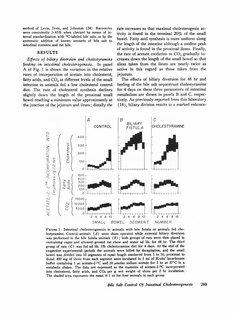
Effects of biliary diversion and cholestyraminefeeding on intestinal cholesterogenesis. In panelA of Fig. 1 is shown the variation in the relativerates of incorporation of acetate into cholesterol,fatty acids, and CO2 at different levels of the smallintestine in animals fed a low cholesterol controldiet. The rate of cholesterol synthesis declinesslightly down the length of the proximal smallbowel reaching a minimum value approximately atthe junction of the jejunum and ileum; distally the
0
F-
0
0
C)CL
-R!
I:
0
C))
O- .c.c: .u
r(Ni l c
E
N~
o C)=t
A
400 i-
300 1-
CONTROL
200
100
0
500400300200100
0
rate increases so that maximal cholesterogenic ac-tivity is found in the terminal 20% of the smallbowel. Fatty acid synthesis is more uniform alongthe length of the intestine although a modest peakof activity is found in the proximal ileum. Finally,the rate of acetate oxidation to CO., gradually in-creases down the length of the small bowel so thatslices taken from the ileum are nearly twice asactive in this regard as those taken from thejejunum.The effects of biliary diversion for 48 hr and
feeding of the bile salt sequestrant cholestyraminefor 4 days on these three parameters of intestinalmetabolism are shown in panels B and C, respec-tively. As previously reported from this laboratory(14), biliary division results in a marked enhance-
15000
10000 o5000LI
2 4 6 810 2 4 6 810 2 4 6 810
SMALL BOWEL SEGMENT NUMBER
FIGURE 1 Intestinal cholesterogenesis in animals with bile fistula or animals fed cho-lestyramine. Control animals (A) were sham operated while external biliary diversionwas performed in the bile fistula animals (B); both groups of rats were then placed inrestraining cages and allowed ground rat chow and water ad lib. for 48 hr. The thirdgroup of rats (C) was fed ad lib. 5% cholestyramine diet for 4 days. At the end of therespective experimental periods the animals were killed by decapitation, and the smallbowel was divided into 10 segments of equal length numbered from 1 to 10, proximal todistal. 400 mg of slices from each segment were incubated in 5 ml of Krebs' bicarbonatebuffer containing 1 1c acetate-2-`C and 10 /Amoles sodium acetate for 2 hr at 370C in a
metabolic shaker. The data are expressed as the m/Amoles of acetate-2-1'C incorporatedinto cholesterol, fatty acids, and CO2 per g wet weight of slices per 2 hr incubation.The shaded area represents the mean ± 1 SE for four animals in each group.
Bile Salt Control Of Intestinal Cholesterogenesis 289
ment of the rate of acetate incorporation intosterols by intestinal slices taken from every levelof the small bowel; this increase equals nearly ten-fold in the mid-intestinal segments. A similar, al-though less marked, increase in sterol synthesiscan be produced in the intact rat by feedingcholestyramine, as shown in panel C.
It should be emphasized that both of these ex-perimental manipulations alter greatly the rate ofcholesterogenesis without affecting the rate ofacetate incorporation into fatty acids or CO2, asshown in the lower panels of Fig. 1. Furthermore,careful analysis at various times throughout the2 hr incubation revealed that the rate of incorpora-tion of acetate into cholesterol became linear, i.e.achieved zero order kinetics, within 10 min andremained linear throughout the subsequent 110 minof incubation. This was true for intestinal slicesobtained both from control animals and from ani-mals with biliary diversion.
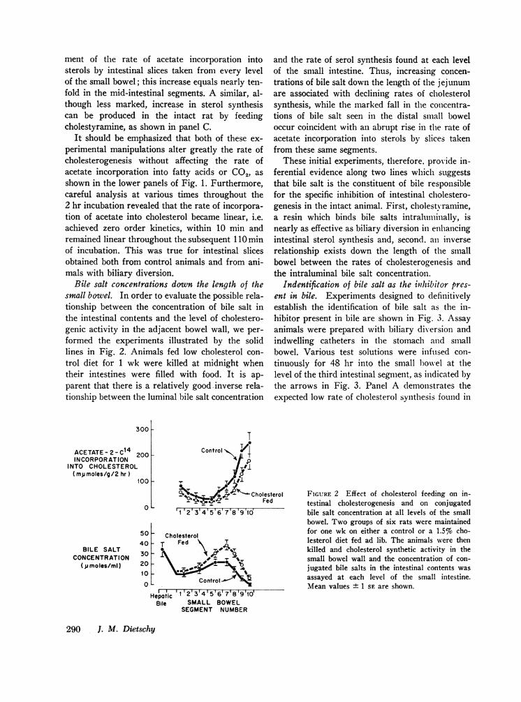
Bile salt concentrations down the length of thesmall bowel. In order to evaluate the possible rela-tionship between the concentration of bile salt inthe intestinal contents and the level of cholestero-genic activity in the adjacent bowel wall, we per-formed the experiments illustrated by the solidlines in Fig. 2. Animals fed low cholesterol con-trol diet for 1 wk were killed at midnight whentheir intestines were filled with food. It is ap-parent that there is a relatively good inverse rela-tionship between the luminal bile salt concentration
and the rate of serol synthesis found at each levelof the small intestine. Thus, increasing concen-trations of bile salt down the length of the jejunumare associated with declining rates of cholesterolsynthesis, while the marked fall in the concentra-tions of bile salt seen in the distal small boweloccur coincident with an abrupt rise in the rate ofacetate incorporation into sterols by slices takenfrom these same segments.
These initial experiments, therefore, provide in-ferential evidence along two lines which suggeststhat bile salt is the constituent of bile responsiblefor the specific inhibition of intestinal cholestero-genesis in the intact animal. First, cholestyramine,a resin which binds bile salts intraluminally, isnearly as effective as biliary diversion in enhancingintestinal sterol synthesis and, second. an inverserelationship exists down the length of the smallbowel between the rates of cholesterogenesis andthe intraluminal bile salt concentration.
Indentification of bile salt as the inhibitor pres-ent in bile. Experiments designed to definitivelyestablish the identification of bile salt as the in-hibitor present in bile are shown in Fig. 3. Assayanimals were prepared with biliary diversion andindwelling catheters in the stomach and smallbowel. Various test solutions were infused con-tinuously for 48 hr into the small bowel at thelevel of the third intestinal segment, as indicated bythe arrows in Fig. 3. Panel A demonstrates theexpected low rate of cholesterol synthesis found in
T200
Control
aco~~~~X#100
cO 0 CholesterolFed
0 '12'3'4'5'6'7'8'9'ld
50 - Cholesterol40T Fed 7
30 A20'I0~ ~ #'
10 Control
Hepatic 1 2 3 4 5 617181910Bile SMALL BOWEL
SEGMENT NUMBER
FIGURE 2 Effect of cholesterol feeding on in-testinal cholesterogenesis and on conjugatedbile salt concentration at all levels of the smallbowel. Two groups of six rats were maintainedfor one wk on either a control or a 1.5%o cho-lesterol diet fed ad lib. The animals were thenkilled and cholesterol synthetic activity in thesmall bowel wall and the concentration of con-jugated bile salts in the intestinal contents wasassayed at each level of the small intestine.Mean values + 1 SE are shown.
290 J. M. Dietschy
300 L
ACETATE-2-C14INCORPORATION
INTO CHOLESTEROL(mpumoles/g/2 hr)
BILE SALTCONCENTRATION
(j moles/ml)
I

CONTROLA
SALINEINFU SION
600
400
o200
0 v( 400
-- 200LL<
0
1o,0,00o 5000
234567
BILE FISTULA WITH INFUSION OF -
B
SALINE
*1"
S I h
C
WHOLEBILE
V'-ski44"*b
DTAUROCHOLIC
ACID(35 pmoles/mi
II~ ~~~~~IIi-III1234567 1234567 1234567
SMALL BOWEL SEGMENT
E
CHOLESTYRAMINE-TREATED BILE
_ t
1 234567
NUMBER
FIGURE 3 Inhibition of intestinal cholesterogenesis by bile salts infused in vivo. Assay animalswere prepared with external bile fistula (except for control animals) and with indwellingstomach and small bowel catheters. With the animals in restraining cages, various test solutionswere infused into the small intestine at 1 ml/hr for 48 hr. During this period the rats were
allowed water ad lib. and were fed a sucrose-casein solution, 2 ml every 6 hr, through the in-dwelling stomach tube. The animals were then killed and intestinal slices were prepared fromthe first seven segments and were incubated with acetate-2-1C. The arrows indicate the levelof the infusion in the small intestine (segmentshown.
the intestines of control animals with intact biliarysystems and with intra-intestinal infusions ofsaline. For comparison, panel B shows the greatlyenhanced rates of synthesis seen in animals withbiliary diversion whose proximal small intestinesalso were perfused with only saline. In contrast,when suich animals were perfused with whole ratbile, as shown in panel C, there was inhibition ofcholesterogenesis distal to the infusion point to thelow rates seen in control animals. However, ofparticular importance is the observation demon-strated in panel D that the infusion of taurocholatealone, at the physiologic concentration of 35mmoles/liter, resulted in as much inhibition ofsterol synthesis as the infusion of whole rat bile.
Finally, the importance of bile salt as the in-hibitory constituent of whole bile is confirmed bythe observations shown in the last two panels ofFig. 3. If whole rat bile is put through a chromato-graphic column packed with cholestyramine to de-plete it of its bile salt content. then essentially all
No. 3). The data from individual animals are
of the inhibitory activity of the bile is lost, as
shown in panel E. When, however, the bile saltcontent of such cholestyramine-treated bile is re-
stored to 35 mmoles/liter by the addition of puretaurocholate, then there is complete restoration ofthe inhibitory activity, as shown in panel F.Therefore, these studies provide strong evidencethat bile salt is the constituent of bile responsiblefor inhibition of intestinal cholesterogenesis in theintact animal. Again, it should be emphasizedthat this is an effect of bile salt specific forcholesterol synthesis, for as shown in the lowerpanels of Fig. 3, bile salt exerts no regulatory ef-fects upon fatty acid synthesis or acetate oxidation.
Relationship of inhibition to the intraluminal bilesalt concentration. In order to determine the rela-tionship between the steady-state intraluminal bilesalt concentration and the rate of cholesterol syn-thesis in the adjacent wall, we infused assay ani-mals intra-jejunally for 48 hr with solutions con-
taining varying concentrations of taurocholate.
Bile Salt Control Of Intestinal Cholesterogenesis
0
z
z0Tou
0a)
-
u-iO D
a,
0iO E,
_~ E
_
_jL&
FCHOLESTYRAMINE-TREATED BILE
+TCA(35 p moles/ ml)
m,,-..,
..iAt,.4- V-1I'tz~
135o
1 234567
291

The rate of cholesterogenesis in the jejunumplotted as a function of the steady-state concentra-tion of taurocholate in the lumen of this same areaof the bowel is shown in Fig. 4. It is apparentthat an inverse relationship exists between thesetwo variables; furthermore, it should be empha-sized that the greatest effects upon the rate ofintestinal cholesterogenesis occur over a range ofintraluminal bile salt concentrations which is en-countered physiologically in this area of the smallbowel. Thus, variations in the luminal bile saltconcentration would be expected to modulateeffectively sterol synthesis in the adjacent wall.
Other bile salts in addition to taurocholate wereassayed for inhibitory activity in a similar manner.The infusion of glycocholate, taurochenodeoxy-cholate, taurodeoxycholate, and unconjugatedcholate resulted in specific inhibition of intestinalcholesterogenesis to approximately the same de-gree, at comparable intraluminal concentrations,as that shown for taurocholate in Fig. 4. The in-fusion of unconjugated deoxycholate, however, re-sulted in nonspecific inhibition of several param-eters of mucosal cell metabolism in addition tosterol synthesis.
ACETATE - 2- C14INCORPORATION
INTO CHOLESTEROL(mpumoles /g /2 hr)
70C
60C
50C
40C
30C
20C
1c
Possible role of cholesterol in mediating the in-hibitory effect of bile salt. While these data clearlydemonstrate that bile salt is the inhibitory con-stituent of bile, it does not necessarily follow thatit is bile salt per se which is directly responsiblefor the inhibition of cholesterogenesis at the intra-cellular level. An alternative possibility is that bilesalt acts only indirectly in regulating mucosal cellmetabolism by promoting the intracellular absorp-tion of a second substance which in turn directlyaffects the cholesterol biosynthetic sequence. Theknown role of cholesterol in feedback control ofhepatic cholesterogenesis and the absolute require-ment of bile salt for the absorption of this 3-,8-OHsterol (25) raises the possibility that cholesterolmight be the agent directly responsible for thefeedback inhibition of intestinal cholesterogenesis.To test this important point, we performed the ex-periments shown in Fig. 2 and Table I.When a comparison was made between the rates
of cholesterol synthesis in the small intestines ofanimals fed either a low cholesterol diet or a highcholesterol diet, it is apparent, as shown in theupper panel of Fig. 2, that cholesterogenesis islower in the intestines of animals fed cholesterol.
0'0
0 00 0
0 00
00 0
0) °O I o
O 5 10 15 20 25TAUROCHOLATE CONCENTRATION
IN INTESTINAL LUMEN( p moles /m I)
FIGURE 4 Relation of the steady-state concentration of taurocholatein the intestinal lumen to the rate of intestinal cholesterogenesis. As-say animals with biliary diversion and indwelling stomach and smallbowel catheters were used in this study. The animals were fed a
sucrose-casein solution intragastrically, 2 ml every 6 hr, while theirsmall intestines were perfused continuously with isosmotic taurocho-late solutions of varying concentrations (2.5-60 mmoles/liter) at therate of 1.5 ml/hr. At the end of the 48 hr experimental period, luminalfluid from the mid-jejunum was aspirated for determination of theintraluminal taurocholate concentration and slices from this same area
of small bowel were assayed for cholesterogenic activity. Each pointrepresents the results from one animal.
292 J. M. Dietschy
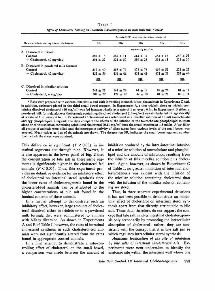
TABLE I
Effect of Cholesterol Feeding on Intestinal Cholesterogenesis in Rats with Bile Fistula*
Acetate-2-"4C incorporation into cholesterol
Means of administering infused cholesterol SB2 SB4 SB6 S8, SBio
Mrnmoles/g per 2 hrA. Dissolved in triolein
Control 266 -- 8 245 ±- 14 313 4- 5 332 ±- 17 237 4- 26+ Cholesterol, 40 mg/day 294 -± 22 324 4 59 359 ±- 25 334 4- 18 222 it 20
B. Dissolved in powdered milk formulaControl 316 ±- 40 368 -- 76 477 -± 78 419 ±- 52 272 4- 27+ Cholesterol, 40 mg/day 425 it 56 434 A- 44 428 -± 48 421 -- 21 252 4- 40
SB3 SB4 SB6 SBe SB7
C. Dissolved in micellar solutionControl 241 ±- 27 147 29 64 -- 12 99 -± 26 84 ±t 17+ Cholesterol, 4 mg/day 207 ±t 22 127 ± 12 39 -- 10 81 -± 21 80 ±- 14
* Rats were prepared with external bile fistula and with indwelling stomach tubes; the animals in Experiment C had,in addition, catheters placed in the third small bowel segment. In Experiment A, either triolein alone or triolein con-taining dissolved cholesterol (10 mg/ml) was fed intragastrically at a rate of 1 ml every 6 hr. In Experiment B either apowdered milk formula alone or the formula containing dissolved cholesterol (10 mg/ml) was similarly fed intragastricallyat a rate of 1 ml every 6 hr. In Experiment C cholesterol was solubilized in a micellar solution of 15 mM taurocholateand egg phospholipid, 2 mg/ml; the data compare the effects of the infusion of the taurocholate-phospholipid solutionalone or of this solution containing solubilized cholesterol (0.11 mg/ml) into the small intestine at 1.5 ml/hr. After 48 hrall groups of animals were killed and cholesterogenic activity of slices taken from various levels of the small bowel wasassayed. Mean values ± 1 SE of six animals are shown. The designation SB. indicates the small bowel segment numberfrom which the slices were obtained.
This difference is significant (P < 0.05) in in-testinal segments six through nine. However, itis also apparent in the lower panel of gig. 2 thatthe concentration of bile salt in these ,same seg-ments is significantly higher in the cholegterol-fedanimals (P < 0.05). Thus, this experiment pro-vides no definitive evidence for an inhibitory effectof cholesterol on intestinal sterol synthesis sincethe lower rates of cholesterogenesis found in thecholesterol-fed animals can be attributed to thehigher concentrations of bile salt found in theluminal contents of these animals.
In a further attempt to demonstrate such aninhibitory effect, however, large amounts of choles-terol dissolved either in triolein or in a powderedmilk formula diet were administered to animalswith biliary diversion. As shown in ExperimentsA and B of Table I, however, the rates of intestinalcholesterol synthesis in such cholesterol-fed ani-mals were not significantly altered from the ratesfound in appropriate control animals.
In a final attempt to demonstrate a rate-con-trolling effect of cholesterol on the small bowel,a comparison was made between the amount of
inhibition produced by the intra-intestinal infusionof a micellar solution of taurocholate and phospho-lipid and the amount of inhibition resulting fromthe infusion of this micellar solution plus choles-terol. Again, however, as shown in Experiment Cof Table I, no greater inhibition of intestinal cho-lesterogenesis was evident with the infusion ofthe micellar solution containing cholesterol thanwith the infusion of the micellar solution contain-ing no sterol.
Thus, in three separate experimental situationsit has not been possible to demonstrate an inhibi-tory effect of cholesterol on intestinal sterol syn-thesis apart from that directly attributable to bilesalt. These data, therefore, do not support the con-cept that bile salt inhibits intestinal cholesterogene-sis only secondarily by promoting the intracellularabsorption of cholesterol; rather, they are con-sistent with the concept that it is bile salt per sewhich regulates intracellular sterol synthesis.Anatomic localization of the site of inhibition
by bile salts of intestinal cholesterogenesis. Ex-periments were next undertaken to identify theanatomic site within the intestinal wall where bile
Bile Salt Control Of Intestinal Cholesterogenesis 293

TABLE I ILocalization of the Anatomic Site of FeedBack Control of Intestinal Cholesterogenesis*
Acetate-2-14C incorporation into
Cholesterol Fatty acids CO2
Villi Crypts Villi Crypts Villi Crypts
mj&moles/IO cm per 2 hr-A. Control 3 i 1 33 ± 5 13 ± 5 73 ± 12 628± 50 3392 ± 555B. Bile fistula 32 i 9 235 ± 15 20 + 8 106 ± 15 521 i 163 3191 ± 117
* Groups of six animals were either sham operated or prepared with external bile fistula; after 48 hr the animalswere killed and their jejunums were excised. Segments 10 cm long were cut open along the mesenteric border and scrapedto yield preparations of intestinal villi and crypts which were then incubated separately. The data are expressed as themean ± 1 SE values of the mpumoles of acetate-2-'4C incorporated into cholesterol, fatty acids, and CO2 by the amountsof villi or crypts obtained from each 10 cm long intestinal segment.
salt feeds back to inhibit the rate of sterol syn-thesis; the results of these experiments are shownin Table II. As previously reported from this lab-oratory (14), nearly all of the cholesterogenic ac-tivity of the intestine was found in the mucosalcell layer; the anatomic site of this activity in themucosa could be localized further, as shown inline A, to the cells of the intestinal crypt. Similarly,these same cells were far more active than thoseof the villi in incorporating acetate into fatty acidsand COO.
After diversion of bile salts from the intestinallumen, nearly all of the greatly enhanced sterolsynthetic activity found in the small bowel couldbe accounted for by a marked increase in the rateof synthesis in the intestinal crypts, as demon-strated in line B of Table II. The intestinal villialso showed some increased synthetic activity, butthis was attributable to contamination of thesepreparations by small amounts of crypt tissue.Again, the inhibitory effect of bile salt' on cryptcell cholesterogenesis was specific,' for, 'as also.shown in line B of Table II, biliary diversion didnot alter the rates of incorporation of acetate intofatty acid or CO2 by this tissue.
Biochemical localization of the site of inhibitionby bile salt of intestinal cholesterogenesis. In or-der to localize the site of feedback inhibition bybile salt on the cholesterol biosynthetic sequence,we compared the rates of incorporation of acetate-2-14C and of mevalonate-2-14C into cholesterol inintestinal slices obtained from control and frombile-diverted animals. As shown in Table III, therate of incorporation of acetate-2-_4C into sterol
was enhanced nearly sixfold, from 1350 + 107 to7940 + 317 cpm/2 hr, after biliary diversion; incontrast, the rate of incorporation of mevalonate-2-14C into cholesterol increased only slightly, from1524 + 110 to 1985 + 189 cpm/2 hr, after elim-ination of bile salt from the intestinal lumen. Thesedata indicate that bile salt inhibits sterol synthe-sis primarily at a step before the formation ofmevalonate. Since biliary diversion does not alterthe rates of incorporation of acetate into fatty acidor CO2 (Fig. 1), neither the intracellular penetra-tion of acetate nor its activation to acetyl CoA israte limiting under the conditions of these experi-ments. Therefore, the point of feedback inhibitionby bile salt on intestinal cholesterogenesis can belocalized more precisely to one of the few enzy-matic steps between the condensation of acetyl
TABLE IIILocalization of the Biochemical Site of Feedback Control
of Intestinal Cholesterogenesis*
Acetate-2-14C Mevalonate-2-14Cincorporation incorporationinto cholesterol into cholesterol
cpm/2 hr
A. Control 1350 ± 107 1524 ± 110B. Bilefistula 7940 ± 317 1985 ± 189
* Intestinal slices were prepared from sham-operatedanimals and from animals which had biliary diversion for48 hr. 400 mg aliquots of these slices were incubated in5 ml of Krebs' bicarbonate buffer containing 1 lsc of eitheracetate-2-"4C or mevalonate-2-14C for 2 hr. The datarepresent the mean ± 1 SE of 10 determinations in eachgroup.
294 1. M. Dietschy
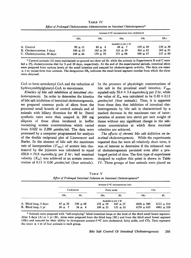
TABLE IV
Effect of Prolonged Cholestyramine Administration on Intestinal Cholesterogenesis*
Acetate-2-14C incorporation into cholesterol
SB2 SB4 SB. SBs SBio
mpmoles/g per 2 hrA. Control 90 4 11 40 ± 4 46 ± 7 129 49 236 36B. Cholestyramine, 5 days 100 10 165 ± 20 321 A 29 363 ± 62 263 ± 51C. Cholestyramine, 30 days 149 ± 44 170 ± 70 271 ± 68 400 ± 47 237 ± 60
* Control animals (A) were maintained on ground rat chow ad lib. while the animals in Experiments B and C werefed a 3% cholestyramine diet for 5 and 30 days, respectively. At the end of the experimental periods, intestinal sliceswere prepared from various levels of the small intestine and assayed for cholesterogenic activity. The data are mean± 1 SE values from four animals. The designation SB. indicates the small bowel segment number from which the sliceswere obtained.
CoA to form acetoAcetyl CoA and the reduction ofhydroxymethylglutaryl-CoA to mevalonate.
Kinetics of bile salt inhibition of intestinal cho-lesterogenesis. In order to determine the kineticsof bile salt inhibition of intestinal cholesterogenesis,we prepared common pools of slices from theproximal small bowels of control animals and ofanimals with biliary diversion for 48 hr. Sterolsynthetic rates were then assayed in 200 mgaliquots of these slices incubated in buffercontaining acetate concentrations which variedfrom 0.022 to 2.200 ,umoles/ml. The data wereprocessed by a computer programmed for analysisof the double reciprocal plot of Lineweaver andBurke. In the absence of bile salt the maximumrate of incorporation (Vmax) of acetate into cho-lesterol by the jejunum was calculated to equal450.8 + 74.8 mpmoles/g per 2 hr; half maximalvelocity (Ki) was achieved at an acetate concen-tration of 0.11 + 0.06 unmoles/ml (four animals).
In the presence of physiologic concentrations of1bile salt in the proximal small intestine, Vmaxequaled only 59.4 ± 5.4 mpumoles/g per 2 hr, whilethe value of Km was calculated to be 0.30 + 0.11pmoles/ml (four animals). Thus, it is apparentfrom these data that inhibition of intestinal cho-lesterogenesis by bile salt is characterized by amarked decrease in the maximum rate of incor-poration of acetate into sterol per unit weight oftissue without any significant change in the sub-strate concentration at which these maximalvelocities are achieved.The effects of chronic bile salt depletion on in-
testinal cholesterogenesis. While the experimentsreported thus far were all relatively short term, itwas of interest to determine if the enhanced rateof cholesterogenesis persisted even after a pro-longed period of time. The first type of experimentdesigned to explore this point is shown in TableIV. Three groups of four animals were placed on
TABLE VEffect of Prolonged Intestinal Isolation on Intestinal Cholesterogenesis*
Acetate-2-14C incorporation into
Cholesterol Fatty acids C02
SB3 BL SB: BL SB: BL
mpmoles/g per 2 hrA. Blind loop, 5 days 67 ± 20 256 ± 68 155 ± 19 147 ± 27 3838 ± 580 4222 ± 514B. Blind loop, I yr 56 ± 7 34 ± 4 149 ± 22 131 ±f 33 4279 ± 635 4962 + 526
* Animals were prepared with "self-emptying" blind intestinal loops at the level of the third small bowel segment.After 5 days (A) or 1 yr (B), slices were prepared from the blind loop (BL) and from the third small bowel segment(SB3) and assayed for their ability to incorporate acetate-2-'4C into cholesterol, fatty acids, and CO2. Data representthe mean ± 1 SE of four animals in each group.
Bile Salt Control Of Intestinal Cholesterogenesis 295
either a control diet or on a 3%o cholestyraminediet for 5 days or for 30 days. It is apparent thatthe same degree of enhanced cholesterol synthesiswas present at the end of one month of chole-styramine feeding as after only 5 days of feeding.
In a second type of experiment, animals wereprepared with a "self-emptying" blind pouch ofjejunum which was still in continuity with the re-mainder of the proximal small bowel through aRoux-en-Y anastamosis. Peristalsis continuallyemptied the isolated loop so that little bile salt wasin contact with this area of intestinal mucosa,whereas normal concentrations of bile salt werepresent in the lumen of the adjacent jejunum. Asshown in Table V, 5 days after such an operation,sterol synthesis in the blind loop was selectivelyenhanced fourfold. However, after the blind loophad been in place 1 yr, cholesterol synthesis mark-edly decreased to a level which was even lowerthan that found in the adjacent jejunum. Thus,even though bile salt concentration in this area re-mained very low, when the mucosa was isolatedfrom the main fecal stream for 1 yr the enhancedrate of sterol synthesis was not maintained. Suchdata have relevance to sterol balance in humans inwhich ileal bypass has been performed to promoteweight loss or to lower serum cholesterol levels.
DISCUSSION
Even though every tissue which has been tested inthe mammal is capable of at least some degree ofcholesterogenesis, only two, the liver and gastroin-testinal tract, are now considered to contribute sig-nificantly to the circulating cholesterol pool (4-9).Knowledge of the mechanisms which control therates of synthesis in these two organs is thereforeessential for an understanding of over-all sterolsynthesis in the intact animal.
Hepatic cholesterogenesis is now known to beaffected by several dietary and operative manipula-tions. Since the initial observations of Gould (4),it has been confirmed repeatedly that the rate ofcholesterol synthesis is dramatically suppressed bycholesterol feeding (2, 3, 11-14, 17). In a recentpublication by Siperstein and Fagan, the point offeedback inhibition by exogenous cholesterol inthe biosynthetic sequence has been shown to beat the conversion of hydroxymethylglutarate tomevalonic acid (26). Because of the resemblanceof several features of this feedback inhibition to
those previously described in bacterial systems,these authors suggested that exogenous cholesterolprimarily suppressed hepatic synthesis by allostericinhibition of the rate-limiting enzyme in the cho-lesterol biosynthetic sequence, i.e., hydroxymethyl-glutaryl reductase. Fasting for periods of 48-72hr causes a similar suppression of hepatic choles-terogenesis (2, 3, 10, 14, 27, 28). The report ofBucher, Overath, and Lynen suggests that the de-crease in synthesis seen after deprivation is alsodue to depressed enzymatic activity at the level ofhydroxymethylglutaryl reductase (28). In contrastto the inhibitory effects of cholesterol feeding andfasting, biliary diversion has been shown to enhancehepatic cholesterogenesis two to threefold (15).The implication has been drawn from these experi-ments that the bile salt pool, which is depletedafter biliary diversion, in some way also exerts arate-controlling effect upon sterol synthesis fromacetate in the liver; however, it is likely that thiseffect is due to an effect of bile salt on cholesterolabsorption (29 and unpublished observations). Insummary, the rate of hepatic cholesterol synthesisdepends upon the total caloric intake, the choles-terol content of the diet, and the enterohepaticcirculation of bile salt.The situation is quite different with respect to
intestinal cholesterogenesis. Several early publica-tions demonstrated that cholesterol feeding did notsignificantly suppress sterol synthesis in the smallintestine (12, 14, 16, 17). This finding was con-firmed in both the rat and the monkey in recentpublications from this laboratory (2, 3), and eventhough in the present study lower rates of synthe-sis were found in segments of the proximal andmidileum of cholesterol-fed animals, this differencewas attributable to a higher bile salt concentrationin these same segments and not directly to thehigh cholesterol content of the diet. In addition, itwas demonstrated in the rat and monkey that fast-ing for 48-96 hr had little effect upon the rate ofintestinal cholesterogenesis, even though the rateof hepatic cholesterogenesis in these animals wassuppressed 12- to 15-fold (2, 3). Thus, neither ofthe dietary manipulations which so dramaticallyalter cholesterol synthesis in the liver plays animportant role in the regulation of intestinal sterolsynthesis.However, diversion of bile from the intestinal
lumen results in a five to tenfold increase in the
296 J. Al. Dietschy

rate of incorporation of acetate into sterols at alllevels of the small intestine both in the rat and inthe monkey. Furthermore, in these two species thiseffect is specific for the small bowel since the ratesof acetate incorporation into cholesterol by theesophagus, stomach, and colon are not altered bybiliary diversion (2, 3).
It should be stressed that these alterations in therates of acetate-2-14C incorporation into digitoninprecipitable sterols brought about by biliary diver-sion represent changes in the actual rates of sterolsynthesis. The constancy of the incorporation ofacetate into CO2 and fatty acids, as shown in Fig.1, make it unlikely that changes in the rate ofacetate penetration into the cell, the tissue acetatepool, or the rate of formation of acetyl CoA ac-count for the specific stimulation of the incorpora-tion of acetate-2-14C into sterols seen after diver-sion of bile from the gastrointestinal tract. Fur-thermore, the demonstration that the appearanceof labeled acetate in sterols becomes linear with.respect to time within 10 min after initiation ofthe incubation of intestinal slices from both controlanimals and animals with biliary diversion indi-cates that in both of these preparations all func-tionally important intermediate pools betweenacetyl CoA and the digitonin precipitable sterolshave attained isotopic equilibrium. Finally, it alsoshould be emphasized that since the value of Kmfor the incorporation of acetate into cholesterolequals approximately 0.30 /Lmoles/ml, sterol syn-thesis in these experiments was measured at a sub-strate concentration approximately seven times theK.; thus, in slices from both control rats and ratswith biliary diversion rates of cholesterogenesiswere measured at essentially zero order kinetics.
Thus, it is clear that some component of wholebile maintains cholesterogenesis in the small bowelin a relatively suppressed state in the intact animal.As shown by the data in Fig. 3, this inhibitory con-stituent is demonstrated by the present studies tobe conjugated bile salt. Infusion of taurocholate,for example, causes as much specific inhibition ofintestinal cholesterogenesis as the infusion of wholebile. Furthermore, removal of bile salt from wholebile results in loss of inhibitory activity whereasrestoration of the bile salt content of this depletedbile with pure taurocholate restores all of the sup-pressive activity on intestinal sterol synthesis.
Finally, the degree of inhibition is a function ofthe steady-state bile salt concentration in the in-testinal lumen, as shown in Fig. 4, so that halfmaximal suppression of cholesterogenesis in theintestine is attained at a luminal bile salt concen-tration of approximately 5 ,umoles/ml.
Conjugated bile salt has been reported to sup-press cholesterol synthesis when added'in vitro tointestinal slices (30). However, as pointed out byPope, Parkinson, and Olson, it is now apparentthat many of the inhibitory effects ascribed to con-jugated bile salt in in vitro experiments are arti-facts due to contamination of these preparationswith small amounts of unconjugated bile salts suchas deoxycholate (31). As demonstrated by theseauthors as well as in this laboratory, pure con-jugated bile salts do not inhibit intestinal choles-terogenesis when added to intestinal slices in vitro(31-33). Such results are not at variance with thebile salt feedback inhibition described in the pres-ent study, for it also has been shown that no in-hibition* of intestinal sterol synthesis becomesmanifest under in vivo conditions until after 6 hrof intestinal perfusion with whole bile (14). There-fore, no inhibition would be expected in the short-term in vitro experiments.While these studies clearly establish bile salt as
the inhibitory constituent of whole bile, it does notnecessarily follow that it is bile salt per se whichacts at the cellular level to inhibit intestinal choles-terogenesis. An alternative possibility is that bilesalt acts in this inhibitory system only by facilitat-ing the entrance of a second substance into themucosal cell; the known role of cholesterol in feed-back inhibition of the liver (26) and the absoluterequirement of bile salt for cholesterol absorptionacross the intestinal wall (25) make this sterol apossible candidate for this hypothetical intracellularinhibitor. In addition, even though cholesterol wasnot present in the infusates in these studies, ap-proximately 2-4 mg is sloughed into the intestinallumen during the 48 hr duration of these experi-ments and so would be available for absorption.On the other hand, a strong argument against cho-lesterol being the actual inhibitor of intestinalcholesterogenesis is presented by the data in TableI where no direct inhibitory effect of cholesterolon cholesterol synthesis by the intestine was de-monstrable. Even when 8 mg of cholesterol was
Bile Salt Control Of Intestinal Cholesterogenesis 297
infused in a micellar solution of bile salt, nogreater inhibition resulted than could be attributedto the infusion of bile salt alone.
There is ample evidence that bile salt is able topenetrate the intestinal mucosa at every level;proximally, this occurs by way of passive ionicdiffusion, whereas distally, both passive ionic andnonionic diffusion as well as active transport actin concert to promote the transmural movement ofbile salts (34). In the jejunum, the rate of intra-cellular movement due to passive diffusion is re-lated to the concentration of bile salt in the in-testinal lumen; the rate of penetration, in turn,should determine the steady state intracellular con-centration and, hence, the degree of inhibition ofcholesterogenesis. This hypothetical scheme cor-relates well with the experimental findings that thesteady-state concentration of bile salt in the jejunallumen, both in control and in experimental animals,was always inversely related to the rate of choles-terol syntehsis in the adjacent wall.
All of these various findings, then, support theconclusion not only that bile salt is the constituentof bile responsible for inhibition of intestinal cho-lesterogenesis, but, in addition, that it is very likelythat it is bile salt per se which acts intracellularlyto specifically inhibit' the conversion of acetate tocholesterol.As shown in Table II, feedback inhibition by
bile salt is directed at synthesis in the cells of theintestinal crypt, the major site for cholesterogene-sis in the intestine. In addition, the point of inhibi-tion in the cholesterol biosynthetic sequence isbefore the formation of mevalonic acid as demon-strated by the data in Table III. If the enzymerelationships in the mucosal cell are the same asthose which have been described for liver ( 17, 26),then the only rate-limiting step between acetyl'CoAand mevalonate is at the reduction of hydroxy-tnethylglutarate; these data, therefore, further im-ply that the site of feedback inhibition by bile saltis at the enzymatic step mediated by hydroxy-methylglutaryl reductase. Finally, the kinetics ofthis inhibition indicate that bile salts act by sup-pressing the amount of effective enzyme per unit'weight of tissue at the rate-limiting step withoutaltering the substrate binding characteristics ofthis enzyme.As pointed out previously, Siperstein and Fagan
have suggested that the cholesterol negative feed-back manifested in liver may involve allostericinhibition of the rate-limiting enzyme, hydroxy-methylglutaryl reductase (26). While precisedefinition of the mode of inhibition involved in thebile salt negative feedback in intestine described inthe present investigation must await studies usingpurified, isolated enzyme systems, several charac-teristics of this inhibition, nevertheless. allowspeculation as to the mechanism of metabolic con-trol. Thus, the latent period between the initiationof bile salt infusion into the bowel of the intactanimal and the appearance of inhibition, the inabil-ity to inhibit sterol synthesis in intestinal slices bythe addition of bile salt in vitro, and the kineticsof this inhibition which indicate a marked changein Vmax without a significant alteration in K,,, areall consistent with enzyme repression at the geneticlevel rather than with allosteric inhibition or anegative effector action of bile salt on the rate-limiting enzyme.These studies emphasize the close interrelation-
ship which exists between the control of cholesterolsynthesis in the liver and in the intestine. Theendogenous synthesis of cholesterol by the liver issensitive to the amount of exogenous cholesterolreaching this organ from the diet. The endogenouissynthesis of cholesterol by the intestine, on theother hand, is controlled by bile salt, the catabolicend product of hepatic degradation of cholesterol.Under conditions of low cholesterol intake, the rateof hepatic cholesterogenesis is higher than thatfound in intestine since the bile salt pool normallypresent in the small bowel maintains intestinalsynthesis at a relatively suppressed level. Afterthe administration of a high cholesterol diet, cho-lesterol synthesis in the liver essentially stops, but,as demonstrated by Wilson in the rat, the produc-tion of bile acids increases (35). As a consequence,bile acid concentration in the snmall bowel increases,as shown in Fig. 2, and this, in turn, is associatedwith slight additional suppression of cholestero-genesis in the ileum. Nevertheless, exogenous cho-lesterol most dramatically inhibits hepatic cho-lesterogenesis so that in the cholesterol-fed animalthe intestine becomes the most active site of sterolsynthesis. Finally, interruption of the enterohepaticcirculation of bile salt increases sterol synthesisboth in the liver, probably by decreasing choles-
298 1. M. Dietschy
terol absorption, and in the small intestine by thedirect effect demonstrated in the present study.Since cholesterogenesis is enhanced to a greaterdegree in intestine (five to tenfold) than in liver(two to threefold), the rate of intestinal synthesismay equal or even exceed the rate of hepatic sterolsynthesis in such animals.
ACKNOWLEDGMENTSThe author would like to thank Dr. Marvin D. Sipersteinfor his continued interest and suggestions during thesestudies and to acknowledge the technical assistance ofJoyce Eckles and Herbert Weaver.
This investigation was supported by U. S. PublicHealth Service Research Grant HE-09610, TrainingGrant TI-AM-5490, and a John and Mary R. MarkleFoundation Scholarship.
REFERENCES1. Srere, P. A., I. L. Chaikoff, S. S. Treitman, and
L. S. Burstein. 1950. The extrahepatic synthesis ofcholesterol. J. Biol. Chem. 182: 629.
2. Dietschy, J. M., and M. D. Siperstein. 1967. Effectof cholesterol feeding and fasting on sterol synthesisin seventeen tissues of the rat. J. Lipid Res. 8: 97.
3. Dietschy, J. M., and J. D. Wilson. 1968. Cholesterolsynthesis in the squirrel monkey: Relative rates ofsynthesis in various tissues and mechanisms of con-trol. J. Clin. Invest. 47: 166.
4. Gould, R. G. 1951. Lipid metabolism and atherosclero-sis. Am. J. Med. 11: 209.
5. Friedman, M., S. 0. Byers, and F. Michaelis. 1951.Production and excretion of cholesterol in mam-mals. IV. Role of liver in restoration of plasma cho-lesterol after experimentally induced hypocholestere-mia. Amn. J. Physiol. 164: 789.
6. Harper, P. V., Jr., W. B. Neal, Jr., and G. R.Hlavacek. 1953. Lipid synthesis and transport in thedog. Mletab. Clin. Exptl. 2: 69.
7. Hotta, S., and I. L. Chaikoff. 1955. The role of theliver in the turnover of plasma cholesterol. Arch.Bioclcnm. Biophys. 56: 28.
8. Lindsey, C. A., Jr., and J. D. Wilson. 1965. Evidencefor a contribution by the intestinal wall to the serumcholesterol of the rat. J. Lipid Res. 6: 173.
9. Wilson, J. D. 1968. The biosynthestic origin of serumcholesterol in the squirrel monkey: Evidence for acontribution by the intestinal wall. J. Clin. Invest. 47:175.
10. Tomkins, G. M., and I. L. Chaikoff. 1952. Cholesterolsynthesis by liver. I. Influence of fasting and of diet.J. Biol. Chemn. 196: 569.
11. Tomkins, G. M., H. Sheppard, and I. L. Chaikoff.1953. Cholesterol synthesis by liver. III. Its regula-tion by ingested cholesterol. J. Biol. Chem. 201: 137.
12. Gould, R. G., C. B. Taylor, J. S. Hagerman, I.Warner, and D. J. Campbell. 1953. Cholesterol me-
tabolism. I. Effect of dietary cholesterol on the syn-thesis of cholesterol in dog tissue in vitro. J. Biol.Chem. 201: 519.
13. Langdon, R. G., and K. Bloch. 1953. The effect ofsome dietary additions on the synthesis of cholesterolfrom acetate in vitro. J. Biol. Chem. 202: 77.
14. Dietschy, J. M., and M. D. Siperstein. 1965. Choles-terol synthesis by the gastrointestinal tract: Locali-zation and mechanisms of control. J. Clin. Invest. 44:1311.
15. Myant, N. B., and H. A. Eder. 1961. The effect ofbiliary drainage upon the synthesis of cholesterol inthe liver. J. Lipid Res. 2: 363.
16. Cox, G. E., L. G. Nelson, W. B. Wood, and C. B.Taylor. 1954. Effect of dietary cholesterol on cho-lesterol synthesis in monkeys' tissue in vitro. Federa-tion Proc. 13: 31. (Abstr.)
17. Siperstein, M. D., and M. J. Guest. 1960. Studieson the site of the feedback control of cholesterol syn-thesis. J. Clin. Invest. 39: 642.
18. Cameron, D. G., G. M. Watson, and L. J. Witts.1950. The alimentary tract of rats with intestinalculs-de-sac. Brit. J. Exptl. Pathol. 31: 349.
19. Hofmann, A. F. 1962. Thin-layer adsorption chro-matography of free and conjugated bile acids onsilicic acid. J. Lipid Res. 3: 127.
20. Eneroth, P. 1963. Thin-layer chromatography of bileacids. J. Lipid Res. 4: 11.
21. Gregg, J. A. 1966. New solvent systems for thin-layerchromatography of bile acids. J. Lipid Res. 7: 579.
22. Pope, J. L. 1967. Crystallization of sodium taurocho-late. J. Lipid Res. 8: 146.
23. Bray, G. A. 1960. A simple efficient liquid scintillatorfor counting aqueous solutions in a liquid scintillationcounter. Anal. Biochem. 1: 279.
24. Levin, S. J., J. L. Irvin, and C. G. Johnston. 1961.Spectrofluorometric determination of total bile acidsin bile. Anal. Chem. 33: 856.
25. Siperstein, M. D., I. L. Chaikoff, and W. 0. Rein-hardt. 1952. C"-cholesterol. V. Obligatory functionof bile in intestinal absorption of cholesterol. J. Biol.Chem. 198: 11-1.
26. Siperstein, M. D., and V. M. Fagan. 1966. Feedbackcontrol of mevalonate synthesis by dietary choles-terol. J. Biol. Chem. 241: 602.
27. Bucher, N. L. R., K. McGarrahan, E. Gould, andA. V. Loudb 1959. Cholesterol biosynthesis in prepa-rations of liver from normal, fasting, X-irradiated,cholesterol-fed, Triton, or A'-cholesten-3-one-treatedrats. J. Biol. Chem. 234: 262.
28. Bucher, N. L. R., P. Overath, and F. Lynen. 1960.3-hydroxy-fi-methylglutaryl coenzyme A reductase,cleavage and condensing enzymes in relation to cho-lesterol formation in rat liver. Biochim. Biophys.Acta. 40: 491.
29. Beher, W. T., and G. D. Baker. 1959. Build-up andregression of inhibitory effects of cholic acid on invivo liver cholesterol synthesis. Proc. Soc. Exptl.Biol. Med. 101: 214.
Bile Salt Control Of Intestinal Cholesterogenesis 299
30. Parkinson, T. M., and J. A. Olson. 1963. Inhibitoryeffects of bile acids on the uptake, metabolism, andtransport of water-soluble substances in the small in-testine of the rat. Life Sci. 2: 393.
31. Pope, J. L., T. M. Parkinson, and J. A. Olson. 1966.Action of bile salts on the metabolism and transportof water-soluble nutrients by perfused rat jejunumin vitro. Biochim. Biophys. Acta. 130: 218.
32. Siperstein, M. D. 1960. The homeostatic control ofcholesterol synthesis in liver. Am. J. Clin. Nutr. 8:645.
33. Dietschy, J. M. The effects of bile salts on intermedi-ate metabolism of the intestinal mucosa. FederationProc. In press.
34. Dietschy, J. M., H. S. Salomon, and M. D. Siperstein.1966. Bile acid metabolism. I. Studies on the mecha-nisms of intestinal transport. J. Clin. Invest. 45: 832.
35. Wilson, J. D. 1964. The quantification of cholesterolexcretion and degradation in the isotopic steady statein the rat: the influence of dietary cholesterol. J.Lipid Res. 5: 409.
300 J. M. Dietschy


![4-Phenylbutyrate modulates ubiquitination of ... · bile salts into bile [2], the MRP2/Mrp2-dependent secretion of these solutes provides the osmotic driving force for the formation](https://static.fdocuments.net/doc/165x107/5eac2e0da3ab5b4fad4f2f47/4-phenylbutyrate-modulates-ubiquitination-of-bile-salts-into-bile-2-the-mrp2mrp2-dependent.jpg)