Intestinal Bile Acid Absorption
12
0 1993 by The American Soeiety for Biochemistry and Molecular Biology, Inc. THE JOURNAL OF BIOLOGICAL CHEMISTRY Vol. 268, No. 24, Issue of August 25. pp. 18035-18046,1993 Printed in U.S.A. Intestinal Bile Acid Absorption Na+-DEPENDENT BILE ACID TRANSPORT ACTIVITY IN RABBIT SMALL INTESTINE CORRELATES WITH THE COEXPRESSION OF AN INTEGRAL 93-kDa AND A PERIPHERAL 14-kDa BILE ACID-BINDING MEMBRANE PROTEIN ALONG THE DUODENUM-ILEUM AXIS* (Received for publication, December 21, 1992, and in revised form, March 29, 1993) Werner KramerS, Frank Girbig, Ulrike Gutjahr, Simone Kowalewski, Karin Jouvenal, Gunter Muller, Dominique Tripier, and Giinther Wess From Hoechst Aktiengesellschuft, 0-65926 Frankfurt am Main, Germany The anatomical localization of the Na+/bile acid co- transport system from rabbit small intestine was de- termined using brush border membrane vesicles pre- pared from eight different segments of the small intes- tine. Na+-dependent transport activity for bile acids, both for [SH]taurocholate and [‘Hlcholate, was found in the distal segment 8 only representing the terminal 12% of the small intestine. In contrast, the Na+-de- pendent D-ghcose transporter and the H+-dependent oligopeptide transporter were found over the whole length of rabbit small intestine in all segments. Photo- affinity labeling with 7,7-azo- and 3,3-azo-derivatives of taurocholate with subsequent fluorographic detec- tion of labeled polypeptides after one- and two-dimen- sional gel electrophoresis showed that an integral membrane polypeptide of M, 87,000 is present in the entire small intestine, whereasan integral membrane protein of M, 93,000 together with a peripheral mem- brane protein of M. 14,000 are exclusively expressed in the distal small intestine correlating with Na+-de- pendent bile acid transport activity. Photoaffinity labeling with the cationic bile acid derivative 1-(7,7- azo-3a,l2a-dihydroxy-58[3B-9H]cbolan-24-oy~)-1,2- diaminoethane hydrochloride and 7,7-azo-3&,12& dihydroxy-S~[12a-SH]cholan-24-oic acid did not result in a specific labeling of the above mentioned proteins, demonstrating their specificity for physiological bile acids. Photoaffinity labeling of the 93- and 14-kDa bile acid-binding proteins was strongly Na+-depend- ent. Significant labeling of the 93- and 14-kDa pro- teins occurred only in the presence of Na+ ions with maximal labeling above 100 mM [Na+] showing a par- allel [Na+] dependence to transport activity. Inactiva- tion of Na+-dependent [3HJtaurocholate uptake by treatment of ileal brush border membrane vesicles with 4-nitrobenzo-2-oxa-1,3-diazol chloride led to a parallel decrease in the extent of photoaffinity labeling of both the 93- and 14-kDa protein. Sequence analysis of the membrane-bound 14-kDa bile acid-binding pro- tein surprisingly revealed its identity with gastrotro- pin, a hydrophobic ligand-binding protein exclusively found in the cytosol from ileocytes and thought to be involved in the intracellular transport of bile acids from the brush border membrane to the basolateral pole of the ileocyte. In conclusion, the present studies * The costs of publication of this article were defrayed in part by the payment of page charges. This article must therefore be hereby marked “crdvertisement” in accordance with 18 U.S.C. Section 1734 solely to indicate this fact. $ TO whom correspondence should be addressed SBU Metabolism, Hoechst Aktiengesellschaft, D-65926 W-6230 Frankfurt am Main, Germany. Tel.: 0049-69-305-3557. suggest that both an integral 93- and a peripheral 14- kDa membrane protein, identified as gastrotropin, and both exclusively expressed in the terminal ileum, are essential components of the Na+/bile acid cotransport system in rabbit terminal ileum. Bile acids are synthesized from cholesterol in the liver, secreted with bile into the small intestine where they are absorbed, and recirculated back to the liver with portal blood (1-3). This enterohepatic circulation of bile acids showing a strict organotropism involving the liver and the small intes- tine under normal physiological conditions is explained by the existence of specific Na+-dependent bile acid transport systems in the sinusoidal membrane of the hepatocyte (4, 5) and the brush border membrane of ileocytes (6-8). The pu- tative protein components of the bile acid transport systems in blood (9, lo), plasma membranes and cytosol of liver (10- 18), and small intestinal (19-22) and kidney (23) cells have been identified with photolabile azi- and azido-derivatives of bile acids (24-26). The bile acids reabsorbed in the ileum and recirculating to the liver inhibit by a feedback mechanism the conversion of cholesterol to bile acids by modulating the activity of choles- terol 7cu-hydroxylase,the rat-limiting enzyme for the conver- sion of cholesterol into bile acids (27). Bile acids are absorbed either by ionic or non-ionic diffusion across the intestinal and colonic epithelia and by an active transport in the terminal ileum. It was generally accepted that under normal physiolog- ical conditions the bulk of bile acids is absorbed in the terminal ileum (3, 6, 7), but some recent studies suggested that a major or even predominant fraction of bile acids may be absorbed by the proximal jejunum (28). Because of its remarkable substrate specificity the ileal bile acid transport system is a putative pharmaceutical target for the treatment of hypercholesterolemia by interruption of the enterohepatic circulation with specific transport inhibitors. An understanding of the transportmechanism and thestruc- ture/activity relationships of bile acids for intestinal absorp- tion is essential for the rational design of putative transport inhibitors. Such a characterization on a molecular level in- volves the identification of the protein components of the transport system and the amino acids essential for molecular recognition together with purification, cloning, and reconsti- tution of the transportsystem. In the present paper we investigated the anatomical Iocal- ization of the Na+-dependent bile acid uptake system of rabbit small intestine using brush border membrane vesicles pre- pared from different segments of the small intestine, proximal 18035
Transcript of Intestinal Bile Acid Absorption
0 1993 by The American Soeiety for Biochemistry and Molecular Biology, Inc. THE JOURNAL OF BIOLOGICAL CHEMISTRY Vol. 268, No. 24, Issue of August 25. pp. 18035-18046,1993 Printed in U.S.A.
Intestinal Bile Acid Absorption Na+-DEPENDENT BILE ACID TRANSPORT ACTIVITY IN RABBIT SMALL INTESTINE CORRELATES WITH THE COEXPRESSION OF AN INTEGRAL 93-kDa AND A PERIPHERAL 14-kDa BILE ACID-BINDING MEMBRANE PROTEIN ALONG THE DUODENUM-ILEUM AXIS*
(Received for publication, December 21, 1992, and in revised form, March 29, 1993)
Werner KramerS, Frank Girbig, Ulrike Gutjahr, Simone Kowalewski, Karin Jouvenal, Gunter Muller, Dominique Tripier, and Giinther Wess From Hoechst Aktiengesellschuft, 0-65926 Frankfurt am Main, Germany
The anatomical localization of the Na+/bile acid co- transport system from rabbit small intestine was de- termined using brush border membrane vesicles pre- pared from eight different segments of the small intes- tine. Na+-dependent transport activity for bile acids, both for [SH]taurocholate and [‘Hlcholate, was found in the distal segment 8 only representing the terminal 12% of the small intestine. In contrast, the Na+-de- pendent D-ghcose transporter and the H+-dependent oligopeptide transporter were found over the whole length of rabbit small intestine in all segments. Photo- affinity labeling with 7,7-azo- and 3,3-azo-derivatives of taurocholate with subsequent fluorographic detec- tion of labeled polypeptides after one- and two-dimen- sional gel electrophoresis showed that an integral membrane polypeptide of M, 87,000 is present in the entire small intestine, whereas an integral membrane protein of M, 93,000 together with a peripheral mem- brane protein of M. 14,000 are exclusively expressed in the distal small intestine correlating with Na+-de- pendent bile acid transport activity. Photoaffinity labeling with the cationic bile acid derivative 1-(7,7- azo-3a,l2a-dihydroxy-58[3B-9H]cbolan-24-oy~)-1,2- diaminoethane hydrochloride and 7,7-azo-3&,12& dihydroxy-S~[12a-SH]cholan-24-oic acid did not result in a specific labeling of the above mentioned proteins, demonstrating their specificity for physiological bile acids. Photoaffinity labeling of the 93- and 14-kDa bile acid-binding proteins was strongly Na+-depend- ent. Significant labeling of the 93- and 14-kDa pro- teins occurred only in the presence of Na+ ions with maximal labeling above 100 mM [Na+] showing a par- allel [Na+] dependence to transport activity. Inactiva- tion of Na+-dependent [3HJtaurocholate uptake by treatment of ileal brush border membrane vesicles with 4-nitrobenzo-2-oxa-1,3-diazol chloride led to a parallel decrease in the extent of photoaffinity labeling of both the 93- and 14-kDa protein. Sequence analysis of the membrane-bound 14-kDa bile acid-binding pro- tein surprisingly revealed its identity with gastrotro- pin, a hydrophobic ligand-binding protein exclusively found in the cytosol from ileocytes and thought to be involved in the intracellular transport of bile acids from the brush border membrane to the basolateral pole of the ileocyte. In conclusion, the present studies
* The costs of publication of this article were defrayed in part by the payment of page charges. This article must therefore be hereby marked “crdvertisement” in accordance with 18 U.S.C. Section 1734 solely to indicate this fact.
$ TO whom correspondence should be addressed SBU Metabolism, Hoechst Aktiengesellschaft, D-65926 W-6230 Frankfurt am Main, Germany. Tel.: 0049-69-305-3557.
suggest that both an integral 93- and a peripheral 14- kDa membrane protein, identified as gastrotropin, and both exclusively expressed in the terminal ileum, are essential components of the Na+/bile acid cotransport system in rabbit terminal ileum.
Bile acids are synthesized from cholesterol in the liver, secreted with bile into the small intestine where they are absorbed, and recirculated back to the liver with portal blood (1-3). This enterohepatic circulation of bile acids showing a strict organotropism involving the liver and the small intes- tine under normal physiological conditions is explained by the existence o f specific Na+-dependent bile acid transport systems in the sinusoidal membrane of the hepatocyte (4, 5) and the brush border membrane of ileocytes (6-8). The pu- tative protein components of the bile acid transport systems in blood (9, lo), plasma membranes and cytosol of liver (10- 18), and small intestinal (19-22) and kidney (23) cells have been identified with photolabile azi- and azido-derivatives of bile acids (24-26).
The bile acids reabsorbed in the ileum and recirculating to the liver inhibit by a feedback mechanism the conversion of cholesterol to bile acids by modulating the activity of choles- terol 7cu-hydroxylase, the rat-limiting enzyme for the conver- sion of cholesterol into bile acids (27). Bile acids are absorbed either by ionic or non-ionic diffusion across the intestinal and colonic epithelia and by an active transport in the terminal ileum. It was generally accepted that under normal physiolog- ical conditions the bulk of bile acids is absorbed in the terminal ileum (3, 6, 7), but some recent studies suggested that a major or even predominant fraction of bile acids may be absorbed by the proximal jejunum (28).
Because of its remarkable substrate specificity the ileal bile acid transport system is a putative pharmaceutical target for the treatment of hypercholesterolemia by interruption of the enterohepatic circulation with specific transport inhibitors. An understanding of the transport mechanism and the struc- ture/activity relationships of bile acids for intestinal absorp- tion is essential for the rational design of putative transport inhibitors. Such a characterization on a molecular level in- volves the identification of the protein components of the transport system and the amino acids essential for molecular recognition together with purification, cloning, and reconsti- tution of the transport system.
In the present paper we investigated the anatomical Iocal- ization of the Na+-dependent bile acid uptake system of rabbit small intestine using brush border membrane vesicles pre- pared from different segments of the small intestine, proximal
18035
18036 Protein Components of the Ileal Na+/Bile Acid Cotransport System
to distal, from the duodenum to the terminal ileum and identified the respective bile acid-binding proteins by photo- affinity labeling.
EXPERIMENTAL PROCEDURES
Materials Photoaffinity labeling was carried out with the photolabile bile
acid derivatives (7,7-azo-3a,12a-dihydroxy-5~[3~-3H]cholan-24-oy~)- 2-aminoethanesulfonic acid (specific radioactivity, 20.25 Ci/mmol), (3,3-az0-7a,12a-dihydroxy-5~[7~,12~-~H]cholan-24-oyl)-2-aminoeth- anesulfonic acid (specific radioactivity, 5.9 Ci/mmol), 3,3-azo-7a,12a- dihydro~y-5p[7p-~H]cholan-24-oic acid (specific radioactivity, 1.3 Ci/ mmol), 7,7-a~o-3a,12~-dihydroxy-5~[12a-3H]cholan-24-oic acid (spe- cific radioactivity, 1.5 Ci/mmol) synthesized as described (24-26). The cationic photolabile bile acid derivative 1-(7,7-azo-3a,12a-di- hydroxy-5~[3~-3H]cholan-24-oyl-1,2-diaminoethane hydrochloride (specific radioactivity, 2.95 Ci/mmol) was synthesized by a reaction of the mixed anhydride formed from 7,7-azo-3-oxo-12a-hydroxy-58- cholan-24-oic acid (25) and ethyl chloroformate with 1,2-diamino- ethane, subsequent reduction with Na[3H]BH4, and purification by thin layer chromatography. (U-2,4-3H]Cholic acid (specific radioac- tivity, 42 Ci/mmol), [G-3H]taurocholic acid (specific radioactivity, 2.1 Ci/mmol), and D-[u-"Cc] glucose (specific radioactivity, 258.5 mCi/mmol) were from DuPont-New England Nuclear. D-Cephalexin, bile acids, and marker proteins for the determination of molecular weights were from Sigma. Triton X-100, Triton X-114, n-OCtyl-0-D- glucopyranoside, Servalytes, Serva Blue R-250, and other materials for electrophoresis were from Serva (Heidelberg, Germany). Cellulose nitrate filters for transport studies (ME 25; 0.45 p ~ ; 25 mm diameter) were from Schleicher & Schiill, and scintillators Quickzint 501 and 361 were from Zinsser Analytik GmbH (Frankfurt, Germany). Kit 3359 for the determination of the activity of the marker enzyme aminopeptidase N was from Merck. Solvents for HPLC' were from Riedel de Haen (Hannover, Germany). Protein was determined ac- cording to Bradford (29) using the Bio-Rad kit (Miinchen, Germany). Prefilled columns for HPLC chromatography (Nucleosil 120, 7 pm, Cia) were obtained from Machery-Nagel (Duren, Germany). All other materials were of analytical grade and obtained from the usual commercial sources. Nitrocellulose sheets, Hybond C extra, poly- clonal and affinity-purified donkey anti-rabbit ['251]IgG, and sheep anti-mouse ["51]IgG were purchased from Amersham-Buchler (Braunschweig, Germany). Polyclonal and affinity-purified anti-rat 8-COP and anti-rat Na+/K'-ATPase (a-subunit) antisera raised in rabbits were kind gifts from Dr. T. Kreis, EMBL, Heidelberg and Dr. R. Mercer, Washington University School of Medicine, St. Louis, MO. Monoclonal antibodies against docking protein (a-subunit) from dog pancreas microsomes were kindly donated by T. A. Rapoport, Zentralinstitut fur Molekularbiologie, Berlin). Polyclonal antibodies against purified rat liver mitochondrial porin* were raised in rabbits according to standard procedures and affinity-purified. Preimmune rabbit sera were used in control experiments and did not generate signals in immunoblotting.
Preparation of Brush Border Membrane Vesicles
Brush border membrane vesicles from the small intestine of male New Zealand White rabbits (weighing 4-5 kg) were prepared by the Mg2+ precipitation method (30) as described previously (8, 32-35). The entire small intestine between the PYIONS and the cecum was removed, rinsed with ice-cold sodium phosphate-buffered saline, and divided into 8 segments of equal length, numbered 1-8, proximal to distal. From each segment the brush border membrane vesicles were prepared, and the enrichment in the marker enzyme aminopeptidase N (EC 3.4.11.2) was determined. The vesicles were used for transport and labeling studies immediately after preparation. This multiseg- mental preparation was performed from four different animals, and consistent results were found in all preparations. In other experi- ments, jejunal vesicles were prepared from segments 2 to 5 and ileal vesicles from the segments 7 to 10 of rabbit small intestine, divided
' The abbreviations used are: HPLC, high pressure liquid chro- matography; NBD, 4-nitrobenzo-2-oxa-l,3-diazol; PAGE, polyacryl- amide gel electrophoresis; TEMED, N,N,N',N'-tetramethylethylene- diamine; DIDS, 4,4'-diisothiocyanostilbene-2,2'-disulfonic acid; TBS, Tris-buffered saline.
G. Muller, unpublished results.
in 10 segments of equal length, proximal to distal. Immediately after preparation, these vesicles were stored in liquid nitrogen without loss of transport and enzymatic activity for at least 4 weeks. The enrich- ment of brush border membranes in the final preparation compared with homogenate was assessed by enzymatic and immunological marker determinations. Leucine aminopeptidase (aminopeptidase N, EC 3.4.11.2) and y-glutamyltransferase (EC 2.3.2.2) were measured using the Merckotest kits 3359 and 3394 (Merck). Glucose 6-phos- phatase activity was determined according to Arion (36). Acidic phosphatase as lysosomal and lactate dehydrogenase as cytosolic marker were analyzed using test combination kits from Boehringer Mannheim (acidic phosphatase) and Merck (lactate dehydrogenase). N-Acetyl-glucosaminidase was determined according to Mahrun (37) and protein according to Bradford (29) using the Bio-Rad kit (Bio- Rad, Miinchen, Germany). The purity of the brush border membrane vesicle preparation was further analyzed by immunoblotting of freshly prepared ileum homogenate and brush border membrane vesicles after SDS-PAGE with specific antibodies against organelle specific proteins. Antibodies against the following antigens were used: Na+/ K+-ATPase (marker for basolateral membrane, a-subunit) (38); dock- ing protein (marker for endoplasmic reticulum) (39); @COP, a micro- tubule-binding protein associated with the cytoplasmic face of golgi membranes (40, 41); porin (marker for mitochondria) (42). The intactness of the vesicles was determined by measuring Na+-depend- ent D-glucose uptake after 15 s of incubation. Usually, the overshoot uptake at 15 s was greater than 20-fold.
Immunoblotting 10, 25, 50, and 100 pg of protein was dissolved in Laemmli buffer
containing 2% mercaptoethanol, heated (95 "C, 5 min), separated by SDS-PAGE, and transferred electrophoretically to nitrocellulose sheets (43) using a semi-dry blotting apparatus. The transfer effi- ciency was controlled by staining of the gel with Coomassie Blue and of the filter sheet with Ponceau S in each case. The sheets were blocked with Tris-buffered saline (TBS) containing 5% (w/v) defatted milk powder and 0.2% (w/v) Tween 20 for 2 h at room temperature. After addition of the antibodies diluted in blocking buffer (anti-porin, 1:8000; anti-docking protein, 1:250; anti-0-COP, 1:750; anti-Na+/K+- ATPase, 1:2000) the incubation was continued for 16 h at 4 'C. Subsequently, the sheets were washed successively (30 min each) with TBS containing 0.25 M NaC1, TBS containing 0.2% Tween (two times), and finally TBS (two times). Bound antibodies were detected by incubation (1 h, 4 "C) with lz5I-anti-rabbit IgG from donkey or anti-mouse IgG from sheep (for anti-docking protein) diluted 1:lOOO in blocking buffer. After washing (see above) the sheets were air- dried and subjected to autoradiography (Kodak X-Omat AR-5 films). The films were developed after 36-72 h of exposure at -80 "C. Immunoreactivity was quantitated by cutting rectangular strips, cor- responding to the locations of the radiolabeled protein bands (iden- tified by autoradiography), from the nitrocellulose filters and count- ing the filter strips for radioactivity in a liquid scintillation counter. Specific radiolabeling was determined after subtracting half of the sum of background radioactivities present on size-matched strips of the filter cut from an area above and below each immunoreactive band. The values are given as the sum of radioactivities for the four different amounts of protein applied onto the gel in each case. Linearity between the amount of protein and the radioactive signal was established for each experiment.
Transport Measurements
Uptake of radioactive or unlabeled substrates by brush border membrane vesicles was determined by the membrane filtration method (30) as described previously (8, 31-35). Typically, the trans- port reaction was initiated by adding 10 pl of the vesicle suspension (50-100 pg of protein) equilibrated with 10 mM Tris-Hepes buffer (pH 7.4)/300 mM mannitol with 90 p1 of incubation medium contain- ing the substrates kept at 30 "C. The composition of the incubation medium for measurements in the presence of an inwardly directed Na+ gradient usually was 10 mM Tris-Hepes (pH 7.41, 100 mM NaCl, 100 mM mannitol, and in the absence of a Na+ gradient, 10 mM Tris- Hepes (pH 7.4), 100 mM KC], 100 mM mannitol. For measurement of bile acid uptake, these media contained 50 pM (0.75 pci) [3H] taurocholate or 50 k~ (0.75 pCi) [3H]cholate and for glucose uptake 19 /IM (1 pci) D-[U-'4C]glUCOSe. The uptake of D-cephalexin as a prototype substrate for the intestinal oligopeptide transport system (32-35) was measured in the presence of an inwardly directed H' gradient; the composition of the incubation medium in the presence
Protein Components of the Ileal Na+/BiZe Acid Cotransport System 18037
of a H+ gradient was 20 mM citrate-Tris (pH 6.0), 140 mM KCl, and in the absence of a H+ gradient 20 mM Tris-HC1 (pH 7.41, 140 mM KCl. At desired time points, the transport reaction was terminated by the addition of 1 ml of ice-cold stop solution (10 mM Tris-Hepes (pH 7 4 , 150 mM KCl). The entire content was pipetted onto the middle of a prewashed, prechilled filter kept under suction with the aid of a vacuum controller. The filters were rinsed immediately with 5 ml of ice-cold stop solution. The amount of radioactively labeled substrates taken up by the vesicles was measured after dissolving the filters in 4 ml of scintillator (Quickszint 361) by liquid scintillation counting in a Packard Tri-Carb 2200 j3-counter (Packard Instrument Co.). D-Cephalexin taken up by the vesicles was eluted with 300 p1 of water and quantified by HPLC analysis as described (32-35). AS eluent, a mixture of 75% (by volume) solvent A (30 mM sodium phosphate (pH 6.2), 10 mM tetramethylammonium chloride) and 27% (by volume) solvent B (400 g of solvent A and 468 g of acetonitrile) was used. Under these conditions, D-cephalexin eluted with a reten- tion time of 4.5 min and was detected by absorbance at 262 nm. Uptake values are in triplicate and given as mean & S.D.
Photoaffinity Labeling Photoaffinity labeling with photoreactive bile acids was performed
as described previously (9, 12,20,24,32,33). Typically, the necessary amount of brush border membrane vesicles equilibrated with 10 mM Tris-Hepes buffer (pH 7.4), 300 mM mannitol was added in the dark to incubation buffer (10 mM Tris-Hepes buffer (pH 7.4), 100 mM NaCl, 100 mM mannitol) containing the radioactively labeled photo- reactive bile acid derivatives. After 5 min of preincubation in the dark, the suspensions were irradiated for 10 min at 350 nm in a Rayonet photochemical reactor RPR 100 (Southern Ultraviolet Co., Hamden, CT) equipped with 16 RPR 3500-A lamps. Afterward, the suspensions were diluted with ice-cold buffer (IO mM Tris-Hepes (pH 7.4), 300 mM mannitol) and centrifuged at 48,000 X g for 30 min, and the pellets were resuspended in 10 mM Tris-Hepes buffer (pH 7.41, 300 mM mannitol and used for further experiments. Proteins were precipitated according to Wessel and Fliigge (44).
Discrimination between Integral and Peripheral Membrane Proteins To determine whether the photolabeled bile acid-binding proteins
are integral or peripheral membrane proteins, the following investi- gations were performed using membrane vesicles photolabeled with 7,7-azo- or 3,3-azo-derivatives of taurocholate.
Alkaline Extraction-Brush border membrane vesicles (340 pg of protein) suspended in 300 p1 of Tris-Hepes buffer (pH 7.4), 300 mM mannitol were centrifuged at 48,000 X g for 30 min. The supernatant was discarded, and the pellet was resuspended in 300 pl of 100 mM sodium hydrogen carbonate solution containing 4 mM phenylmeth- ylsulfonyl fluoride, 4 mM iodacetamide, 4 mM EDTA and kept at 0 "C for 30 min. After centrifugation at 48,000 X g for 30 min, the supernatant and the pellet were carefully separated, delipidated as described above, and submitted to SDS-PAGE.
Solubilization with Triton X-100-Brush border membrane vesicles (230 pg of protein) were suspended in 200 pl of 10 mM Tris-Hepes buffer (pH 7.4), 300 mM mannitol, 0.1% (v/v) Triton X-100 and kept at 0 "C for 30 min. After centrifugation at 48,000 X g for 30 min, both the supernatant and the pellet were delipidated and submitted to
Treatment with Triton X-114"Treatment of membranes with Triton X-I14 was performed using a modification of the method described by Bordier (45). Brush border membrane vesicles (340 pg of protein) were suspended in 250 p1 of 10 mM Tris-HC1 buffer (pH 7.3), 1% (v/v) Triton X-114, 150 mM NaCl and kept a t 0 "C for 15 min. After centrifugation at 48,000 X g for 30 min at 4 "C the supernatant was carefully overlaid on a cushion of 100 pl of 10 mM Tris-HC1 buffer (pH 7.3), 6% (w/v) sucrose, 150 mM NaCl, 0.1% (v/ v) Triton X-114 in an Eppendorf tube. After 5 min of incubation at 37 "C the probe was centrifuged at 3,000 X g for 5 min. The upper detergent-poor phase containing hydrophilic proteins was carefully separated from the lower detergent-rich phase containing hydropho- bic proteins. Both fractions were diluted with water to a volume of 300 pl, and subsequently the proteins were precipitated as described above and submitted to SDS-PAGE.
SDS-PAGE.
Gel Electrophoresis SDS-PAGE was carried out in vertical slab gels (20 X 17 X 0.15
m) using an electrophoresis system LE 2/4 (Pharmacia LKB Bio- technology) as described. Usually a gel concentration of 12% was
used at a ratio of 97.2% acrylamide and 2.8% N,N-bismethyleneac- rylamide. For two-dimensional electrophoresis, photolabeled brush border membrane vesicles (1.5 mg of protein) were solubilized in 100- 150 pl of lysis solution (9 M (w/v) urea, 2% (w/v) Triton X-100, 2% (v/v) Servalyte 3-10, 5% (w/v) 2-mercaptoethanol) and kept under vortexing at 4 "C for 30 min. After centrifugation at 48,000 X g for 30 min, the clear and particle-free supernatants were applied to isoelectric focusing performed in glass tubes (170 x 4 mm, inner diameter) with a gel height of 13 cm. The focusing gels were prepared by polymerization of a freshly prepared solution of 5.5 g of urea, 2 ml of 10% (w/v) Triton X-100,3.23 ml of water, 0.5 ml of 40% Servalyte 3-10 solution, 400 mg of acrylamide-bisacrylamide premix (5% his- acrylamide), and 25 p1 of TEMED with 25 pl of a 10% (w/v) solution of ammonium peroxodisulfate. The polymerizing solution was over- laid with water-saturated isobutanol. After polymerization the tubes were mounted into a Bio-Rad model 175 tube cell. A 100-p1 volume of lysis buffer applied on the gel surface was carefully overlaid with 200 pl of a solution of 4 M urea, 2% Servalyte 3-10, and subsequently prefocusing was performed for 500 V-h with a maximum current of 1 mA/tube. Phosphoric acid (25 mM) and sodium hydroxide (50 mM) solutions were used as cathode and anode buffers, respectively. After prefocusing, the lysis and overlay solutions were removed, and the samples were applied. The sample solution was overlaid with 200 pl of 4 M urea, 2% Servalyte 3-10 followed by 50 mM sodium hydroxide solution. Isoelectric focusing was performed for 10,000 V-h at a maximum current of 1 mA/tube and a maximum voltage of 600 V. After 10,000 V-h the voltage was increased to 800 V for 1 h, and subsequently the gels were submitted to SDS-PAGE using 12% gels. The gel rods were applied to the separation gel of discontinuous SDS slab gels and overlaid with a warm (60 'C) solution of 1% (w/v) agarose in 62.5 mM Tris-HC1 buffer (pH 6.8), 2% (w/v) SDS, 5% (w/ v) 2-mercaptoethanol, 10% (v/v) glycerol, 0.001% (w/v) bromphenol blue. After cooling the agarose, elution of proteins from the focusing gels was performed at 40 V, and subsequently voltage was increased to 60 V. Gels were fixed with 12.5% (w/v) trichloroacetic acid, and proteins were stained with Serva Blue R 250. One-dimensional gels were scanned with a densitometer CD 50 (DESAGA, Heidelberg, Germany). For detection of radioactivity, either the individual lanes were cut into slices of 2-mm thickness and submitted to liquid scintillation counting after digestion of the proteins with 250 pl of tissue solubilizer Biolute S or the gels were submitted to fluorography (46). For fluorography, the gels were immersed for 20 min in a 1 M solution of sodium salicylate in 70% methanol (47). After drying of the gels with a Bio-Rad gel dryer, the gels were exposed to Kodak X- Omat AR films preflashed with red light (48) at -70 "C.
Peptide Analysis After separation of the proteins on SDS-PAGE and staining, the
protein band containing the 14-kDa protein was excised, thoroughly washed with water (3-4 changes, the last one overnight), sliced, dried (Eppendorf cones, Savant centrifuge, 4 h), and reswollen with a trypsin solution (5% w/w from the estimated protein quantity) in a digestion buffer (0.2 M N-methylmorpholine/acetate (pH 7.8), 1 M urea). After the total absorption of the trypsin solution, buffer was added up to saturation of the gel pieces. Incubation was performed for 16 h at 37 'C. The peptides were eluted from the matrix together with the dye by addition of 500 pl of digestion buffer followed by centrifugation (12,000 X g, 4 'C, 2.5 h) (49, 50). The elution was completed by a second step (30 min) with an acidic solution (500 p1 of 0.1% trifluoroacetic acid). It is useful to prepare a trypsin blank with a comparable trypsin quantity loaded on an unstained gel piece. The mixed eluates were injected portionwise (200 pl) on a 1 X 250 mm reversed phase HPLC column (50 pl/min). After the last injection and stabilization of the base line the column was equilibrated with buffer A (5% acetonitrile in 0.1% trifluoroacetic acid), and the gra- dient was developed at 0.5%/min up to 66% of buffer B (90% acetonitrile in 0.1% trifluoroacetic acid). The peaks were collected manually. Serva Blue R 250 produced 4-5 major blue-colored peaks positioned toward the end of the chromatogram, and trypsin peptides were identified by comparison with the blank. Sequence analysis was carried out on a pulsed-liquid automatic sequenator (447, Applied Biosystems, Inc.) fitted with an on-line phenylthiohydantoin-ana- lyzer (120A, Applied Biosystems, Inc.).
RESULTS
Anatomical Localization of the Na'-dependent Bile Acid, Na+-dependent D-Glucose, and H+-dependent Oligopeptide
18038 Protein Components of the Ikal Na+/Bile Acid Cotransport System
Transport System in Rabbit Small Intestine Proximal to Dk- tal-To determine the exact anatomical localization of the functional Na+-dependent bile acid transport system along the small intestine, brush border membrane vesicles were prepared from different anatomical sites of the small intes- tine. The entire small intestine of rabbits was removed, start- ing directly at the pylorus and ending at the cecum, and was segmented into 8 pieces of identical length, numbered 1-8, proximal to distal. Subsequently from each segment brush border membrane vesicles were prepared. Brush border rnem- brane vesicles isolated according to the Mg2+ or Ca’+ precip- itation method are essentially enriched in specific markers for the brush border membrane such as aminopeptidase N, 7- glutamyltransferase, or sucrase, whereas other cellular com- ponents such as basolateral membranes, mitochondria, en- doplasmic reticulum, lysosomes, or cytosol are not (52-54). The brush border membrane vesicles isolated from the differ- ent small intestinal segments were highly enriched (18-28- fold) in aminopeptidase N as a characteristic marker enzyme for apical membranes. To detect possible contaminations by cellular components other than apical membranes, the final brush border membrane vesicle preparation was analyzed by a combination of enzymatic and immunological methods (Table 1). Whereas the apical membrane marker enzymes, aminopeptidase N and y-glutamyltransferase, were highly enriched (19- and 14-f0ld), acidic phosphatase as lysosomal marker and glucose 6-phosphatase as an indicator for the endoplasmic reticulum showed only a slight (3.6- and 2.1- fold) enrichment in the specific activity compared with freshly prepared homogenate. A more specific marker for lysosomes, N-acetylglucosaminidase, however, shows no enrichment in the specific activity of the enzyme in the brush border mem- brane preparation. Lactate dehydrogenase as a cytosolic marker also was decreased in its specific activity in the membrane preparation indicating no contamination by cyto- solic proteins. A much more sensitive and conclusive method to detect contamination of the vesicle preparation by other cellular components is possible with specific antibodies raised against organelle-specific proteins. Immunablot analysis of homogenate and brush border membrane vesicles revealed that Na’/K’-ATPase, assayed with a specific antibody against the a-subunit, as a specific marker for basolateral plasma membranes (55) , docking protein (SRP receptor), a protein exclusively located at the endoplasmic reticulum (39), and porin as a mitochondrium-specific protein (42) revealed no clear enrichment of these proteins in the brush border membranes compared with homogenate. Antibodies against
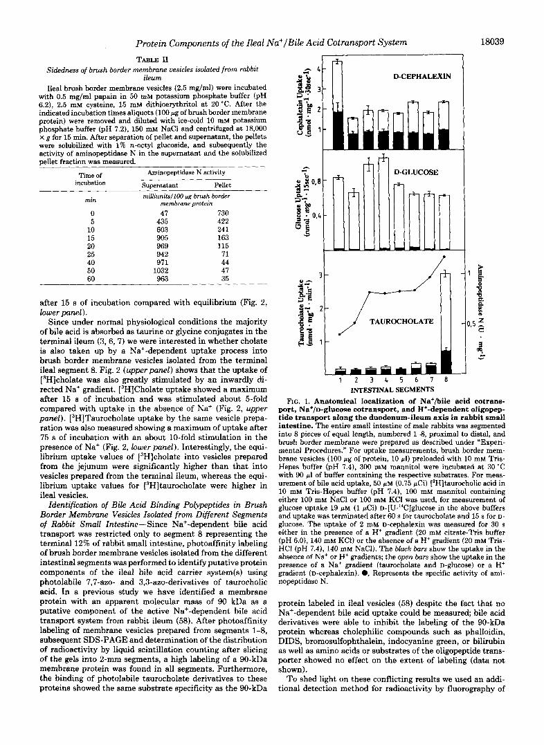
the clathrin p-COP (40,41) indicated a low contamination of the brush border vesicle preparation with Golgi membranes. Taken together, these findings indicate a high purity of the brush border membrane preparation without significant con- tamination by other membranes ensuring that the biochemi- cal investigations outlined in this paper really reflect molec- ular events at the brush border membrane of the enterocytes. Preparation of brush border membrane vesicles from intes- tinal mucosa and kidney cortex by the Ca2+ or M2+ precipi- tation method yields nearly exclusively right-side out vesicles, as was shown with electron microscopic freeze-fracture tech- nique and immunological methods (56). Nevertheless we de- termined the sidedness of our final intestinal brush border membrane vesicle preparation by solubilization of the ectoen- zyme aminopeptidase N with papain (57). Table 11 shows that within 1 h, more than 95% of the aminopeptidase activity was solubilized indicating a portion of >95% of vesicles oriented right side out.
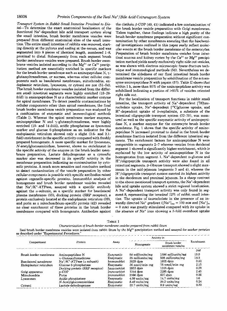
For the localization of transport functions in rabbit small intestine, the transport activity of Na’-dependent L3H]tau- rocholate uptake, Na+-dependent [‘4C]glucose uptake, and H+-dependent uptake of D-cephalexin, a substrate of the intestinal oligopeptide transport system (32-35), was meas- ured as well as the specific enzymatic activity of aminopepti- dase N, a marker enzyme for the enterocyte brush border membrane. Fig. 1 shows that the specific activity of amino- peptidase N increased proximal to distal in the brush border membrane fraction isolated from the different intestinal seg- ments. The enrichment factors for aminopeptidase N were comparable in segments 2-7 whereas vesicles from duodenal segment 1 showed a significantly higher enrichment, which is explained by the low activity of aminopeptidase N in the homogenates from segment 1. Na+-dependent D-glucose and K+/oligopeptide transport activity were also found in all intestinal segments. D-Glucose transport showed a slight max- imum in the mid-jejunum (segments 3 and 4), whereas the H+/oligopeptide transport system exerted its highest activity in the duodenum and proximal jejunum. In a sharp contrast to the above mentioned transport systems, the Na+-dependent bile acid uptake system showed a strict regional localization. A Na+-dependent transport activity was only found in seg- ment 8, representing the terminal 12% of rabbit small intes- tine. The uptake of taurocholate in the presence of an in- wardly directed Na+ gradient ( [Na+JOut = 100 mM and (Na+ji, = 0 mM) was greatly stimulated compared with its uptake in the absence of Na+ ions showing a %fold overshoot uptake
TABLE I Characterization of brush-border membrane vesicles prepared from rabbit ibum
Ileal brush border membrane vesicles were isolated from rabbit ileum by the M e precipitation method and assayed for marker proteins as described under “Experimental Procedures.”
Activity in -__
Compartment Protein Assay Homogenate
border Enrichment membrane vesicles
Brush border membrane Aminopeptidase N yGlutamyltransferase
Basolateral membrane Na+/K+-ATPase ( a subunit) Endoplasmic reticulum Glucose 6-phosphatase
Golgi apparatus p-COP Mitochondria Porin Lysosomes Acidic phosphatase
Cvtosol Lactate dehydrogenase
Docking protein (SRP receptor)
N-Acetylglucosaminidase
Enzymatic Enzymatic Immunoblot Enzymatic Immunoblot Immunoblot Immunoblot Enzymatic Enzymatic Enzymatic
84 milliunits/mg 35 milliunits/mg 3029 dpm 36 nmol/min. mg 2853 dpm 1344 dprn 2386 dpm 4.09 units/mg 8.49 units/mg 32.7 units/mg
1596 milliunits/mg 508 milliunits/mg 1605 dprn 78 nmol/min. mg 1105 dpm 3295 dpm 637 dpm 14.7 units/mg 20.2 units/mg 9.9 units/mg
-fold 19.0 14.5 0.53 2.15 0.38 2.45 0.26 3.6 0.24 0.30
Protein Components of the Ileal Na+/Bile Acid Cotransport System TABLE I1
Sidedness of brush border membrane vesicles isolated from rabbit ileum
Ileal brush border membrane vesicles (2.5 mg/ml) were incubated with 0.5 mg/ml papain in 50 mM potassium phosphate buffer (pH 6.2), 2.5 mM cysteine, 15 mM dithioerythritol at 20 "c. After the indicated incubation times aliquots (100 pg of brush border membrane protein) were removed and diluted with ice-cold 10 mM potassium phosphate buffer (pH 7.2), 150 mM NaCl and centrifuged at 18,000 X g for 15 min. After separation of pellet and supernatant, the pellets were solubilized with 1% n-octyl glucoside, and subsequently the activity of aminopeptidase N in the supernatant and the solubilized pellet fraction was measured.
Time of Aminopeptidase N activity incubation Supernatant Pellet
milliunits/I00 pg brush border membrane protein
0 47 730 5 435 422
10 603 241 15 905 163 20 969 115 25 942 71 40 971 44 50 1032 47 60 963 35
min
after 15 s of incubation compared with equilibrium (Fig. 2, lower panel).
Since under normal physiological conditions the majority of bile acid is absorbed as taurine or glycine conjugates in the terminal ileum (3,6,7) we were interested in whether cholate is also taken up by a Na+-dependent uptake process into brush border membrane vesicles isolated from the terminal ileal segment 8. Fig. 2 (upper panel) shows that the uptake of [3H]cholate was also greatly stimulated by an inwardly di- rected Na+ gradient. [3H]Cholate uptake showed a maximum after 15 s of incubation and was stimulated about 5-fold compared with uptake in the absence of Na' (Fig. 2, upper panel). [3H]Taurocholate uptake by the same vesicle prepa- ration was also measured showing a maximum of uptake after 75 s of incubation with an about 10-fold stimulation in the presence of Na+ (Fig. 2, lower panel). Interestingly, the equi- librium uptake values of [3H]cholate into vesicles prepared from the jejunum were significantly higher than that into vesicles prepared from the terminal ileum, whereas the equi- librium uptake values for [3H]taurocholate were higher in ileal vesicles.
Identification of Bile Acid Binding Polypeptides in Brush Border Membrane Vesicles Isolated from Different Segments of Rabbit Small Intestine-Since Na+-dependent bile acid transport was restricted only to segment 8 representing the terminal 12% of rabbit small intestine, photoaffinity labeling of brush border membrane vesicles isolated from the different intestinal segments was performed to identify putative protein components of the ileal bile acid carrier system(s) using photolabile 7,7-azo- and 3,3-azo-derivatives of taurocholic acid. In a previous study we have identified a membrane protein with an apparent molecular mass of 90 kDa as a putative component of the active Na+-dependent bile acid transport system from rabbit ileum (58). After photoaffinity labeling of membrane vesicles prepared from segments 1-8, subsequent SDS-PAGE and determination of the distribution of radioactivity by liquid scintillation counting after slicing of the gels into 2-mm segments, a high labeling of a 90-kDa membrane protein was found in all segments. Furthermore, the binding of photolabile taurocholate derivatives to these proteins showed the same substrate specificity as the 90-kDa
D-CEPHALEXIN
I T
1 - 1
1 2 3 4 5 6 7 0 INTESTINAL SEGMENTS
FIG. 1. Anatomical localization of Na+/bile acid cotrans- port, Na+/D-glucose cotransport, and H+-dependent oligopep- tide transport along the duodenum-ileum axis in rabbit small intestine. The entire small intestine of male rabbits was segmented into 8 pieces of equal length, numbered 1-8, proximal to distal, and brush border membrane were prepared as described under "Experi- mental Procedures." For uptake measurements, brush border mem- brane vesicles (100 pg of protein, 10 pl) preloaded with 10 mM Tris- Hepes buffer (pH 7.4), 300 mM mannitol were incubated at 30 "C with 90 pl of buffer containing the respective substrates. For meas- urement of bile acid uptake, 50 p M (0.75 pCi) [3H]taurocholic acid in 10 mM Tris-Hepes buffer (pH 7.4), 100 mM mannitol containing either 100 mM NaCl or 100 mM KC1 was used, for measurement of glucose uptake 19 pM (1 pCi) D-[U-"C]glucose in the above buffers and uptake was terminated after 60 s for taurocholate and 15 s for D- glucose. The uptake of 2 mM D-cephalexin was measured for 30 s either in the presence of a H+ gradient (20 mM citrate-Tris buffer (pH 6.0), 140 mM KC1) or the absence of a H+ gradient (20 mM Tris- HCl (pH 7.4), 140 mM NaCl). The black bars show the uptake in the absence of Na+ or H+ gradients; the open bars show the uptake in the presence of a Na+ gradient (taurocholate and D-glUCOSe) or a H+ gradient (D-cephalexin). 0, Represents the specific activity of ami- nopeptidase N.
protein labeled in ileal vesicles (58) despite the fact that no Na+-dependent bile acid uptake could be measured; bile acid derivatives were able to inhibit the labeling of the 90-kDa protein whereas cholephilic compounds such as phalloidin, DIDS, bromosulfophthalein, indocyanine green, or bilirubin as well as amino acids or substrates of the oligopeptide trans- porter showed no effect on the extent of labeling (data not shown).
To shed light on these conflicting results we used an addi- tional detection method for radioactivity by fluorography of

18040 Protein Components of the Ileal Na+/Bile Acid Cotransport System
k# .i."-JT p""-----~--i ~, $, Time (min)
FIG. 2. Na+-dependent uptake of [SH]cholate (upper panel) and [SH]taurocholate (lower panel) into brush border mem- brane vesicles from rabbit small intestine. Ileal or jejunal brush border membrane vesicles (10 pl, 50 pg of protein) loaded with 10 mM Tris-Hepes buffer (pH 7.4), 300 mM mannitol were incubated at 30 "C with 90 pl either of 10 mM Tris-Hepes buffer (pH 7.4), 100 mM mannitol, (100 mM NaCl (0) or 10 mM Tris-Hepes buffer (pH 7.4), 100 mM mannitol, 100 mM KC1 (0) containing either 50 p M (0.75 pCi) [3H]cholate (upperpanel) or 50 p~ (0.75 pCi) [3H]taurocholate (lower panel). Uptake was measured after the indicated incubation times and corrected for unspecific binding of 3H-bile acids to the filters. 0, Uptake into ileal vesicles in the presence of a Na+ gradient; 0, uptake into ileal vesicles in the absence of a Na+ gradient; U, uptake into jejunal vesicles in the presence of a Na' gradient; 0, uptake into jejunal vesicles in the absence of a Na+ gradient.
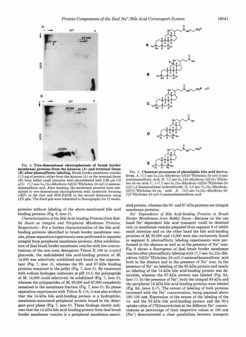
the dried gels and varied the gel concentration from 7 to 18% to ensure that no labeled polypeptide escapes detection. Fig. 3A shows the polypeptide pattern of brush border membrane vesicles from the respective small intestinal segments. Fig. 3B shows a fluorogram of brush border membrane vesicles iso- lated from segments 1-8 photolabeled with (7,7-azo-3a,12a- dihydroxy-5~[3~-3H]cholan-24-oyl)-2-aminoethanesulfonic acid after SDS-PAGE on a 12% gel. It is evident that a bile acid-binding protein with M, 87,000 was present in all seg- ments, although very weak in segment 1. In segment 8 only, two additional bile acid-binding proteins with M , 93,000 and 14,000 were found. The appearance of the 14-kDa protein is also evident from the Serva-Blue-stained polypeptide pattern (Fig. 3A). These findings were confirmed by two dimensional gel electrophoresis of jejunal (segments 3-5) and ileal (seg- ment 8) brush border membrane vesicles. In jejunal vesicles only one radiolabeled region of M, 87,000 was present (Fig. a), whereas in membrane vesicles isolated from the terminal ileum a second region of higher PI value and M, 93,000 (Fig. 4B) was detectable. In addition, a high amount of radioactivity was found in a polypeptide of M, 14,000. The radioactivity of the photolabeled 87 and 93 kDa bile acid-binding proteins was not found in a sharply focused spot on the two-dimen- sional gels but was distributed over a pH range of about 0.8 units indicating a microheterogeneity of these proteins.
To obtain information about the specificity of these pro- teins for bile acids of different structure we also performed
A 1 2 3 4 5 6 7 8
6 1 2 3 4 5 6 7 8
116 97 66 - 43 -
% 3 6 - . 29 - i 24 -
20
14
e14 kDa
,93 KDa
R7 KDa
I 14 KDa
FIG. 3. Anatomical localization of bile acid-binding pro- teins along the duodenum-ileum axis in small intestinal brush border membrane vesicles after photoaffinity labeling. Brush border membrane vesicles (150 pg of protein) prepared lrom the intestinal segments 1-8, loaded with 10 mM Tris-Hepes buffer (pH 7.4), 300 mM mannitol were incubated for 5 min in the dark with 185 pl of 10 mM Tris-Hepes buffer (pH 7.4), 100 mM NaC1, 100 mM mannitol containing 0.49 p~ (2 pCi) (7,7-azo-3n,12a-dihydroxy- 5fl[3fl-3H]cholan-24-oyl)-2-aminoethanesulfonic acid and subse- quently irradiated at 350 nm for 10 min. After washing the vesicles, membrane proteins were separated by SDS-PAGE using 12% gels. The dried gels were submitted to fluorography for 12 weeks. A, Serva Blue R 250-stained polypeptides; R, fluorogram.
photoaffinity labeling of ileal brush border membrane vesicles with other photoreactive bile acid derivatives (Fig. 5). Photo- affinity labeling with (3,3-azo-7a,12a-dihydroxy-5P(78,12P- 3H]cholan-24-oyl)-2-aminoethanesulfonic acid resulted in an identical labeling pattern as with the 7,7-azo isomer (data not shown). The unconjugated 3,3-azo analogue of cholic acid (Fig. 6, lune 4 ) also led to a labeling of the 93-, 87-, and 14- kDa bile acid-binding protein but to a significantly lesser extent as with photolabile taurocholic acid derivatives. 7,7- Azo-3cu,l2/3-dihydroxy-5P[12a-"H]cholan-24-0ic acid, how- ever, was not able to label to the above mentioned bile acid- binding proteins (Fig. 6, lane 2), indicating that an a-oriented hydroxyl group at position 12 of the steroid nucleus is impor- tant for optimal molecular recognition of a bile acid by the ileal bile acid transporter, thus explaining the low ileal ab- sorption of 12g-hydroxy bile acids such as lagodeoxycholic acid (59). Bile acids with a cationic side chain are weak inhibitors of ileal bile acid uptake but are not taken up by the ileal Na+-dependent bile acid transporter (7, 60). Photo- affinity labeling of ileal brush border membrane vesicles with a cationic photolabile bile acid derivative where the negatively charged taurine residue in (7,7-azo-3a,12a-dihydroxy-5~-cho- lan-24-oyl)-2-aminoethanesulfonic acid was replaced by the cationic ethylenediamine structure conserving the distance between the photoreactive group and the charge in the side chain resulted in a diffuse labeling of various membrane
Protein Components of the Ileal Na+/Bile Acid Cotransport System 18041
A "
s
v) 0 VI
1- - B
v) 0 VI
IEF +
IEF +
FIG. 4. Two-dimensional electrophoresis of brush border membrane proteins from the jejunum ( A ) and terminal ileum ( B ) after photoaffinity labeling. Brush border membrane vesicles (1.5 mg of protein) either from the jejunum ( A ) or the terminal ileum ( B ) from rabbit small intestine were photolabeled with 0.99 pM (10 pCi) (7,7-azo-3a,12~-dihydroxy-5j3[3~-3HJcholan-24-oyl)-2-aminoe- thanesulfonic acid. After washing, the membrane proteins were sub- mitted to two-dimensional electrophoresis with isoelectric focusing ( I E F ) in the first and SDS-PAGE in the second dimension using 12% gels. The dried gels were submitted to fluorography for 12 weeks.
proteins without labeling of the above-mentioned bile acid binding proteins (Fig. 6, lane 3) .
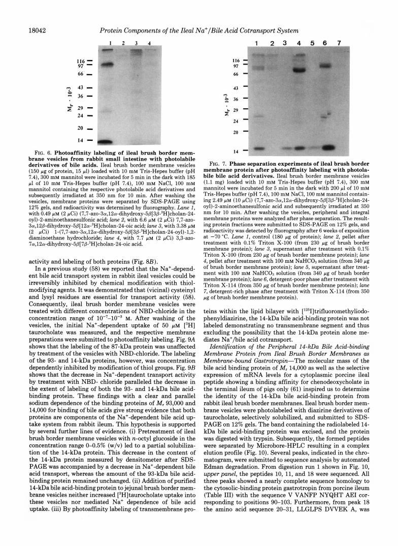
Characterization of the Bile Acid-binding Proteins from Rab- bit Ileum as Integral and Peripheral Membrane Proteins, Respectively-For a further characterization of the bile acid- binding proteins identified in brush border membrane vesi- cles, phase separation experiments were performed to separate integral from peripheral membrane proteins. After solubiliza- tion of ileal brush border membrane vesicles with low concen- trations of the non-ionic detergents Triton X-100 or n-octyl glucoside, the radiolabeled bile acid-binding protein of M, 14,000 was selectively solubilized and found in the superna- tant (Fig. 7, lane 3 ) , whereas the 93- and 87-kDa binding proteins remained in the pellet (Fig. 7, lane 2). By treatment with sodium hydrogen carbonate at pH 10.5, the polypeptide of M, 14,000 could selectively be solubilized (Fig. 7, lane 5 ) , whereas the polypeptides of M, 93,000 and 87,000 completely remained in the membrane fraction (Fig. 7, lane 4) . By phase separation experiments with Triton X-114, it was also evident that the 14-kDa bile acid-binding protein is a hydrophilic, membrane-associated peripheral protein found in the deter- gent-poor phase (Fig. 7, lane 6). These findings clearly indi- cate that the 14-kDa bile acid-binding protein from ileal brush border membrane vesicles is a peripheral membrane-associ-
B
C sHm OH n NsN
D
0
FIG. 5. Chemical structures of photolabile bile acid deriva- tives. A, (7,7-azo-3a,12a-dihydroxy-5~[3j3-3HJcholan-24-oyl)-2-ami- noethanesulfonic acid; B, 7,7-az0-3a,12j3-dihydroxy-5j3[12a-~H]cho- Ian-24-oic acid; C, 1-(7,7-azo-3a,12a-dihydroxy-5j3[3~-3H]cholan-24- oyl)-1,2-diaminoethane hydrochloride; D, 3,3-azo-7a,12a-dihydroxy- 5j3[7j3-DH]cholan-24-oic acid; E, (3,3-azo-7a,12a-dihydroxy-5@- [7j3-3H]cholan-24-oyl)-2-aminoethanesulfonic acid.
ated protein, whereas the 93- and 87-kDa proteins are integral membrane proteins.
Na+ Dependence of Bile Acid-binding Proteins in Brush Border Membranes from Rabbit Ileum-Because on the one hand Na+-dependent bile acid transport could be detected only in membrane vesicles prepared from segment 8 of rabbit small intestine and on the other hand the bile acid-binding proteins of M, 93,000 and 14,000 were also exclusively found in segment 8, photoaffinity labeling experiments were per- formed in the absence as well as in the presence of Na+ ions. Fig. 8 shows a fluorogram of ileal brush border membrane vesicles after photoaffinity labeling with (7,7-azo-3a,12a-dih- ydroxy-5~[3~-3H]cholan-24-oyl)-2-aminoethanesulfonic acid both in the absence and in the presence of Na+ ions. In the presence of Na+ no labeling of the 93-kDa protein and nearly no labeling of the 14-kDa bile acid-binding protein was de- tectable, whereas the 87-kDa protein was labeled (Fig. 8A, lane 1 ). In the presence of Na+, both the integral 93-kDa and the peripheral 14-kDa bile acid-binding proteins were labeled (Fig. 8A, lanes 2-7). The extent of labeling of both proteins increased with the Na+ concentration, being maximal above 100-120 mM. Expression of the extent of the labeling of the 14- and the 93-kDa bile acid-binding protein and the 30-9 uptake value of [3H]taurocholate at the different Na+ concen- trations as percentage of their respective values at 100 mM [Na+] demonstrated a clear parallelism between transport
18042 Protein Components of the Ileal Na+/Bile Acid Cotransport System
. 1 2
116 = 97 66 - 43 - - 36 -
2 29 - 24 - 20 I
14 - ,
2
FIG. 6. Photoaffinity labeling of ileal brush border mem- brane vesicles from rabbit small intestine with photolabile derivatives of bile acids. Ileal brush border membrane vesicles (150 pg of protein, 15 pl) loaded with 10 mM Tris-Hepes buffer (pH 7.4), 300 mM mannitol were incubated for 5 min in the dark with 185 pl of 10 mM Tris-Hepes buffer (pH 7.4), 100 mM NaC1, 100 mM mannitol containing the respective photolabile acid derivatives and subsequently irradiated a t 350 nm for 10 min. After washing the vesicles, membrane proteins were separated by SDS-PAGE using 12% gels, and radioactivity was determined by fluorography. Lune 1 , with 0.49 p~ (2 pCi) (7,7-az0-3a,12a-dihydroxy-5@[3~-~H]cholan-24- oyl)-2-aminoethanesulfonic acid; lane 2, with 6.6 p M (2 pCi) 7,7-aZO- 3a,12@-dihydroxy-5@[12a-3H]cholan-24-oic acid; lane 3, with 3.38 p~ (2 pCi) 1-(7,7-azo-3a,12a-dihydroxy-5~[3~-3H]cholan-24-oyl)-l,2- diaminoethane hydrochloride; lane 4, with 7.7 p~ (2 pCi) 3,3-azo- 7a,12a-dihydro~y-5@[7~-~H]cholan-24-oic acid.
activity and labeling of both proteins (Fig. 8B). In a previous study (58) we reported that the Na+-depend-
ent bile acid transport system in rabbit ileal vesicles could be irreversibly inhibited by chemical modification with thiol- modifying agents. It was demonstrated that (vicinal) cysteinyl and lysyl residues are essential for transport activity (58). Consequently, ileal brush border membrane vesicles were treated with different concentrations of NBD-chloride in the concentration range of 10-7-10-3 M. After washing of the vesicles, the initial Na+-dependent uptake of 50 PM [3H] taurocholate was measured, and the respective membrane preparations were submitted to photoaffinity labeling. Fig. 9A shows that the labeling of the 87-kDa protein was unaffected by treatment of the vesicles with NBD-chloride. The labeling of the 93- and 14-kDa proteins, however, was concentration dependently inhibited by modification of thiol groups. Fig. 9B shows that the decrease in Na+-dependent transport activity by treatment with NBD- chloride paralleled the decrease in the extent of labeling of both the 93- and 14-kDa bile acid- binding protein. These findings with a clear and parallel sodium dependence of the binding proteins of M, 93,000 and 14,000 for binding of bile acids give strong evidence that both proteins are components of the Na+-dependent bile acid up- take system from rabbit ileum. This hypothesis is supported by several further lines of evidence. (i) Pretreatment of ileal brush border membrane vesicles with n-octyl glucoside in the concentration range 0-0.5% (w/v) led to a partial solubiliza- tion of the 14-kDa protein. This decrease in the content of the 14-kDa protein measured by densitometer after SDS- PAGE was accompanied by a decrease in Na+-dependent bile acid transport, whereas the amount of the 93-kDa bile acid- binding protein remained unchanged. (ii) Addition of purified 14-kDa bile acid-binding protein to jejunal brush border mem- brane vesicles neither increased [3H]taurocholate uptake into these vesicles nor mediated Na+ dependence of bile acid uptake. (iii) By photoaffinity labeling of transmembrane pro-
1 2 3 4 5 6 7 m - = " " ~ U ~
116 - 97 - 66-
24 - 20 - 14 -
FIG. 7. Phase separation experiments of ileal brush border membrane protein after photoaffinity labeling with photola- bile bile acid derivatives. Ileal brush border membrane vesicles (1.1 mg) loaded with 10 mM Tris-Hepes buffer (pH 7.4), 300 mM mannitol were incubated for 5 min in the dark with 200 pl of 10 mM Tris-Hepes buffer (pH 7.4), 100 mM NaC1,lOO mM mannitol contain- ing 2.49 p~ (10 pCi) (7,7-az0-3a,12a-dihydroxy-5@[3@-~H]cholan-24- oyl)-2-aminoethanesulfonic acid and subsequently irradiated at 350 nm for 10 min. After washing the vesicles, peripheral and integral membrane proteins were analyzed after phase separation. The result- ing protein fractions were submitted to SDS-PAGE on 12% gels, and radioactivity was detected by fluorography after 6 weeks of exposition at -70 "C. Lune I , control (180 pg of protein); lane 2, pellet after treatment with 0.1% Triton X-100 (from 230 pg of brush border membrane protein); lane 3, supernatant after treatment with 0.1% Triton X-100 (from 230 pg of brush border membrane protein); lane 4, pellet after treatment with 100 mM NaHC03 solution (from 340 pg of brush border membrane protein); lane 5, supernatant after treat- ment with 100 mM NaHC03 solution (from 340 pg of brush border membrane protein); lane 6, detergent-poor phase after treatment with Triton X-114 (from 350 pg of brush border membrane protein); lane 7, detergent-rich phase after treatment with Triton X-114 (from 350 pg of brush border membrane protein).
teins within the lipid bilayer with ['2sI]trifluoromethyliodo- phenyldiazirine, the 14-kDa bile acid-binding protein was not labeled demonstrating no transmembrane segment and thus excluding the possibility that the 14-kDa protein alone me- diates Na+/bile acid cotransport.
Identification of the Peripheral 14-kDa Bile Acid-binding Membrane Protein from Ileal Brush Border Membranes as Membrane-bound Gastrotropin-The molecular mass of the bile acid binding protein of M, 14,000 as well as the selective expression of mRNA levels for a cytoplasmic porcine ileal peptide showing a binding affinity for chenodeoxycholate in the terminal ileum of pigs only (61) inspired us to determine the identity of the 14-kDa bile acid-binding protein from rabbit ileal brush border membranes. Ileal brush border mem- brane vesicles were photolabeled with diazirine derivatives of taurocholate, selectively solubilized, and submitted to SDS- PAGE on 12% gels. The band containing the radiolabeled 14- kDa bile acid-binding protein was excised, and the protein was digested with trypsin. Subsequently, the formed peptides were separated by Microbore-HPLC resulting in a complex elution profile (Fig. 10). Several peaks, indicated in the chro- matogram, were submitted to sequence analysis by automated Edman degradation. From digestion run 1 shown in Fig. 10, upper panel, the peptides 10, 11, and 18 were sequenced. All three peaks showed a nearly complete sequence homology to the cytosolic-binding protein gastrotropin from porcine ileum (Table 111) with the sequence V VANFP NYQHT AEI cor- responding to positions 90-103. Furthermore, from peak 18 the amino acid sequence 20-31, LLGLPS DVVEK A, was

Protein Components of the Ileal Na+/Bile Acid Cotransport System
A I 2 3 4 5 6 7
205-
97-
66-
43- m b 36- c(
29-
24-
20 - 14-
0 30 60 90 120
(mM) FIG. 8. Na+ dependence of Na+/bile acid cotransport and
photoaffinity labeling of bile acid binding polypeptides in rabbit ileal brush border membranes. A , ileal brush border mem- brane vesicles (150 pg of protein, 15 pl) loaded with 10 mM Tris- Hepes buffer (pH 7.4), 300 mM mannitol were mixed for 5 min in the dark with 185 pl of 10 mM Tris-Hepes buffer (pH 7.4), 100 mM mannitol, containing 0.49 pM (2 pci) (7,7-az0-3a,lPa-dihydroxy- 5~[3~-3H]cholan-24-oyl)-2-amincethanesulfonic acid and 0 ( l a n e I ) , 6 ( l a n e 2), 12 ( l a n e 3), 30 ( l a n e 4) , 60 ( l a n e 51,102 ( l a n e 61, and 132 ( l a n e 7) mM NaCl and (132 - [Na+]) mM KC1 and subsequently irradiated at 350 nm for 10 min. After washing the vesicles, membrane proteins were analyzed by SDS-PAGE on 12% gels and subsequent fluorography for 6 weeks, a t -70 "C. E , ileal brush border membrane vesicles (10 pl, 50 pg of protein) loaded with 10 mM Tris-Hepes buffer (pH 7.4), 300 mM mannitol were incubated at 30 "C with 90 pl of 10 mM Tris-Hepes buffer (pH 7.4) with 3.75, 7.5,15,30,48, 75, 105, and 120 mM NaCl and (120 - [Na+]) mM KC1 containing 50 p M (0.75 pCi) [3H]taurocholate, and uptake was measured for 30 s. [3H]Tau- rocholate uptake (W) at the different [Na'] concentrations was ex- pressed as uptake at 120 mM NaCI. The extent of photoaffinity labeling of 93- (0) and 14-kDa (0) protein was expressed as a percentage of the extent of labeling at 132 mM NaCI, respectively.
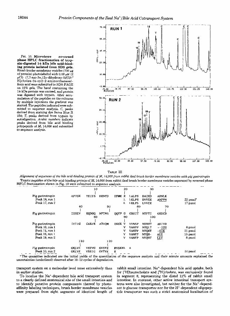
determined corresponding to RLALPS DAIKD A from por- cine gastrotropin. In a second digest obtained from the 14- kDa band of a separate SDS gel (Fig. 10, run 2), we obtained sequence information from peaks 10, 17, and 19 (Table 111) coding for the amino acid sequences 20-30 (peak 17), 90-103 (peak 19), and 106-121 (peak 10). The sequence of gastrotro- pin from rabbit ileum was not available, but the high degree of sequence homology with gastrotropin from pig ileal cytosol strongly suggests that the bile acid-binding protein of M , 14,000 identified in brush border membrane vesicles from segment 8 of rabbit small intestine is identical with gastrotro- pin. The identity of our 14-kDa protein with gastrotropin is proved by the structure of the peptide contained in peak 10 from run 2. Compared with gastrotropin the fatty acid-binding protein family exhibits an insertion after position 116 (Table IV) .
A 1 2 3 4 5 6
116, 97 - 66 - 43 - 36 - ;
* 29'
20 -
18043
0 7 6 5 4 3 -log[NLlD-chloride]
FIG. 9. Effect of NBD-chloride treatment on photoaffinity labeling of bile acid-binding proteins in brush border mem- brane proteins and on [SH]taurocholate uptake. Brush border membrane vesicles (10 mg/ml) were treated with the indicated con- centrations of NBD-chloride for 1 h at 20 "C. After washing, the uptake of 50 p~ (0.75 pCi) of [3H]taurocholate was measured for 30 s in the presence of an inwardly directed Na' gradient. Brush border membrane vesicles (150 pg of protein) were photolabeled with 0.49 p M (2 pCi) (7,7-az0-3a,12a-dihydroxy-5~[3~-~H]cholan-24-oyl)-2- aminoethanesulfonic acid, and subsequently submitted to SDS-PAGE and detection of radioactivity by fluorography for 6 weeks. A , fluo- rogram of ileal brush border membrane proteins pretreated with NBD-chloride and subsequent photoaffinity labeling. Lane 1, control; lane 2, after pretreatment with lo-' M NBD-chloride; lane 3, after pretreatment with M NBD-chloride; lane 4, after pretreatment with M NBD-chloride; lane 5, after pretreatment with lo" M NBD-chloride; lane 6, after pretreatment with lo-' M NBD-chloride. E , concentration dependence of NBD-chloride treatment on [3H] taurocholate uptake and photoaffinity labeling of the bile acid-bind- ing proteins of M, 93,000 and 14,000. Transport activity (30 s) and intensity of labeled 93- and 14-kDa protein on the fluorograms shown in A were expressed as percentage of the respective controls without NBD-chloride treatment. W, Transport, 0, labeling of the 93-kDa protein; 0, labeling of the 14-kDa protein.
A contamination of the brush border membrane by cytosolic proteins is unlikely because the intensity of the 14-kDa band on SDS gels showed a relatively constant ratio to the intensity of other bands such as the 43-kDa band of actin independently on the membrane preparation or several washings of the brush border membrane vesicles.
DISCUSSION
The transport of polar charged compounds across biological membranes occurs carrier-mediated with the aid of specific transport systems. Bile acids undergo an enterohepatic cir- culation and are reabsorbed by more than 95% in the small intestine, predominantly by a Na+-dependent uptake process located in the terminal ileum. Since the understanding of this reabsorption process is an essential step for the rationale design of putative transport inhibitors, we investigated this
18044 Protein Components of the Ileal Na+/Bile Acid Cotransport System
FIG. 10. Microbore reversed phase HPLC fractionation of tryp- sin-digested 14-kDa bile acid-bind- ing protein isolated from SDS gels. Brush border membrane vesicles (150 Fg of protein) photolabeled with 0.49 p~ (2 pCi) (7,7-azo-3~~,12n-dihydroxy-5fl[3@-~ H]cholan-24-oyl)-2-aminoethanesul- fonic acid were submitted to SDS-PAGE on 12% gels. The band containing the 14-kDa protein was excised, and protein was digested with trypsin. After accu- mulation of the peptides on the columns by multiple injections the gradient was started. The peptides indicated were sub- mitted to sequence analysis. C, peaks derived from staining dye Serva Blue R 250; T, peaks derived from trypsin by autodigestion. Arabic numbers indicate peaks derived from bile acid binding polypeptide of M, 14,000 and submitted to sequence analysis.
70..00 78.75 87.50 96.25 105.00 113.75 122.50 131.25 140.00 mlnutes
50.00 I RUN
39.44 I I
7.77 i t
-2.77l I I I I I I I 1 , I I I , I I I 20..00 32.50 45.00 57.50 70.00 82.50 95.00 107.50 120.00 m~nutes
TABLE 111 Alignment of sequences of the bite acid-binding protein of M, 14,000 from rabbit &ai brush border membrane uesicles with pig gastrotropin Tryptic peptides of the bile acid-binding protein of M, 14,000 from rabbit ileal brush border membrane vesicles separated by reversed phase
HPLC fractionation shown in Fig. 10 were submitted to sequence analysis.
10 20 I I
30 I
Pig gastrotropin AFTGK Y E I E S EKNYD EFMK R LALPS DAIKD ARNLK Peak 18, run 1 L L G L P S D W E K A N N N Peak 17, run 2 L L G L P S L W E K
40 50 60 70
22 pmol" 17 pmol
I I I I Pig gastrotropin I I S E V KQDGQ NFTWS QQYP G G H S I T N T F T I GKECD
80 I
90 I I
100
Pig gastrotropin I E T I G GKKFK ATVQM EGGK V VVNSP NYHHT AEIVD Peak 10, run 1 V VANFP NYQLT - -E Peak 11, run 1
8 pmol V VANFP NYQHT - E I K
Peak 18, run 1 21 pmol
V VANFP NYQH- 15 pmol Peak 19, run 2 V VANFP NYQHT LEI 8 pmol
110
Pig gastrotropin GKLVE VSTVG GVSYE RVSKKL A I
120 I
Peak 10, run 2 GKLVE V S S I G GVTYE R 14 pmol "The quantities indicated are the initial yields of the quantitation of the sequence analysis and their minute amounts explained the
uncertainties (underlined) observed after 10-12 cycles of degradation.
transport system on a molecular level more extensively than rabbit small intestine. Na+-dependent bile acid uptake, both in earlier studies. for [3H]taurocholate and [3H]cholate, was exclusively found
To localize the Na+-dependent bile acid transport system in segment 8, representing the distal 12% of rabbit small to a clearly defined anatomical site of the small intestine and intestine. In contrast, other active intestinal transport sys- to identify putative protein components thereof by photo- tems were also investigated, but neither for the Na+-depend- affinity labeling techniques, brush border membrane vesicles ent D-ghCOSe transporter nor for the H+-dependent oligopep- were prepared from eight segments of identical length of tide transporter was such a strict anatomical localization of

Protein Components of the Ileal Na+/Bile Acid Cotransport System 18045
TABLE IV Sequence comparison of porcine and rabbit gastrotropin with fatty
acid-binding protein from rat liver 110 116 120
Porcine gastrotropin GKLVEVSTVGG-VSYER
Rabbit gastrotropin GKLVEVSSIGG-VTYER Peak 10, run 2 FABP-L (rat liver) GDTITN'IMTLGDIWKR
1 1 0 120
I I I
I I
I I
transport activity found. Identification of putative protein components of the Na+-dependent bile acid reabsorption sys- tem with photolabile 3 and 7-azi-derivatives of taurocholic and cholic acid also revealed a strict and exclusive localization of two bile acid-binding proteins with apparent molecular weights of 93,000 and 14,000 to the brush border membranes isolated from the distal segment 8, whereas a bile acid-binding membrane protein of M , 87,000 was found in the brush border membrane vesicles from all segments. The appearance of two bile acid binding polypeptides with similar molecular weights of 93,000 and 87,000 could only be detected by fluorography of dried gels, whereas by slicing the gels into 2-mm pieces both proteins could not be distinguished, and hence a molec- ular weight of 90,000 was determined (58). Two-dimensional gel electrophoresis revealed that in brush border membrane vesicles showing Na+-dependent bile acid transport, an inten- sive spot of photolabeled 14-kDa protein was present, whereas the 93- and 87-kDa proteins appeared as microheterogenous proteins. In contrast, in brush border membrane vesicles isolated from the upper small intestine, only one region with radiolabeled 87-kDa protein could be detected. Phase sepa- ration experiments showed that the polypeptides of M , 93,000 and 87,000 are integral membrane proteins whereas that of M, 14,000 was a peripheral membrane-associated protein. A direct role of both the integral 93-kDa protein and the periph- eral 14-kDa protein in Na+-dependent bile acid uptake was made probable by photoaffinity labeling experiments in the absence and presence of Na+ ions. A binding and labeling of the 93-kDa protein occurred only in the presence of Na' ions, whereas the labeling of the 87-kDa protein was independent of the presence of Na+ ions. The labeling of the 14-kDa polypeptide by 7,7-azo- or 3,3-azo-taurocholate was also sig- nificantly increased by the presence of Na+ indicating an increase in its affinity to bind bile acids by Na'. The Na+- dependent bile acid transport is very sensitive to the modifi- cation of thiol groups and is inhibited by treatment with NBD- chloride (58). The concentration-dependent decrease in Na+- dependent bile acid uptake by NBD-chloride runs parallel to the decrease in photoaffinity labeling of the 93- and 14-kDa proteins. Due to its relative high abundance in ileal brush border membrane vesicles, the protein band with an apparent M, 14,000 could be submitted to sequence analysis. Sequence analysis of tryptic peptides revealed a very surprising result, namely a nearly complete homology of the amino acid se- quence of this bile acid-binding protein with a cytosolic bind- ing protein for bile acids and amphiphilic organic anions, gastrotropin. Gastrotropin is a 14-kDa protein nearly exclu- sively expressed in the terminal ileum (61) and initially thought to be an "intestinal phase hormone" or enterooxyntin (62-64). Sequence analysis revealed that gastrotropin is a member of the family of cytoplasmic hydrophobic ligand- binding proteins (65), which are involved in the intracellular transport of ileal epithelium. A 14-kDa polypeptide in rat ileal cytosol has been identified as the major intestinal cytosolic bile acid-binding protein (22, 66), and sequence analysis re-
vealed its identity with gastrotropin (67). In the present study, membrane-bound gastrotropin was exclusively found in the brush border membrane of vesicles prepared from small in- testinal segment 8, and furthermore the appearance of gastro- tropin correlated with the onset of Na+-dependent bile acid transport along the duodenum-ileum axis. The existence of integral membrane bile acid-binding proteins in both the jejunum and the ileum of the rabbit is similar to the rat as we have shown previously (19, 20). Because of an identical sub- strate specificity of the jejunal and the ileal proteins, it is tempting to speculate that the rabbit small intestine has two different uptake mechanisms for bile acids, a passive one in the jejunum as well the ileum and an active one exclusively in the ileum (68). The bile acid-binding protein of M, 87,000 may be (a component of) a passive facilitated diffusion carrier system for bile acids. The Na+ dependence of the 93- and the 14-kDa protein for binding of bile acids, as well as their appearance together with Na+-dependent bile acid transport along the duodenum-ileum axis, strongly suggests that both proteins are involved in Na+-dependent bile acid transport and are putative protein components of the ileal bile acid transporter. Purification, sequencing, and cloning of the 93- and 87-kDa proteins are under way to determine whether these proteins are related, and studies are also under investi- gation to evaluate the molecular interaction of membrane- bound gastrotropin with the 93-kDa bile acid-binding protein.
Acknowledgments-We are grateful to Susanne Winkler for excel- lent secretarial assistance and preparation of the manuscript. We thank Dr. Hans-Jorg Burger and Dr. Horst Grotsch for help in enzymatic determinations.
REFERENCES 1. Matern, S. & Gerok, W. (1979) Rev. Physiol. Biochem. Pharmacol. 85,126-
2. Hofman, A. F. (1976) in Advances in Internal Medicine (Stollerman, G. H., 204
3. Vlahcevic. 2. R., Heuman, D. M. & Hylemon, P. B (1990) in Hepatology ed), pp. 501-503, Year Book Medical Publishers, Chicago, IL
(Zakim, D. & Boyer, T. D., eds) pp. 341-377, W. B. Saunders, Philadel-
4. Anwer, M. S., Kroker, R. & Hegner, D. (1986) Hoppe-Seylerk Z . Physiol. phia, PA
Chem. 375,1477-1486 5. Schwarz, L. R., Burr, R., Schwenk, M., Pfaff, E. & Greim, H. (1975) Eur.
6. Wilson, F. A. (1981) Am. J. Physiol. 2 4 1 , 683-692 J. Biochem. 55,617-623
7. Lack, L. (1979) Enuiron. Health Perspect. 33, 79-90 8. Burckhardt, G., Kramer, W., Kurz, G. & Wilson, F. A. (1983) J . Bid. Chem.
9. Kramer, W.. Buscher, H.-P. Gerok, W. & Kurz, G. (1979) Eur. J. Biochem. 258,3618-3622
1 0 2 , l - 9 10. Abberger, H., Bickel, U., Buscher, H.-P., Fuchte, K., Gerok, W., Kramer,
W. & Kurz, G. (1981) in Bile Acids and Li ids (Paumgartner, G., Stiehl, A. & Gerok, W., e&) pp. 233-246, M4P Press, Lancaster, United KinPdom
11.
12.
13. 14.
15.
16.
17.
18.
19.
20. Kramer, W., Birckhardt, G., Wilson, F. A,, & Kurz, G. (1982) J . Biol. ~~, .~
Chem. 258.3623-3627
Kram;?or;-W., Bickel, U., Buscher, H.-P., Gerok, W. & Kurz, G. (1980)
Kramer, W., Bickel, U., Buscher, H.-P., Gerok, W. & Kurz, G. (1982) Eur.
von Dippe, P..& Levy, D. (1983) J. Biol. Chem. 258,8896-8901 Ziegler, K., Frlmmer, M., Miillner, S. & Fasold, H. (1989) Biockm. Biophys.
Wieland, T., Nassal, M., Kramer, W., Fricker, G., Bickel, U. & Kurz, G. Acta 980,161-168
Buscher, H.-P., Fricker G., Gerok, W., Kramer, W., Kurz, G., Muller, M. (1984) Proc. Natl. Acad. Sci. U. S. A. 81,5232-5236
& Schneider, S. (1986) in Receptor-mediated Uptake in the Liver (Greten, H., Winder, E. & Beisiegel, U., eds) pp. 189-199, Springer-Verlag, Berlin
Fricker,.G., Schneider, S., Gerok, W. & Kurz, G. (1987) Hoppe-Seyler's Z . Physrol. Chem. 368, 1143-1150
Ahberger, H., Buscher, H.-P., Fuchte, K., Gerok, W., Giese U., Kramer W., Kurz, G. & Zanger, U. (1983) in Bile Acids and Choles&rol in Health' and Duease (Paumgartner, G., Stiehl, A. & Gerok, W., e&.) pp. 77-87, MTP Press, Lancaster, United Kingdom
Kramer, W., Burckhardt, G., Wilson, F. A. & Kurz, G. (1982) Hop* Sevler's 2. Phvsiol. Chem. 363.901
Hoppe-Seyler's 2, Physiol. Chem. 361, 1307
J. Bwckm. 129,13-24
21. Lin, M. C.,~Weinberg,~S. L., Kramer, W., Burckhardt, G. & Wilson, F. A.
22. Lin, M. C., Kramer, W. & Wilson, F. A. (1990) J. Bid. Chem. 265,14986-
23. Burckhardt. G.. Kramer. W.. Kurz. G. & Wilson. F. A. 119871 RiorhPm
(1988) J . Membr. Biol. 106,1-11
14995
Biophys. Res.'Cornmun. 143,~101i)-1023 24. Kramer, W. (1981) Die Identifizierung galkmliaurebindender polypeptide
durch photoaffinitatsnarkierung. Synthese und anwendung phntolabiler
. ~ ~ , . ~. . ,
18046 Protein Components of the Ileal Na+/Bile Acid Cotransport System deriuate der gallensauren. Inaugural dissertation, pp. 1-316, Universitat Frieburg
25. Kramer, W. & Kurz, G . (1983) J. Lipid Res. 24,910-923
27. Einarrson, K. & Angelin, B. (1991) Curr. Opin. Lipidol. 2, 190-196 26. Kramer, W. & Schneider, S. (1989) J. L@id Res. 30,1281-1288
29. Bradford, M. M. (1976) Anal. Biochem. 72, 248.254 28. McClintock, C. & Shiau, Y.-F. (1983) Am. J. Physiol. 244, G507-G514
30. Houfer. U.. Nelson. K.. Perrotto. J. & Isselbacher. K. J. (1973) J. Biol. them. 248,25-32
31. Kramer, W., Wess, G., Schubert, G., Bickel, M., Girbig, F., Gutjahr, U., Kowalewski, S., Baringhaus, K.-H., Enhsen, A., Glombik, H., Mullner, S., Neckermann, G., Schulz, S. & Petzinger, E. (1992) J. Biol. Chem. 267, 18598-18604
32. Kramer, W. (1987) Biochim. Biophys. Acta 905,65-74 33. Kramer, W., Girbig, F., Leipe, I. & Petzoldt, E. (1988) Biochem. Pharmacal.
34. Kramer, W., Girbig, F., Gutjabr, U., Kowalewski, S., Adam, F. & Schiebler,
35. Kramer, W., Girbig, F., Petzoldt, E. & Leipe, I. (1988) Biochim. Biophys.
37,2427-2435
W. (1992) Eur. J. Biochem. 204,923-930
Acta 943.288-296 ~
36. Arion. W. J. (1989) Methods Gnzvmol. 174,58-67 ". ~~- -~ -
37. Mahkn, D. (1976) Clin. Chim. Acta 73,453-461 38. Shull, G. E., Schwartz, A. & Lengrel, J. B. (1985) Nature 316,691-695 39. Tajima, S., Lauffer, L., Rath, V. L. & Walter, P. (1986) J. Cell. Biol. 103,
1167-117A 40. Allan, V. J. & Kreis, T. E. (1986) J. Cell. Biol. 103, 2229-2239 41. Piper, R. C., Tai, C., Slot, J. W., Hahn, C. S., Rice, C. M., Huang, H. &
42. Linden, M., Gellerfors, P. & Nelson, B. D. (1982) Biochem. J. 208, 77-82 43. Towbin, H., Staehelin, T. & Gordon, J. (1979) Proc. Natl. Acd. Sci. U. S. A.
44. Wessel, D. & Fliigge, U. J. (1984) A d . Biochern. 138,141-143
46. Bonner, W. M. & Laskey, R. A. (1974) Eur. J. Biochem. 46,83-88 45. Bordier, C. (1981) J. Biol. Chem. 256, 1604-1607
47. Chamberlain, J. P. (1979) Awl. Biochern. 9 8 , 132-135
49. Eckerskorn, C. & Lottspeich, F. (1989) Chromatographia 28, 92-94 48. Laskey, R. A. & Mills, A. D. (1975) Eur. J. Biochem. 56,335-341
James, D. E. (1992) J. Cell. Btol. 117, 729-743
76,4350-4354
50. Eggert, M., Radomski, N., Tripier, D., Traub, P. & Jost, E. (1991) FEBS
51. Keelan. M.. Walker. K. & Thomson. A. B. R. (1985) Can. J. Phvsiol. Lett. 292,205-209
Pharmacal. 63, 1528-1532 52. Yakvmvshvn. L. M.. Walker. K. & Thomson. A. B. R. (1982) Biochim.
B;ophys."Acta 690,269-281
54. Barnard, J. A. & Gishan, F. K. (1987) Gastroenterology 93,925-933 53. Pind, S. & Kuksis, A. (1987) Biochim. Biophys. Acta 901,78-87
55. Murer, H., Ammann, E., Biber, J. & Hopfer, U. (1976) Biochim. Biophys.
56. Haase, W., Schiifer, A., Murer, H. & Kinne, R. (1978) Biochem. J. 172,
57. Louvard, D., Maroux, S., Vannier, C. & Desnuelle, P. (1975) Biochim.
58. Kramer, W., Nicol, S.-B., Girbig, F., Gutjahr, U., Kowalewski, S. & Fasold,
59. Schmassmann. A.. Anpellotti. M. A,. Clerici. C.. Hofmann. A. F.. Ton-Nu.
Acta 433,509-519
57-62
Biophys. Acta 375,236-248
H. (1992) Biochirn. Biophys. Acta 11 11,93-102 ~ ~ ~ ~~~~. ~ ~~
H.-T., Schteingart, 3. D., 'Marcus, S. N.,' Hagey, L. R.; Rossi, S. S. & Aigner, A. (1990) Gastroenterology 9 9 , 1092T1104
60. Fimi. A.. Walker. J. T. & Lack. L. (1973) J. Ltmd Res. 16,379-384 61. Saichetiini, J. C.; Hauft, S. M.; Van Camp, S. L., Cistola, D. P. & Gordon,
J. I. (1990) J. Biol. Chem. 265. 19199-19207 62. Wider,'M:.-Vinik, A. & Heldsin&, A. (1984) Endocrinology 115, 1484-
149i 63. Defize, J., Wider, M. D., Walz, D. & Hunt, R. M. (1988) Endocrimlogy
64. Walz. D. A,. Wider. M. D.. Snow. J. W.. Dass. C. & Desiderio. D. M. (1988) 123, 2578-2584
~ J. hid. Chrn. 263, 14189-14195 . .
65. Gantz, J., Nothwehr, S. F., Lucey, M., Sacchettini, J. C., DelValle, J., Banaszak. L. J.. Naud, M.. Gordon, J. I. & Yamada, T. (1989) J. Biol. Chem. 264,20248-20254 '
66. Lin, M. C., Gong, Y., Geoghegan, K. F. & Wilson, F. A. (1991) Biochim. Biophys. Acta 1078,329-335
67. Vodenlich, A. D., Jr., Gong, Y.-Z., Geoghegan, K. F., Lin, M. C., Lanzetti, A. J. & Wilson. F. A. (1991) Biochem. Bioph.ys. Res. Commun. 177, . . 1147-1154
. .
68. Lewis, M. C. & Root, C. (1990) Am. J. Physiol. 259, G233-G238


