The Respiratory Systems3.amazonaws.com/scschoolfiles/198/chapter_13.pdf · Elaine N. Marieb Chapter...
70
Essentials of Human Anatomy & Physiology Seventh Edition Elaine N. Marieb Chapter 13 The Respiratory System Copyright © 2003 Pearson Education, Inc. publishing as Benjamin Cummings Slides 13.1 – 13.30 The Respiratory System Lecture Slides in PowerPoint by Jerry L. Cook
Transcript of The Respiratory Systems3.amazonaws.com/scschoolfiles/198/chapter_13.pdf · Elaine N. Marieb Chapter...

Essentials of Human Anatomy & Physiology
Seventh Edition
Elaine N. Marieb
Chapter 13
The Respiratory System
Copyright © 2003 Pearson Education, Inc. publishing as Benjamin Cummings
Slides 13.1 – 13.30
The Respiratory System
Lecture Slides in PowerPoint by Jerry L. Cook
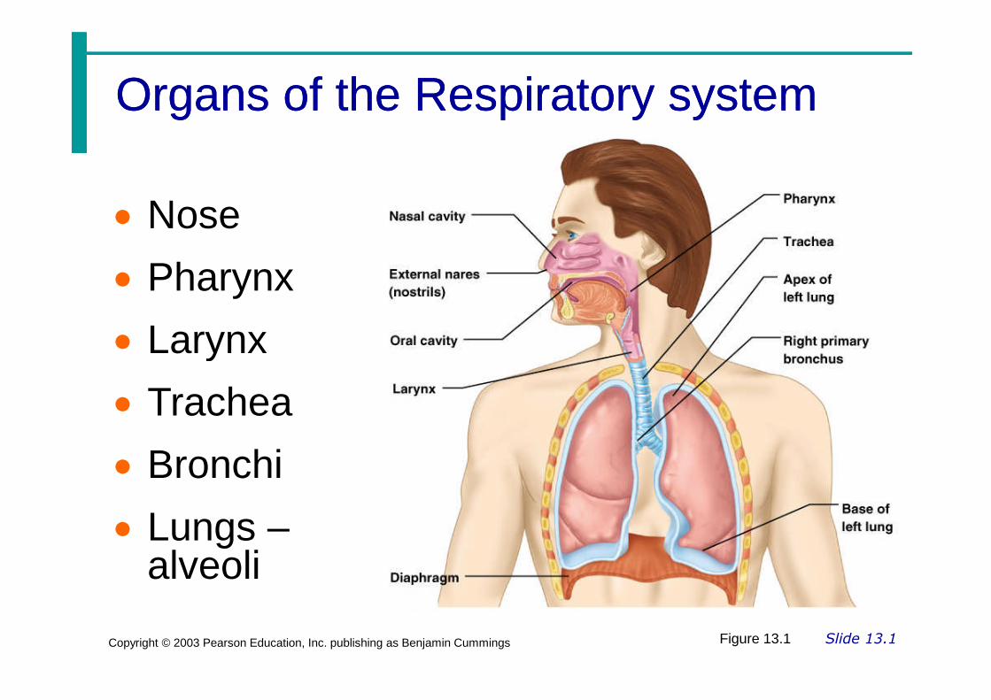
Organs of the Respiratory systemOrgans of the Respiratory system
Nose
Pharynx
Larynx
Slide 13.1Copyright © 2003 Pearson Education, Inc. publishing as Benjamin Cummings
Larynx
Trachea
Bronchi
Lungs –alveoli
Figure 13.1

Function of the Respiratory SystemFunction of the Respiratory System
Oversees gas exchanges between theblood and external environment
Exchange of gasses takes place within
Slide 13.2Copyright © 2003 Pearson Education, Inc. publishing as Benjamin Cummings
Exchange of gasses takes place withinthe lungs in the alveoli
Passageways to the lungs purify, warm,and humidify the incoming air
The NoseThe Nose
The only externally visible part of therespiratory system
Air enters the nose through the external
Slide 13.3aCopyright © 2003 Pearson Education, Inc. publishing as Benjamin Cummings
Air enters the nose through the externalnares (nostrils)
The interior of the nose consists of anasal cavity divided by a nasal septum

Upper Respiratory TractUpper Respiratory Tract
Slide 13.3bCopyright © 2003 Pearson Education, Inc. publishing as Benjamin Cummings
Figure 13.2

Anatomy of the Nasal CavityAnatomy of the Nasal Cavity
Olfactory receptors are located in themucosa on the superior surface
The rest of the cavity is lined with
Slide 13.4aCopyright © 2003 Pearson Education, Inc. publishing as Benjamin Cummings
The rest of the cavity is lined withrespiratory mucosa
Moistens air
Traps incoming foreign particles

Anatomy of the Nasal CavityAnatomy of the Nasal Cavity
Lateral walls have projections calledconchae
Increases surface area
Increases air turbulence within the nasal
Slide 13.4bCopyright © 2003 Pearson Education, Inc. publishing as Benjamin Cummings
Increases air turbulence within the nasalcavity
The nasal cavity is separated from theoral cavity by the palate
Anterior hard palate (bone)
Posterior soft palate (muscle)

Paranasal SinusesParanasal Sinuses
Cavities within bones surrounding thenasal cavity
Frontal bone
Slide 13.5aCopyright © 2003 Pearson Education, Inc. publishing as Benjamin Cummings
Frontal bone
Sphenoid bone
Ethmoid bone
Maxillary bone

Paranasal SinusesParanasal Sinuses
Function of the sinuses
Lighten the skull
Slide 13.5bCopyright © 2003 Pearson Education, Inc. publishing as Benjamin Cummings
Act as resonance chambers for speech
Produce mucus that drains into the nasalcavity

Pharynx (Throat)Pharynx (Throat)
Muscular passage from nasal cavity tolarynx
Three regions of the pharynx
Nasopharynx – superior region behindnasal cavity
Slide 13.6Copyright © 2003 Pearson Education, Inc. publishing as Benjamin Cummings
Nasopharynx – superior region behindnasal cavity
Oropharynx – middle region behind mouth
Laryngopharynx – inferior region attachedto larynx
The oropharynx and laryngopharynx arecommon passageways for air and food

Structures of the PharynxStructures of the Pharynx
Auditory tubes enter the nasopharynx
Tonsils of the pharynx
Pharyngeal tonsil (adenoids) in the
Slide 13.7Copyright © 2003 Pearson Education, Inc. publishing as Benjamin Cummings
Pharyngeal tonsil (adenoids) in thenasopharynx
Palatine tonsils in the oropharynx
Lingual tonsils at the base of the tongue

Larynx (Voice Box)Larynx (Voice Box)
Routes air and food into properchannels
Plays a role in speech
Slide 13.8Copyright © 2003 Pearson Education, Inc. publishing as Benjamin Cummings
Plays a role in speech
Made of eight rigid hyaline cartilagesand a spoon-shaped flap of elasticcartilage (epiglottis)

Structures of the LarynxStructures of the Larynx
Thyroid cartilage
Largest hyaline cartilage
Protrudes anteriorly (Adam’s apple)
Slide 13.9aCopyright © 2003 Pearson Education, Inc. publishing as Benjamin Cummings
Protrudes anteriorly (Adam’s apple)
Epiglottis
Superior opening of the larynx
Routes food to the larynx and air towardthe trachea

Structures of the LarynxStructures of the Larynx
Vocal cords (vocal folds)
Vibrate with expelled air to create sound
Slide 13.9bCopyright © 2003 Pearson Education, Inc. publishing as Benjamin Cummings
Vibrate with expelled air to create sound(speech)
Glottis – opening between vocal cords

Trachea (Windpipe)Trachea (Windpipe)
Connects larynx with bronchi
Lined with ciliated mucosa
Beat continuously in the opposite direction ofincoming air
Slide 13.10Copyright © 2003 Pearson Education, Inc. publishing as Benjamin Cummings
incoming air
Expel mucus loaded with dust and otherdebris away from lungs
Walls are reinforced with C-shapedhyaline cartilage

Primary BronchiPrimary Bronchi
Formed by division of the trachea
Enters the lung at the hilus(medial depression)
Slide 13.11Copyright © 2003 Pearson Education, Inc. publishing as Benjamin Cummings
Right bronchus is wider, shorter,and straighter than left
Bronchi subdivide into smallerand smaller branches

LungsLungs
Occupy most of the thoracic cavity
Apex is near the clavicle (superior portion)
Base rests on the diaphragm (inferior
Slide 13.12aCopyright © 2003 Pearson Education, Inc. publishing as Benjamin Cummings
Base rests on the diaphragm (inferiorportion)
Each lung is divided into lobes by fissures
Left lung – two lobes
Right lung – three lobes

LungsLungs
Slide 13.12bCopyright © 2003 Pearson Education, Inc. publishing as Benjamin Cummings
Figure 13.4b

Coverings of the LungsCoverings of the Lungs
Pulmonary (visceral) pleura covers thelung surface
Parietal pleura lines the walls of the
Slide 13.13Copyright © 2003 Pearson Education, Inc. publishing as Benjamin Cummings
Parietal pleura lines the walls of thethoracic cavity
Pleural fluid fills the area betweenlayers of pleura to allow gliding

Respiratory Tree DivisionsRespiratory Tree Divisions
Primary bronchi
Secondary bronchi
Slide 13.14Copyright © 2003 Pearson Education, Inc. publishing as Benjamin Cummings
Tertiary bronchi
Bronchioli
Terminal bronchioli

BronchiolesBronchioles
Slide 13.15aCopyright © 2003 Pearson Education, Inc. publishing as Benjamin Cummings
Figure 13.5a
Smallestbranches ofthe bronchi

BronchiolesBronchioles
Slide 13.15bCopyright © 2003 Pearson Education, Inc. publishing as Benjamin Cummings
Figure 13.5a
All but the smallestbranches havereinforcing cartilage

BronchiolesBronchioles
Slide 13.15cCopyright © 2003 Pearson Education, Inc. publishing as Benjamin Cummings
Terminalbronchioles endin alveoli
Figure 13.5a
Respiratory ZoneRespiratory Zone
Structures
Respiratory bronchioli
Slide 13.16Copyright © 2003 Pearson Education, Inc. publishing as Benjamin Cummings
Alveolar duct
Alveoli
Site of gas exchange

AlveoliAlveoli
Structure of alveoli
Alveolar duct
Alveolar sac
Slide 13.17Copyright © 2003 Pearson Education, Inc. publishing as Benjamin Cummings
Alveolar sac
Alveolus
Gas exchange takes place within the alveoliin the respiratory membrane

Respiratory MembraneRespiratory Membrane(Air(Air--Blood Barrier)Blood Barrier)
Thin squamous epithelial layer liningalveolar walls
Slide 13.18aCopyright © 2003 Pearson Education, Inc. publishing as Benjamin Cummings
alveolar walls
Pulmonary capillaries cover externalsurfaces of alveoli

Respiratory MembraneRespiratory Membrane(Air(Air--Blood Barrier)Blood Barrier)
Slide 13.18bCopyright © 2003 Pearson Education, Inc. publishing as Benjamin Cummings
Figure 13.6

Gas ExchangeGas Exchange
Gas crosses the respiratory membraneby diffusion
Oxygen enters the blood
Slide 13.19Copyright © 2003 Pearson Education, Inc. publishing as Benjamin Cummings
Carbon dioxide enters the alveoli
Macrophages add protection
Surfactant coats gas-exposed alveolarsurfaces

Events of RespirationEvents of Respiration
Pulmonary ventilation – moving air in andout of the lungs
Slide 13.20aCopyright © 2003 Pearson Education, Inc. publishing as Benjamin Cummings
out of the lungs
External respiration – gas exchangebetween pulmonary blood and alveoli

Events of RespirationEvents of Respiration
Respiratory gas transport – transport ofoxygen and carbon dioxide via thebloodstream
Slide 13.20bCopyright © 2003 Pearson Education, Inc. publishing as Benjamin Cummings
bloodstream
Internal respiration – gas exchangebetween blood and tissue cells insystemic capillaries
Mechanics of BreathingMechanics of Breathing(Pulmonary Ventilation)(Pulmonary Ventilation)
Completely mechanical process
Depends on volume changes in thethoracic cavity
Slide 13.21aCopyright © 2003 Pearson Education, Inc. publishing as Benjamin Cummings
thoracic cavity
Volume changes lead to pressurechanges, which lead to the flow ofgases to equalize pressure

Mechanics of BreathingMechanics of Breathing(Pulmonary Ventilation)(Pulmonary Ventilation)
Two phases
Inspiration – flow of air into lung
Slide 13.21bCopyright © 2003 Pearson Education, Inc. publishing as Benjamin Cummings
Inspiration – flow of air into lung
Expiration – air leaving lung

InspirationInspiration
Diaphragm and intercostal musclescontract
Slide 13.22aCopyright © 2003 Pearson Education, Inc. publishing as Benjamin Cummings
The size of the thoracic cavity increases
External air is pulled into the lungs due toan increase in intrapulmonary volume

InspirationInspiration
Slide 13.22bCopyright © 2003 Pearson Education, Inc. publishing as Benjamin Cummings
Figure 13.7a

ExhalationExhalation
Largely a passive process which dependson natural lung elasticity
As muscles relax, air is pushed out of the
Slide 13.23aCopyright © 2003 Pearson Education, Inc. publishing as Benjamin Cummings
As muscles relax, air is pushed out of thelungs
Forced expiration can occur mostly bycontracting internal intercostal muscles todepress the rib cage

ExhalationExhalation
Slide 13.23bCopyright © 2003 Pearson Education, Inc. publishing as Benjamin Cummings
Figure 13.7b

Pressure Differences in thePressure Differences in theThoracic CavityThoracic Cavity
Normal pressure within the pleuralspace is always negative (intrapleuralpressure)
Slide 13.24Copyright © 2003 Pearson Education, Inc. publishing as Benjamin Cummings
pressure)
Differences in lung and pleural spacepressures keep lungs from collapsing

Nonrespiratory Air MovementsNonrespiratory Air Movements
Can be caused by reflexes or voluntaryactions
Examples
Cough and sneeze – clears lungs of debris
Slide 13.25Copyright © 2003 Pearson Education, Inc. publishing as Benjamin Cummings
Cough and sneeze – clears lungs of debris
Laughing
Crying
Yawn
Hiccup

Respiratory Volumes and CapacitiesRespiratory Volumes and Capacities
Normal breathing moves about 500 ml of airwith each breath (tidal volume [TV])
Many factors that affect respiratory capacity
A person’s size
Slide 13.26Copyright © 2003 Pearson Education, Inc. publishing as Benjamin Cummings
A person’s size
Sex
Age
Physical condition
Residual volume of air – after exhalation,about 1200 ml of air remains in the lungs
Respiratory Volumes and CapacitiesRespiratory Volumes and Capacities
Inspiratory reserve volume (IRV)
Amount of air that can be taken in forciblyover the tidal volume
Slide 13.27aCopyright © 2003 Pearson Education, Inc. publishing as Benjamin Cummings
Usually between 2100 and 3200 ml
Expiratory reserve volume (ERV)
Amount of air that can be forcibly exhaled
Approximately 1200 ml

Respiratory Volumes and CapacitiesRespiratory Volumes and Capacities
Residual volume
Air remaining in lung after expiration
Slide 13.27bCopyright © 2003 Pearson Education, Inc. publishing as Benjamin Cummings
Air remaining in lung after expiration
About 1200 ml

Respiratory Volumes and CapacitiesRespiratory Volumes and Capacities
Vital capacity
The total amount of exchangeable air
Vital capacity = TV + IRV + ERV
Slide 13.28Copyright © 2003 Pearson Education, Inc. publishing as Benjamin Cummings
Vital capacity = TV + IRV + ERV
Dead space volume
Air that remains in conducting zone andnever reaches alveoli
About 150 ml

Respiratory Volumes and CapacitiesRespiratory Volumes and Capacities
Functional volume
Air that actually reaches the respiratoryzone
Slide 13.29Copyright © 2003 Pearson Education, Inc. publishing as Benjamin Cummings
zone
Usually about 350 ml
Respiratory capacities are measuredwith a spirometer

Respiratory CapacitiesRespiratory Capacities
Slide 13.30Copyright © 2003 Pearson Education, Inc. publishing as Benjamin Cummings
Figure 13.9

Respiratory SoundsRespiratory Sounds
Sounds are monitored with astethoscope
Bronchial sounds – produced by air
Slide 13.31Copyright © 2003 Pearson Education, Inc. publishing as Benjamin Cummings
Bronchial sounds – produced by airrushing through trachea and bronchi
Vesicular breathing sounds – softsounds of air filling alveoli

External RespirationExternal Respiration
Oxygen movement into the blood
The alveoli always has more oxygen thanthe blood
Slide 13.32aCopyright © 2003 Pearson Education, Inc. publishing as Benjamin Cummings
the blood
Oxygen moves by diffusion towards thearea of lower concentration
Pulmonary capillary blood gains oxygen

External RespirationExternal Respiration
Carbon dioxide movement out of theblood
Blood returning from tissues has higherconcentrations of carbon dioxide than air in
Slide 13.32bCopyright © 2003 Pearson Education, Inc. publishing as Benjamin Cummings
concentrations of carbon dioxide than air inthe alveoli
Pulmonary capillary blood gives up carbondioxide
Blood leaving the lungs is oxygen-richand carbon dioxide-poor

Gas Transport in the BloodGas Transport in the Blood
Oxygen transport in the blood
Inside red blood cells attached to
Slide 13.33aCopyright © 2003 Pearson Education, Inc. publishing as Benjamin Cummings
Inside red blood cells attached tohemoglobin (oxyhemoglobin [HbO2])
A small amount is carried dissolved in theplasma

Gas Transport in the BloodGas Transport in the Blood
Carbon dioxide transport in the blood
Most is transported in the plasma as
Slide 13.33bCopyright © 2003 Pearson Education, Inc. publishing as Benjamin Cummings
Most is transported in the plasma asbicarbonate ion (HCO3
–)
A small amount is carried inside red bloodcells on hemoglobin, but at different bindingsites than those of oxygen

Internal RespirationInternal Respiration
Exchange of gases between blood andbody cells
An opposite reaction to what occurs in
Slide 13.34aCopyright © 2003 Pearson Education, Inc. publishing as Benjamin Cummings
An opposite reaction to what occurs inthe lungs
Carbon dioxide diffuses out of tissue toblood
Oxygen diffuses from blood into tissue

Internal RespirationInternal Respiration
Slide 13.34bCopyright © 2003 Pearson Education, Inc. publishing as Benjamin Cummings
Figure 13.11

External Respiration,External Respiration,Gas Transport, andGas Transport, andInternal RespirationInternal RespirationSummarySummary
Slide 13.35Copyright © 2003 Pearson Education, Inc. publishing as Benjamin Cummings
Figure 13.10

Neural Regulation of RespirationNeural Regulation of Respiration
Activity of respiratory muscles is transmittedto the brain by the phrenic and intercostalnerves
Neural centers that control rate and depth arelocated in the medulla
Slide 13.36Copyright © 2003 Pearson Education, Inc. publishing as Benjamin Cummings
located in the medulla
The pons appears to smooth out respiratoryrate
Normal respiratory rate (eupnea) is 12–15respirations per minute
Hypernia is increased respiratory rate oftendue to extra oxygen needs

Neural Regulation of RespirationNeural Regulation of Respiration
Slide 13.37Copyright © 2003 Pearson Education, Inc. publishing as Benjamin Cummings
Figure 13.12

Factors Influencing RespiratoryFactors Influencing RespiratoryRate and DepthRate and Depth
Physical factors
Increased body temperature
Exercise
Slide 13.38Copyright © 2003 Pearson Education, Inc. publishing as Benjamin Cummings
Exercise
Talking
Coughing
Volition (conscious control)
Emotional factors

Factors Influencing RespiratoryFactors Influencing RespiratoryRate and DepthRate and Depth
Chemical factors
Carbon dioxide levels
Level of carbon dioxide in the blood is the
Slide 13.39aCopyright © 2003 Pearson Education, Inc. publishing as Benjamin Cummings
Level of carbon dioxide in the blood is themain regulatory chemical for respiration
Increased carbon dioxide increasesrespiration
Changes in carbon dioxide act directly onthe medulla oblongata

Factors Influencing RespiratoryFactors Influencing RespiratoryRate and DepthRate and Depth
Chemical factors (continued)
Oxygen levels
Slide 13.39bCopyright © 2003 Pearson Education, Inc. publishing as Benjamin Cummings
Oxygen levels
Changes in oxygen concentration in theblood are detected by chemoreceptors inthe aorta and carotid artery
Information is sent to the medulla oblongata
Respiratory Disorders: ChronicRespiratory Disorders: ChronicObstructive Pulmonary DiseaseObstructive Pulmonary Disease(COPD)(COPD)
Exemplified by chronic bronchitis andemphysema
Slide 13.40aCopyright © 2003 Pearson Education, Inc. publishing as Benjamin Cummings
emphysema
Major causes of death and disability inthe United States

Respiratory Disorders: ChronicRespiratory Disorders: ChronicObstructive Pulmonary DiseaseObstructive Pulmonary Disease(COPD)(COPD)
Features of these diseases
Patients almost always have a history of
Slide 13.40bCopyright © 2003 Pearson Education, Inc. publishing as Benjamin Cummings
Patients almost always have a history ofsmoking
Labored breathing (dyspnea) becomesprogressively more severe
Coughing and frequent pulmonaryinfections are common

Respiratory Disorders: ChronicRespiratory Disorders: ChronicObstructive Pulmonary DiseaseObstructive Pulmonary Disease(COPD)(COPD)
Features of these diseases (continued)
Slide 13.40cCopyright © 2003 Pearson Education, Inc. publishing as Benjamin Cummings
Most victimes retain carbon dioxide, arehypoxic and have respiratory acidosis
Those infected will ultimately developrespiratory failure

EmphysemaEmphysema
Alveoli enlarge as adjacent chambers breakthrough
Chronic inflammation promotes lung fibrosis
Airways collapse during expiration
Slide 13.41Copyright © 2003 Pearson Education, Inc. publishing as Benjamin Cummings
Airways collapse during expiration
Patients use a large amount of energy toexhale
Overinflation of the lungs leads to apermanently expanded barrel chest
Cyanosis appears late in the disease

Chronic BronchitisChronic Bronchitis
Mucosa of the lower respiratorypassages becomes severely inflamed
Mucus production increases
Pooled mucus impairs ventilation and
Slide 13.42Copyright © 2003 Pearson Education, Inc. publishing as Benjamin Cummings
Pooled mucus impairs ventilation andgas exchange
Risk of lung infection increases
Pneumonia is common
Hypoxia and cyanosis occur early
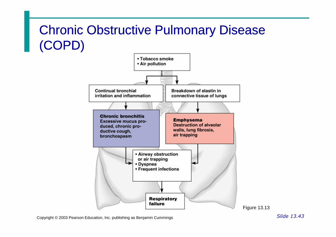
Chronic Obstructive Pulmonary DiseaseChronic Obstructive Pulmonary Disease(COPD)(COPD)
Slide 13.43Copyright © 2003 Pearson Education, Inc. publishing as Benjamin Cummings
Figure 13.13

Lung CancerLung Cancer
Accounts for 1/3 of all cancer deaths inthe United States
Increased incidence associated withsmoking
Slide 13.44Copyright © 2003 Pearson Education, Inc. publishing as Benjamin Cummings
smoking
Three common types
Squamous cell carcinoma
Adenocarcinoma
Small cell carcinoma

Sudden Infant Death syndromeSudden Infant Death syndrome(SIDS)(SIDS)
Apparently healthy infant stopsbreathing and dies during sleep
Some cases are thought to be a
Slide 13.45Copyright © 2003 Pearson Education, Inc. publishing as Benjamin Cummings
Some cases are thought to be aproblem of the neural respiratory controlcenter
One third of cases appear to be due toheart rhythm abnormalities

AsthmaAsthma
Chronic inflamed hypersensitivebronchiole passages
Slide 13.46Copyright © 2003 Pearson Education, Inc. publishing as Benjamin Cummings
bronchiole passages
Response to irritants with dyspnea,coughing, and wheezing

Developmental Aspects of theDevelopmental Aspects of theRespiratory SystemRespiratory System
Lungs are filled with fluid in the fetus
Lungs are not fully inflated with air untiltwo weeks after birth
Slide 13.47aCopyright © 2003 Pearson Education, Inc. publishing as Benjamin Cummings
two weeks after birth
Surfactant that lowers alveolar surfacetension is not present until late in fetaldevelopment and may not be present inpremature babies

Developmental Aspects of theDevelopmental Aspects of theRespiratory SystemRespiratory System
Important birth defects
Cystic fibrosis – oversecretion of thick
Slide 13.47bCopyright © 2003 Pearson Education, Inc. publishing as Benjamin Cummings
Cystic fibrosis – oversecretion of thickmucus clogs the respiratory system
Cleft palate

Aging EffectsAging Effects
Elasticity of lungs decreases
Vital capacity decreases
Blood oxygen levels decrease
Slide 13.48Copyright © 2003 Pearson Education, Inc. publishing as Benjamin Cummings
Blood oxygen levels decrease
Stimulating effects of carbon dioxidedecreases
More risks of respiratory tract infection

Respiratory Rate ChangesRespiratory Rate ChangesThroughout LifeThroughout Life
Newborns – 40 to 80 respirations perminute
Infants – 30 respirations per minute
Slide 13.49Copyright © 2003 Pearson Education, Inc. publishing as Benjamin Cummings
Age 5 – 25 respirations per minute
Adults – 12 to 18 respirations perminute
Rate often increases somewhat with oldage


