The pathogenesis of transfusion-related acute lung injury (TRALI)
12
The pathogenesis of transfusion-related acute lung injury (TRALI) Ju ¨rgen Bux 1 and Ulrich J. H. Sachs 2 1 DRK-Blood Service West of the German Red Cross, Hagen, and 2 Institute for Clinical Immunology and Transfusion Medicine, Justus Liebig-University, Giessen, Germany Summary In recent years, transfusion-related acute lung injury (TRALI) has developed from an almost unknown transfusion reaction to the most common cause of transfusion-related major morbidities and fatalities. A clinical definition of TRALI was established in 2004, based on acute respiratory distress, non- cardiogenic lung oedema temporal association with trans- fusion and hypoxaemia. Histological findings reveal lung oedema, capillary leucostasis and neutrophil extravasation. However, the pathogenesis of TRALI remains controversial. Leucocyte antibodies, present in fresh frozen plasma and platelet concentrates from multiparous donors, and neutrophil priming agents released in stored cellular blood components have been considered to be causative. As neutrophils and endothelial cells are pivotal in the pathogenesis of TRALI, a threshold model was established to try to unify the various reported findings on pathogenesis. This model comprises the priming of neutrophils and/or endothelium by the patient’s co-morbidity, neutrophil and/or endothelial cell activation by the transfused blood component, and the severity of the TRALI reaction. Keywords: transfusion-related acute lung injury, transfusion, granulocytes, neutrophils. Non-cardiogenic lung oedema as a result of blood transfu- sion was first described by Barnard (1951). Popovsky et al (1983) recognised this transfusion reaction as a distinct clinical entity and coined the term transfusion-related acute lung injury (TRALI). Two years later Popovsky and Moore (1985) analysed a series of 36 patients with TRALI. Minimal criteria for the diagnosis of TRALI were: acute respiratory distress and new bilateral lung infiltrations in the chest X-ray within 6 h of blood transfusion and the absence of evidence for the presence of volume overload or cardiac malfunction. They reported a ventilation rate of 70%, a fatality rate of 6% and the presence of leucocyte, i.e. neutrophil and human leucocyte antigen (HLA), antibodies in the blood of 89% of implicated donors. Silliman et al (1997) reported the association of biologically active lipids with the development of TRALI. Meanwhile, several neutrophil-priming agents have been identified in stored cellular blood components. In a prospective study of 90 TRALI reactions in 81 patients, patients with haematological malignancies and cardiac disease were identified as patients at risk for TRALI (Silliman et al, 2003a). All TRALI reactions were secondary to the transfusion of stored platelets and red cells, with one exception. Mechanical ventilation was required in only 3% of the cases; TRALI lead to death in one patient. However, in this study lung oedema was mainly diagnosed by auscultation and not by chest X-ray. Despite these reports, TRALI remained little known. In 2004 the European Haemo- vigilance Network (EHN) and the Canadian Consensus Conference (Kleinman et al, 2004) proposed criteria for the diagnosis of TRALI, which are summarised in Table I. It has been thanks to the British Serious Hazards of Transfusion (SHOT) haemovigilance system that the recognition of TRALI has gained increased recognition. In recent years, TRALI has been reliably shown to be the most common cause of transfusion-related fatalities in the United States and in the United Kingdom (Holness et al, 2004; Stainsby et al, 2004). This review summarises the current knowledge on the pathogenesis of TRALI. Pathological findings in TRALI patients Histological findings in patients who died from TRALI are consistent with early acute respiratory distress syndrome (ARDS), showing interstitial and intra-alveolar oedema (Felbo & Jensen, 1962; Flury & Reutter, 1966; Kernoff et al, 1972; Wolf & Canale, 1976; Popovsky & Moore, 1985; Silliman et al, 1997; Dry et al, 1999) and an extravasation of neutrophils into the interstitial and air spaces (Kernoff et al, 1972; Wolf & Canale, 1976; Silliman et al, 1997; Dry et al, 1999). In addition, hyaline membranes and destruction of the pulmonary architecture have been reported (Wolf & Canale, 1976; Silliman et al, 1997). More important, an increased number of neutrophils within the pulmonary capillary vasculature and small pulmonary vessels Correspondence: Dr Ju ¨rgen Bux, MD PhD, DRK-Blutspendedienst West Feithstrasse 182 D-58097 Hagen, Germany. E-mail: [email protected] review ª 2007 The Authors doi:10.1111/j.1365-2141.2007.06492.x Journal Compilation ª 2007 Blackwell Publishing Ltd, British Journal of Haematology, 136, 788–799
Transcript of The pathogenesis of transfusion-related acute lung injury (TRALI)

The pathogenesis of transfusion-related acute lung injury(TRALI)
Jurgen Bux1 and Ulrich J. H. Sachs2
1DRK-Blood Service West of the German Red Cross, Hagen, and 2Institute for Clinical Immunology and Transfusion Medicine, Justus
Liebig-University, Giessen, Germany
Summary
In recent years, transfusion-related acute lung injury (TRALI)
has developed from an almost unknown transfusion reaction
to the most common cause of transfusion-related major
morbidities and fatalities. A clinical definition of TRALI was
established in 2004, based on acute respiratory distress, non-
cardiogenic lung oedema temporal association with trans-
fusion and hypoxaemia. Histological findings reveal lung
oedema, capillary leucostasis and neutrophil extravasation.
However, the pathogenesis of TRALI remains controversial.
Leucocyte antibodies, present in fresh frozen plasma and
platelet concentrates from multiparous donors, and neutrophil
priming agents released in stored cellular blood components
have been considered to be causative. As neutrophils and
endothelial cells are pivotal in the pathogenesis of TRALI, a
threshold model was established to try to unify the various
reported findings on pathogenesis. This model comprises the
priming of neutrophils and/or endothelium by the patient’s
co-morbidity, neutrophil and/or endothelial cell activation by
the transfused blood component, and the severity of the TRALI
reaction.
Keywords: transfusion-related acute lung injury, transfusion,
granulocytes, neutrophils.
Non-cardiogenic lung oedema as a result of blood transfu-
sion was first described by Barnard (1951). Popovsky et al
(1983) recognised this transfusion reaction as a distinct clinical
entity and coined the term transfusion-related acute lung
injury (TRALI). Two years later Popovsky and Moore (1985)
analysed a series of 36 patients with TRALI. Minimal criteria
for the diagnosis of TRALI were: acute respiratory distress and
new bilateral lung infiltrations in the chest X-ray within 6 h of
blood transfusion and the absence of evidence for the presence
of volume overload or cardiac malfunction. They reported
a ventilation rate of 70%, a fatality rate of 6% and the presence
of leucocyte, i.e. neutrophil and human leucocyte antigen
(HLA), antibodies in the blood of 89% of implicated donors.
Silliman et al (1997) reported the association of biologically
active lipids with the development of TRALI. Meanwhile,
several neutrophil-priming agents have been identified in
stored cellular blood components. In a prospective study of 90
TRALI reactions in 81 patients, patients with haematological
malignancies and cardiac disease were identified as patients at
risk for TRALI (Silliman et al, 2003a). All TRALI reactions
were secondary to the transfusion of stored platelets and red
cells, with one exception. Mechanical ventilation was required
in only 3% of the cases; TRALI lead to death in one patient.
However, in this study lung oedema was mainly diagnosed by
auscultation and not by chest X-ray. Despite these reports,
TRALI remained little known. In 2004 the European Haemo-
vigilance Network (EHN) and the Canadian Consensus
Conference (Kleinman et al, 2004) proposed criteria for the
diagnosis of TRALI, which are summarised in Table I. It has
been thanks to the British Serious Hazards of Transfusion
(SHOT) haemovigilance system that the recognition of TRALI
has gained increased recognition. In recent years, TRALI has
been reliably shown to be the most common cause of
transfusion-related fatalities in the United States and in the
United Kingdom (Holness et al, 2004; Stainsby et al, 2004).
This review summarises the current knowledge on the
pathogenesis of TRALI.
Pathological findings in TRALI patients
Histological findings in patients who died from TRALI are
consistent with early acute respiratory distress syndrome
(ARDS), showing interstitial and intra-alveolar oedema (Felbo
& Jensen, 1962; Flury & Reutter, 1966; Kernoff et al, 1972; Wolf
& Canale, 1976; Popovsky & Moore, 1985; Silliman et al, 1997;
Dry et al, 1999) and an extravasation of neutrophils into the
interstitial and air spaces (Kernoff et al, 1972; Wolf & Canale,
1976; Silliman et al, 1997; Dry et al, 1999). In addition, hyaline
membranes and destruction of the pulmonary architecture have
been reported (Wolf & Canale, 1976; Silliman et al, 1997). More
important, an increased number of neutrophils within the
pulmonary capillary vasculature and small pulmonary vessels
Correspondence: Dr Jurgen Bux, MD PhD, DRK-Blutspendedienst
West Feithstrasse 182 D-58097 Hagen, Germany.
E-mail: [email protected]
review
ª 2007 The Authorsdoi:10.1111/j.1365-2141.2007.06492.x Journal Compilation ª 2007 Blackwell Publishing Ltd, British Journal of Haematology, 136, 788–799

was observed in lung sections from TRALI patients (Felbo &
Jensen, 1962; Dry et al, 1999), and a positive correlation between
the degree of capillary leucostasis and the amount of proteina-
ceous fluid within the alveolar air spaces has been demonstrated.
On electron microscopic pictures, neutrophils were degranulat-
ed and focally in direct contact with denuded stretches of the
capillary wall; in addition, a correlation between capillary
leucostasis and desquamated epithelial cells were reported
(Dry et al, 1999). From these observations, it seems reasonable
to speculate that after sequestration in the early stages of TRALI,
neutrophils and endothelial cells of the pulmonary microvascu-
lature establish close contacts, which lead to firm neutrophil
adhesion and retention. Once the neutrophils are immobilised in
the alveolar capillaries, they activate their microbicidal arsenal
and induce endothelial damage and capillary leak. This allows
transit of proteinaceous fluid from the vessels into the air spaces,
resulting in acute pulmonary oedema. In the later stages,
especially of severe TRALI, neutrophils can extravasate from the
capillary into the alveoli and induce pulmonary injury. Accord-
ingly, neutrophils and endothelial cells of the lung capillaries are
the key players in the pathogenesis of TRALI.
Physiological aspects of neutrophil passagethrough the pulmonary microvasculature
Neutrophil transit
An adult human with a cardiac output of 5 l/min pumps
approximately 7200 l of blood through the pulmonary circu-
lation in 24 h. As each litre of circulating blood contains about
109 leucocytes, their traffic through pulmonary microvessels is
enormous. The alveolar capillary bed is a complex intercon-
necting network of short capillary segments. From quantitative
histological studies it was calculated that each of the estimated
100 million alveoli in the adult human lung contains about
1000 capillary segments. The path from arteriole to venule
crosses several alveolar walls (often >8) so that a blood cell
encounters >50 capillary segments, each with an average length
of 14Æ5 lm. Approximately 50% of the pulmonary capillaries
(2–15 lm) are narrower than the diameter of a spherically
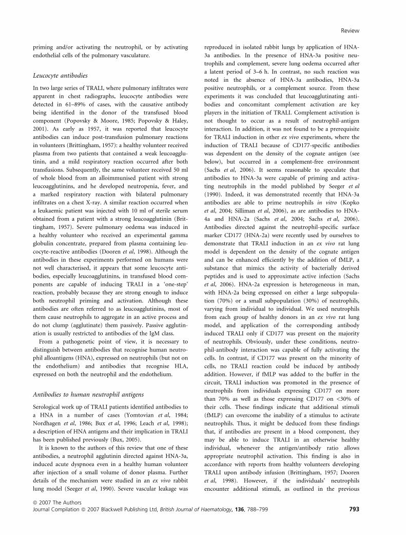
shaped neutrophil (6–8 lm). Thus, neutrophils often encoun-
ter a capillary segment that forces them to pause and to deform
and assume a ‘sausage’ shape before squeezing through the
narrow capillary segment (Fig. 1). Median transit times of
neutrophils were 26 s compared with 1Æ4–4Æ2 s of erythrocytes,
because, although the erythrocyte has a discoid shape with
a similar diameter to the spherical neutrophil, the erythrocyte
can rapidly reduce its diameter by folding when it encounters
a narrow capillary (Gebb et al, 1995). Computational models
of the capillary bed support the concept that the structure of
the capillary bed and the deformation of neutrophils are
critical under normal conditions (Huang et al, 2001). Conse-
quently, the transit time of neutrophils through the pulmonary
capillary bed is mainly affected by their deformation time. The
increased transit time also accounts for the significant
neutrophil accumulation (‘marginated pool’) in the lungs
(Doerschuk, 1999). The pulmonary circulation contains about
28% of the blood neutrophil pool that is available on demand
for host defence against bacterial infections (Peters, 1998).
Neutrophil sequestration and retention
In the microvasculature of many organs, neutrophil recruit-
ment from blood into tissue at sites of inflammation occurs in
postcapillary venules. In contrast, the principal site of leuco-
cyte emigration in the lung is the capillary bed (Loosli & Baker,
1962; Downey et al, 1993; Lee & Downey, 2001). As
neutrophils do not roll in lung capillaries, the conventional
model of neutrophil tethering, rolling and arrest on inflamed
endothelium is not applicable to lung capillaries (Burns et al,
2003). The stimulus-induced decrease in deformability is
thought to be important for neutrophil sequestration in the
lung and probably replaces the role of selectin-mediated rolling
in stopping neutrophils within capillaries (Doerschuk, 1999;
Burns et al, 2003; Reutershan & Ley, 2004). The role of
Table I. Criteria for the clinical diagnosis of transfusion-related acute
lung injury (TRALI).
TRALI Consensus Conference Committee 2004 and European
Haemovigilance Network
Acute respiratory distress
Bilateral lung infiltrations in the chest radiograph
Occurrence during or within 6-h after completion of transfusion
No evidence of transfusion-associated circulatory
overload/cardiogenic lung oedema
Hypoxaemia (PaO2/FiO2 <300 mmHg or O2 saturation
<90% or other clinical evidence)
New acute lung injury (ALI) and no other ALI risk factors
present (aspiration, multiple trauma, pneumonia,
cardiopulmonary bypass, burn injury, toxic inhalation, lung
contusion, pancreatitis, drug overdose, near drowning, shock,
severe sepsis)
If one or more ALI risk factors are present, possible TRALI should be
diagnosed.
~ ~ ~ ~ ~ ~ ~ ~
Arteriole Capillary Venule
6–8 µm ~2–15 µm
Fig 1. Neutrophil passage through pulmonary microvasculature.
During passage of the pulmonary microvasculature, neutrophils have
to pass more than 50 segments of the alveolar capillary bed. As the
diameter of spherical neutrophil is larger than about 50% of the
capillary segments, neutrophils, on their path from arteriole to venule,
encounter many segments that require them to change shape in order
to squeeze through. In contrast to the vasculature of many other or-
gans, the principal site of neutrophil emigration in response to
inflammation is the alveolar capillary bed.
Review
ª 2007 The AuthorsJournal Compilation ª 2007 Blackwell Publishing Ltd, British Journal of Haematology, 136, 788–799 789

mechanical factors in the initial sequestration of neutrophils in
the alveolar capillaries is supported by direct evidence that
neither l-selectin nor b2-integrins are required (Kubo et al,
1999). Activated neutrophils lose their ability to deform mainly
because of the intracellular polymerisation of actin filaments.
In contrast, events following the initial sequestration, i.e.
retention and transendothelial migration, are apparently
influenced by adhesion molecules. As mediator-induced
decreases in neutrophil deformability are temporally correlated
with a conformational change of b2-integrins from non-
adhesive to adhesive, physical trapping goes along with
neutrophil attraction to the endothelial surface. Intercellular
adhesion molecule 2 (ICAM-2; CD102) which is constitutively
expressed on vascular cells, is a ligand of the b2-integrin
CD11b/CD18 and appears to play a role in neutrophil
transendothelial migration (Issekutz et al, 1999). Transendot-
helial migration occurs mainly by penetrating interendothelial
junctions at bicellular or tricellular corners of endothelial
cells, although there is an alternative transcellular route (Burns
et al, 2003). Platelet/endothelial cell adhesion molecule-1
(PECAM-1; CD31), junctional adhesion molecules, vascular/
endothelial-cadherin, and CD99 are thought to regulate
neutrophil transendothelial migration (Chavakis et al, 2003).
Fluid leakage in the pulmonary microvasculature
Fluid and protein leakage occur in the normal lung primarily
through small gaps between capillary endothelial cells. The
filtered fluid enters the alveolar interstitial space but not the
alveoli because the alveolar epithelium is composed of very
tight junctions. From the alveolar interstitium the fluid moves
into the peribronchovascular space where it is removed by the
lymphatics and returned to the systemic circulation (Ware &
Matthay, 2005). Increased transvascular fluid filtration is the
hallmark of acute cardiogenic or volume overload oedema
(transudate). By contrast, non-cardiogenic pulmonary oedema
is caused by an increase in the vascular permeability resulting
in an increased flux of fluid and protein into the lung
interstitium and air spaces. Non-cardiogenic pulmonary
oedema has therefore high protein content (exudate).
Pathogenic aspects of TRALI: priming andactivation of neutrophils
In general, contacts with injurious agents are a prerequisite of
neutrophil activation. Activated neutrophils will respond by
phagocytosis, the release of preformed granular enzymes and
proteins, and by the de novo synthesis of a range of ephemeral,
but highly toxic reactive oxygen species (ROS). In order to be
become fully activated, neutrophils need to be primed.
Priming
Circulating neutrophils do not express anywhere near their full
microbicidal capacity when challenged with biological activating
agents unless they have first been primed. Priming refers to a
process whereby the response of neutrophils to an activating
stimulus is potentiated; it facilitates the clustering of relevant
surface receptors, such as FccRIIa and b2-integrins, and the
formation of the NADPH oxidase complex, which is respon-
sible for the synthesis of ROS. The release of harmful ROS that
occurs in response to an agonist is enhanced up to 20-fold
by prior exposure of the cell to a priming agent (Guthrie et al,
1984).
Priming agents do not elicit effector functions on their own,
except when they are applied at very high concentrations. As
neutrophil activation is of central importance in the patho-
genesis of TRALI, we have to assume that neutrophils
experience both priming and activation, usually triggered by
different stimuli, and that one of these stimuli must come from
the transfused blood component.
Priming of neutrophils owing to underlying co-morbidity
A number of studies and epidemiological data from haemo-
vigilance systems indicate that a large percentage of patients
who develop TRALI had recent surgery. In the initial study by
Popovsky and Moore (1985), 31 out of 36 TRALI cases
occurred in patients with surgical procedures, requiring blood
transfusion either during surgery (n ¼ 24) or caused by
postoperative blood loss (n ¼ 7), and only five out of 36
TRALI cases occurred in patients with anaemia for chronic or
stable conditions. These data were corroborated by others, and
active infection, cardiovascular disease and leukaemia were
identified as additional risk factors (Silliman et al, 1997;
Silliman et al, 2003a; Holness et al, 2004). It should be kept in
mind that linking TRALI to any specific clinical event needs
valid denominator data. However, some of these observations
are in accordance with in vivo evidence from studies demon-
strating that surgical procedures and active infections induce
neutrophil priming in patients (Bass et al, 1986; Krause et al,
1988; Kawahito et al, 2000). A variety of neutrophil priming
agents that are released either by dying/necrotic cells or by
stimulated endothelial cells, monocytes and lymphocytes have
been described, including platelet activating factor (PAF)
(Vercellotti et al, 1988), tumour necrosis factor-a (TNF-a)
(Berkow et al, 1987), interleukin 8 (IL-8) (Daniels et al, 1992),
granulocyte/macrophage-colony stimulating factor (Fleisch-
mann et al, 1986), and interferon-c (Tennenberg et al, 1993).
Neutrophils can also be primed by a number of infectious
agents, such as influenza A virus (Busse et al, 1991) and
bacteria-derived lipopolysaccharides (LPS) (Guthrie et al,
1984).
Consequences of circulating primed neutrophils withregard to TRALI
In response to priming agents, neutrophils undergo polarisa-
tion or shape change, a process that has originally been
suggested to represent frustrated chemotaxis (Haslett et al,
Review
ª 2007 The Authors790 Journal Compilation ª 2007 Blackwell Publishing Ltd, British Journal of Haematology, 136, 788–799

1985). This ‘stiffening’ of the cells augments the physiological
mechanism of mechanical retention of neutrophils within the
pulmonary capillary bed (sequestration), and prolongs the
process of neutrophil squeezing through the narrow capillaries
(Worthen et al, 1989). Prolonged, close contact of the stiff, i.e.
primed, neutrophil with the endothelial cells allows the
neutrophil to effectively sample the endothelial surface and
provides a micro-environment in which transmembrane
receptors and released mediators of each cell type can easily
influence the other. Exogenous stimuli present in the blood
bag can have impact on both, neutrophils and endothelial cells
(Fig. 2). In addition, after having been primed in the
circulation, sequestered neutrophils can now be activated to
express their full microbicidal activity by exogenous activating
substances present in the blood bag. These include neutrophil-
binding antibodies, cytokines and bioactive lipids (see below).
If a stimulus is no longer present, the primed neutrophil will
be deprimed. Studies on human peripheral blood neutrophils
in vitro suggest that the primed status is maintained for at least
24 h (Ichinose et al, 1990), a finding that fits well with the
epidemiological observation that surgery (as a priming event)
is a risk factor for TRALI if it was performed recently, i.e. <48-
h previous to the transfusion event (Silliman et al, 1997).
As mentioned above, some priming substances may also
induce full activation of the neutrophil when applied at high
concentrations. Besides chemokines/cytokines, cross-linking
antibodies to neutrophil surface receptors, namely to l-selectin
and CD18, have been demonstrated to induce neutrophil
priming efficiently (Waddell et al, 1994; Liles et al, 1995).
More important, ligation of neutrophil receptors may not only
result in neutrophil priming, but can also afford neutrophil
activation either in concert with or independent of soluble
activators (Berton et al, 1992; Crockett-Torabi et al, 1995). We
and others have demonstrated that antibodies to neutrophil-
specific antigens involved in TRALI cases are able to prime and
even activate neutrophils as well (Kopko et al, 2004; Sachs
et al, 2004; Sachs et al, 2006; Silliman et al, 2006). This, as
discussed below, explains why even completely healthy indi-
viduals can develop TRALI upon neutrophil antibody infusion
(Dooren et al, 1998).
Pathogenic aspects of TRALI: activation ofpulmonary endothelial cells
There is both clinical and experimental evidence that TRALI
induction does not always start from the primed neutrophil,
but may also be triggered by an activated pulmonary
endothelium (Fig. 2, right panel). In addition to the consti-
tutively expressed ICAM-2, activated endothelial cells upreg-
ulate surface membrane receptors that allow neutrophil
adhesion, including ligands for l-selectin (Spertini et al,
1991), p-selectin, and ICAM-1 (Gerritsen & Bloor, 1993; Klein
et al, 1995; Scholz et al, 1996). Neutrophils that pass through
the narrow lung capillaries may well get stuck if the endothe-
lium is activated and ligands for l-selectin have been
upregulated, as the process of retaining neutrophils within
the pulmonary capillaries has been described to be selectin-
dependent (Yamaguchi et al, 1997; Kubo et al, 1999). Once
sreggiTrsdipil
senikotyc/senikomehcseidobitnagnidnib-lihportuen
CECE
ILARTActivatedneutrophil
Primedneutrophil
Activatedneutrophil
ActivatedEC
Trappedneutrophil,
primed
Primedneutrophil,
trapped
Neutrophil EC
Fig 2. Possible pathomechanism of transfusion-related acute lung injury (TRALI). Neutrophils and pulmonary cells are key players in TRALI.
Activation of each cell type may lead to TRALI. On the one hand, neutrophils may become primed, most probably as a result of endogenous triggers,
such as those present during infections. Primed cells are trapped in the lungs’ microvasculature, where they experience activation via substances
present in the blood component, e.g. antibodies or bioactive lipids. On the other hand, an activated endothelial cell can induce neutrophil trapping
within the lungs, where they are primed and finally activated because of triggers present in the blood component. In either case, neutrophil/
endothelial cell interaction is necessary to finally induce TRALI.
Review
ª 2007 The AuthorsJournal Compilation ª 2007 Blackwell Publishing Ltd, British Journal of Haematology, 136, 788–799 791

trapped, the neutrophil will incorporate selectin-dependent
signals with other extracellular inflammatory stimuli in order
to achieve a preactivated (primed) status (Williams &
Solomkin, 1999). Additional stimuli come from endothelial
cells as well, because, once activated, they produce a broad
range of chemokines/cytokines, including PAF, leukotriene B4,
and IL-8. These substances are not only secreted, but remain
also bound to the endothelial cell surface, where they have
access to their corresponding receptors on the surface of
sequestered and/or adherent neutrophils (Kuijpers et al, 1992).
Various substances have been identified as endothelial cell
activators, including TNF-a, IL-1b, and other mediators
released during inflammatory processes, as well as exogenous
LPS and antibodies.
One interesting example of TRALI because of antibody-
mediated endothelial cell activation was reported by Dykes
et al (2000). In a patient who underwent lung transplantation
and became dyspneic after transfusion of two units red blood
cells, a chest X-ray revealed a unilateral white-out of the
transplanted lung. An antibody to HLA-B44 was present in the
donor of one of the transfused units, and the cognate antigen
was expressed on the transplanted lung but not on the patients’
tissues. Obviously, the TRALI reaction was triggered by
antibody binding to the endothelium of the transplanted lung.
Recently, Looney et al (2006) presented in vivo data on the
mechanism of endothelial cell-dependent TRALI in a mouse
model. Transfusion of a major histocompatibility complex
(MHC) class I monoclonal antibody to mice expressing the
cognate antigen induced TRALI and acute peripheral blood
neutropenia. Mice lacking neutrophils and mice lacking the
Fcc-receptor (FcRc)/) mice) were resistant to MHC class I
antibody-induced TRALI, but transfer of wild-type neutrophils
into FcRc)/) mice restored TRALI following antibody infu-
sion. Accordingly, disease pathogenesis in this model was
a result of immune recognition of MHC class I antibodies
bound to lung endothelium, as the protection observed in
FcRc)/) mice argues against direct neutrophil activation by the
antibody. The antibody seems to bind to the endothelial cell of
the lungs (the first vascular bed encountered after injection);
subsequently, neutrophils become sequestered to the lung via
Fcc-receptor interaction, which then leads to neutrophil
activation and lung injury. Elevation of a number of cytokines
in this murine model (including TNF-a and the murine IL-8
homologues) indicates that endothelial cell activation may
participate in additional neutrophil recruitment and activa-
tion.
Primary activation of endothelial cells has also been
suggested as the mechanism responsible for TRALI induction
after infusion of bioactive lipids (Silliman et al, 1998; Silliman
et al, 2003b). In this model, rats were treated with LPS to
simulate active infection. Lungs were ventilated, isolated and
perfused with buffer or plasma containing bioactive lipids that
can develop during storage of blood components (Silliman
et al, 1994). Lungs from LPS-treated animals perfused with
plasma from stored packed red blood cells or stored platelet
concentrates, but not plasma from identical fresh packed red
blood cells or platelet concentrates, caused TRALI. Lungs
pretreated with vehicle instead of LPS did not develop TRALI
with any of the perfusates. Although LPS is a well-known
stimulator of the endothelium, it also efficiently primes
neutrophils (Guthrie et al, 1984). Thus, although these
experiments demonstrate clearly that bioactive lipids are
capable of inducing TRALI, it cannot be dissected whether
activation of the endothelium, priming of the rat neutrophils,
or both is necessary to allow these lipids to commence the
TRALI reaction.
Pathogenic aspects of TRALI: neutrophil/endothelial cell interplay
Although in early TRALI it might be possible to differentiate
between those mechanisms that primarily lead to neutrophil
priming, trapping and activation and those which primarily
lead to endothelial cell activation, neutrophil trapping/priming
and activation (Fig. 2), it is likely that the spatio-temporal
interplay between neutrophils and endothelial cells, once it has
been started, contributes largely to lung damage. Neutrophils
respond to endothelial cell-derived mediators by activating and
expressing integrins and by releasing pro-inflammatory medi-
ators and granule contents. Released mediators activate the
endothelium, endothelial cells mobilise selectins, upregulate
adhesion proteins, and produce inflammatory mediators;
thereby, they enhance neutrophil adhesion and neutrophil
priming and activation. It is within this interplay that the lung
barrier breaks down and allows transit of proteinaceous fluid
and, later, of neutrophils into the alveolar space. ROS may play
a relevant role in this process, as it is likely that they
accumulate in this situation. Both, neutrophil specific anti-
bodies and bioactive lipids have been demonstrated to prime
the formyl-methionylleucylphenylalanine (fMLP)-activated
respiratory burst reaction of the neutrophil (Silliman et al,
1997; Sachs et al, 2006). These partially reduced molecules of
oxygen are known to induce several adhesion receptors on
endothelial cells in vitro, including PECAM-1, an important
molecule in neutrophil transmigration (Rattan et al, 1997).
Upregulation of PECAM-1 has been observed on endothelial
cells of pulmonary vasculature from a patient who died from
TRALI (Kao et al, 2003). More important, in an animal model
of lung injury, blockade of ROS protected the lungs from
oedema and vascular leak (Mulligan et al, 1992), indicating
that ROS-dependent activation of the endothelium does not
only induce efficient recruitment of additional neutrophils, but
is also involved in permitting generous transit of both protein-
rich fluid and white blood cells into the alveolar space.
Priming and activating substances present inblood components
As outlined in the previous section, substances that are
contained in blood components can induce TRALI by either
Review
ª 2007 The Authors792 Journal Compilation ª 2007 Blackwell Publishing Ltd, British Journal of Haematology, 136, 788–799
priming and/or activating the neutrophil, or by activating
endothelial cells of the pulmonary vasculature.
Leucocyte antibodies
In two large series of TRALI, where pulmonary infiltrates were
apparent in chest radiographs, leucocyte antibodies were
detected in 61–89% of cases, with the causative antibody
being identified in the donor of the transfused blood
component (Popovsky & Moore, 1985; Popovsky & Haley,
2001). As early as 1957, it was reported that leucocyte
antibodies can induce post-transfusion pulmonary reactions
in volunteers (Brittingham, 1957): a healthy volunteer received
plasma from two patients that contained a weak leucoagglu-
tinin, and a mild respiratory reaction occurred after both
transfusions. Subsequently, the same volunteer received 50 ml
of whole blood from an alloimmunised patient with strong
leucoagglutinins, and he developed neutropenia, fever, and
a marked respiratory reaction with bilateral pulmonary
infiltrates on a chest X-ray. A similar reaction occurred when
a leukaemic patient was injected with 10 ml of sterile serum
obtained from a patient with a strong leucoagglutinin (Brit-
tingham, 1957). Severe pulmonary oedema was induced in
a healthy volunteer who received an experimental gamma
globulin concentrate, prepared from plasma containing leu-
cocyte-reactive antibodies (Dooren et al, 1998). Although the
antibodies in these experiments performed on humans were
not well characterised, it appears that some leucocyte anti-
bodies, especially leucoagglutinins, in transfused blood com-
ponents are capable of inducing TRALI in a ‘one-step’
reaction, probably because they are strong enough to induce
both neutrophil priming and activation. Although these
antibodies are often referred to as leucoagglutinins, most of
them cause neutrophils to aggregate in an active process and
do not clump (agglutinate) them passively. Passive agglutin-
ation is usually restricted to antibodies of the IgM class.
From a pathogenetic point of view, it is necessary to
distinguish between antibodies that recognise human neutro-
phil alloantigens (HNA), expressed on neutrophils (but not on
the endothelium) and antibodies that recognise HLA,
expressed on both the neutrophil and the endothelium.
Antibodies to human neutrophil antigens
Serological work up of TRALI patients identified antibodies to
a HNA in a number of cases (Yomtovian et al, 1984;
Nordhagen et al, 1986; Bux et al, 1996; Leach et al, 1998);
a description of HNA antigens and their implication in TRALI
has been published previously (Bux, 2005).
It is known to the authors of this review that one of these
antibodies, a neutrophil agglutinin directed against HNA-3a,
induced acute dyspnoea even in a healthy human volunteer
after injection of a small volume of donor plasma. Further
details of the mechanism were studied in an ex vivo rabbit
lung model (Seeger et al, 1990). Severe vascular leakage was
reproduced in isolated rabbit lungs by application of HNA-
3a antibodies. In the presence of HNA-3a positive neu-
trophils and complement, severe lung oedema occurred after
a latent period of 3–6 h. In contrast, no such reaction was
noted in the absence of HNA-3a antibodies, HNA-3a
positive neutrophils, or a complement source. From these
experiments it was concluded that leucoagglutinating anti-
bodies and concomitant complement activation are key
players in the initiation of TRALI. Complement activation is
not thought to occur as a result of neutrophil-antigen
interaction. In addition, it was not found to be a prerequisite
for TRALI induction in other ex vivo experiments, where the
induction of TRALI because of CD177-specific antibodies
was dependent on the density of the cognate antigen (see
below), but occurred in a complement-free environment
(Sachs et al, 2006). It seems reasonable to speculate that
antibodies to HNA-3a were capable of priming and activa-
ting neutrophils in the model published by Seeger et al
(1990). Indeed, it was demonstrated recently that HNA-3a
antibodies are able to prime neutrophils in vitro (Kopko
et al, 2004; Silliman et al, 2006), as are antibodies to HNA-
4a and HNA-2a (Sachs et al, 2004; Sachs et al, 2006).
Antibodies directed against the neutrophil-specific surface
marker CD177 (HNA-2a) were recently used by ourselves to
demonstrate that TRALI induction in an ex vivo rat lung
model is dependent on the density of the cognate antigen
and can be enhanced efficiently by the addition of fMLP, a
substance that mimics the activity of bacterially derived
peptides and is used to approximate active infection (Sachs
et al, 2006). HNA-2a expression is heterogeneous in man,
with HNA-2a being expressed on either a large subpopula-
tion (70%) or a small subpopulation (30%) of neutrophils,
varying from individual to individual. We used neutrophils
from each group of healthy donors in an ex vivo rat lung
model, and application of the corresponding antibody
induced TRALI only if CD177 was present on the majority
of neutrophils. Obviously, under these conditions, neutro-
phil-antibody interaction was capable of fully activating the
cells. In contrast, if CD177 was present on the minority of
cells, no TRALI reaction could be induced by antibody
addition. However, if fMLP was added to the buffer in the
circuit, TRALI induction was promoted in the presence of
neutrophils from individuals expressing CD177 on more
than 70% as well as those expressing CD177 on <30% of
their cells. These findings indicate that additional stimuli
(fMLP) can overcome the inability of a stimulus to activate
neutrophils. Thus, it might be deduced from these findings
that, if antibodies are present in a blood component, they
may be able to induce TRALI in an otherwise healthy
individual, whenever the antigen/antibody ratio allows
appropriate neutrophil activation. This finding is also in
accordance with reports from healthy volunteers developing
TRALI upon antibody infusion (Brittingham, 1957; Dooren
et al, 1998). However, if the individuals’ neutrophils
encounter additional stimuli, as outlined in the previous
Review
ª 2007 The AuthorsJournal Compilation ª 2007 Blackwell Publishing Ltd, British Journal of Haematology, 136, 788–799 793

section (and mimicked by fMLP in our experiments), it
appears that TRALI can develop more readily, a finding that
is in accordance with the fact that most TRALI patients are
not ‘healthy’, but suffer from an active infection or had
recent surgery.
Antibodies to human leucocyte antigens
In contrast to antibodies to HNA antigens, HLA antibodies
are reported frequently. However, only few studies have
investigated the mechanisms by which these antibodies can
induce a TRALI reaction. In contrast to HNA antibodies,
which will exclusively bind to neutrophils or leucocytes, HLA
class I antigens are also present on the surface of endothelial
cells. An elegant study performed on mice implicates that
a major mechanism induced by MHC class I antibodies is,
once they have bound to the endothelium, accumulation of
neutrophils within the lung capillaries via Fc receptors, and
subsequent activation of neutrophils and endothelial cells
(Looney et al, 2006). In this study, it appeared that a direct
(Fab-dependent) interaction between MHC class I antibodies
and the neutrophils does not contribute to the TRALI
reaction. One constraint of this model is that a monoclonal
antibody was used which probably was unable to induce
neutrophil priming and/or activation. Unfortunately, there is
no systematic evaluation of HLA class I antibodies with
regard to their priming activity. However, it is known that
some HLA antibodies, such as anti-HLA-A2, induce neutro-
phil aggregation in vitro, whereas others do not. As neutro-
phil aggregation implies neutrophil activation, these HLA
antibodies must be able to prime and activate neutrophils. In
addition, it has to be kept in mind that antibodies of the
same specificity are not always functionally alike, as we have
demonstrated for human antibodies recognising HNA-4a on
CD11b. Some of these antibodies were capable of inducing
the respiratory burst reaction, whereas others were not,
although their serological reactivity was identical (Sachs et al,
2004). It should thus not be excluded that antibodies to HLA
class I, besides their possible role in trapping neutrophils via
Fc receptors and activating the pulmonary endothelium, can
also prime and/or activate neutrophils; currently, there are
only case reports from neutrophil transfusions indicating that
HLA antibodies are capable of interacting with neutrophils
directly (see below), and this issue awaits further experimen-
tal clarification.
In 2001, antibodies to HLA class II were reported to be
associated with TRALI in a series of 11 patients (Kopko et al,
2001). In this study, HLA class II antibodies were detected in
combination with HLA class I antibodies in five cases, and as
the only entity in two cases. Although there is growing
epidemiological evidence for TRALI induction by HLA class
II antibodies, the biological mechanism by which these
antibodies induce TRALI remains to be elucidated. Corres-
ponding HLA class II antigens are not expressed on resting
human neutrophils, although they may be expressed upon
neutrophil stimulation (Gosselin et al, 1993). Expression of
HLA class II antigens has also been described for the
activated endothelium. However, expression of HLA class II
antigens was not present on vascular endothelium of
pulmonary capillaries or intravascular neutrophils in
a patient who experienced fatal TRALI (Kao et al, 2003).
Thus, binding of HLA class II antibodies to monocytes with
subsequent release of cytokines and neutrophil activation has
been suggested to constitute an alternate pathway of TRALI
induction (Kopko et al, 2003). Further evidence is required
especially regarding whether local monocytes are able to
produce sufficient amounts of cytokines to really alter the
activity of neutrophils and/or the endothelium. Furthermore,
the question needs to be addressed whether this multistep-
pathway is fast enough to explain the rapid onset of TRALI
(within 1–2 h). Finally, HLA class II can be found on intra-
alveolar macrophages, and anti-HLA class II binding to these
cells may induce release of cytokines and subsequent
activation of neutrophils and/or endothelial cells. However,
it seems unlikely that antibodies have access to the alveolar
space through an intact endothelium. Still, once the endo-
thelium has been destroyed, such a reaction may exacerbate
TRALI, but none of these hypotheses has yet been investi-
gated. Finally, antibodies to HNA and HLA could act as
surrogates for antibodies to other cell types, e.g. monocytes.
Alloantibodies to these or other cells might explain some
apparently antibody-negative cases.
Bioactive lipids
Blood components are biological material derived from
humans and may thus, as outlined above, contain antibodies
to antigens present on neutrophils, endothelial cells, or both.
In addition to these unwanted alloantibodies, blood compo-
nents may accumulate intermediate metabolic products, such
as bioactive lipids, during storage. These substances are
breakdown products of membrane lipids, including lyso-
phosphatidylcholine species (C16, C18 lyso-PAF), and act on
neutrophils through the cells’ PAF receptors in order to
prime the respiratory burst reaction (Silliman et al, 1994). As
these neutrophil-priming agents do not develop in stored
acellular plasma, their generation is dependent on the
presence of blood cells. In a small series of TRALI patients,
it has been demonstrated that post-transfusion sera from
these patients contained significantly more neutrophil-pri-
ming activity than the controls (Silliman et al, 1997), and
they have also been reported to induce TRALI after the
transfusion of stored autologous blood (Covin et al, 2004).
As outlined above, in an ex vivo rat lung model of TRALI,
addition of both LPS and plasma containing neutrophil-
priming lipids obtained from stored red blood cells was
necessary to induce TRALI (Silliman et al, 1998), which is
well in accordance with the pathophysiological model that
priming (in this model, via LPS) advances efficient activation
of neutrophils (in this model, via biologically active lipids).
Review
ª 2007 The Authors794 Journal Compilation ª 2007 Blackwell Publishing Ltd, British Journal of Haematology, 136, 788–799

Later, the same group demonstrated that plasma from stored
platelet concentrates, which also contains neutrophil-priming
lipids, causes TRALI in an identical animal model (Silliman
et al, 2003b).
Other factors
Another neutrophil-priming breakdown product that does not
belong to the family of lipids was identified recently, CD40-
ligand (CD40L). CD40L is a primarily platelet-derived pro-
inflammatory mediator found in cell-associated and soluble
(sCD40L) forms. It has been described to be present in platelet
concentrates, where it accumulates during storage (Phipps
et al, 2001). CD40L binds to CD40, which is present on the
surface of monocytes and macrophages; only recently, CD40
was also reported to be expressed by neutrophils (Khan et al,
2006). These authors demonstrated that CD40L primes
neutrophils through CD40, and identified CD40L as a possible
co-factor in TRALI because its concentration in transfused
platelet concentrates that were involved in TRALI cases was
significantly higher than in the control units. In vitro, human
microvascular endothelial cells (HMVECs) preincubated with
LPS experienced severe damage when sCD40L-primed neu-
trophils were added, whereas unprimed neutrophils did not
induce HMVEC damage.
Activation of neutrophils by immune complexes (ICs) has
been demonstrated to activate neutrophils as well, because,
after incubation with ICs, they produced TNF-a and induced
the apoptosis of HMVECs in vitro (Nishimura et al, 2004).
However, it remains speculative whether antibodies present in
blood components form ICs with their corresponding soluble
HNA or HLA antigens in the recipient’s circulation.
‘Inverse TRALI’: transfusion of neutrophils
In most cases of TRALI, antibodies or neutrophil-priming
agents present in the blood component are causative for the
pulmonary reaction. However, it should not go unmentioned
that TRALI has also been described in alloimmunised patients
receiving blood components which contain neutrophils. This
has particular relevance to patients receiving neutrophil
transfusions (O’Connor et al, 1988; Sachs & Bux, 2003). In
one of the case reports, a recipient was immunised against
HLA-A2 and the transfused neutrophils were HLA-A2 positive
(Sachs & Bux, 2003). It must be claimed that these HLA-
antibodies interacted directly with the transfused cells,
because, as an alloantibody, these antibodies will not bind
to autologous endothelial cells to commence the pathological
cascade reported by Looney et al (2006). Rather, these
antibodies bind to the cognate alloantigen on the surface of
the transfused neutrophils where they induce neutrophil
priming, sequestration within the lungs, and development of
TRALI. In vivo studies performed with 111-indium labelled
neutrophils that have been transfused to patients with
neutrophil agglutinins showed similar findings, as these cells
were abnormally sequestered in the lungs (McCullough et al,
1986). Most likely, after antibody-dependent neutrophil
priming, ‘stiff’ neutrophils were trapped in the pulmonary
capillaries.
Viable neutrophils may still be present in other blood
components; Popovsky and Moore (1985) reported that 6% of
all TRALI cases were a result of antibodies present in the
recipient. However, as general leucocyte depletion is intro-
duced in more and more countries, antibody binding to
contaminating leucocytes in platelet and erythrocyte concen-
trates will not be of importance in the future.
Prevention of TRALI
The donor of the implicated blood component in antibody-
mediated TRALI is usually a multiparous woman who had
several exposures to paternal leucocyte antigens from the
foetus during pregnancy. The clinical significance of plasma
from multiparous donors was confirmed by Palfi et al (2001).
They showed that, in a prospective randomised controlled
trial of 100 intensive care patients receiving a unit of control
plasma and, 4-h later, a plasma unit from a multiparous
donor or vice versa, transfusion of plasma from multiparous
donors was associated with significantly lower oxygen
saturation and higher TNF-a concentrations than transfusion
of control plasma. Therefore, in late 2003 the UK National
Blood Service introduced a policy of using male donors
whenever possible to produce fresh frozen plasma (Chapman
et al, 2006). However, the exclusion of multiparous female
donors would result in the loss of approximately of 30% of
donors (Densmore et al, 1999; Webert & Blajchman, 2003).
For apheresis donors, the disqualification of all multiparous
donors seems to be disproportionate and initiating screening
of parous female donors for the presence of leucocyte
antibodies appears to be more reasonable. Such a practice will
cause a loss of approximately 6–8% of apheresis platelets, for
which blood centres should be able to compensate (Insunza
et al, 2004). The use of leucocyte-depleted cellular blood
components protects against the very rare cases of TRALI
because of leucocyte antibodies in the recipient and possibly
also against other neutrophil priming agent-mediated TRALI
reactions. For the prevention of the latter, it has been
suggested that either fresh components should be used for
patients at risk or that neutrophil-priming agents should be
removed by washing the components before transfusion
(Silliman et al, 2005). However, the patients at risk still need
to be characterised in more detail and the impact of different
preparation methods, including leucocyte depletion, on
neutrophil-priming agent release as well as the critical storage
time, need to be determined. Finally, the risk of bacterial
contamination, quality impairments and delays in the pro-
vision of either fresh or washed cellular blood components
must be weighed against the potential benefits of preventing
mainly milder cases of neutrophil priming agent-mediated
TRALI.
Review
ª 2007 The AuthorsJournal Compilation ª 2007 Blackwell Publishing Ltd, British Journal of Haematology, 136, 788–799 795

Conclusion: a threshold model of TRALI
Neutrophils contribute essentially to the pathogenesis of
TRALI: without neutrophils, TRALI does not occur. As
summarised in this article, a broad range of substances is
capable of priming or activating neutrophils, either directly or
via an activated pulmonary endothelium, or both. These
substances are, by definition, present in the transfused blood
component, but often are also endogenously present in the
transfusion recipient, because of surgery, infection, or other
inflammatory responses. Thus, pathophysiology of TRALI
makes it difficult to exactly define which and how many
substances the neutrophil has to encounter before lung damage
occurs. We therefore suggest that TRALI evolves once the
neutrophil has overcome a threshold (Fig. 3). Compared with
a quiescent neutrophil, a primed neutrophil has reached
a certain level of activation, still below the threshold; how
strong the neutrophil has been activated depends on the
number of priming agents and their (cumulative) potency. In
the event of transfusion, the agents present in the transfused
compound may activate the neutrophil further, which leads to
TRALI. However, it is also possible that the stimuli present in
the transfused blood component are not strong enough to
overcome the threshold – which would explain why blood
components obtained from a single immunised donor and
transfused to different patients do not always elicit a TRALI
reaction (Kopko et al, 2002; Toy et al, 2004): the potency of
this stimulus is too weak to overcome the threshold if the level
of preactivation is too low. From epidemiological observations,
it seems also reasonable to state that a mild TRALI reaction (in
which oxygen support is sufficient) needs a lower level of
activation than does a severe TRALI reaction (where the
patient requires mechanical ventilation). However, a single
transfusion of an antibody-containing blood component to
a healthy volunteer might be powerful enough to overcome the
threshold straight away (Brittingham, 1957; Dooren et al,
1998).
References
Barnard, R.D. (1951) Indiscriminate transfusion: a critique of case
reports illustrating hypersensitivity reactions. New York State Journal
of Medicine, 51, 2399–2402.
Bass, D.A., Olbrantz, P., Szejda, P., Seeds, M.C. & McCall, C.E. (1986)
Subpopulations of neutrophils with increased oxidative product
formation in blood of patients with infection. Journal of Im-
munology, 136, 860–866.
Berkow, R.L., Wang, D., Larrick, J.W., Dodson, R.W. & Howard, T.H.
(1987) Enhancement of neutrophil superoxide production by pre-
incubation with recombinant human tumor necrosis factor. Journal
of Immunology, 139, 3783–3791.
Berton, G., Laudanna, C., Sorio, C. & Rossi, F. (1992) Generation of
signals activating neutrophil functions by leukocyte integrins:
LFA-1 and gp150/95, but not CR3, are able to stimulate the re-
spiratory burst of human neutrophils. Journal of Cell Biology, 116,
1007–1017.
Brittingham, T.E. (1957) Immunologic studies on leukocytes. Vox
Sanguinis, 2, 242–248.
Burns, A.R., Smith, C.W. & Walker, D.C. (2003) Unique structural
features that influence neutrophil emigration into the lung. Phy-
siological Reviews, 83, 309–336.
Busse, W.W., Vrtis, R.F., Steiner, R. & Dick, E.C. (1991) In vitro in-
cubation with influenza virus primes human polymorphonuclear
leukocyte generation of superoxide. American Journal of Respiratory
Cell and Molecular Biology, 4, 347–354.
Bux, J. (2005) Transfusion-related acute lung injury (TRALI): a serious
adverse event of blood transfusion. Vox Sanguinis, 89, 1–10.
Bux, J., Becker, F., Seeger, W., Kilpatrick, D., Chapman, J. & Waters, A.
(1996) Transfusion-related acute lung injury due to HLA-A2-spe-
cific antibodies in recipient and NB1-specific antibodies in donor
blood. British Journal of Haematology, 93, 707–713.
Chapman, C.E., Williamson, L.M., Cohen, H., Stainsby, D. & Jones, H.
(2006) The impact of using male donor plasma on haemovigliance
reports of transfusion related acute lung injury (TRALI) in the UK.
Vox Sanguinis, 91, 227.
Chavakis, T., Preissner, K.T. & Santoso, S. (2003) Leukocyte trans-
endothelial migration: JAMs add new pieces to the puzzle. Throm-
bosis and Haemostasis, 89, 13–17.
Covin, R.B., Ambruso, D.R., England, K.M., Kelher, M.R.,
Mehdizadehkashi, Z., Boshkov, L.K., Masuno, T., Moore, E.E., Kim,
F.J. & Silliman, C.C. (2004) Hypotension and acute pulmonary in-
sufficiency following transfusion of autologous red blood cells
during surgery: a case report and review of the literature. Transfusion
Medicine, 14, 375–383.
Crockett-Torabi, E., Sulenbarger, B., Smith, C.W. & Fantone, J.C.
(1995) Activation of human neutrophils through l-selectin and
Mac-1 molecules. Journal of Immunology, 154, 2291–2302.
Daniels, R.H., Finnen, M.J., Hill, M.E. & Lackie, J.M. (1992) Re-
combinant human monocyte IL-8 primes NADPH-oxidase and
laudividninoitisopsiderp
A B
activated primedneutrophils / endothelial cells
resting
patients at riskhealthy individuals
severe TRALI
mild TRALI
strength of transfusion-related mediators
Fig 3. Threshold model of transfusion-related acute lung injury
(TRALI). The TRALI threshold model proposes that a certain
threshold must be overcome to induce a TRALI reaction. The
threshold of mild TRALI, in which oxygen supply is sufficient, is lower
than that of severe TRALI, where patients require mechanical venti-
lation (horizontal lines). In order to overcome these thresholds,
numerous factors must act together. These factors can be summarised
as the strength of transfusion-related mediators (light box) and as the
individual predisposition of the patient (grey box). The strength of
transfused mediators depends on their neutrophil/endothelial priming
or activating capacity. The individual predisposition covers both
constitutive (genetic) and co-morbidity-related factors. A transfused
strong neutrophil antibody can induce a TRALI reaction even if the
individual predisposition is low, e.g. in an otherwise healthy recipient
(A). In contrast, in an individual ‘at risk’, such as a patient with a co-
morbidity-related activation of the pulmonary endothelium, the
transfusion of mediators with a relatively low neutrophil-priming
activity will be sufficient to overcome the threshold (B).
Review
ª 2007 The Authors796 Journal Compilation ª 2007 Blackwell Publishing Ltd, British Journal of Haematology, 136, 788–799

phospholipase A2 activation in human neutrophils. Immunology, 75,
157–163.
Densmore, T.L., Goodnough, L.T., Ali, S., Dynis, M. & Chaplin, H.
(1999) Prevalence of HLA sensitization in female apheresis donors.
Transfusion, 39, 103–106.
Doerschuk, C.M. (1999) Neutrophil rheology and transit through
capillaries and sinusoids. American Journal of Respiratory and Crit-
ical Care Medicine, 159, 1693–1695.
Dooren, M.C., Ouwehand, W.H., Verhoeven, A.J., dem Borne, A.E. &
Kuijpers, R.W. (1998) Adult respiratory distress syndrome after
experimental intravenous gamma-globulin concentrate and mono-
cyte-reactive IgG antibodies. Lancet, 352, 1601–1602.
Downey, G.P., Worthen, G.S., Henson, P.M. & Hyde, D.M. (1993)
Neutrophil sequestration and migration in localized pulmonary
inflammation. Capillary localization and migration across the in-
teralveolar septum. The American Review of Respiratory Disease, 147,
168–176.
Dry, S.M., Bechard, K.M., Milford, E.L., Churchill, W.H. & Benjamin,
R.J. (1999) The pathology of transfusion-related acute lung injury.
American Journal of Clinical Pathology, 112, 216–221.
Dykes, A., Smallwood, D., Kotsimbos, T. & Street, A. (2000)
Transfusion-related acute lung injury (TRALI) in a patient with a
single lung transplant. British Journal of Haematology, 109,
674–676.
Felbo, M. & Jensen, K.G. (1962) Death in childbirth following trans-
fusion of leukocyte-incompatible blood. Acta Haematologica, 27,
113–119.
Fleischmann, J., Golde, D.W., Weisbart, R.H. & Gasson, J.C. (1986)
Granulocyte-macrophage colony-stimulating factor enhances
phagocytosis of bacteria by human neutrophils. Blood, 68,
708–711.
Flury, R. & Reutter, F. (1966) Lethal anaphylactic shock during the
transfusion of a thrombocyte concentrate. Schweizerische Medizi-
nische Wochenschrift. Journal Suisse de Medecine, 96, 918–920.
Gebb, S.A., Graham, J.A., Hanger, C.C., Godbey, P.S., Capen, R.L.,
Doerschuk, C.M. & Wagner, W.W. (1995) Sites of leukocyte se-
questration in the pulmonary microcirculation. Journal of Applied
Physiology, 79, 493–497.
Gerritsen, M.E. & Bloor, C.M. (1993) Endothelial cell gene expression
in response to injury. FASEB Journal, 7, 523–532.
Gosselin, E.J., Wardwell, K., Rigby, W.F. & Guyre, P.M. (1993) In-
duction of MHC class II on human polymorphonuclear neutrophils
by granulocyte/macrophage colony-stimulating factor, IFN-gamma,
and IL-3. Journal of Immunology, 151, 1482–1490.
Guthrie, L.A., McPhail, L.C., Henson, P.M. & Johnston, R.B. (1984)
Priming of neutrophils for enhanced release of oxygen metabolites
by bacterial lipopolysaccharide. Evidence for increased activity of the
superoxide-producing enzyme. Journal of Experimental Medicine,
160, 1656–1671.
Haslett, C., Guthrie, L.A., Kopaniak, M.M., Johnston, Jr, R.B. &
Henson, P.M. (1985) Modulation of multiple neutrophil functions
by preparative methods or trace concentrations of bacterial lipo-
polysaccharide. American Journal of Pathology, 119, 101–110.
Holness, L., Knippen, M.A., Simmons, L. & Lachenbruch, P.A. (2004)
Fatalities caused by TRALI. Transfusion Medicine Reviews, 18,
184–188.
Huang, Y., Doerschuk, C.M. & Kamm, R.D. (2001) Computational
modeling of RBC and neutrophil transit through the pulmonary
capillaries. Journal of Applied Physiology, 90, 545–564.
Ichinose, Y., Hara, N., Ohta, M., Aso, H., Chikama, H., Kawasaki, M.,
Kubota, I., Shimizu, T. & Yagawa, K. (1990) Recombinant granu-
locyte colony-stimulating factor and lipopolysaccharide maintain
the phenotype of and superoxide anion generation by neutrophils.
Infection and Immunity, 58, 1647–1652.
Insunza, A., Roman, I., Gonzalez-Ponte, M.L., Hoyos, A., Pastor, J.M.,
Iriondo, A. & Hermosa, V. (2004) Implementation of a strategy to
prevent TRALI in a regional blood centre. Transfusion Medicine, 14,
157–164.
Issekutz, A.C., Rowter, D. & Springer, T.A. (1999) Role of ICAM-1 and
ICAM-2 and alternate CD11/CD18 ligands in neutrophil transen-
dothelial migration. Journal of Leukocyte Biology, 65, 117–126.
Kao, G.S., Wood, I.G., Dorfman, D.M., Milford, E.L. & Benjamin, R.J.
(2003) Investigations into the role of anti-HLA class II antibodies in
TRALI. Transfusion, 43, 185–191.
Kawahito, K., Kobayashi, E., Ohmori, M., Harada, K., Kitoh, Y.,
Fujimura, A. & Fuse, K. (2000) Enhanced responsiveness of
circulatory neutrophils after cardiopulmonary bypass: increased
aggregability and superoxide producing capacity. Artificial Organs,
24, 37–42.
Kernoff, P.B., Durrant, I.J., Rizza, C.R. & Wright, F.W. (1972) Severe
allergic pulmonary oedema after plasma transfusion. British Journal
of Haematology, 23, 777–781.
Khan, S.Y., Kelher, M.R., Heal, J.M., Blumberg, N., Boshkov, L.K.,
Phipps, R., Gettings, K.F., McLaughlin, N.J. & Silliman, C.C. (2006)
Soluble CD40 ligand accumulates in stored blood components,
primes neutrophils through CD40, and is a potential cofactor in the
development of transfusion-related acute lung injury. Blood, 108,
2455–2462.
Klein, C.L., Bittinger, F., Skarke, C.C., Wagner, M., Kohler, H., Wal-
genbach, S. & Kirkpatrick, C.J. (1995) Effects of cytokines on the
expression of cell adhesion molecules by cultured human omental
mesothelial cells. Pathobiology, 63, 204–212.
Kleinman, S., Caulfield, T., Chan, P., Davenport, R., McFarland, J.,
McPhedran, S., Meade, M., Morrison, D., Pinsent, T., Robillard, P.
& Slinger, P. (2004) Toward an understanding of transfusion-related
acute lung injury: statement of a consensus panel. Transfusion, 44,
1774–1789.
Kopko, P.M., Popovsky, M.A., MacKenzie, M.R., Paglieroni, T.G.,
Muto, K.N. & Holland, P.V. (2001) HLA class II antibodies
in transfusion-related acute lung injury. Transfusion, 41, 1244–1248.
Kopko, P.M., Marshall, C.S., MacKenzie, M.R., Holland, P.V. & Po-
povsky, M.A. (2002) Transfusion-related acute lung injury: report of
a clinical look-back investigation. JAMA, 287, 1968–1971.
Kopko, P.M., Paglieroni, T.G., Popovsky, M.A., Muto, K.N., Mac-
Kenzie, M.R. & Holland, P.V. (2003) TRALI: correlation of antigen-
antibody and monocyte activation in donor-recipient pairs. Trans-
fusion, 43, 177–184.
Kopko, P.M., Curtis, B.R., Kelher, M., McLaughlin, N. & Silliman, C.C.
(2004) Merging the pathogenesis of transfusion-related acute lung
injury: the priming activity of the 5b (HNA-3) antibody. Transfu-
sion, 44 (Suppl. S1), 22A.
Krause, P.J., Maderazo, E.G., Bannon, P., Kosciol, K. & Malech, H.M.
(1988) Neutrophil heterogeneity in patients with blunt trauma.
Journal of Laboratory and Clinical Medicine, 112, 208–215.
Kubo, H., Doyle, N.A., Graham, L., Bhagwan, S.D., Quinlan, W.M. &
Doerschuk, C.M. (1999) l- and p-selectin and CD11/CD18 in in-
tracapillary neutrophil sequestration in rabbit lungs. American
Journal of Respiratory and Critical Care Medicine, 159, 267–274.
Review
ª 2007 The AuthorsJournal Compilation ª 2007 Blackwell Publishing Ltd, British Journal of Haematology, 136, 788–799 797

Kuijpers, T.W., Hakkert, B.C., Hart, M.H. & Roos, D. (1992) Neu-
trophil migration across monolayers of cytokine-prestimulated en-
dothelial cells: a role for platelet-activating factor and IL-8. Journal
of Cell Biology, 117, 565–572.
Leach, M., Vora, A.J., Jones, D.A. & Lucas, G. (1998) Transfusion-
related acute lung injury (TRALI) following autologous stem cell
transplant for relapsed acute myeloid leukaemia: a case report and
review of the literature. Transfusion Medicine, 8, 333–337.
Lee, W.L. & Downey, G.P. (2001) Neutrophil activation and acute lung
injury. Current Opinion in Critical Care, 7, 1–7.
Liles, W.C., Ledbetter, J.A., Waltersdorph, A.W. & Klebanoff, S.J.
(1995) Cross-linking of CD18 primes human neutrophils for acti-
vation of the respiratory burst in response to specific stimuli: im-
plications for adhesion-dependent physiological responses in
neutrophils. Journal of Leukocyte Biology, 58, 690–697.
Looney, M.R., Su, X., Van Ziffle, J.A., Lowell, C.A. & Matthay, M.A.
(2006) Neutrophils and their Fc gamma receptors are essential in a
mouse model of transfusion-related acute lung injury. Journal of
Clinical Investigation, 116, 1615–1623.
Loosli, C.G. & Baker, R.F. (1962) Acute experimental pneumococcal
(type I) pneumonia in the mouse: the migration of leucocytes from
the pulmonary capillaries into the alveolar spaces as revealed by the
electron microscope. Transactions of the American Clinical and Cli-
matological Association, 74, 15–28.
McCullough, J., Clay, M., Hurd, D., Richards, K., Ludvigsen, C. &
Forstrom, L. (1986) Effect of leukocyte antibodies and HLA
matching on the intravascular recovery, survival, and tissue locali-
zation of 111-indium granulocytes. Blood, 67, 522–528.
Mulligan, M.S., Varani, J., Warren, J.S., Till, G.O., Smith, C.W., An-
derson, D.C., Todd, III, R.F. & Ward, P.A. (1992) Roles of beta 2
integrins of rat neutrophils in complement- and oxygen radical-
mediated acute inflammatory injury. Journal of Immunology, 148,
1847–1857.
Nishimura, M., Ishikawa, Y. & Satake, M. (2004) Activation of poly-
morphonuclear neutrophils by immune complex: possible involve-
ment in development of transfusion-related acute lung injury.
Transfusion Medicine, 14, 359–367.
Nordhagen, R., Conradi, M. & Dromtorp, S.M. (1986) Pulmonary
reaction associated with transfusion of plasma containing anti-5b.
Vox Sanguinis, 51, 102–107.
O’Connor, J.C., Strauss, R.G., Goeken, N.E. & Knox, L.B. (1988) A
near-fatal reaction during granulocyte transfusion of a neonate.
Transfusion, 28, 173–176.
Palfi, M., Berg, S., Ernerudh, J. & Berlin, G. (2001) A randomized
controlled trial of transfusion-related acute lung injury: is plasma
from multiparous blood donors dangerous? Transfusion, 41,
317–322.
Peters, A.M. (1998) Just how big is the pulmonary granulocyte pool?
Clinical Science (London), 94, 7–19.
Phipps, R.P., Kaufman, J. & Blumberg, N. (2001) Platelet derived
CD154 (CD40 ligand) and febrile responses to transfusion. Lancet,
357, 2023–2024.
Popovsky, M.A. & Haley, N.R. (2001) Further characterization of
transfusion-related acute lung injury: demographics, clinical and la-
boratory features, and morbidity. Immunohematology, 17, 157–159.
Popovsky, M.A. & Moore, S.B. (1985) Diagnostic and pathogenetic
considerations in transfusion-related acute lung injury. Transfusion,
25, 573–577.
Popovsky, M.A., Abel, M.D. & Moore, S.B. (1983) Transfusion-related
acute lung injury associated with passive transfer of antileukocyte
antibodies. The American Review of Respiratory Disease, 128, 185–189.
Rattan, V., Sultana, C., Shen, Y. & Kalra, V.K. (1997) Oxidant stress-
induced transendothelial migration of monocytes is linked to
phosphorylation of PECAM-1. American Journal of Physiology, 273,
E453–E461.
Reutershan, J. & Ley, K. (2004) Bench-to-bedside review: acute re-
spiratory distress syndrome – how neutrophils migrate into the lung.
Critical Care, 8, 453–461.
Sachs, U.J. & Bux, J. (2003) TRALI after the transfusion of cross-
match-positive granulocytes. Transfusion, 43, 1683–1686.
Sachs, U.J., Chavakis, T., Fung, L., Lohrenz, A., Bux, J., Reil, A., Ruf, A.
& Santoso, S. (2004) Human alloantibody anti-Mart interferes with
Mac-1-dependent leukocyte adhesion. Blood, 104, 727–734.
Sachs, U.J., Hattar, K., Weissmann, N., Bohle, R.M., Weiss, T., Sibelius,
U. & Bux, J. (2006) Antibody-induced neutrophil activation as a
trigger for transfusion-related acute lung injury in an ex vivo rat lung
model. Blood, 107, 1217–1219.
Scholz, D., Devaux, B., Hirche, A., Potzsch, B., Kropp, B., Schaper, W.
& Schaper, J. (1996) Expression of adhesion molecules is specific and
time-dependent in cytokine-stimulated endothelial cells in culture.
Cell and Tissue Research, 284, 415–423.
Seeger, W., Schneider, U., Kreusler, B., von Witzleben, E., Walmrath,
D., Grimminger, F. & Neppert, J. (1990) Reproduction of transfu-
sion-related acute lung injury in an ex vivo lung model. Blood, 76,
1438–1444.
Silliman, C.C., Clay, K.L., Thurman, G.W., Johnson, C.A. & Ambruso,
D.R. (1994) Partial characterization of lipids that develop during the
routine storage of blood and prime the neutrophil NADPH oxidase.
Journal of Laboratory and Clinical Medicine, 124, 684–694.
Silliman, C.C., Paterson, A.J., Dickey, W.O., Stroneck, D.F., Po-
povsky, M.A., Caldwell, S.A. & Ambruso, D.R. (1997) The asso-
ciation of biologically active lipids with the development of
transfusion-related acute lung injury: a retrospective study.
Transfusion, 37, 719–726.
Silliman, C.C., Voelkel, N.F., Allard, J.D., Elzi, D.J., Tuder, R.M.,
Johnson, J.L. & Ambruso, D.R. (1998) Plasma and lipids from stored
packed red blood cells cause acute lung injury in an animal model.
Journal of Clinical Investigation, 101, 1458–1467.
Silliman, C.C., Boshkov, L.K., Mehdizadehkashi, Z., Elzi, D.J., Dickey,
W.O., Podlosky, L., Clarke, G. & Ambruso, D.R. (2003a) Transfu-
sion-related acute lung injury: epidemiology and a prospective
analysis of etiologic factors. Blood, 101, 454–462.
Silliman, C.C., Bjornsen, A.J., Wyman, T.H., Kelher, M., Allard, J., Bie-
ber, S. & Voelkel, N.F. (2003b) Plasma and lipids from stored platelets
cause acute lung injury in an animal model. Transfusion, 43, 633–640.
Silliman, C.C., Ambruso, D.R. & Boshkov, L.K. (2005) Transfusion-
related acute lung injury. Blood, 105, 2266–2273.
Silliman, C.C., Curtis, B.R., Kopko, P.M., Khan, S.Y., Kelher, M.R.,
Schuller, R.M., Sannoh, B. & Ambruso, D.R. (2006) Donor anti-
bodies to HNA-3a implicated in TRALI reactions prime neutrophils
and cause PMN-mediated damage to human pulmonary micro-
vascular endothelial cells in a two-event, in vitro model. Blood,
doi:10.1182/blood-2006-05-025106, [Epub ahead of print].
Spertini, O., Luscinskas, F.W., Kansas, G.S., Munro, J.M., Griffin, J.D.,
Gimbrone, Jr, M.A. & Tedder, T.F. (1991) Leukocyte adhesion
molecule-1 (LAM-1, l-selectin) interacts with an inducible en-
Review
ª 2007 The Authors798 Journal Compilation ª 2007 Blackwell Publishing Ltd, British Journal of Haematology, 136, 788–799

dothelial cell ligand to support leukocyte adhesion. Journal of Im-
munology, 147, 2565–2573.
Stainsby, D., Jones, H., Milkins, C., Ginson, B., Norfolk, D.R., Revill, J.,
Atterbury, C.L., Cohen, H., Knowles, S., Chapman, C., Davison, K.,
Taylor, C., Asher, D. & Gray, A. (2004) Serious Hazards of Trans-
fusion (SHOT). Available at: http://www.shot.uk.org (accessed on 23
January 2007).
Tennenberg, S.D., Fey, D.E. & Lieser, M.J. (1993) Oxidative priming of
neutrophils by interferon-gamma. Journal of Leukocyte Biology, 53,
301–308.
Toy, P., Hollis-Perry, K.M., Jun, J. & Nakagawa, M. (2004) Recipients
of blood from a donor with multiple HLA antibodies: a lookback
study of transfusion-related acute lung injury. Transfusion, 44,
1683–1688.
Vercellotti, G.M., Yin, H.Q., Gustafson, K.S., Nelson, R.D. & Jacob,
H.S. (1988) Platelet-activating factor primes neutrophil responses to
agonists: role in promoting neutrophil-mediated endothelial da-
mage. Blood, 71, 1100–1107.
Waddell, T.K., Fialkow, L., Chan, C.K., Kishimoto, T.K. & Downey,
G.P. (1994) Potentiation of the oxidative burst of human neu-
trophils. A signaling role for l-selectin. Journal of Biological Chem-
istry, 269, 18485–18491.
Ware, L.B. & Matthay, M.A. (2005) Clinical practice. Acute pulmonary
edema. New England Journal of Medicine, 353, 2788–2796.
Webert, K.E. & Blajchman, M.A. (2003) Transfusion-related acute lung
injury. Transfusion Medicine Reviews, 17, 252–262.
Williams, M.A. & Solomkin, J.S. (1999) Integrin-mediated signaling in
human neutrophil functioning. Journal of Leukocyte Biology, 65,
725–736.
Wolf, C.F. & Canale, V.C. (1976) Fatal pulmonary hypersensitivity
reaction to HL-A incompatible blood transfusion: report of a case
and review of the literature. Transfusion, 16, 135–140.
Worthen, G.S., Schwab, III, B., Elson, E.L. & Downey, G.P. (1989)
Mechanics of stimulated neutrophils: cell stiffening induces reten-
tion in capillaries. Science, 245, 183–186.
Yamaguchi, K., Nishio, K., Sato, N., Tsumura, H., Ichihara, A., Kudo, H.,
Aoki, T., Naoki, K., Suzuki, K., Miyata, A., Suzuki, Y. & Morooka, S.
(1997) Leukocyte kinetics in the pulmonary microcirculation: ob-
servations using real-time confocal luminescence microscopy coupled
with high-speed video analysis. Laboratory Investigation, 76, 809–822.
Yomtovian, R., Kline, W., Press, C., Clay, M., Engman, H., Ham-
merschmidt, D. & McCullough, J. (1984) Severe pulmonary hy-
persensitivity associated with passive transfusion of a neutrophil-
specific antibody. Lancet, 1, 244–246.
Review
ª 2007 The AuthorsJournal Compilation ª 2007 Blackwell Publishing Ltd, British Journal of Haematology, 136, 788–799 799