The new england journal medicine - STICH · The new england journal of medicine 1706 n engl j med...
25
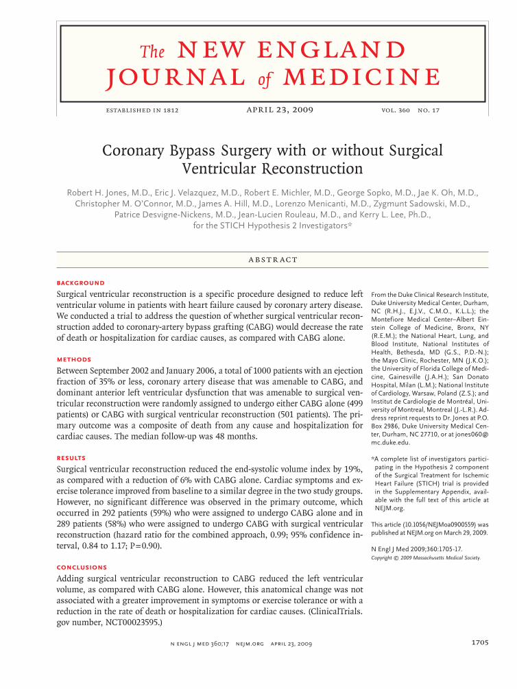
n engl j med 360;17 nejm.org april 23, 2009 1705 The new england journal of medicine established in 1812 april 23, 2009 vol. 360 no. 17 Coronary Bypass Surgery with or without Surgical Ventricular Reconstruction Robert H. Jones, M.D., Eric J. Velazquez, M.D., Robert E. Michler, M.D., George Sopko, M.D., Jae K. Oh, M.D., Christopher M. O’Connor, M.D., James A. Hill, M.D., Lorenzo Menicanti, M.D., Zygmunt Sadowski, M.D., Patrice Desvigne-Nickens, M.D., Jean-Lucien Rouleau, M.D., and Kerry L. Lee, Ph.D., for the STICH Hypothesis 2 Investigators* Abstract From the Duke Clinical Research Institute, Duke University Medical Center, Durham, NC (R.H.J., E.J.V., C.M.O., K.L.L.); the Montefiore Medical Center–Albert Ein- stein College of Medicine, Bronx, NY (R.E.M.); the National Heart, Lung, and Blood Institute, National Institutes of Health, Bethesda, MD (G.S., P.D.-N.); the Mayo Clinic, Rochester, MN (J.K.O.); the University of Florida College of Medi- cine, Gainesville (J.A.H.); San Donato Hospital, Milan (L.M.); National Institute of Cardiology, Warsaw, Poland (Z.S.); and Institut de Cardiologie de Montréal, Uni- versity of Montreal, Montreal (J.-L.R.). Ad- dress reprint requests to Dr. Jones at P.O. Box 2986, Duke University Medical Cen- ter, Durham, NC 27710, or at jones060@ mc.duke.edu. *A complete list of investigators partici- pating in the Hypothesis 2 component of the Surgical Treatment for Ischemic Heart Failure (STICH) trial is provided in the Supplementary Appendix, avail- able with the full text of this article at NEJM.org. This article (10.1056/NEJMoa0900559) was published at NEJM.org on March 29, 2009. N Engl J Med 2009;360:1705-17. Copyright © 2009 Massachusetts Medical Society. Background Surgical ventricular reconstruction is a specific procedure designed to reduce left ventricular volume in patients with heart failure caused by coronary artery disease. We conducted a trial to address the question of whether surgical ventricular recon- struction added to coronary-artery bypass grafting (CABG) would decrease the rate of death or hospitalization for cardiac causes, as compared with CABG alone. Methods Between September 2002 and January 2006, a total of 1000 patients with an ejection fraction of 35% or less, coronary artery disease that was amenable to CABG, and dominant anterior left ventricular dysfunction that was amenable to surgical ven- tricular reconstruction were randomly assigned to undergo either CABG alone (499 patients) or CABG with surgical ventricular reconstruction (501 patients). The pri- mary outcome was a composite of death from any cause and hospitalization for cardiac causes. The median follow-up was 48 months. Results Surgical ventricular reconstruction reduced the end-systolic volume index by 19%, as compared with a reduction of 6% with CABG alone. Cardiac symptoms and ex- ercise tolerance improved from baseline to a similar degree in the two study groups. However, no significant difference was observed in the primary outcome, which occurred in 292 patients (59%) who were assigned to undergo CABG alone and in 289 patients (58%) who were assigned to undergo CABG with surgical ventricular reconstruction (hazard ratio for the combined approach, 0.99; 95% confidence in- terval, 0.84 to 1.17; P = 0.90). Conclusions Adding surgical ventricular reconstruction to CABG reduced the left ventricular volume, as compared with CABG alone. However, this anatomical change was not associated with a greater improvement in symptoms or exercise tolerance or with a reduction in the rate of death or hospitalization for cardiac causes. (ClinicalTrials. gov number, NCT00023595.)
Transcript of The new england journal medicine - STICH · The new england journal of medicine 1706 n engl j med...
n engl j med 360;17 nejm.org april 23, 2009 1705
The new england journal of medicineestablished in 1812 april 23, 2009 vol. 360 no. 17
Coronary Bypass Surgery with or without Surgical Ventricular Reconstruction
Robert H. Jones, M.D., Eric J. Velazquez, M.D., Robert E. Michler, M.D., George Sopko, M.D., Jae K. Oh, M.D., Christopher M. O’Connor, M.D., James A. Hill, M.D., Lorenzo Menicanti, M.D., Zygmunt Sadowski, M.D.,
Patrice Desvigne-Nickens, M.D., Jean-Lucien Rouleau, M.D., and Kerry L. Lee, Ph.D., for the STICH Hypothesis 2 Investigators*
A bs tr ac t
From the Duke Clinical Research Institute, Duke University Medical Center, Durham, NC (R.H.J., E.J.V., C.M.O., K.L.L.); the Montefiore Medical Center–Albert Ein-stein College of Medicine, Bronx, NY (R.E.M.); the National Heart, Lung, and Blood Institute, National Institutes of Health, Bethesda, MD (G.S., P.D.-N.); the Mayo Clinic, Rochester, MN (J.K.O.); the University of Florida College of Medi-cine, Gainesville ( J.A.H.); San Donato Hospital, Milan (L.M.); National Institute of Cardiology, Warsaw, Poland (Z.S.); and Institut de Cardiologie de Montréal, Uni-versity of Montreal, Montreal (J.-L.R.). Ad-dress reprint requests to Dr. Jones at P.O. Box 2986, Duke University Medical Cen-ter, Durham, NC 27710, or at [email protected].
*A complete list of investigators partici-pating in the Hypothesis 2 component of the Surgical Treatment for Ischemic Heart Failure (STICH) trial is provided in the Supplementary Appendix, avail-able with the full text of this article at NEJM.org.
This article (10.1056/NEJMoa0900559) was published at NEJM.org on March 29, 2009.
N Engl J Med 2009;360:1705-17.Copyright © 2009 Massachusetts Medical Society.
Background
Surgical ventricular reconstruction is a specific procedure designed to reduce left ventricular volume in patients with heart failure caused by coronary artery disease. We conducted a trial to address the question of whether surgical ventricular recon-struction added to coronary-artery bypass grafting (CABG) would decrease the rate of death or hospitalization for cardiac causes, as compared with CABG alone.
Methods
Between September 2002 and January 2006, a total of 1000 patients with an ejection fraction of 35% or less, coronary artery disease that was amenable to CABG, and dominant anterior left ventricular dysfunction that was amenable to surgical ven-tricular reconstruction were randomly assigned to undergo either CABG alone (499 patients) or CABG with surgical ventricular reconstruction (501 patients). The pri-mary outcome was a composite of death from any cause and hospitalization for cardiac causes. The median follow-up was 48 months.
Results
Surgical ventricular reconstruction reduced the end-systolic volume index by 19%, as compared with a reduction of 6% with CABG alone. Cardiac symptoms and ex-ercise tolerance improved from baseline to a similar degree in the two study groups. However, no significant difference was observed in the primary outcome, which occurred in 292 patients (59%) who were assigned to undergo CABG alone and in 289 patients (58%) who were assigned to undergo CABG with surgical ventricular reconstruction (hazard ratio for the combined approach, 0.99; 95% confidence in-terval, 0.84 to 1.17; P = 0.90).
Conclusions
Adding surgical ventricular reconstruction to CABG reduced the left ventricular volume, as compared with CABG alone. However, this anatomical change was not associated with a greater improvement in symptoms or exercise tolerance or with a reduction in the rate of death or hospitalization for cardiac causes. (ClinicalTrials.gov number, NCT00023595.)
T h e n e w e ngl a nd j o u r na l o f m e dic i n e
n engl j med 360;17 nejm.org april 23, 20091706
Coronary artery disease is the pre-dominant cause of heart failure, which is a major cause of death and disability through-
out the world. Evidence-based medical therapy has been shown to reduce symptoms and increase sur-vival in patients with heart failure and coronary artery disease.1 In addition, selected patients may benefit from surgical revascularization by means of coronary-artery bypass grafting (CABG), espe-cially if the coronary anatomy is suitable for such surgery and if there is evidence of myocardial vi-ability.2,3
The reduction in left ventricular function that can occur after myocardial infarction is typically accompanied by left ventricular remodeling, a pro-cess that includes left ventricular enlargement and changes in chamber geometry. Left ventricular remodeling is correlated with progression of heart failure and a poor prognosis,4,5 and the benefi-cial effects of therapeutic agents such as angio-tensin-converting–enzyme (ACE) inhibitors and beta-blockers are associated with their effect on remodeling.4,6-9 These findings have generated considerable interest in the possibility that a sur-gical approach to remodeling through left ven-tricular volume reduction could improve outcomes for patients with coronary artery disease and heart failure.10
Surgical ventricular reconstruction is a specific surgical procedure developed for the management of heart failure with left ventricular remodeling caused by coronary artery disease.11 This opera-tion has been shown to reduce the left ventricu-lar volume, increase the ejection fraction, and im-prove ventricular function.12,13 On the basis of a small, nonrandomized, case–control study,14 it has been suggested that surgical ventricular recon-struction that is performed together with CABG may reduce the rate of hospitalization and im-prove ventricular function to a greater degree than CABG alone.
The Surgical Treatment for Ischemic Heart Fail-ure (STICH) trial was designed to define the role of cardiac surgery in the treatment of patients with heart failure and coronary artery disease.15,16 One of the two major hypotheses of this trial (Hypoth-esis 2) was that surgical ventricular reconstruction, when added to CABG, would decrease the rate of death or hospitalization for a cardiac event, as com-pared with CABG alone.
Me thods
Study Design
We conducted a multicenter, nonblinded, random-ized trial at 127 clinical sites in 26 countries.15 The trial was sponsored by the National Heart, Lung, and Blood Institute (NHLBI) of the National In-stitutes of Health. Additional support was provided by Abbott Laboratories, Chase Medical, and CV Therapeutics, which had no role in the design, con-duct, or reporting of the trial. The trial protocol was designed by the authors in collaboration with the NHLBI and was approved by the appropriate institutional review board or ethics committee at each study center. Trial operations, site manage-ment and monitoring, and data collection and analysis were coordinated by the Duke Clinical Research Institute. Oversight was provided by an independent data and safety monitoring board. A clinical events committee whose members were unaware of study-group assignments adjudicated primary outcome events. The authors wrote the manuscript and vouch for the completeness and accuracy of the data and the analyses.
Selection of Patients and Randomization
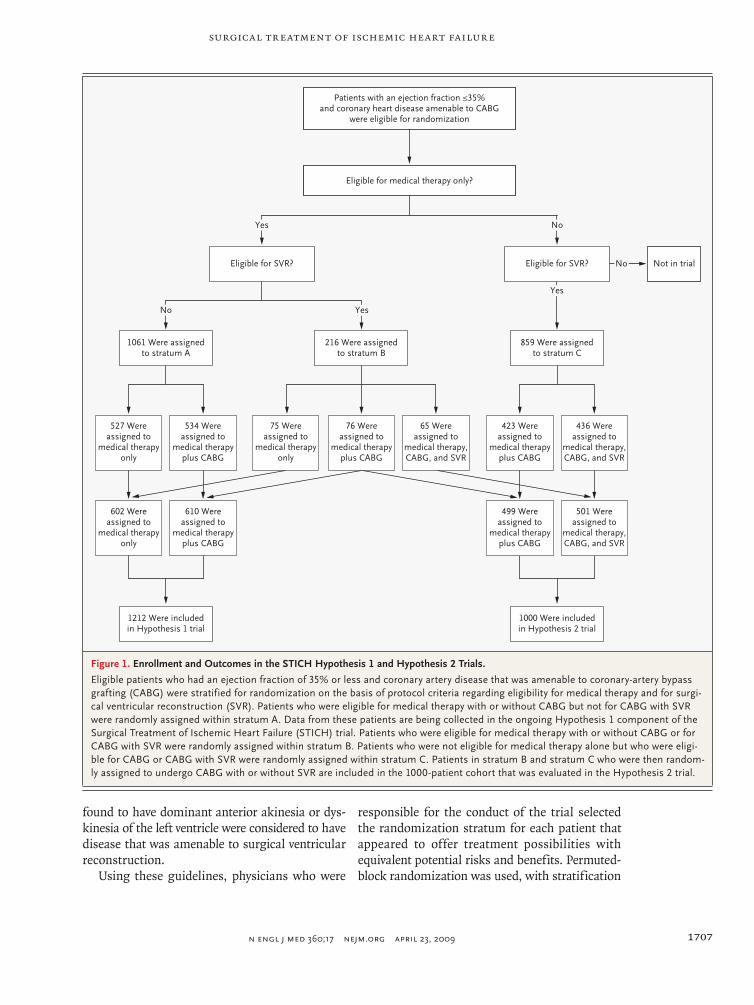
Patients were eligible for enrollment if they had coronary artery disease that was amenable to CABG and if they had a left ventricular ejection fraction of 35% or less (Fig. 1). Exclusion criteria were a recent myocardial infarction, a need for aortic-valve replacement, a planned percutaneous coronary in-tervention (PCI), and coexisting noncardiac dis-ease resulting in a life expectancy of less than 3 years. All patients provided written informed consent.
After initial determination of overall eligibil-ity for the trial, patients were evaluated to deter-mine which component of the STICH program was appropriate for them on the basis of suitable therapeutic options for that patient (medical ther-apy alone, CABG alone, or CABG plus surgical ventricular reconstruction) (Fig. 1). Patients who had stenosis of the left main coronary artery of 50% or more or who had angina of Canadian Car-diovascular Society (CCS) class III or IV while re-ceiving medical therapy were not eligible for medical therapy alone. All patients underwent car-diac imaging for assessment of left ventricular function and wall motion. Patients who were
Surgical Treatment of Ischemic Heart Failure
n engl j med 360;17 nejm.org april 23, 2009 1707
found to have dominant anterior akinesia or dys-kinesia of the left ventricle were considered to have disease that was amenable to surgical ventricular reconstruction.
Using these guidelines, physicians who were
responsible for the conduct of the trial selected the randomization stratum for each patient that appeared to offer treatment possibilities with equivalent potential risks and benefits. Permuted-block randomization was used, with stratification
39p6
Eligible for medical therapy only?
Patients with an ejection fraction ≤35%and coronary heart disease amenable to CABG
were eligible for randomization
Eligible for SVR?Eligible for SVR? Not in trial
1061 Were assignedto stratum A
216 Were assignedto stratum B
859 Were assignedto stratum C
527 Wereassigned to
medical therapyonly
75 Wereassigned to
medical therapyonly
534 Wereassigned to
medical therapyplus CABG
76 Wereassigned to
medical therapyplus CABG
423 Wereassigned to
medical therapyplus CABG
65 Wereassigned to
medical therapy,CABG, and SVR
436 Wereassigned to
medical therapy,CABG, and SVR
602 Wereassigned to
medical therapyonly
610 Wereassigned to
medical therapyplus CABG
1212 Were includedin Hypothesis 1 trial
1000 Were includedin Hypothesis 2 trial
499 Wereassigned to
medical therapyplus CABG
501 Wereassigned to
medical therapy,CABG, and SVR
AUTHOR:
FIGURE:
JOB: ISSUE:
4-CH/T
RETAKE
SIZE
ICM
CASE
EMail LineH/TCombo
Revised
AUTHOR, PLEASE NOTE: Figure has been redrawn and type has been reset.
Please check carefully.
REG F
Enon
1st2nd
3rd
Jones
1 of 4
04-23-09
ARTIST: ts
36017
Yes
No
No
Yes
Yes
No
Figure 1. Enrollment and Outcomes in the STICH Hypothesis 1 and Hypothesis 2 Trials.
Eligible patients who had an ejection fraction of 35% or less and coronary artery disease that was amenable to coronary-artery bypass grafting (CABG) were stratified for randomization on the basis of protocol criteria regarding eligibility for medical therapy and for surgi-cal ventricular reconstruction (SVR). Patients who were eligible for medical therapy with or without CABG but not for CABG with SVR were randomly assigned within stratum A. Data from these patients are being collected in the ongoing Hypothesis 1 component of the Surgical Treatment of Ischemic Heart Failure (STICH) trial. Patients who were eligible for medical therapy with or without CABG or for CABG with SVR were randomly assigned within stratum B. Patients who were not eligible for medical therapy alone but who were eligi-ble for CABG or CABG with SVR were randomly assigned within stratum C. Patients in stratum B and stratum C who were then random-ly assigned to undergo CABG with or without SVR are included in the 1000-patient cohort that was evaluated in the Hypothesis 2 trial.
T h e n e w e ngl a nd j o u r na l o f m e dic i n e
n engl j med 360;17 nejm.org april 23, 20091708
according to clinical site and according to whether the patient was a candidate for SVR, for medical therapy alone, or for both (Fig. 1).
The resulting trial included two major compo-nents. Patients in the Hypothesis 1 component were randomly assigned to receive either medi-cal therapy alone or medical therapy plus CABG. The Hypothesis 1 component of the trial is ongo-ing. Patients in the Hypothesis 2 component were randomly assigned to receive either medical ther-apy plus CABG or medical therapy plus CABG and surgical ventricular reconstruction. The results of the Hypothesis 2 component are the subject of this report.
Treatment
Guideline-based recommendations for drug and device use were emphasized for all patients. The lead cardiologist at each site was responsible for monitoring to ensure that ACE inhibitors, angio-tensin-receptor blockers, beta-blockers, aldoster-one antagonists, antiplatelet agents, statins, diuret-ics, digitalis, pacemakers (for bradyarrhythmias or for cardiac resynchronization), and implant-able cardioverter–defibrillators were used prop-erly throughout the study.
Cardiac surgeons were individually certified to participate in the trial if they met prespecified performance criteria. For CABG certification, sur-geons were required to provide data on at least 25 patients with a left ventricular ejection fraction of 40% or less who underwent CABG, with a death rate of 5% or less. Education in the operative tech-nique for surgical ventricular reconstruction and perioperative management was made available be-fore patient enrollment and during investigator meetings. Certification of individual surgeons for performing surgical ventricular reconstruction re-quired evidence of a consistent postoperative de-crease in left ventricular volume in five consecu-tive patients who survived the operation.
During CABG, arterial grafting for stenosis of the left anterior descending coronary artery was required for all patients without specific contrain-dications. The use of additional arterial conduits supplemented by vein grafts was recommended for revascularization of all major vessels with clini-cally significant stenoses. Concurrent mitral-valve surgery for regurgitation was performed at the discretion of the surgeon.
The technique of surgical ventricular recon-struction has been described previously.11,12,17 For patients who were assigned to undergo surgical
ventricular reconstruction, this component of the operation was most commonly performed dur-ing a single period of cardioplegic arrest after construction of bypass grafts. However, the pro-cedure could also be performed with the heart beating in order to facilitate identification of the noncontractile zone of scarring. In this procedure, after an anterior left ventriculotomy is centered in the zone of anterior asynergy, a suture is placed in the interior of the ventricle to encircle the scar at the boundary between the akinetic and viable tissue. Tightening of this suture brings the healthy portions of the ventricular walls together. Visual inspection and palpation facilitate the judgment of whether a patch is needed to optimize the chamber size without deforming the left ventri-cle during closure of the ventriculotomy.
Primary and Secondary Outcomes
Major perioperative events and specified end points were recorded at discharge or at 30 days for pa-tients remaining in the hospital. Patients were eval-uated at 4-month intervals after randomization during the first year and thereafter at 6-month intervals.
Symptoms of angina and heart failure were assessed at each follow-up visit. All patients who were able to do so performed a 6-minute walk test at baseline, at 4 months, and annually thereafter. Left ventricular volumes and function were as-sessed with the use of echocardiography, cardiac magnetic resonance imaging, or single photon emission computed tomography at baseline, at 4 months, and at 2 years.
The primary outcome was the time to death from any cause or hospitalization for cardiac causes. Secondary outcomes included death from any cause at 30 days, hospitalization for any cause and for cardiovascular causes, myocardial infarc-tion, and stroke.
Statistical Analysis
We calculated that we would need to enroll 1000 patients in the trial for a power of 90% to detect a 20% reduction in the relative risk of death or hos-pitalization for cardiac causes, assuming a 3-year event rate in the CABG-only group of 45% or more and allowing for a crossover rate of up to 20%.
All major study-group comparisons were per-formed according to the intention-to-treat prin-ciple. Supplementary analyses tabulated postoper-ative complications and clinical events occurring within 30 days, according to the type of opera-
Surgical Treatment of Ischemic Heart Failure
n engl j med 360;17 nejm.org april 23, 2009 1709
tion that was performed. All statistical tests were two-tailed. Cumulative event rates from the time of randomization were calculated with the use of the Kaplan–Meier method.18 The log-rank test for time-to-event data was used for the statistical com-parison of study groups with respect to the pri-mary outcome and overall mortality.19 Hazard ra-tios with associated 95% confidence intervals were derived with the use of the Cox proportional-hazards model.20 The Cox model was also used to assess the consistency of treatment effects by testing for interactions between the type of sur-gery and prespecified baseline characteristics.
Eight interim analyses of the data were per-formed and reviewed by the data and safety moni-toring board. Interim comparisons between study groups were monitored with the use of two-sided, symmetric O’Brien–Fleming boundaries gener-ated with the alpha-spending-function approach to group-sequential testing.21,22 A P value of 0.05 or less was considered to indicate statistical sig-nificance. Because of the sequential monitoring, the level of significance that was required for the primary analysis at the completion of the study was 0.04.
R esult s
Study Population
Between July 24, 2002, and May 5, 2007, 2136 patients were enrolled in the overall STICH trial (Fig. 1). Of these patients, 1000 were enrolled in the Hypothesis 2 component at 96 clinical sites between September 12, 2002, and January 24, 2006, and were randomly assigned to undergo either CABG alone (499 patients) or CABG with surgi-cal ventricular reconstruction (501 patients), with follow-up continued through December 31, 2008. No significant differences between the two study groups were observed in baseline demographic or clinical characteristics (Table 1). The median age was 62 years, and 147 of the 1000 patients were women. The median left ventricular ejection frac-tion was 28%. The median end-systolic volume index was 82 ml per square meter of body-surface area. Multivessel coronary artery disease was pres-ent in 913 patients; 197 patients had stenosis of the left main coronary artery.
Surgical Procedures
Of the 499 patients who were assigned to undergo CABG alone, 463 (93%) underwent the assigned procedure; 9 did not undergo any surgery, and 27
underwent CABG with surgical ventricular re-construction (Table 1 in the Supplementary Ap-pendix, available with the full text of this article at NEJM.org). Of the 501 patients who were as-signed to undergo CABG with surgical ventricu-lar reconstruction, 454 (91%) underwent the as-signed procedure; 12 patients did not undergo any surgery, and 35 patients underwent CABG with-out surgical ventricular reconstruction.
Of the 979 patients who underwent surgery, the procedure was elective in 819 patients (84%), urgent in 127 (13%), performed for ongoing isch-emia in 21 (2%), and performed under emergency conditions in 11 (1%) (Table 2 in the Supplemen-tary Appendix). Mitral-valve surgery was performed in 178 patients (18%) undergoing surgery. More arterial conduits were used in patients under-going CABG alone than in patients undergoing CABG with surgical ventricular reconstruction (P = 0.008). Surgical ventricular reconstruction add-ed a median of 27 minutes of cardiopulmonary bypass time to the CABG procedure (P<0.001). The duration of aortic cross-clamping, the time to endotrachael extubation, and the duration of post-operative hospitalization were longer for patients undergoing CABG with surgical ventricular re-construction (P<0.001 for all comparisons).
Follow-up
The median follow-up for all surviving patients was 48 months (minimum, 30). Only four patients withdrew consent, and six patients were lost to follow-up before the last visit. Of the 1000 patients, 990 (99%) underwent complete follow-up that be-gan at randomization and concluded between Au-gust 1 and December 31, 2008.
Left Ventricular Volume
A core-laboratory quantitative assessment of the end-systolic volume index on echocardiography was performed at baseline and at 4 months in a total of 373 patients (212 patients who were assigned to undergo CABG alone and 161 who were assigned to undergo CABG with surgical ventricular recon-struction). The mean end-systolic volume index in patients assigned to undergo CABG alone de-creased by an average of 5 ml per square meter, from 82 to 77 ml per square meter (a reduction of 6%). For patients who were assigned to undergo CABG with surgical ventricular reconstruction, the average decrease was 16 ml per square meter, from 83 to 67 ml per square meter (a reduction of 19%) (Fig. 1 in the Supplementary Appendix). The
T h e n e w e ngl a nd j o u r na l o f m e dic i n e
n engl j med 360;17 nejm.org april 23, 20091710
Table 1. Baseline Characteristics of the Patients.*
VariableCABG Alone
(N = 499)
CABG with Surgical Ventricular Reconstruction
(N = 501)
Demographic characteristics
Age — yr
Median 62 62
Interquartile range 54–69 55–69
Female sex — no. (%) 78 (16) 69 (14)
Race — no. (%)†
White 451 (90) 460 (92)
Black or other 48 (10) 41 (8)
Body-mass index
Median 27 27
Interquartile range 25–30 24–30
Medical history
Myocardial infarction — no. (%) 435 (87) 437 (87)
Hyperlipidemia — no. (%) 367 (74) 351 (70)
Hypertension — no. (%) 289 (58) 296 (59)
Diabetes — no. (%) 173 (35) 171 (34)
Current smoker — no. (%) 117 (23) 100 (20)
Previous percutaneous coronary intervention — no. (%) 100 (20) 95 (19)
Chronic renal insufficiency — no. (%) 42 (8) 43 (9)
Stroke — no. (%) 28 (6) 28 (6)
Previous CABG — no. (%) 15 (3) 9 (2)
Current Canadian Cardiovascular Society angina class — no. (%)
No angina 121 (24) 128 (26)
I 36 (7) 35 (7)
II 94 (19) 94 (19)
III 203 (41) 205 (41)
IV 45 (9) 39 (8)
Current New York Heart Association heart failure class — no. (%)
I 36 (7) 50 (10)
II 222 (44) 207 (41)
III 210 (42) 218 (44)
IV 31 (6) 26 (5)
Blood pressure — mm Hg
Systolic
Median 120 120
Interquartile range 110–130 110–130
Diastolic
Median 71 74
Interquartile range 65–80 66–80
Pulse — beats/min
Median 70 72
Interquartile range 64–80 64–80
Surgical Treatment of Ischemic Heart Failure
n engl j med 360;17 nejm.org april 23, 2009 1711
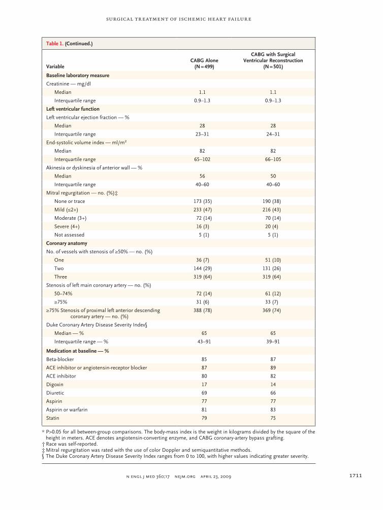
Table 1. (Continued.)
VariableCABG Alone
(N = 499)
CABG with Surgical Ventricular Reconstruction
(N = 501)
Baseline laboratory measure
Creatinine — mg/dl
Median 1.1 1.1
Interquartile range 0.9–1.3 0.9–1.3
Left ventricular function
Left ventricular ejection fraction — %
Median 28 28
Interquartile range 23–31 24–31
End-systolic volume index — ml/m2
Median 82 82
Interquartile range 65–102 66–105
Akinesia or dyskinesia of anterior wall — %
Median 56 50
Interquartile range 40–60 40–60
Mitral regurgitation — no. (%)‡
None or trace 173 (35) 190 (38)
Mild (≤2+) 233 (47) 216 (43)
Moderate (3+) 72 (14) 70 (14)
Severe (4+) 16 (3) 20 (4)
Not assessed 5 (1) 5 (1)
Coronary anatomy
No. of vessels with stenosis of ≥50% — no. (%)
One 36 (7) 51 (10)
Two 144 (29) 131 (26)
Three 319 (64) 319 (64)
Stenosis of left main coronary artery — no. (%)
50–74% 72 (14) 61 (12)
≥75% 31 (6) 33 (7)
≥75% Stenosis of proximal left anterior descending coronary artery — no. (%)
388 (78) 369 (74)
Duke Coronary Artery Disease Severity Index§
Median — % 65 65
Interquartile range — % 43–91 39–91
Medication at baseline — %
Beta-blocker 85 87
ACE inhibitor or angiotensin-receptor blocker 87 89
ACE inhibitor 80 82
Digoxin 17 14
Diuretic 69 66
Aspirin 77 77
Aspirin or warfarin 81 83
Statin 79 75
* P>0.05 for all between-group comparisons. The body-mass index is the weight in kilograms divided by the square of the height in meters. ACE denotes angiotensin-converting enzyme, and CABG coronary-artery bypass grafting.
† Race was self-reported.‡ Mitral regurgitation was rated with the use of color Doppler and semiquantitative methods.§ The Duke Coronary Artery Disease Severity Index ranges from 0 to 100, with higher values indicating greater severity.
T h e n e w e ngl a nd j o u r na l o f m e dic i n e
n engl j med 360;17 nejm.org april 23, 20091712
difference between the two groups in the change from baseline was significant (P<0.001).
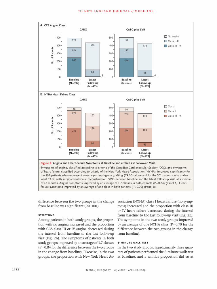
Symptoms
Among patients in both study groups, the propor-tion with no angina increased and the proportion with CCS class III or IV angina decreased during the interval from baseline to the last follow-up visit (Fig. 2A). The symptoms of patients in both study groups improved by an average of 1.7 classes (P = 0.84 for the difference between the two groups in the change from baseline). Likewise, in the two groups, the proportion with New York Heart As-
sociation (NYHA) class I heart failure (no symp-toms) increased and the proportion with class III or IV heart failure decreased during the interval from baseline to the last follow-up visit (Fig. 2B). The symptoms in the two study groups improved by an average of one NYHA class (P = 0.70 for the difference between the two groups in the change from baseline).
6-Minute Walk Test
In the two study groups, approximately three quar-ters of patients performed the 6-minute walk test at baseline, and a similar proportion did so at
33p9
241
222
36
80
190
165
244
207
50
62
188
179
B NYHA Heart Failure Class
A CCS Angina Class
AUTHOR:
FIGURE:
JOB:
4-CH/T
RETAKE
SIZE
ICM
CASE
EMail LineH/TCombo
Revised
AUTHOR, PLEASE NOTE: Figure has been redrawn and type has been reset.
Please check carefully.
REG F
Enon
1st2nd
3rd
Jones
2 of 4
04-23-09
ARTIST: ts
36015 ISSUE:
No.
of P
atie
nts
500
400
300
100
200
0Baseline(N=499)
LatestFollow-up(N=435)
CABG
500
400
300
100
200
0Baseline(N=501)
LatestFollow-up(N=428)
CABG plus SVR
No angina
Class I–II
Class III–IV
No.
of P
atie
nts
500
400
300
100
200
0Baseline(N=499)
LatestFollow-up(N=435)
CABG
500
400
300
100
200
0Baseline(N=501)
LatestFollow-up(N=429)
CABG plus SVR
Class I
Class II
Class III–IV
8
248
130
121
88
339
6
244
129
128
83
339
Figure 2. Angina and Heart-Failure Symptoms at Baseline and at the Last Follow-up Visit.
Symptoms of angina, classified according to criteria of the Canadian Cardiovascular Society (CCS), and symptoms of heart failure, classified according to criteria of the New York Heart Association (NYHA), improved significantly for the 499 patients who underwent coronary-artery bypass grafting (CABG) alone and for the 501 patients who under-went CABG with surgical ventricular reconstruction (SVR) between baseline and the latest follow-up visit, at a median of 48 months. Angina symptoms improved by an average of 1.7 classes in both cohorts (P = 0.84) (Panel A). Heart-failure symptoms improved by an average of one class in both cohorts (P = 0.70) (Panel B).
Surgical Treatment of Ischemic Heart Failure
n engl j med 360;17 nejm.org april 23, 2009 1713
4 months (Fig. 2 in the Supplementary Appendix). The median distance walked was 350 m at base-line and 350 m at 4 months for patients assigned to undergo CABG and 358 m at baseline and 410 m at 4 months for those assigned to undergo CABG with surgical ventricular reconstruction. The in-crease in the median distance walked was simi-lar in the two groups (48 m among patients who were assigned to undergo CABG and 52 m among patients assigned to undergo CABG with surgical ventricular reconstruction, P = 0.80). Among pa-tients assigned to undergo CABG who performed the 6-minute walk test and were assessed for symp-toms, 34% were symptomatic during the base-
line test and 9% were symptomatic at 4 months. The corresponding rates among patients assigned to undergo CABG with surgical ventricular recon-struction were 33% and 11%.
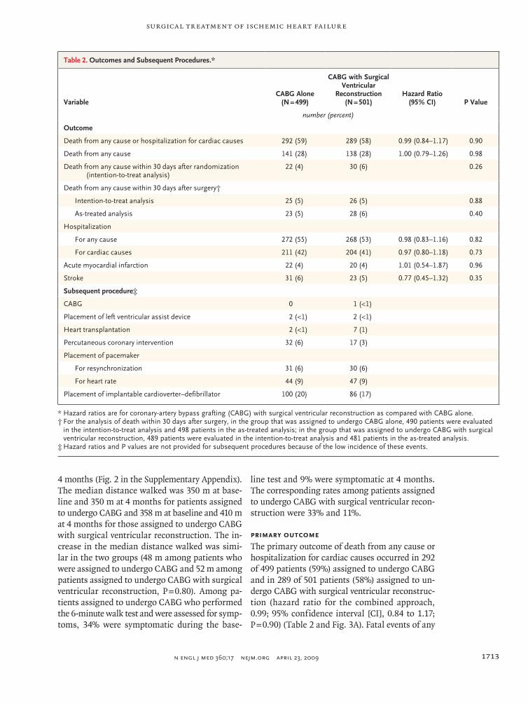
Primary Outcome
The primary outcome of death from any cause or hospitalization for cardiac causes occurred in 292 of 499 patients (59%) assigned to undergo CABG and in 289 of 501 patients (58%) assigned to un-dergo CABG with surgical ventricular reconstruc-tion (hazard ratio for the combined approach, 0.99; 95% confidence interval [CI], 0.84 to 1.17; P = 0.90) (Table 2 and Fig. 3A). Fatal events of any
Table 2. Outcomes and Subsequent Procedures.*
VariableCABG Alone
(N = 499)
CABG with Surgical Ventricular
Reconstruction(N = 501)
Hazard Ratio(95% CI) P Value
number (percent)
Outcome
Death from any cause or hospitalization for cardiac causes 292 (59) 289 (58) 0.99 (0.84–1.17) 0.90
Death from any cause 141 (28) 138 (28) 1.00 (0.79–1.26) 0.98
Death from any cause within 30 days after randomization (intention-to-treat analysis)
22 (4) 30 (6) 0.26
Death from any cause within 30 days after surgery†
Intention-to-treat analysis 25 (5) 26 (5) 0.88
As-treated analysis 23 (5) 28 (6) 0.40
Hospitalization
For any cause 272 (55) 268 (53) 0.98 (0.83–1.16) 0.82
For cardiac causes 211 (42) 204 (41) 0.97 (0.80–1.18) 0.73
Acute myocardial infarction 22 (4) 20 (4) 1.01 (0.54–1.87) 0.96
Stroke 31 (6) 23 (5) 0.77 (0.45–1.32) 0.35
Subsequent procedure‡
CABG 0 1 (<1)
Placement of left ventricular assist device 2 (<1) 2 (<1)
Heart transplantation 2 (<1) 7 (1)
Percutaneous coronary intervention 32 (6) 17 (3)
Placement of pacemaker
For resynchronization 31 (6) 30 (6)
For heart rate 44 (9) 47 (9)
Placement of implantable cardioverter–defibrillator 100 (20) 86 (17)
* Hazard ratios are for coronary-artery bypass grafting (CABG) with surgical ventricular reconstruction as compared with CABG alone. † For the analysis of death within 30 days after surgery, in the group that was assigned to undergo CABG alone, 490 patients were evaluated
in the intention-to-treat analysis and 498 patients in the as-treated analysis; in the group that was assigned to undergo CABG with surgical ventricular reconstruction, 489 patients were evaluated in the intention-to-treat analysis and 481 patients in the as-treated analysis.
‡ Hazard ratios and P values are not provided for subsequent procedures because of the low incidence of these events.

T h e n e w e ngl a nd j o u r na l o f m e dic i n e
n engl j med 360;17 nejm.org april 23, 20091714
cause occurred in 141 patients (28%) assigned to undergo CABG and in 138 patients (28%) assigned to undergo CABG with surgical ventricular recon-struction (hazard ratio, 1.00; 95% CI, 0.79 to 1.26; P = 0.98) (Table 2 and Fig. 3B). Hospitalization for cardiac causes occurred in 211 patients (42%) and in 204 patients (41%), respectively (P = 0.73). Haz-
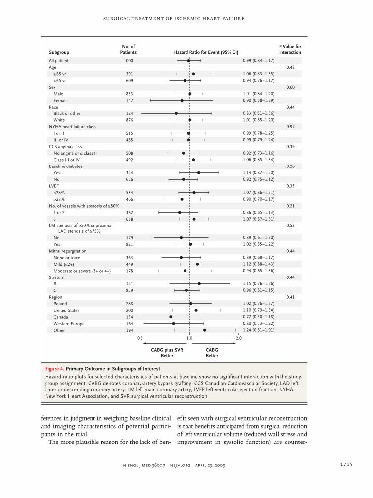
ard-ratio plots showed no interaction for the pri-mary outcome between study-group assignment and baseline characteristics of interest (Fig. 4).
Secondary Outcomes and Events
Operative rates of death (i.e., deaths occurring within 30 days after the procedure) did not differ significantly between the two study groups, ac-cording to either the intention-to-treat analysis or the as-treated analysis (Table 2). The rates of seven secondary procedures, of acute myocardial infarc-tion (P = 0.96), and of stroke (P = 0.35) were low and similar in the two groups.
Discussion
We compared the efficacy of CABG alone with that of CABG combined with surgical ventricular reconstruction in patients with coronary artery disease and left ventricular systolic dysfunction. As anticipated, the addition of surgical ventricu-lar reconstruction resulted in a significantly great-er reduction in left ventricular volume than was achieved with CABG alone. However, this improve-ment in ventricular volume did not translate into a measurable benefit for the patients. Symptom-atic improvement after surgery was similar in the two study groups. There was no significant be-tween-group difference in the primary outcome of death or hospitalization for cardiac causes or in any other clinical outcome. Operative, intuba-tion, and initial hospitalization times were longer in patients treated with the combined procedure. The findings of this study do not support the use of surgical ventricular reconstruction in the pop-ulation studied.
Two reasons might be offered for the negative outcome of this trial. Perhaps experienced sur-geons decided to enroll patients for whom they recognized that surgical ventricular reconstruction would prove unnecessary but offered this proce-dure directly, instead of enrollment in the trial, to all concurrently evaluated patients for whom they were confident the procedure would be benefi-cial. Making such precise decisions about patient selection is not consistent with the diverse opin-ions about the eligibility of specific patients for randomized assignment to surgical ventricular re-construction, as discussed at STICH investigator meetings. Participating investigators appeared to make randomization decisions from a broad spec-trum of positions of equipoise on the basis of dif-
22p3
0.7Pr
obab
ility
0.5
0.6
0.4
0.3
0.1
0.2
0.0
0.7
Prob
abili
ty
0.5
0.6
0.4
0.3
0.1
0.2
0.0
0 1 2 3 4 5
CABG
CABG plus SVR
Years since Randomization
B Death from Any Cause
A Death from Any Cause or Hospitalization for Cardiac Causes
0 1 2 3 4 5
CABG
CABG plus SVR
Years since Randomization
P=0.98
No. at RiskCABGCABG plus SVR
499501
No. at RiskCABGCABG plus SVR
499501
319319
434429
270275
417404
220216
363352
99111
201193
2323
5953
AUTHOR:
FIGURE:
JOB:
4-CH/T
RETAKE
SIZE
ICM
CASE
EMail LineH/TCombo
Revised
AUTHOR, PLEASE NOTE: Figure has been redrawn and type has been reset.
Please check carefully.
REG F
Enon
1st2nd
3rd
Jones
3 of 4
xx-xx-09
ARTIST: ts
360xx ISSUE:
P=0.90
Figure 3. Kaplan–Meier Estimates of Outcomes.
Panel A shows the probability of the primary outcome (death from any cause or hospitalization for cardiac causes), which did not differ signifi-cantly between the two groups. The primary outcome occurred in 292 pa-tients (59%) assigned to undergo coronary-artery bypass grafting (CABG) alone and in 289 patients (58%) assigned to undergo CABG with surgical ventricular reconstruction (SVR) (hazard ratio, 0.99; 95% CI, 0.84 to 1.17). Panel B shows the probability of death from any cause, which occurred in 141 patients (28%) assigned to undergo CABG and in 138 patients (28%) as-signed to undergo CABG with SVR (hazard ratio, 1.00; 95% CI, 0.79 to 1.26).
Surgical Treatment of Ischemic Heart Failure
n engl j med 360;17 nejm.org april 23, 2009 1715
ferences in judgment in weighing baseline clinical and imaging characteristics of potential partici-pants in the trial.
The more plausible reason for the lack of ben-
efit seen with surgical ventricular reconstruction is that benefits anticipated from surgical reduction of left ventricular volume (reduced wall stress and improvement in systolic function) are counter-
33p9
1.0 2.0
CABGBetter
CABG plus SVRBetter
All patients
Age
≥65 yr
<65 yr
Sex
Male
Female
Race
Black or other
White
NYHA heart failure class
I or II
III or IV
CCS angina class
No angina or ≤ class II
Class III or IV
Baseline diabetes
Yes
No
LVEF
≤28%
>28%
No. of vessels with stenosis of ≥50%
1 or 2
3
LM stenosis of ≥50% or proximalLAD stenosis of ≥75%
No
Yes
Mitral regurgitation
None or trace
Mild (≤2+)
Moderate or severe (3+ or 4+)
Stratum
B
C
Region
Poland
United States
Canada
Western Europe
Other
No. ofPatients Hazard Ratio for Event (95% CI)Subgroup
0.94 (0.76–1.17)
0.77 (0.50–1.18)
1.10 (0.79–1.54)
1.02 (0.76–1.37)
0.94 (0.65–1.36)
0.89 (0.68–1.17)
1.12 (0.88–1.43)
0.89 (0.61–1.30)
1.02 (0.85–1.22)
0.90 (0.70–1.17)
1.07 (0.86–1.31)
0.86 (0.65–1.13)
1.07 (0.87–1.31)
1.15 (0.76–1.76)
0.96 (0.81–1.15)
0.5
0.80 (0.53–1.22)
1.24 (0.81–1.91)
0.92 (0.75–1.12)
1.06 (0.85–1.34)
1.14 (0.87–1.50)
0.92 (0.73–1.16)
0.99 (0.78–1.25)
0.99 (0.79–1.24)
1.01 (0.85–1.20)
0.83 (0.51–1.36)
0.90 (0.58–1.39)
1.01 (0.84–1.20)
1.06 (0.83–1.35)
0.99 (0.84–1.17)
P Value forInteraction
1000
391
609
853
147
124
876
515
485
508
492
344
656
534
466
362
638
179
821
363
449
178
141
859
288
200
154
164
194
0.48
0.60
0.44
0.97
0.39
0.20
0.33
0.21
0.53
0.44
0.44
0.41
AUTHOR:
FIGURE:
JOB:
4-CH/T
RETAKE
SIZE
ICM
CASE
EMail LineH/TCombo
Revised
AUTHOR, PLEASE NOTE: Figure has been redrawn and type has been reset.
Please check carefully.
REG F
Enon
1st
2nd
3rd
Jones
4 of 4
04-23-09
ARTIST: ts
36015 ISSUE:
Figure 4. Primary Outcome in Subgroups of Interest.
Hazard-ratio plots for selected characteristics of patients at baseline show no significant interaction with the study-group assignment. CABG denotes coronary-artery bypass grafting, CCS Canadian Cardiovascular Society, LAD left anterior descending coronary artery, LM left main coronary artery, LVEF left ventricular ejection fraction, NYHA New York Heart Association, and SVR surgical ventricular reconstruction.
T h e n e w e ngl a nd j o u r na l o f m e dic i n e
n engl j med 360;17 nejm.org april 23, 20091716
balanced by a reduction in diastolic distensibility. Previous work has shown that an important pre-dictor of survival in patients with systolic dysfunc-tion is the left ventricular ejection fraction during exercise.23 The most favorable dynamic response of the ventricle to the demand for increased car-diac output is associated with both an increase in end-diastolic volume and a decrease in end-systolic volume. Surgical ventricular reconstruction may impede this enhanced filling response.
One limitation of the design of the STICH trial was that physicians and surgeons caring for pa-tients were aware of the treatment received. We sought to mitigate this limitation as much as pos-sible by seeking complete follow-up for all pa-tients and by selecting trial end points that were not primarily subjective ones. It is conceivable that the decision to hospitalize a patient during the follow-up period could have been influenced by the physician’s knowledge of the specific opera-tion performed. However, rates of death, myo-cardial infarction, and stroke were similar in the two study groups.
A total of 83 of 1000 patients (8%) did not re-ceive the assigned treatment. Patients who were
identified before randomization as eligible for ei-ther treatment could be expected to have a higher preoperative crossover rate than patients for whom one operative strategy was preferred. Moreover, physician-directed crossovers were similar in the two study groups. Analyses that were performed on the intention-to-treat principle and according to the surgery received had similar results.
In summary, our trial compared the efficacy of CABG alone with that of CABG with surgical ventricular reconstruction in patients with coro-nary artery disease and left ventricular systolic dysfunction. There was no significant difference between the two study groups in the degree of symptom improvement or in the rate of death or hospitalization for cardiac causes.
Supported by grants (5U01-HL-69015, 5U01-HL-69013, and 5U01-HL-69010) from the National Heart, Lung, and Blood In-stitute.
Dr. Velazquez reports receiving grant support from Cardioki-netix; Dr. O’Connor, receiving grant support from Scios and NovaCardia; and Dr. Rouleau, receiving consulting fees from Novartis, Pfizer, and Scios. No other potential conflict of inter-est relevant to this article was reported.
We thank Vanessa Moore for her assistance in the preparation of the manuscript and Kerry Bassett and Anthony Doll for their work in the initial preparation of the figures.
References
Hunt SA, Abraham WT, Chin MH, et 1. al. ACC/AHA 2005 guideline update for the diagnosis and management of chronic heart failure in the adult: a report of the American College of Cardiology/American Heart Association Task Force on Practice Guidelines (Writing Committee to Update the 2001 Guidelines for the Evaluation and Management of Heart Failure): developed in collaboration with the American Col-lege of Chest Physicians and the Interna-tional Society for Heart and Lung Trans-plantation: endorsed by the Heart Rhythm Society. Circulation 2005;112(12):e154-e235.
Bax JJ, van der Wall EE, Harbinson M. 2. Radionuclide techniques for the assess-ment of myocardial viability and hiberna-tion. Heart 2004;90:Suppl 5:v26-v33.
Eagle KA, Guyton RA, Davidoff R, et 3. al. ACC/AHA 2004 guideline update for coronary artery bypass graft surgery: a re-port of the American College of Cardiol-ogy/American Heart Association Task Force on Practice Guidelines (Committee to Up-date the 1999 Guidelines for Coronary Artery Bypass Graft Surgery). Circulation 2004;110(14):e340-e437. [Erratum, Circu-lation 2005:111:2014.]
Sutton MGS, Sharpe N. Left ventricu-4. lar remodeling after myocardial infarc-tion: pathophysiology and therapy. Circu-lation 2000;101:2981-8.
Cohn JN, Ferrari R, Sharpe N. Cardiac 5. remodeling — concepts and clinical im-plications: a consensus paper from an in-ternational forum on cardiac remodeling. J Am Coll Cardiol 2000;35:569-82.
Pfeffer MA, Lamas GA, Vaughan DE, 6. Parisi AF, Braunwald E. Effect of captopril on progressive ventricular dilatation after anterior myocardial infarction. N Engl J Med 1988;319:80-6.
Greenberg B, Quinones MA, Koilpillai 7. C, et al. Effects of long-term enalapril therapy on cardiac structure and function in patients with left ventricular dysfunc-tion: results of the SOLVD echocardiogra-phy substudy. Circulation 1995;91:2573-81.
Doughty RN, Whalley GA, Gamble G, 8. MacMahon S, Sharpe N. Left ventricular remodeling with carvedilol in patients with congestive heart failure due to ische-mic heart disease. J Am Coll Cardiol 1997; 29:1060-6.
Doughty RN, Whalley GA, Walsh HA, 9. Gamble GD, López-Sendón J, Sharpe N.
Effects of carvedilol on left ventricular re-modeling after acute myocardial infarc-tion: the CAPRICORN Echo Substudy. Circulation 2004;109:201-6.
Alfieri O, Maisano F, Schreuder JJ. 10. Surgical methods to reverse left ventricu-lar remodeling in congestive heart failure. Am J Cardiol 2003;91:Suppl 9A:81F-87F.
Athanasuleas CL, Stanley AWH Jr, 11. Buckberg GD. Restoration of contractile function in the enlarged left ventricle by exclusion of remodeled akinetic anterior segment: surgical strategy, myocardial pro-tection, and angiographic results. J Card Surg 1998;13:418-28.
Athanasuleas CL, Buckberg GD, Stan-12. ley AWH, et al. Surgical ventricular resto-ration in the treatment of congestive heart failure due to post-infarction ventricular dilation. J Am Coll Cardiol 2004;44:1439-45.
Menicanti L, Castelvecchio S, Ranucci 13. M, et al. Surgical therapy for ischemic heart failure: single-center experience with surgical anterior ventricular restora-tion. J Thorac Cardiovasc Surg 2007;134: 433-41.
Prucz RB, Weiss ES, Patel ND, Nwa-14. kanna LU, Baumgartner WA, Conte JV.
Surgical Treatment of Ischemic Heart Failure
n engl j med 360;17 nejm.org april 23, 2009 1717
Coronary artery bypass grafting with or without surgical ventricular restoration: a comparison. Ann Thorac Surg 2008;86: 806-14.
Velazquez EJ, Lee KL, O’Connor CM, 15. et al. The rationale and design of the Sur-gical Treatment for Ischemic Heart Fail-ure (STICH) trial. J Thorac Cardiovasc Surg 2007;134:1540-7.
Jones RH. Is it time for a randomized 16. trial of surgical treatment of ischemic heart failure? J Am Coll Cardiol 2001;37: 1210-3.
Athanasuleas CL, Stanley AWH Jr, 17.
Buckberg GD, Dor V, Di Donato M, Black-stone EH. Surgical anterior ventricular endocardial restoration (SAVER) in the dilated remodeled ventricle after anterior myocardial infarction. J Am Coll Cardiol 2001;37:1199-209.
Kaplan EL, Meier P. Nonparametric 18. estimation from incomplete observations. J Am Stat Assoc 1958;53:457-81.
Kalbfleisch JD, Prentice RL. The sta-19. tistical analysis of failure time data. 2nd ed. New York: John Wiley, 2002.
Cox DR. Regression models and life-20. tables. J R Stat Soc [B] 1972;34:187-220.
O’Brien PC, Fleming TR. A multiple 21. testing procedure for clinical trials. Bio-metrics 1979;35:549-56.
Lan KKG, DeMets DL. Discrete se-22. quential boundaries for clinical trials. Biometrika 1983;79:659-63.
Lee KL, Pryor DB, Pieper KS, et al. 23. Prognostic value of radionuclide angiog-raphy in medically treated patients with coronary artery disease: a comparison with clinical and catheterization vari-ables. Circulation 1990;82:1705-17.Copyright © 2009 Massachusetts Medical Society.
full text of all journal articles on the world wide web
Access to the complete text of the Journal on the Internet is free to all subscribers. To use this Web site, subscribers should go to the Journal’s home page (NEJM.org) and register by entering their names and subscriber numbers as they appear on their mailing labels. After this one-time registration, subscribers can use their passwords to log on for electronic access to the entire Journal from any computer that is connected to the Internet. Features include a library of all issues since January 1993 and abstracts since January 1975, a full-text search capacity, and a personal archive for saving articles and search results of interest. All articles can be printed in a format that is virtually identical to that of the typeset pages. Beginning 6 months after publication, the full text of all Original Articles and Special Articles is available free to nonsubscribers.
Supplementary Appendix
This appendix has been provided by the authors to give readers additional information about their work.
Supplement to: Jones RH, Velazquez EJ, Michler RE, et al. Coronary bypass surgery with or without surgical ventricular reconstruction. N Engl J Med 2009;360:1705-17. DOI: 10.1056/NEJMoa0900559.
Coronary Bypass Surgery with or without Surgical Ventricular Reconstruction
Online Supplementary Appendix
Robert H. Jones, M.D., Eric J. Velazquez, M.D., Robert E. Michler, M.D., George Sopko, M.D., Jae K. Oh, M.D., Christopher M. O’Connor, M.D., James A. Hill, M.D., Lorenzo
Menicanti, M.D., Zygmunt Sadowski, M.D., Patrice Desvigne-Nickens, M.D., Jean-Lucien Rouleau, M.D., Kerry L. Lee, Ph.D.
for the STICH Hypothesis 2 Investigators
Division of Cardiothoracic Surgery/Department of Surgery, Duke Clinical Research Institute, Duke University Medical Center, Durham, North Carolina (RHJ); Division of Cardiovascular Medicine/Department of Medicine, Duke Clinical Research Institute, Duke University Medical Center, Durham, North Carolina (EJV, CMO); Department of Cardiothoracic Surgery and Department of Surgery, Montefiore Medical Center/Albert Einstein College of Medicine, Bronx, New York (REM); Division of Cardiovascular Diseases, National Heart, Lung, and Blood Institute, National Institutes of Health, Bethesda, Maryland (GS, PD-N); Division of Cardiovascular Diseases, Department of Internal Medicine, Mayo Clinic, Rochester, Minnesota (JKO); Division of Cardiovascular Medicine, University of Florida College of Medicine, Gainesville, Florida (JAH); Department of Cardiac Surgery, San Donato Hospital, Milan, Italy (LM); National Institute of Cardiology, Warsaw, Poland (ZS); Institut de Cardiologie de Montreal, University of Montreal, Montreal, Canada (JLR); Department of Biostatistics and Bioinformatics, Duke Clinical Research Institute, Duke University Medical Center, Durham, North Carolina (KLL) Address for Correspondence: Robert H. Jones, M.D. Mary and Deryl Hart Professor of Surgery P. O. Box 2986, Duke University Medical Center Durham, NC 27710 Telephone: 919-668-8357 Fax: 919-684-5700 Email: [email protected]

Hypothesis 2 Investigators and Trial Committees
The following institutions and principal investigators (PI), lead surgeons (LS), lead cardiologists (LC), and study coordinators (SC) participated in STICH Hypothesis 2 enrollment: Investigators (listed in descending order of the number of randomized patients): Slaski Osrodek Kardiologii, Katowice, Poland: A. Bochenek-PI, M. Krejca-LS, M. Trusz-Gluza-LC, K. Wita-SC; Silesian Medical Academy, Zabrze, Poland: M. Zembala-PI, R. Przybylski-LS, T. Kukulski-SC; Research Institute of Circulation Pathology, Novosibirsk, Russia: A. Cherniavsky-PI, A. Marchenko-LS, A. Romanov-SC; Medical University of Silesia, Katowice, Poland: S. Wos-PI, M. Deja-LS, K. Golba-LC, J. Kot-SC; Toronto General Hospital, Toronto, Canada: V. Rao-PI, M. Iwanochko-LC, J. Renton-SC, S. Hemeon-SC; Medical University of Gdansk, Gdansk, Poland: J. Rogowski-PI, A. Rynkiewicz-LC, P. Betlejewski-SC; Ohio State University Medical Center, Columbus, USA: B. Sun-PI, J. Crestanello-LS, P. Binkley-LC, J. Chang-SC; Ospedali Riuniti di Bergamo, Bergamo, Italy: P. Ferrazzi-PI, A. Gavazzi-LC, M. Senni-SC; John Paul II Hospital, Krakow, Poland: J. Sadowski-PI, B. Kapelak-LS, D. Sobczyk-LC, K. Wrobel-SC; Institute for Clinical and Experimental Medicine, Prague, Czech Republic: J. Pirk-PI, R. Jandova-SC; Duke University Medical Center, Durham, USA: E. Velazquez-PI, P. Smith-LS, C. Milano-LS, P. Adams-SC; San Donato Hospital, Milan, Italy: L. Menicanti-PI, M. Di Donato-LC, S. Castelvecchio-SC; Laval Hospital, Sainte Foy, Canada: F. Dagenais-PI, G. Dussault-SC; University of North Carolina Hospitals, Chapel Hill, USA: C. Dupree-PI, B. Sheridan-LS, C. Schuler-SC; St. Vincent's Hospital Melbourne, Melbourne, Australia: M. Yii-PI, D. Prior-LC, J. Mack-SC; Montreal Heart Institute, Montreal, Canada: N. Racine-PI, D. Bouchard-LS, A. Ducharme-LC, J. Lavoignat-SC; University of Vienna Allgemeines Krankenhaus, Vienna, Austria: G. Maurer-PI, M. Grimm-LS, I. Lang-LC, C. Adlbrecht-SC; National Institute of Cardiology, Warsaw, Poland: Z. Religa-PI, A. Biederman-LS, H. Szwed-LC, Z. Sadowski-SC; Capital Health Queen Elizabeth II Health Sciences Centre, Halifax, Canada: M. Rajda-PI, I. Ali-LS, J. Howlett-LC, M. MacFarlane-SC; University of Freiburg, Freiburg, Germany: M. Siepe-PI, F. Beyersdorf-LS, C. Cuerten-SC; Katedra I Klinika Kardiochirurgii, Szczecin, Poland: S. Wiechowski-PI, K. Mokrzycki-SC; Shands Hospital at the University of Florida, Gainesville, USA: J. Hill-PI, T. Beaver-LS, D. Olitsky-SC; Vancouver General Hospital, Vancouver, Canada: V. Bernstein-PI, M. Janusz-LS, V. O'Neill-SC; Baylor University Medical Center, Dallas, USA: P. Grayburn-PI, R. Hebeler-LS, B. Hamman-LS, S. Aston-SC; Dedinje Cardiovascular Institute, Belgrade, Serbia: S. Gradinac-PI, M. Vukovic-LC, L. Djokovic-SC; Kaunas Medical University Clinics, Kaunas, Lithuania: R. Benetis-PI, L. Jankauskiene-SC; Martin Luther University, Halle, Germany: I. Friedrich-PI, M. Buerke-LC, A. Paraforos-SC; Poliambulanza Hospital, Brescia, Italy: E. Quaini-PI, M. Cirillo-SC; National Heart Centre Singapore, Singapore: L. Chua-PI, C. Lim-LS, B. Kwok-LC, S. Kong-SC; Hesperia Hospital, Modena, Italy: G. Stefanelli-PI, C. Labia-SC; Sahlgrenska University Hospital, Goteborg, Sweden: C. Bergh-PI, C. Gustafsson-SC; Mayo Clinic, Rochester, USA: R. Daly-PI, R. Rodeheffer-LC, S. Nelson-SC; Foothills Medical Centre, Calgary, Canada: A. Maitland-PI, D. Isaac-LC, M. Holland-SC; S. Giovanni Di Dio Ruggi D’Aragona Hospital, Salerno, Italy: G. Di Benedetto-PI, T. Attisano-SC; Universitat Schleswig-Holstein, Lubeck, Germany: H. Sievers-PI, H. Schunkert-LC, U. Stierle-SC; Ottawa Heart Institute, Ottawa, Canada: H. Haddad-PI, P. Hendry-LS, J. Donaldson-SC; Boston V.A. Healthcare System, West Roxbury, USA: V. Birjiniuk-PI, M. Harrington-SC; Chiang Mai University Hospital, Chiang Mai, Thailand: W. Nawarawong-PI, S. Woragidpunpol-LS, S. Kuanprasert-LC, W. Mekara-SC; Saint Mary's Duluth Clinic Health System, Duluth, USA: S. Konda-PI, C. Neva-SC; Mission Hospital, Inc., Asheville, USA: W. Hathaway-PI, M. Groh-LS, J. Blakely-SC; Hamilton General Hospital, Hamilton, Canada: A. Lamy-PI, C. Demers-LC, T. Rizzo-SC; University of Texas Southwestern Medical Center, Dallas, USA:

M. Drazner-PI, J. DiMaio-LS, J. Joy-SC; Na Homolce Hospital, Prague, Czech Republic: J. Benedik-PI, K. Marketa-SC; Ospedale Maggiore, Parma, Italy: C. Beghi-PI, M. De Blasi-SC; Hopital Notre-Dame du CHUM, Montreal, Canada: J. Helou-PI, S. Dallaire-SC; University of Virginia Health System, Charlottesville, USA: I. Kron-PI, J. Kern-LS, J. Bergin-LC, J. Phillips-SC; University of Washington Medical Center, Seattle, USA: G. Aldea-PI, E. Verrier-LS, L. Harrison-SC; Instituto Dante Pazzanese de Cardiologia, Sao Paulo, Brazil: L. Piegas-PI, P. Paulista -LS, P. Farsky-LC, C. Veiga-Kantorowitz-SC; Boston Medical Center, Boston, USA: G. Philippides-PI, R. Shemin-LS, J. Thompson-SC; Auckland City Hospital, Auckland, New Zealand: H. White-PI, P. Alison-LS, R. Stewart-LC, T. Clapham-SC; Sentara Norfolk General Hospital, Norfolk, USA: J. Rich-PI, J. Herre-LC, L. Pine-SC; Fundacao Universitaria de Cardiologia, Porto Alegre, Brazil: R. Kalil-PI, I. Nesralla-LS, M. Santos-LC, M. Pereira de Moraes-SC; Montefiore Medical Center/Albert Einstein College of Medicine, Bronx, USA: R. Michler-PI, R. Swayze-SC; University Hospital-London Health Sciences Center, London, Canada: M. Arnold-PI, N. McKenzie-LS, J. Smith-SC; Instituto do Coracão (InCor) - HC/FMUSP, Sao Paulo, Brazil: J. Nicolau-PI, S. Oliveira-LS, N. Stolf-LS, M. Ferraz-SC; Casa De Galicia, Montevideo, Uruguay: J. Filgueira-PI, C. Batlle-SC; Hospital de Cardiologia de Laranjeiras, Rio de Janeiro, Brazil: A. Rocha-PI, A.Gurgel Camara-SC; Montreal General Hospital, Montreal, Canada: T. Huynh-PI, R. Cecere-LS, S. Finkenbine-SC, B. St-Jacques-SC; Royal Darwin Hospital, Darwin, Australia: M. Ilton-PI, Johns Hopkins Hospital, Baltimore, USA: I. Wittstein-PI, J. Conte-LS, E. Breton-SC; Washington Hospital Center, Washington, DC, USA: J. Panza-PI, S. Boyce-LS, M. McNulty-SC; Los Angeles County and University of Southern California Medical Center, Los Angeles, USA: V. Starnes-PI, B. Lopez-SC; Allegheny General Hospital,Pittsburgh, USA: R. Biederman-PI, J. Magovern-LS, D. Dean-LS, S. Grant-SC; Wake Forest University Health Services, Winston-Salem, USA: J. Hammon-PI, G. Wells-LC, Flinders Medical Centre, Adelaide, Australia: C. De Pasquale-PI, J. Knight-LS, H. Healy-SC; Hospital de Base da Faculdade de Medicina de Sao Jose Rio Preto, Sao Paulo, Brazil: L. Maia-PI, A. Souza-SC; Heartcare Mid West, Peoria, USA: R. McRae-PI, M. Pierson-SC; Rikshospitalet HF, Oslo, Norway: L. Gullestad-PI, G. Sorensen-SC; Portland V.A. Medical Center, Portland, USA: E. Murphy-PI, P. Ravichandran-LS, K. Avalos-SC; Queen Elizabeth Hospital, Woodville, Australia: J. Horowitz-PI, E. Owen-SC; Columbia University Medical Center, New York, USA: D. Ascheim-PI, Y. Naka-LS, M. Yushak-SC; Humanitas-Gavazzeni, Bergamo, Italy: P. Gerometta-PI, V. Arena-LS, E. Borghini-SC; Lund University Hospital, Lund, Sweden: P. Johnsson-PI, B. Ekmehag-LC, K. Engels-SC; Westchester Medical Center-New York Medical College, Valhalla, USA: W. Rosenblum-PI, R. Swayze-SC; Albert Einstein Medical Center, Philadelphia, USA: A. Amanullah-PI, Instytut Kardiologii AM, Lodz, Poland: M. Krzeminska-Pakula-PI, J. Drozdz-SC; Royal Perth Hospital, Perth, Australia: R. Larbalestier-PI, X. Wang-SC; National Medical Center, Budapest, Hungary: C. Busmann-SC; George Gottsegen National Institute of Cardiology, Budapest, Hungary: F. Horkay-PI, L. Szekely-LS, M. Keltai-LC, German Heart Institute Berlin-DHZB, Berlin, Germany: R. Hetzer-PI, C. Knosalla-LS, T. Nienkarken-SC; Policlinico Tor Vergata of Rome, Rome, Italy: L. Chiariello-PI, P. Nardi-SC; Bangkok Heart Hospital, Bangkok, Thailand: K. Arom-PI, P. Ruengsakulrach-SC; St. Vincent's Hospital Sydney, Sydney, Australia: C. Hayward-PI, P. Jansz-LS, S. Stuart-SC; Dokuz Eylul University, Balcova, Turkey: O. Oto-PI, O. Sariomanoglu-SC; Liverpool Hospital, Liverpool, Australia: R. Dignan-PI, J. French-LC, M. Gonzalez-SC; University of Debrecen Medical and Health Science Center, Debrecen, Hungary: I. Edes-PI, V. Szathmarine-SC; National Heart Institute, Kuala Lumpur, Malaysia: M. Yakub-PI, S. Sarip-SC; Zala County Hospital and Pécs University, Zalaegerszeg, Hungary: N. Alotti-PI, G. Lupkovics-SC; Cleveland Clinic Foundation, Cleveland, USA: N. Smedira-PI, J. Pryce-SC; Onassis Cardiac Surgery Center, Athens, Greece: D. Cokkinos-PI, G.
Palatianos-LS, D. Kremastinos-LC; University Hospitals of Cleveland, Cleveland, USA: R. Stewart-PI, L. Rinkes-SC; University of Medicine and Dentistry of New Jersey-Newark, Newark, USA: B. Esrig-PI, M. Baptiste-SC; University of Kentucky Gill Heart Institute, Lexington, USA: D. Booth-PI, C. Ramaiah-LS, V. Ferraris-LS; Lindner Clinical Trial Center, Cincinnati, USA: S. Menon-PI, L. Martin-SC; Brigham and Women's Hospital, Boston, USA: G. Couper-PI, D. Rosborough-SC; UZ Gasthuisberg/University Hospitals, Leuven, Belgium: J. Vanhaecke-PI, A. Strijckmans-SC; CEC Endpoint Committee: P. Carson (chair), C. Dupree, A. Miller, I. Pina, C. Selzman, J. Wertheimer; Data and Safety Monitoring Board: S. Goldstein (chair), F. Cohn, M. Hlatky, K. Kennedy, S. Rankin, R. Robbins, B. Zaret; Executive Council: J. Rouleau (chair), P. Desvigne-Nickens, R. Jones, K. Lee, R. Michler, C. O’Connor, J. Oh, G. Rankin, E. Velazquez; Policy and Publication Committee: J. Hill (chair), F. Beyersdorf, R. Bonow, P. Desvigne-Nickens, R. Jones, K. Lee, J. Oh, J. Panza, J. Rouleau, Z. Sadowski, E. Velazquez, H. White; Coordinating Center Clinical Leadership: R. Jones, E. Velazquez, C. O’Connor; Project Management: G. Rankin, M. Sellers; Site Management: B. Sparrow-Parker, A. McCormick, J. Albright, R. Dandridge, L. Rittenhouse; Data Management: D. Wagstaff, N. Wakeley, S. Burns, M. Williams, D. Bailey, L. Parrish, H. Daniels, G. Grissom, K. Medlin; Statistics: K. Lee, L. She, A. McDaniel, Y. Lokhnygina; Clinical Events Coordination: D. Greene; Project Support: V. Moore; Cardiac Magnetic Resonance Core Laboratory: G. Pohost (director), S. Agarwal, P. Apte, P. Bahukha, M. Chow, X. Chu, M. Doyle, J. Forder, M. Ocon, V. Reddy, N. Santos, R. Tripathi, P. Varadarajan; Echocardiography Core Laboratory: J. Oh (director), F. Blahnik, C. Bruce, G. Lin, B. Manahan, D. Miller, F. Miller, P. Pellikka, R. Springer, J. Welper, H. Wiste; Economics and Quality of Life Core Laboratory: D. Mark (director), K. Anstrom, K. Baloch, A. Burnette, N. Clapp-Channing, P. Cowper, N. Davidson-Ray, L. Drew, T. Harding, V. Hunt, D. Knight, A. Patterson, T. Redick, B. Sanderford; Neurohormone-Cytokine-Genetics Core Laboratory: A. Feldman (director), M. Bristow, T. Chan, M. Diamond, A. Maisel, D. Mann, D. McNamara; Radionuclide Core Laboratory: R. Bonow (director), D. Berman, D. Helmer, T. Holly, S. Leonard, M. Woods; DECIPHER Ancillary Study: J. Panza (director), M. McNulty; MR TEE Ancillary Study: P. Grayburn (director), S. Aston.
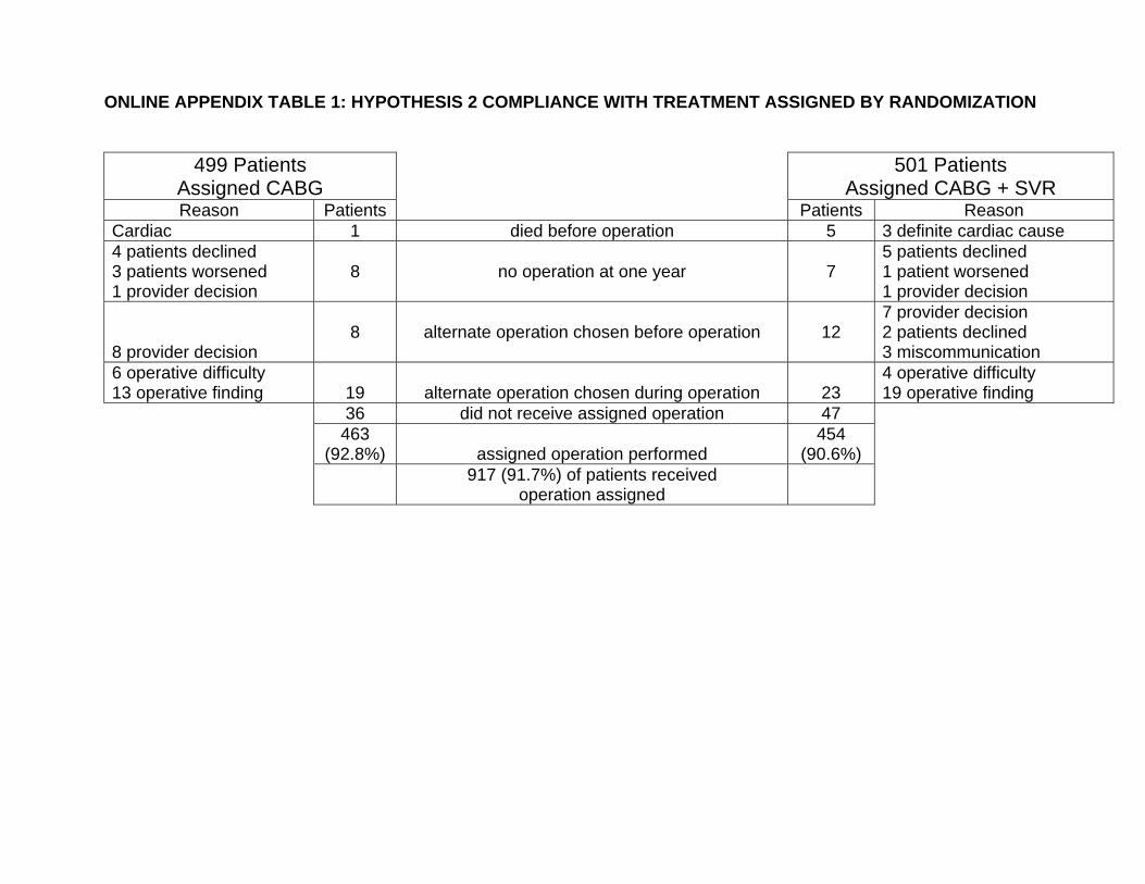
ONLINE APPENDIX TABLE 1: HYPOTHESIS 2 COMPLIANCE WITH TREATMENT ASSIGNED BY RANDOMIZATION
499 Patients Assigned CABG
501 Patients Assigned CABG + SVR
Reason Patients Patients Reason Cardiac 1 died before operation 5 3 definite cardiac cause 4 patients declined 3 patients worsened 1 provider decision
8 no operation at one year 7 5 patients declined 1 patient worsened 1 provider decision
8 provider decision
8 alternate operation chosen before operation 12 7 provider decision 2 patients declined 3 miscommunication
6 operative difficulty 13 operative finding 19 alternate operation chosen during operation 23
4 operative difficulty 19 operative finding
36 did not receive assigned operation 47 463
(92.8%) assigned operation performed 454
(90.6%)
917 (91.7%) of patients received operation assigned

ONLINE APPENDIX TABLE 2: OPERATIVE CONDUCT BY TREATMENT ARM (as randomized)
Variables CABG CABG with SVR P (N = 490) (N = 489) Surgical Data Acuteness at operation Elective 413 (84%) 406 (83%) 0.54 Urgent 62 (13%) 65 (13%) Ongoing ischemia 9 (2%) 12 (2%) Hemodynamic instability 2 (0.4%) 2 (0.4%) Salvage 3 (0.6%) 4 (0.8%) Off-pump bypass 50 (10%) 6 (1%) <0.001 Cardioplegia None 73 (15%) 13 (3%) Crystalloid 90 (18%) 116 (24%) Blood 307 (63%) 338 (69%) Both 19 (4%) 22 (4%) Number of conduits 0.49 1 40 (8%) 33 (7%) 2 134 (27%) 156 (32%) 3 214 (44%) 203 (42%) 4 89 (18%) 86 (18%) 5 11 (2%) 10 (2%) 6 or more 2 (0.4%) 1 (0.2%) Number of arterial conduits 0.008 0 33 (7%) 56 (11%) 1 403 (82%) 392 (80%) 2 42 (9%) 31 (6%) 3 12 (2%) 9 (2%) 4 0 1 (0.2%) 5 or more 0 0
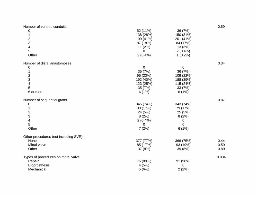
Number of venous conduits 0.59 0 52 (11%) 36 (7%) 1 138 (28%) 150 (31%) 2 199 (41%) 201 (41%) 3 87 (18%) 84 (17%) 4 11 (2%) 13 (3%) 5 0 2 (0.4%) Other 2 (0.4%) 1 (0.2%) Number of distal anastomoses 0.34 0 0 0 1 35 (7%) 36 (7%) 2 95 (20%) 109 (22%) 3 192 (40%) 188 (39%) 4 123 (25%) 115 (24%) 5 35 (7%) 33 (7%) 6 or more 6 (1%) 6 (1%) Number of sequential grafts 0.87 0 345 (74%) 343 (74%) 1 80 (17%) 79 (17%) 2 24 (5%) 25 (5%) 3 8 (2%) 8 (2%) 4 2 (0.4%) 0 5 0 0 Other 7 (2%) 6 (1%) Other procedures (not including SVR) None 377 (77%) 366 (75%) 0.44 Mitral valve 85 (17%) 93 (19%) 0.50 Other 37 (8%) 39 (8%) 0.80 Types of procedures on mitral valve 0.034 Repair 76 (89%) 91 (98%) Bioprosthesis 4 (5%) 0 Mechanical 5 (6%) 2 (2%)
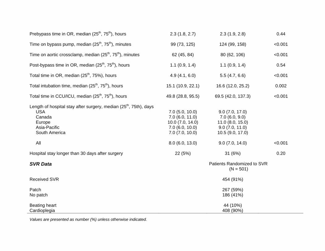
Prebypass time in OR, median (25th, 75th), hours 2.3 (1.8, 2.7) 2.3 (1.9, 2.8) 0.44 Time on bypass pump, median (25th, 75th), minutes 99 (73, 125) 124 (99, 158) <0.001 Time on aortic crossclamp, median (25th, 75th), minutes 62 (45, 84) 80 (62, 106) <0.001 Post-bypass time in OR, median (25th, 75th), hours 1.1 (0.9, 1.4) 1.1 (0.9, 1.4) 0.54 Total time in OR, median (25th, 75%), hours 4.9 (4.1, 6.0) 5.5 (4.7, 6.6) <0.001 Total intubation time, median (25th, 75th), hours 15.1 (10.9, 22.1) 16.6 (12.0, 25.2) 0.002 Total time in CCU/ICU, median (25th, 75th), hours 49.8 (28.8, 95.5) 69.5 (42.0, 137.3) <0.001 Length of hospital stay after surgery, median (25th, 75th), days USA 7.0 (5.0, 10.0) 9.0 (7.0, 17.0) Canada 7.0 (6.0, 11.0) 7.0 (6.0, 9.0) Europe 10.0 (7.0, 14.0) 11.0 (8.0, 15.0) Asia-Pacific 7.0 (6.0, 10.0) 9.0 (7.0, 11.0) South America 7.0 (7.0, 10.0) 10.5 (9.0, 17.0) All 8.0 (6.0, 13.0) 9.0 (7.0, 14.0) <0.001 Hospital stay longer than 30 days after surgery 22 (5%) 31 (6%) 0.20 SVR Data Patients Randomized to SVR
(N = 501) Received SVR 454 (91%) Patch 267 (59%) No patch 186 (41%) Beating heart 44 (10%) Cardioplegia 408 (90%) Values are presented as number (%) unless otherwise indicated.
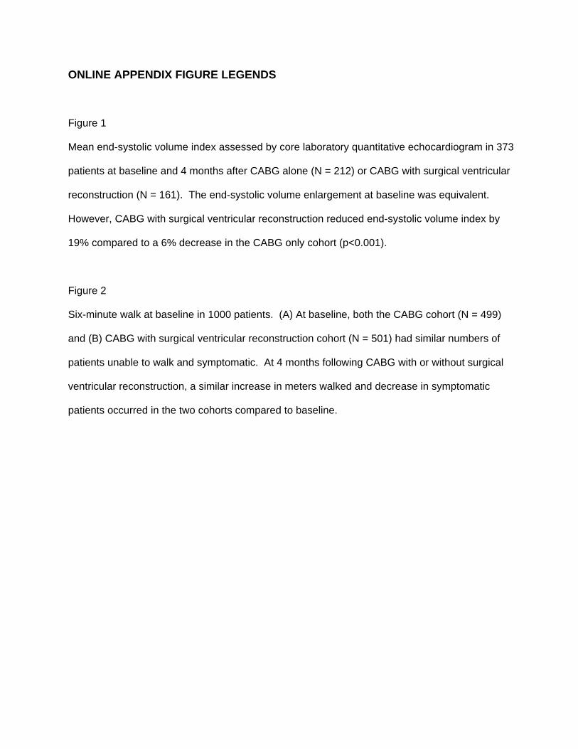
ONLINE APPENDIX FIGURE LEGENDS Figure 1 Mean end-systolic volume index assessed by core laboratory quantitative echocardiogram in 373
patients at baseline and 4 months after CABG alone (N = 212) or CABG with surgical ventricular
reconstruction (N = 161). The end-systolic volume enlargement at baseline was equivalent.
However, CABG with surgical ventricular reconstruction reduced end-systolic volume index by
19% compared to a 6% decrease in the CABG only cohort (p<0.001).
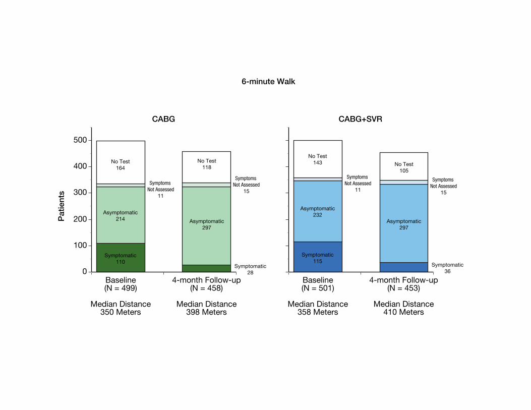
Figure 2
Six-minute walk at baseline in 1000 patients. (A) At baseline, both the CABG cohort (N = 499)
and (B) CABG with surgical ventricular reconstruction cohort (N = 501) had similar numbers of
patients unable to walk and symptomatic. At 4 months following CABG with or without surgical
ventricular reconstruction, a similar increase in meters walked and decrease in symptomatic
patients occurred in the two cohorts compared to baseline.

Baseline 4 Months0
20
40
60
80
CABG(N = 212)
Baseline 4 MonthsCABG+SVR
(N = 161)
82 ml/m2 77 ml/m2 83 ml/m2 67 ml/m2ESVI
0
100
200
300
400
500
Baseline(N = 499)
Median Distance350 Meters
4-month Follow-up(N = 458)
Median Distance398 Meters
Baseline(N = 501)
Median Distance358 Meters
4-month Follow-up(N = 453)
Median Distance410 Meters
CABG
6-minute Walk
CABG+SVR
Symptomatic110
Asymptomatic214
Symptoms Not Assessed
11
No Test164
Symptomatic28
Asymptomatic297
Symptoms Not Assessed
15
No Test118
Symptomatic115
Asymptomatic232
Symptoms Not Assessed
11
No Test143
Symptomatic36
Asymptomatic297
Symptoms Not Assessed
15
No Test105
Pat
ient
s