
THE JOURNAL OF BIOLOGICAL CHEMISTRY © 2001 by · PDF fileHuman Epidermal Growth Factor...
9
Human Epidermal Growth Factor (EGF) Module-containing Mucin- like Hormone Receptor 3 Is a New Member of the EGF-TM7 Family That Recognizes a Ligand on Human Macrophages and Activated Neutrophils* Received for publication, February 6, 2001, and in revised form, March 8, 2001 Published, JBC Papers in Press, March 14, 2001, DOI 10.1074/jbc.M101147200 Martin Stacey‡, Hsi-Hsien Lin§, Katherine L. Hilyard¶, Siamon Gordoni, and Andrew J. McKnighti** From the Sir William Dunn School of Pathology, University of Oxford, South Parks Road, Oxford OX1 3RE, ¶Roche Discovery Welwyn, 40 Broadwater Road, Welwyn Garden City AL7 3AY, and the **Department of Clinical Sciences, Institute of Liver Studies, King’s College Hospital, Bessemer Road, London SE5 9PJ, United Kingdom The epidermal growth factor (EGF)-TM7 subgroup of G-protein-coupled receptors is composed predomi- nantly of leukocyte-restricted glycoproteins defined by their unique hybrid structure, in which extracellular EGF-like domains are coupled to a seven-span trans- membrane moiety via a mucin-like stalk. The EGF-TM7 group comprises mouse F4/80, human EGF module-con- taining mucin-like hormone receptor (EMR) 1, human EMR2, and human and mouse CD97, the genes for which map to human chromosome 19p13 and the syntenic re- gions of the mouse genome. In this study we describe the cloning and characterization of EMR3, a novel human EGF-TM7 molecule, and show the existence of its cellu- lar ligand. The EMR3 gene maps closely to the existing members of the EGF-TM7 family on human chromosome 19p13.1 and, in common with other EGF-TM7 genes, is capable of generating different protein isoforms through alternative splicing. Two alternative splice forms have been isolated: one encoding a 652-amino acid cell surface protein consisting of two EGF-like domains, a mucin stalk, and a putative G-protein-coupled recep- tor domain and the other encoding a truncated soluble form containing only two EGF-like domains. As with other members of the EGF-TM7 family, EMR3 mRNA displays a predominantly leukocyte-restricted expres- sion pattern, with highest levels in neutrophils, mono- cytes, and macrophages. Through the use of soluble EMR3 multivalent probes we have shown the presence of a ligand at the surface of monocyte-derived macro- phages and activated human neutrophils. These inter- actions suggest a potential role for EMR3 in myeloid- myeloid interactions during immune and inflammatory responses. Professional phagocytic cells such as neutrophils and macro- phages play essential roles in a wide range of both immune and homeostatic functions. Many crucial processes such as chemo- taxis and migration, pathogen recognition and phagocytosis, inflammation, and microbial killing are mediated in part by the vast repertoire of leukocyte cell surface receptors. One rapidly growing group of receptors that may be involved in such pro- cesses is that of the epidermal growth factor (EGF) 1 -TM7 family (1, 2). At present the EGF-TM7 family comprises human and mouse CD97, mouse F4/80, human EMR1 (EGF module-con- taining mucin-like hormone receptor 1), and human EMR2 (3– 8). These predominantly leukocyte-restricted glycoproteins are defined by their unique chimeric structures, which consist of varying numbers of N-terminal EGF-like repeats coupled to a family B G-protein-coupled receptor (GPCR)-related moiety via a mucin-like stalk (Fig. 1). The founding member of the EGF-TM7 family, F4/80, comprises seven EGF-like domains, a spacer region, and a hydrophobic seven-span transmembrane (TM7) region. As a result of its macrophage-restricted expres- sion, F4/80 has long been used as an excellent marker for populations of mouse tissue macrophages. EMR1, the likely human homologue of F4/80, possesses six EGF-like domains and shares 68% overall amino acid identity with its murine counterpart. CD97, a B and T cell activation antigen, is ex- pressed constitutively by human granulocytes and monocytes and at low levels by resting T and B cells and is markedly and rapidly up-regulated upon T and B cell activation. CD97 occurs on the cell surface as three major isoforms resulting from alternative RNA splicing. All CD97 isoforms have been shown to bind to a membrane protein of the complement system, CD55 (DAF); however, the isoform containing three EGF-like do- mains binds with higher affinity to CD55 than the other iso- forms. More recently, human EMR2 has been cloned that con- tains EGF-like domains that are highly homologous to those of CD97. Similarly, EMR2 RNA undergoes alternative splicing; however, the isoforms appear to be more myeloid-restricted and are known not to interact with CD55. The EGF-like domain, like those found in EGF-TM7 pro- teins, is a common structural module often occurring as tan- dem repeats in extracellular proteins involved in cellular ad- hesion, receptor-ligand interactions, and blood coagulation. EGF-like domains consist of ;50 amino acids and are defined * The costs of publication of this article were defrayed in part by the payment of page charges. This article must therefore be hereby marked “advertisement” in accordance with 18 U.S.C. Section 1734 solely to indicate this fact. The nucleotide sequence(s) reported in this paper has been submitted to the GenBank TM /EBI Data Bank with accession number(s) AF239764. ‡ Funded through a BBSRC/Roche CASE studentship and a Goodger Scholarship. To whom correspondence should be addressed. Tel.: 44 1865 275532; Fax: 44 1865 275515; E-mail: [email protected]. § Supported by the Wellcome Trust Initiative for Cardiovascular Research. i Supported by grants from the Medical Research Council. 1 The abbreviations used are: EGF, epidermal growth factor; EMR, EGF module-containing mucin-like hormone receptor; GPCR, G-pro- tein-coupled receptor; DAF, decay-accelerating factor; RACE, rapid am- plification of cDNA ends; PCR, polymerase chain reaction; RT-PCR, reverse transcription-PCR; FACS, fluorescence-activated cell sorting. THE JOURNAL OF BIOLOGICAL CHEMISTRY Vol. 276, No. 22, Issue of June 1, pp. 18863–18870, 2001 © 2001 by The American Society for Biochemistry and Molecular Biology, Inc. Printed in U.S.A. This paper is available on line at http://www.jbc.org 18863 by guest on May 26, 2018 http://www.jbc.org/ Downloaded from
Transcript of THE JOURNAL OF BIOLOGICAL CHEMISTRY © 2001 by · PDF fileHuman Epidermal Growth Factor...
Human Epidermal Growth Factor (EGF) Module-containing Mucin-like Hormone Receptor 3 Is a New Member of the EGF-TM7 FamilyThat Recognizes a Ligand on Human Macrophages and ActivatedNeutrophils*
Received for publication, February 6, 2001, and in revised form, March 8, 2001Published, JBC Papers in Press, March 14, 2001, DOI 10.1074/jbc.M101147200
Martin Stacey‡, Hsi-Hsien Lin§, Katherine L. Hilyard¶, Siamon Gordoni,and Andrew J. McKnighti**
From the Sir William Dunn School of Pathology, University of Oxford, South Parks Road, Oxford OX1 3RE, ¶RocheDiscovery Welwyn, 40 Broadwater Road, Welwyn Garden City AL7 3AY, and the **Department of Clinical Sciences,Institute of Liver Studies, King’s College Hospital, Bessemer Road, London SE5 9PJ, United Kingdom
The epidermal growth factor (EGF)-TM7 subgroup ofG-protein-coupled receptors is composed predomi-nantly of leukocyte-restricted glycoproteins defined bytheir unique hybrid structure, in which extracellularEGF-like domains are coupled to a seven-span trans-membrane moiety via a mucin-like stalk. The EGF-TM7group comprises mouse F4/80, human EGF module-con-taining mucin-like hormone receptor (EMR) 1, humanEMR2, and human and mouse CD97, the genes for whichmap to human chromosome 19p13 and the syntenic re-gions of the mouse genome. In this study we describe thecloning and characterization of EMR3, a novel humanEGF-TM7 molecule, and show the existence of its cellu-lar ligand. The EMR3 gene maps closely to the existingmembers of the EGF-TM7 family on human chromosome19p13.1 and, in common with other EGF-TM7 genes, iscapable of generating different protein isoformsthrough alternative splicing. Two alternative spliceforms have been isolated: one encoding a 652-amino acidcell surface protein consisting of two EGF-like domains,a mucin stalk, and a putative G-protein-coupled recep-tor domain and the other encoding a truncated solubleform containing only two EGF-like domains. As withother members of the EGF-TM7 family, EMR3 mRNAdisplays a predominantly leukocyte-restricted expres-sion pattern, with highest levels in neutrophils, mono-cytes, and macrophages. Through the use of solubleEMR3 multivalent probes we have shown the presenceof a ligand at the surface of monocyte-derived macro-phages and activated human neutrophils. These inter-actions suggest a potential role for EMR3 in myeloid-myeloid interactions during immune and inflammatoryresponses.
Professional phagocytic cells such as neutrophils and macro-phages play essential roles in a wide range of both immune and
homeostatic functions. Many crucial processes such as chemo-taxis and migration, pathogen recognition and phagocytosis,inflammation, and microbial killing are mediated in part by thevast repertoire of leukocyte cell surface receptors. One rapidlygrowing group of receptors that may be involved in such pro-cesses is that of the epidermal growth factor (EGF)1-TM7family (1, 2).
At present the EGF-TM7 family comprises human andmouse CD97, mouse F4/80, human EMR1 (EGF module-con-taining mucin-like hormone receptor 1), and human EMR2(3–8). These predominantly leukocyte-restricted glycoproteinsare defined by their unique chimeric structures, which consistof varying numbers of N-terminal EGF-like repeats coupled toa family B G-protein-coupled receptor (GPCR)-related moietyvia a mucin-like stalk (Fig. 1). The founding member of theEGF-TM7 family, F4/80, comprises seven EGF-like domains, aspacer region, and a hydrophobic seven-span transmembrane(TM7) region. As a result of its macrophage-restricted expres-sion, F4/80 has long been used as an excellent marker forpopulations of mouse tissue macrophages. EMR1, the likelyhuman homologue of F4/80, possesses six EGF-like domainsand shares 68% overall amino acid identity with its murinecounterpart. CD97, a B and T cell activation antigen, is ex-pressed constitutively by human granulocytes and monocytesand at low levels by resting T and B cells and is markedly andrapidly up-regulated upon T and B cell activation. CD97 occurson the cell surface as three major isoforms resulting fromalternative RNA splicing. All CD97 isoforms have been shownto bind to a membrane protein of the complement system, CD55(DAF); however, the isoform containing three EGF-like do-mains binds with higher affinity to CD55 than the other iso-forms. More recently, human EMR2 has been cloned that con-tains EGF-like domains that are highly homologous to those ofCD97. Similarly, EMR2 RNA undergoes alternative splicing;however, the isoforms appear to be more myeloid-restrictedand are known not to interact with CD55.
The EGF-like domain, like those found in EGF-TM7 pro-teins, is a common structural module often occurring as tan-dem repeats in extracellular proteins involved in cellular ad-hesion, receptor-ligand interactions, and blood coagulation.EGF-like domains consist of ;50 amino acids and are defined
* The costs of publication of this article were defrayed in part by thepayment of page charges. This article must therefore be hereby marked“advertisement” in accordance with 18 U.S.C. Section 1734 solely toindicate this fact.
The nucleotide sequence(s) reported in this paper has been submittedto the GenBankTM/EBI Data Bank with accession number(s) AF239764.
‡ Funded through a BBSRC/Roche CASE studentship and a GoodgerScholarship. To whom correspondence should be addressed. Tel.: 441865 275532; Fax: 44 1865 275515; E-mail: [email protected].
§ Supported by the Wellcome Trust Initiative for CardiovascularResearch.
i Supported by grants from the Medical Research Council.
1 The abbreviations used are: EGF, epidermal growth factor; EMR,EGF module-containing mucin-like hormone receptor; GPCR, G-pro-tein-coupled receptor; DAF, decay-accelerating factor; RACE, rapid am-plification of cDNA ends; PCR, polymerase chain reaction; RT-PCR,reverse transcription-PCR; FACS, fluorescence-activated cell sorting.
THE JOURNAL OF BIOLOGICAL CHEMISTRY Vol. 276, No. 22, Issue of June 1, pp. 18863–18870, 2001© 2001 by The American Society for Biochemistry and Molecular Biology, Inc. Printed in U.S.A.
This paper is available on line at http://www.jbc.org 18863
by guest on May 26, 2018
http://ww
w.jbc.org/
Dow
nloaded from

by six conserved cysteine residues that form three intra-do-main disulfide bonds. A subset of EGF-like domains contains aCa21-binding consensus sequence, (D/N)X(D/N)(E/Q)Xm(D/N*)Xn(Y/F) (where m and n are variable, and * indicates pos-sible b-hydroxylation) (9). Chelated Ca21 performs a key role inthe orientation of EGF-like pairs by restricting flexibility of theinterdomain linkage, thus allowing the presentation of specificsurfaces for protein-protein interactions to occur (10). The im-portance of EGF-like domains is demonstrated by natural mu-tations in the genes encoding fibrillin-1, low density lipoproteinreceptor, factor IX, and protein S, which are responsible forMarfan syndrome, familial hypercholesterolemia, hemophiliaB, and protein S deficiency, respectively (11–14).
The transmembrane regions of the EGF-TM7 moleculesshow the greatest degree of sequence homology to GPCR Bfamily. GPCRs constitute the largest gene superfamily withinthe genome and have been shown to mediate a range of cellularresponses to an extensive array of stimuli including peptides,amino acid derivatives, lipid analogues, ions, and external sen-sory stimuli such as light, taste, and odors through intracellu-lar signaling (15).
Based upon the restricted expression and hybrid structure ofEGF-TM7 family members, it has been suggested that thesecell surface proteins may play a significant role within theimmune system, mediating adhesion through their EGF-likerepeats with concomitant intracellular signaling via the GPCRmoiety. Although signaling in these molecules has yet to bedemonstrated, CD97 has been shown to bind to a membraneprotein of the complement system, CD55 (DAF), demonstratingits role in cell-cell interactions (16).
In this study we describe the cloning and characterization ofhuman EMR3, a new member of the EGF-TM7 family. TheEMR3 gene maps closely to other members of the EGF-TM7group on human chromosome 19p13.1 and encodes a leukocyte-restricted protein expressed predominantly on neutrophils,monocytes, and macrophages. Multivalent EMR3 probes wereused to demonstrate the presence of a cell surface ligand forEMR3 on both monocyte-derived macrophages and activatedhuman neutrophils, suggesting a potential role for EMR3 inmyeloid cell interactions.
EXPERIMENTAL PROCEDURES
Materials—All chemicals and reagents were obtained from Sigmaunless otherwise specified. Cell culture media and supplements werepurchased from Life Technologies, Inc. X-Vivo 10 medium was fromBioWhittaker (Walkersville, MD). Human buffy coats were purchasedfrom the National Blood Service, Bristol, UK. Cell lines were providedby the cell bank at the Sir William Dunn School of Pathology, Univer-sity of Oxford.
Cloning and Sequence Analysis—Oligonucleotide primers weredesigned from the sequence of a human chromosome 19 cosmid
(GenBankTM accession number AC004262, clone R29368) that showedhomology with the TM1 and membrane proximal region of F4/80 andCD97. 59 and 39 EMR3 cDNA fragments were amplified from humanspleen Marathon Ready cDNA by rapid amplification of cDNA ends(RACE) under conditions recommended by the manufacturer (CLON-TECH). In brief, 59-RACE products amplified from primer E1 (59-CTGGTGTTCTGGATGGCTTTACACAGGAG) and the anchor-specificprimer AP-1 of the manufacturer were used in a secondary nested PCRreaction using E2 (59-GCACGGGATCCTCCTGCACATCGTAGTGG)and the anchor-specific AP-2 primer. The 59-proximal cDNA productwas then subcloned and sequenced using standard molecular tech-niques. The 39-proximal cDNA fragment EMR3 cDNA was obtainedusing E3 (59-TGTGTCTACTGGAAGAGCACAGGGCA) and E4 (59-TGATACACGTGAACAAGAGTCAC) with AP-1 and AP-2 primers intwo rounds of PCR as described above. Full-length EMR3 cDNA wasamplified from human spleen cDNA using primers E5 (59-TTTCTT-GAGCTAGGAAAGGTGGTGGTT) and E6 (59-TTGAAGAGAAATG-CAATTTACCACACA) and HF-polymerase under conditions recom-mended by the manufacturer (CLONTECH). The PCR products weresubcloned into pGEM-T (Promega, Madison, WI), sequenced, and subse-quently excised and ligated into pcDNA3 (Invitrogen) using NotI and ApaIrestriction enzymes.
RNA Blot Analysis and RT-PCR—Total RNA was prepared fromvarious cell lines and human peripheral blood mononuclear cells, poly-morphonuclear cells, monocytes, lymphocytes, as well as day 1 and day7 monocyte-derived macrophages using the acid guanidium thiocya-nate-phenol-chloroform technique (17). Total RNA (10 mg) was electro-phoresed on a 1% formaldehyde-agarose gel, blotted onto a nylon mem-brane (Stratagene), and then hybridized with gene-specific probes aspreviously described (18). Likewise, commercially available humanmultiple tissue Northern blots (CLONTECH) were hybridized with afull-length EMR3 cDNA probe according to the instructions of themanufacturer. Hybridized blots were washed in 0.53 SSC (0.15 M NaCland 0.015 M sodium citrate) containing 1% SDS at 55 °C for 30 min andexposed to x-ray film (Kodak Biomax MR) at 280 °C for 2–4 days.Radioactive probes were stripped from RNA blots by washing in 0.13SSC, 1% SDS for 15 min at 95 °C, and the blots were rehybridized witha human b-actin cDNA probe to compare the amount of RNA loaded ineach lane. RT-PCR data using multiple tissue cDNA panels (CLON-TECH) and RNA templates from various cell lines were also used tostudy the EMR3 expression profile. Advantage polymerase (CLON-TECH) and primers E5 (59-CACAAATGCAGGGACCATTGCT) and E6(59-GGATGTCCATTGAGTTCATTTG) were used to amplify EMR3-spe-cific cDNA fragments of 700 base pairs under conditions recommendedby the manufacturer. b2-microglobulin-specific primers F1 (59-GAT-TCAGGTTTACTCAGG) and R1 (59-CCATGATGCTGCTTACATG) werealso used in both RT-positive and RT-negative PCR reactions to com-pare cDNA levels and show a lack of genomic DNA contaminationwithin each PCR reaction.
Production of Biotinylated EMR3 Recombinant Protein—A cDNAfragment encoding the peptide sequence NSGSLHHILDAQKM-VWNHR*, recognized by the Escherichia coli biotin holoenzyme syn-thetase BirA, was generated by PCR using Bio5 (59-TAGTAGGGATC-CGAATTCCGGATCACTGCATCATATT-39) and Bio3 (59-TAGTAG-GGGCCCTTAACGATGATTCCACACC-39) primers and an HLA-A2plasmid construct, containing the peptide coding sequence as template(19). Following BamHI and ApaI digestion, the cDNA fragment was
FIG. 1. The EGF-TM7 family comprises mouse F4/80 (GenBankTM accession number X93328), human EMR1 (GenBankTM accessionnumber X81479), human EMR2 (GenBankTM accession number AF114491), human EMR3 (GenBankTM accession number AF239764),and human and mouse CD97 (GenBankTM accession numbers U76764 and Y18365). Triangles represent EGF-like domains, filled circlesdenote potential sites of N-linked glycosylation, and membrane-spanning cylinders show transmembrane domains of the putative GPCR moieties.The largest known isoform of each molecule is depicted.
Human EMR3 Is a New Member of the EGF-TM7 Family18864
by guest on May 26, 2018
http://ww
w.jbc.org/
Dow
nloaded from

subcloned immediately downstream of the stalk region of EMR3 inpcDNA3. Soluble biotinylated EMR3 protein comprising the EGF-likedomains and the stalk region was produced as described previously(19). Briefly, conditioned medium collected from transfected 293T cellswas concentrated to ;0.5 ml using a 30-kDa cutoff Centriprep filtrationunit (Millipore), dialyzed against 10 mM Tris-HCl (pH 8) buffer, andincubated with 1 ml of BirA enzyme and manufacturer substrates (Avid-ity) overnight at room temperature. Excess biotin was subsequentlyremoved by dialysis against 10 mM Tris-HCl (pH 7.3) buffer containing10 mM CaCl2 and 150 mM NaCl. The biotinylated proteins were thenaliquoted and stored at 280 °C after quantification by dot-blot analysisusing biotinylated maltose-binding protein (Avidity) as standard.
Isolation of Blood Cells—10 ml of heparinized blood from a healthydonor was overlaid on a Polymorphprepy gradient and centrifuged asdirected by the instructions of the manufacturer (Nycomed Pharma AS,Oslo, Norway). The erythrocyte, mononuclear, and polymorphonuclearcell fractions were collected separately, washed once in 0.53 RPMI andtwice in RPMI, and then resuspended in RPMI medium. Purified neu-trophils were activated by incubation at 37 °C for 30 min with 100 nM
formyl-Met-Leu-Phe.Cell Binding Assay Using Biotinylated Protein-coupled Fluorescent
Beads—Cell binding assays using fluorescent beads coupled to biotiny-lated proteins were performed as previously described (19). In brief,10-ml avidin-coated fluorescent beads (Spherotech Inc.) were washedtwice with phosphate-buffered saline containing 1% bovine serum al-bumin (phosphate-buffered saline/bovine serum albumin) and added to1 mg of biotinylated protein in a total volume of 50 ml. The bead andbiotinylated protein mixture was sonicated for 1 min and then incu-bated at 4 °C for 1 h. Non-binding proteins were removed by washingtwice with phosphate-buffered saline/bovine serum albumin, and thebeads were then resuspended in 50 ml of RPMI, 10% fetal calf serum.The bead-protein complex was sonicated again immediately before add-ing to various cell types in a 96-well plate (5 3 105 cells/50 ml of RPMI,10% fetal calf serum/well). The cell-bead mixture was spun at 1000 3 gat 4 °C for 20 min, incubated for a further 40 min at 4 °C, and finallyresuspended in 500 ml of phosphate-buffered saline for FACS analysis.
RESULTS
In a search for novel members of the EGF-TM7 group ofleukocyte receptors, DNA sequence data bases were screenedfor expressed sequence tags or genomic clones that displayedsequence homology with existing EGF-TM7 family members. Acosmid clone (R29368) derived from human chromosome 19DNA (GenBankTM accession number AC004262) demonstrated;50% amino acid identity to the first transmembrane domainand membrane proximal region of F4/80, EMR1, and CD97.The cosmid sequence was therefore used for primer design(E1–4) (see “Experimental Procedures”) in subsequent 59- and39-RACE reactions. Using human spleen cDNA template andprimers E1–4 plus AP1 and AP2 anchor sequences, two novelEGF-TM7 molecules designated EMR2 (8) and EMR3 wereindependently isolated. The EMR2 DNA sequence was found tomatch the R29368 cosmid sequence, whereas the EMR3 DNAsequence suggests that the gene-specific primers misprimed,resulting in the amplification of a closely related, yet independ-ent cDNA species.
The full-length EMR3 cDNA (Fig. 2) is comprised of 2247nucleotides encoding a predicted 652-amino acid polypeptide.Based on the protein analysis programs from the Expasyserver, the EMR3 sequence is predicted to contain a 21-aminoacid signal peptide (residues 1–21) and two EGF-like domains(residues 25–66 and 67–118), with the N-terminal EGF-likedomain being a non-Ca21-binding module and the second EGF-like domain containing a Ca21-binding consensus sequence.The EGF-like domains are followed by a spacer region (residues118–350) containing several potential N- and O-linked glyco-sylation sites, a putative GPCR proteolytic site (20), a family BGPCR-related TM7 region (residues 350–602), and a shortcytoplasmic tail (residues 602–652).
As well as this membrane-bound form, other cDNA clonescontained a 40-nucleotide deletion (nucleotides 439–479). Thisdeletion, which corresponds to the splicing out of a single exon
(data obtained from a partial genomic clone; GenBankTM ac-cession number AC0022156), results in a frameshift leading toa premature stop codon and the production of a truncatedsoluble form of EMR3 protein that comprises the two EGF-likedomains of the molecule. Multiple sequence alignment usingthe ClustalW software shows EMR3 to be most closely relatedto EMR2, followed by other members of the EGF-TM7 group ofmolecules: F4/80, EMR1, and CD97 (Fig. 3). The TM7 region ofEMR3 shows ;90% sequence identity to EMR2, whereas thesequence identity in the stalk regions and EGF-like domains is;35 and 50%, respectively.
RT-PCR and Northern blot analysis performed on variouscell lines and tissues showed EMR3 mRNA to have a leukocyte-restricted expression profile resembling that of other knownEGF-TM7 molecules (Fig. 4). Northern blot analysis of EMR3identified two major transcripts at 2.4 and 4 kilobases ex-pressed at high levels in immune tissues (spleen, bone marrow,and peripheral blood leukocytes) and at much lower levels inthe placenta and lung (Fig. 4a). RT-PCR was performed usingprimers from the 59 untranslated region (sense) and the stalkregion (antisense) of EMR3 and gave similar results to theNorthern blot data (Fig. 4b). Highest levels of expression wereagain observed in the spleen, peripheral blood leukocytes, andlung, followed by intermediate levels in the placenta and bonemarrow, with low detected levels in heart, kidney, liver, pan-creas, skeletal muscle, lymph node, thymus, tonsil, and fetalliver. EMR3 expression was not detected in the brain.
To dissect the expression profile of EMR3 mRNA in periph-eral blood cells further, RT-PCR was performed on RNA sam-ples from eight hemopoietic cell lines and, as negative controls,two human fibroblast cell lines (Fig. 4d). Following 30 cycles ofPCR amplification, EMR3 expression was undetectable in all ofthe cell lines; however, after 40 cycles of amplification EMR3expression was detected in two monocyte/macrophage cell lines(HL60 and U937), two B cell lines (Daudi and H9), and one Tcell line (Jurkat). No signal was detected in the other cell lines,including the non-hemopoietic cell line HepG2 and 293 cells. Toallow further examination of the expression profile in periph-eral blood leukocytes, RNA was extracted from fractionatedblood including polymorphonuclear cells, peripheral bloodmononuclear cells, and lymphocytes from healthy donors andused in subsequent Northern blot analysis (Fig. 4c). The blotshows that the vast majority of the EMR3 hybridization signalis derived from the polymorphonuclear cell fraction, whichcontains mainly neutrophils, with a small number of eosino-phils and basophils. Much lower levels of EMR3 expressionwere detected in freshly isolated monocytes and culturedmacrophages, whereas no expression was observed in the RNAisolated from lymphocytes.
It has been well documented that many of the cell-cell inter-actions within the immune system are of low affinity andtransient in nature (21). As such, when screening for suchinteractions, it is often necessary to use multivalent probes toincrease the avidity of the molecular association to allow de-tection. In a search for the cellular ligand(s) of EMR3, a previ-ously established sensitive assay system was utilized forscreening cells that may express potential ligand(s). Recombi-nant soluble EMR3 containing a biotinylation sequence recog-nized by the E. Coli enzyme BirA (Fig. 5a) was biotinylated invitro and coupled to avidin-coated fluorescent beads (22). Themultivalent fluorescent bead-biotinylated EMR3 complex (Fig.5b) was then used as a ligand probe in a FACS-based assay.Fluorescent beads coated with CD97 (EGF-1,2,5 isoform) andEMR2 (EGF-1,2 isoform) were also produced and served asbinding and non-binding controls, respectively. Because of thehigh level of EMR3 expression in peripheral blood leukocytes,
Human EMR3 Is a New Member of the EGF-TM7 Family 18865
by guest on May 26, 2018
http://ww
w.jbc.org/
Dow
nloaded from

as measured by RNA analysis, various cell types were isolatedfrom healthy donors by Polymorphprepy separation and sub-sequently screened for interactions with the multivalent EMR3probe. Following the incubation of human erythrocytes withCD97-multivalent fluorescent beads, a predicted shift in fluo-
rescence was observed (Fig. 5c). This indicated that the CD97-coated fluorescent beads effectively bound to the CD97 cellularligand CD55, which is known to be expressed at high levels onerythrocytes and other cells in the blood. In contrast, the FACSprofiles of the EMR3- and the EMR2-coated fluorescent beads
FIG. 2. Full-length cDNA and predicted amino acid sequence of EMR3. Bold type represents the predicted signal peptide (amino acids1–21), shaded areas represent the two EGF-like domains (amino acids 22–118), and the broken underlined sequence shows the nucleotide deletion(nucleotides 439–479) found in the EMR3 soluble alternate splice form. Boxes show potential N-linked glycosylation sites, dotted underliningshows the consensus GPCR proteolytic site sequence (amino acids 320–345), and double underlined sequences show predicted transmembranedomains. The cDNA sequence has been submitted to the GenBankTM/EBI Data Bank with accession number AF239764.
Human EMR3 Is a New Member of the EGF-TM7 Family18866
by guest on May 26, 2018
http://ww
w.jbc.org/
Dow
nloaded from
showed no shift in fluorescence, indicating no detectable inter-actions with human erythrocyte cell surface molecules (Fig. 5c).Similarly, no shift in fluorescence was detected when EMR3-coated fluorescent beads were incubated with either restingpolymorphonuclear cells or peripheral blood mononuclear cells(Fig. 5, d and f). However, a significant shift in fluorescencewas observed when EMR3-coated fluorescent beads were incu-bated with monocyte-derived macrophages and activated neu-trophils following their exposure to 100 nM formyl-Met-Leu-Phe for 30 min (Fig. 5, e and d). These results indicate thepresence of a ligand for EMR3 that is up-regulated on mono-
cyte-derived macrophages and human neutrophils followingactivation. Both the monocyte-derived macrophages and theactivated neutrophils failed to interact with EMR2-coatedbeads, proving the specificity of the interaction between EMR3and a molecule on the surface of the myeloid cells.
DISCUSSION
The data presented show EMR3 to possess the typical char-acteristics displayed by other EGF-TM7 proteins. The EMR3gene is located on chromosome 19 and is capable of encodingdifferent EMR3 protein isoforms through alternative RNA
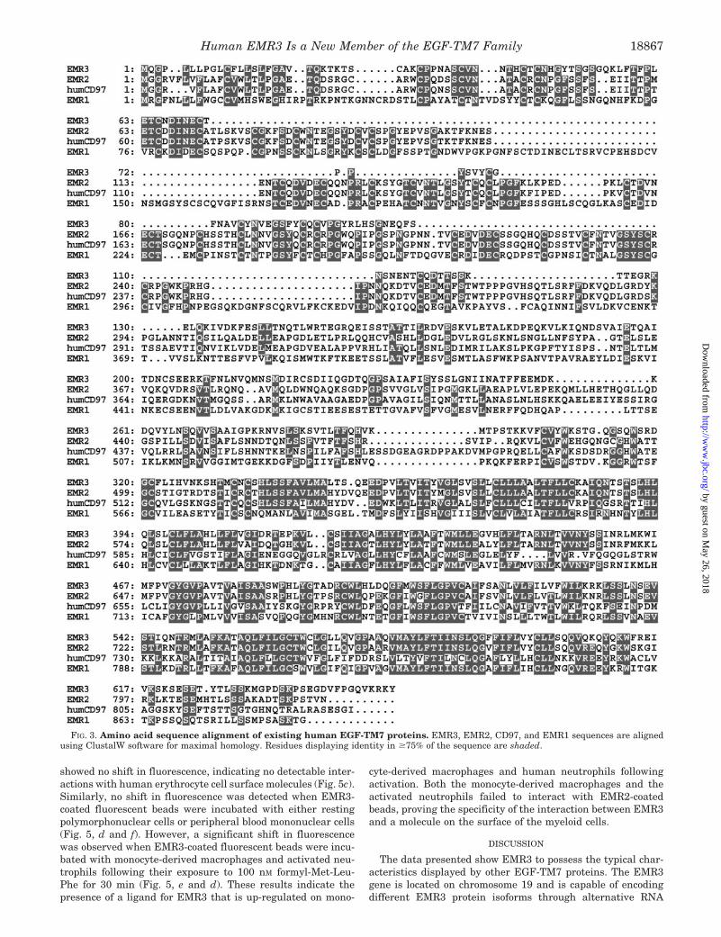
FIG. 3. Amino acid sequence alignment of existing human EGF-TM7 proteins. EMR3, EMR2, CD97, and EMR1 sequences are alignedusing ClustalW software for maximal homology. Residues displaying identity in $75% of the sequence are shaded.
Human EMR3 Is a New Member of the EGF-TM7 Family 18867
by guest on May 26, 2018
http://ww
w.jbc.org/
Dow
nloaded from

splicing. This results in the production of a predominantlyleukocyte-restricted cell surface protein comprised of two EGF-like domains coupled to a seven-span transmembrane moietyvia a mucin-like spacer region as well as its soluble secretedcounterpart lacking both the transmembrane and stalk re-gions. In addition to these data, a sensitive FACS-based assaysystem using multivalent EMR3 fluorescent beads as a ligandprobe demonstrated the presence of an EMR3 ligand on bothhuman monocyte-derived macrophages and activated humanneutrophils.
As with existing members of the EGF-TM7 family, RT-PCRdata and Northern blot analysis show EMR3 to be leukocyte-restricted (Fig. 4). In Northern analysis of fractionated humanblood, the majority of EMR3 signal is derived from polymor-phonuclear cells (Fig. 4c). The main cellular components of thisisolated fraction are neutrophils; however, a signal from thelower number of eosinophils and basophils cannot be ruled out.Lower levels of EMR3 mRNA were also detected in monocytesand culture-derived macrophages, suggesting that EMR3 is amyeloid-restricted molecule. The future development of EMR3-specific antibodies will greatly facilitate further characteriza-tion of expression within these cells. Once such antibodies areavailable, it would be interesting to show whether EMR3 ispresent in intracellular vesicles such as storage granules ormainly at the cell surface and to determine whether the acti-vation state of polymorphonuclear cells, macrophages, or mono-cytes affects the levels or distribution of EMR3.
The significance of the interaction between EMR3 and mono-cyte-derived macrophages and activated neutrophils detectedby FACS has yet to be established. However, because themajority of EMR3 is shown to be expressed by myeloid cellssuch as neutrophils and macrophages (Fig. 4), the interaction
could play an important role in modulating immune and in-flammatory responses by allowing “cross-talk” between mye-loid cells. The ligation of EMR3 with a ligand on macrophagesand activated neutrophils could potentially activate or deacti-vate cellular responses. Resting neutrophils and macrophagesmight be activated via signal transduction through the GPCRmoiety of EMR3 following ligand binding, thereby amplifyingimmune and inflammatory responses, or signaling could possi-bly occur in the opposite direction. The binding of the EMR3ligand to EMR3 could result in the modulation of macrophagesand activated neutrophils, thereby reducing inflammatory cell-mediated tissue damage as a result of negative signaling.
The exact nature of the EMR3 ligand is not yet known;however, a related member of the EGF-TM7 family, CD97, isknown to bind CD55 (DAF), which belongs to the regulators ofcomplement activation family of proteins that are involved inthe prevention of complement-mediated lysis of healthy cells.Although EMR3 does not bind CD55, other candidate proteinscontaining similar short consensus repeat domains includeCD46 (membrane cofactor protein), CD35 (CR1), and CD21(CR2) (23). The further biochemical characterization of EMR3ligand is underway.
Because the multivalent EMR3 probes were produced withsoluble EMR3, it raises the question whether membrane an-choring of EMR3 is necessary for ligand binding in vivo. In-deed, an EMR3 cDNA clone containing a 40-nucleotide deletionhas also been isolated that is predicted to encode a soluble formof EMR3 lacking the transmembrane domains and the mucin-like region. The presence of a putative GPCR proteolytic site inEMR3 (residues 320–345) as defined by Krasnoperov et al. (27)gives further weight to the possibility that a soluble form ofEMR3 mediates receptor ligation. The GPCR proteolytic site
FIG. 4. EMR3 expression is re-stricted predominantly to immunetissues. a, Northern blot showing EMR3mRNA expression profile in various hu-man tissues. b, RT-PCR results of a cDNApanel from various human tissues using aprimer pair from the 59 untranslated re-gion and stalk region of EMR3. c, North-ern blot of total RNA from fractionatedhuman blood cell populations probed withan EMR3-specific cDNA fragment. d,RNA from various human hemopoieticand non-hemopoietic cells was used as atemplate for RT-PCR using EMR3-spe-cific primers and b2-microglobulin (b2M)-specific primers. kb, kilobases; PMN,polymorphonuclear cells; PBMC, periph-eral blood mononuclear cells.
Human EMR3 Is a New Member of the EGF-TM7 Family18868
by guest on May 26, 2018
http://ww
w.jbc.org/
Dow
nloaded from

sequence is believed to be defined by a highly conserved patternof Cys residues and a Leu-Thr site that was initially identifiedin a sperm receptor for egg jelly in sea urchins (Strongylocen-trotus purpuratus) (24). More recently, the site has been shownto be involved in the cleavage of the extracellular domains ofCD97 and latrophilin (CL1) (25–27), resulting in the formationof membrane-associated heterodimers in which the transmem-brane domains are non-covalently associated with the extracel-lular domains. This has led to the suggestion that the subse-quent release of the extracellular domains could then allowinteraction with ligands on neighboring cells and at the sametime allow the binding of other ligands such as peptide hor-mones to the GPCR moiety of the EGF-TM7 proteins.
The cluster of closely related EGF-TM7 genes on humanchromosome 19p13 and on the syntenic regions of the mousegenome suggests an ancient gene family that has arisenthrough gene duplication and exon shuffling (28). Indeed, theserendipitous discovery of EMR3 during the initial cloning ofEMR2 was due to the fact that there is a greater than 90%identity within the exons encoding the transmembrane do-mains of EMR2 (exons 14–19) and EMR3 (8). However, despitethe very high degree of sequence identity shared by EMR3 andEMR2 within their transmembrane domains, the two mole-cules display much lower levels of identity within their mucin-like stalk regions and EGF-like domains (35 and 50%, respec-tively). This leads to the possibility of related proteins having
different ligand binding properties, mediated through theirextracellular regions, yet similar intracellular signaling, medi-ated via their GPCR moieties. The converse could also be pos-tulated for two other EGF-TM7 proteins. Human CD97 andhuman EMR2 contain extremely similar EGF-like domains(differing by only six amino acids within the five EGF-likedomains) yet possess much less similar transmembrane do-main sequences. The temporal and spatial regulation of suchreceptors could allow differential intracellular signaling via thetransmembrane domain to occur by the same ligand binding tothe extracellular domain.
With the complete sequencing and publication of the humangenome in sight and the sequencing of the mouse genome wellunder way, it is interesting to speculate whether more EGF-TM7 genes reside on chromosome 19 or within the mousegenome. If more do exist, the cloning and characterization ofthese proteins and their ligands will facilitate our understand-ing of the physiological importance of the EGF-TM7 proteinswithin the immune system.
REFERENCES
1. Stacey, M., Lin, H. H., Gordon, S., and McKnight, A. J. (2000) Trends Biochem.Sci. 25, 284–289
2. McKnight, A. J., and Gordon, S. (1998) J. Leukocyte Biol. 63, 271–2803. Hamann, J., Eichler, W., Hamann, D., Kerstens, H. M., Poddighe, P. J.,
Hoovers, J. M., Hartmann, E., Strauss, M., and van Lier, R. A. (1995)J. Immunol. 155, 1942–1950
4. Hamann, J., van Zeventer, C., Bijl, A., Molenaar, C., Tesselaar, K., and van
FIG. 5. Demonstration of an EMR3ligand on human macrophages andactivated neutrophils. a, soluble EMR3protein containing an amino acid consen-sus sequence recognized by the BirA en-zyme allowing in vitro biotinylation of thelysine residue (*). b, multivalent EMR3probes produced by coupling soluble bio-tinylated proteins to avidin-coated fluo-rescent beads. c–f, FACS profiles of fluo-rescent beads coated with EMR3, CD97(EGF-1,2,5), or EMR2 (EGF-1,2) incu-bated with erythrocytes, resting andformyl-Met-Leu-Phe-activated neutro-phils, monocyte-derived macrophages,and lymphocytes, respectively.
Human EMR3 Is a New Member of the EGF-TM7 Family 18869
by guest on May 26, 2018
http://ww
w.jbc.org/
Dow
nloaded from

Lier, R. A. (2000) Int. Immunol. 12, 439–4485. Qian, Y. M., Haino, M., Kelly, K., and Song, W. C. (1999) Immunology 98,
303–3116. McKnight, A. J., Macfarlane, A. J., Dri, P., Turley, L., Willis, A. C., and
Gordon, S. (1996) J. Biol. Chem. 271, 486–4897. Baud, V., Chiosse, S. L., Viegas-Pequignot, E., Diriong, S., N9Guyen, V. C.,
Roe, B. A., and Lipinski, M. (1995) Genomics 26, 334–3448. Lin, H.-H., Stacey, M., Hamann, J., Gordon, S., and McKnight, A. J. (2000)
Genomics 67, 188–2009. Handford, P., Mayhew, M., Baron, M., Winship, P. R., Campbell, I. D., and
Brownlee, G. G. (1991) Nature 351, 164–16710. Downing, A. K., Knott, V., Werner, J. M., Cardy, C. M., Campbell, I. D., and
Handford, P. A. (1996) Cell 85, 597–60511. Dietz, H. C., and Pyeritz, R. E. (1995) Hum. Mol. Genet. 4, 1799–180912. Hobbs, H. H., Brown, M. S., and Goldstein, J. L. (1992) Hum. Mutat. 1,
445–46613. Giannelli, F., Green, P. M., Sommer, S. S., Poon, M.-C., Ludwig, M., Schwaab,
R., Reitsma, P. H., Goossens, M., Yoshioka, A., and Browmlee, G. G. (1996)Nucleic Acids Res. 24, 103–118
14. Gandrille, S., Borgel, D., Eschwege-Gufflet, V., Aillaud, M., Dreyfus, M.,Matheron, C., Gaussem, P., Abgrail, J. F., Jude, B., Sie, P., Toulon, P., andAlach, M. (1995) Blood 85, 130–138
15. Bockaert, J., and Pin, J. P. (1999) EMBO J. 18, 1723–172916. Hamann, J., Vogel, B., Van Schijndel, G. M., and van Lier, R. A. (1996) J. Exp.
Med. 184, 1185–1189
17. Chomczynski, P., Mackey, K., Drews, R., and Wilfinger, W. (1997) BioTech-niques 22, 550–553
18. Lin, H. H., Stubbs, L. J., and Mucenski, M. L. (1997) Genomics 41, 301–30819. Brown, M. H., Boles, K., van der Merwe, P. A., Kumar, V., Mathew, P. A., and
Barclay, A. N. (1998) J. Exp. Med. 188, 2083–209020. Lelianova, V. G., Davletov, B. A., Sterling, A., Rahman, M. A., Grishin, E. V.,
Totty, N. F., and Ushkaryov, Y. A. (1997) J. Biol. Chem. 272, 21504–2150821. van der Merwe, P. A., Brown, M. H., Davis, S. J., and Barclay, A. N. (1993)
EMBO J. 12, 4945–495422. Brown, M. H., Preston, S., and Barclay, A. N. (1995) Eur. J. Immunol. 25,
3222–322823. Liszewski, M. K., Farries, T. C., Lublin, D. M., Rooney, I. A., and Atkinson,
J. P. (1996) Adv. Immunol. 61, 201–28324. Moy, G. W., Mendoza, L. M., Schulz, J. R., Swanson, W. J., Glabe, C. G., and
Vacquier, V. D. (1996) J. Cell Biol. 133, 809–81725. Gray, J. X., Haino, M., Roth, M. J., Maguire, J. E., Jensen, P. N., Yarme, A.,
Stetler-Stevenson, M. A., Siebenlist, U., and Kelly, K. (1996) J. Immunol.157, 5438–5447
26. Sugita, S., Ichtchenko, K., Khvotchev, M., and Sudhof, T. C. (1998) J. Biol.Chem. 273, 32715–32724
27. Krasnoperov, V. G., Bittner, M. A., Beavis, R., Kuang, Y., Salnikow, K. V.,Chepurny, O. G., Little, A. R., Plotnikov, A. N., Wu, D., Holz, R. W., andPetrenko, A. G. (1997) Neuron 18, 925–937
28. Carver, E. A., Hamann, J., Olsen, A. S., and Stubbs, L. (1999) Mamm. Genome10, 1039–1040
Human EMR3 Is a New Member of the EGF-TM7 Family18870
by guest on May 26, 2018
http://ww
w.jbc.org/
Dow
nloaded from
McKnightMartin Stacey, Hsi-Hsien Lin, Katherine L. Hilyard, Siamon Gordon and Andrew J.
Human Macrophages and Activated NeutrophilsReceptor 3 Is a New Member of the EGF-TM7 Family That Recognizes a Ligand on Human Epidermal Growth Factor (EGF) Module-containing Mucin-like Hormone
doi: 10.1074/jbc.M101147200 originally published online March 14, 20012001, 276:18863-18870.J. Biol. Chem.
10.1074/jbc.M101147200Access the most updated version of this article at doi:
Alerts:
When a correction for this article is posted•
When this article is cited•
to choose from all of JBC's e-mail alertsClick here
http://www.jbc.org/content/276/22/18863.full.html#ref-list-1
This article cites 28 references, 11 of which can be accessed free at
by guest on May 26, 2018
http://ww
w.jbc.org/
Dow
nloaded from


![Modul Mekanika Tanah II [TM7]](https://static.fdocuments.net/doc/165x107/563db808550346aa9a8fef04/modul-mekanika-tanah-ii-tm7.jpg)

