Gallstone disease Q&S briefing paper2 | Gallstone disease ...
Asian Pacific Journal of Cancer Prevention, Vol 17, 2016 467
DOI:http://dx.doi.org/10.7314/APJCP.2016.17.2.467Gallstone Etiopathogenesis, Lith and Mucin Genes and New Treatment Modalities
Asian Pac J Cancer Prev, 17 (2), 467-471
Introduction
Gallstones are more common among women (3/1); 6.3 million men and 14.2 million women aged 20 to 74 years in the US have gallstones (Everhart et al., 1999). They are a major risk factor for gallbladder cancer development (Rajic et al., 2014; Gupta et al., 2015). Annual new case incidence is 1,000,000 persons and the prevalence is about 20,000,000. Number of yearly cholecystectomies is more than 700.000. Gallstones are more frequent in the US, Australia and in some European countries (10-15%) and less common in China, Russia, Iran, India and in some African countries.(1-5%). Gallstone disease was first described in ancient Egypt and the first treatment was reported in Chinese (Shennong Bencao Jing) and Indian sources. There are still some Chinese treatments. The first cholecystectomy was performed in Berlin in 1882 (Everhart et al., 1999).
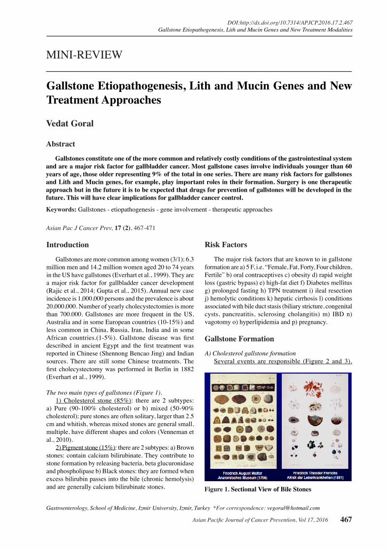
The two main types of gallstones (Figure 1).1) Cholesterol stone (85%): there are 2 subtypes:
a) Pure (90-100% cholesterol) or b) mixed (50-90% cholesterol); pure stones are often solitary, larger than 2.5 cm and whitish, whereas mixed stones are general small, multiple, have different shapes and colors (Venneman et al., 2010).
2) Pigment stone (15%): there are 2 subtypes: a) Brown stones: contain calcium bilirubinate. They contribute to stone formation by releasing bacteria, beta glucuronidase and phospholipase b) Black stones: they are formed when excess bilirubin passes into the bile (chronic hemolysis) and are generally calcium bilirubinate stones.
Gastroenterology, School of Medicine, Izmir University, Izmir, Turkey *For correspondence: [email protected]
Abstract
Gallstones constitute one of the more common and relatively costly conditions of the gastrointestinal system and are a major risk factor for gallbladder cancer. Most gallstone cases involve individuals younger than 60 years of age, those older representing 9% of the total in one series. There are many risk factors for gallstones and Lith and Mucin genes, for example, play important roles in their formation. Surgery is one therapeutic approach but in the future it is to be expected that drugs for prevention of gallstones will be developed in the future. This will have clear implications for gallbladder cancer control. Keywords: Gallstones - etiopathogenesis - gene involvement - therapeutic approaches
MINI-REVIEW
Gallstone Etiopathogenesis, Lith and Mucin Genes and New Treatment Approaches
Vedat Goral
Risk Factors
The major risk factors that are known to in gallstone formation are a) 5 F, i.e. “Female, Fat, Forty, Four children, Fertile” b) oral contraceptives c) obesity d) rapid weight loss (gastric bypass) e) high-fat diet f) Diabetes mellitus g) prolonged fasting h) TPN treatment i) ileal resection j) hemolytic conditions k) hepatic cirrhosis l) conditions associated with bile duct stasis (biliary stricture, congenital cysts, pancreatitis, sclerosing cholangitis) m) IBD n) vagotomy o) hyperlipidemia and p) pregnancy.
Gallstone Formation
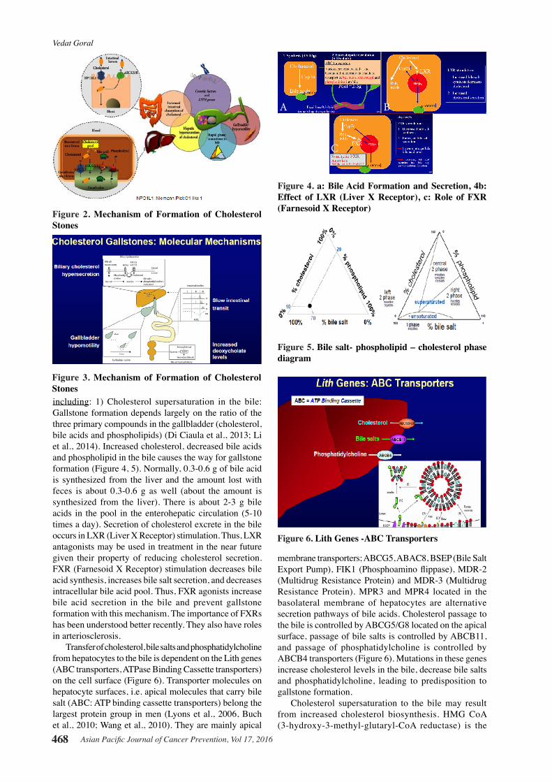
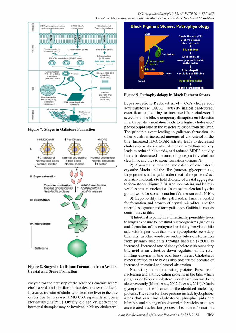
A) Cholesterol gallstone formationSeveral events are responsible (Figure 2 and 3),
Figure 1. Sectional View of Bile Stones
Vedat Goral
Asian Pacific Journal of Cancer Prevention, Vol 17, 2016468
including: 1) Cholesterol supersaturation in the bile: Gallstone formation depends largely on the ratio of the three primary compounds in the gallbladder (cholesterol, bile acids and phospholipids) (Di Ciaula et al., 2013; Li et al., 2014). Increased cholesterol, decreased bile acids and phospholipid in the bile causes the way for gallstone formation (Figure 4, 5). Normally, 0.3-0.6 g of bile acid is synthesized from the liver and the amount lost with feces is about 0.3-0.6 g as well (about the amount is synthesized from the liver). There is about 2-3 g bile acids in the pool in the enterohepatic circulation (5-10 times a day). Secretion of cholesterol excrete in the bile occurs in LXR (Liver X Receptor) stimulation. Thus, LXR antagonists may be used in treatment in the near future given their property of reducing cholesterol secretion. FXR (Farnesoid X Receptor) stimulation decreases bile acid synthesis, increases bile salt secretion, and decreases intracellular bile acid pool. Thus, FXR agonists increase bile acid secretion in the bile and prevent gallstone formation with this mechanism. The importance of FXRs has been understood better recently. They also have roles in arteriosclerosis.
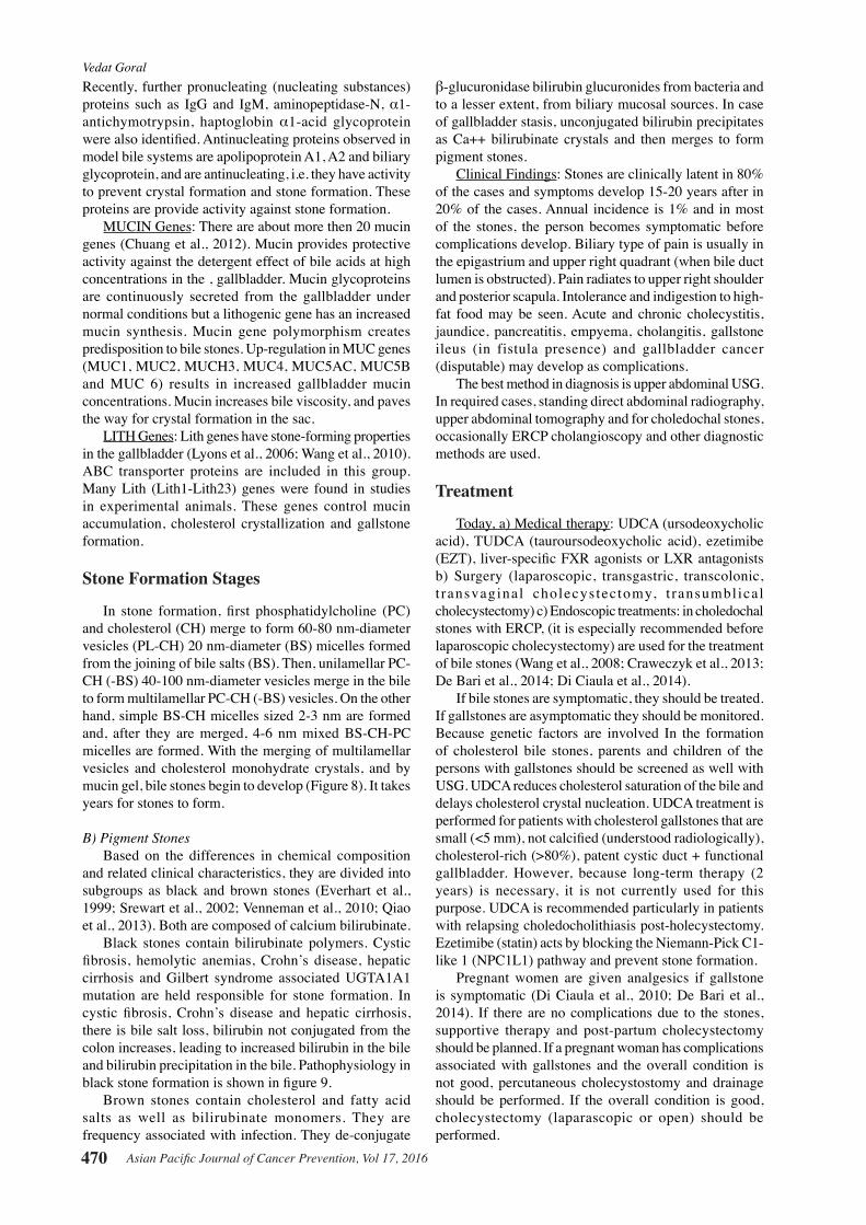
Transfer of cholesterol, bile salts and phosphatidylcholine from hepatocytes to the bile is dependent on the Lith genes (ABC transporters, ATPase Binding Cassette transporters) on the cell surface (Figure 6). Transporter molecules on hepatocyte surfaces, i.e. apical molecules that carry bile salt (ABC: ATP binding cassette transporters) belong the largest protein group in men (Lyons et al., 2006, Buch et al., 2010; Wang et al., 2010). They are mainly apical
membrane transporters; ABCG5, ABAC8, BSEP (Bile Salt Export Pump), FIK1 (Phosphoamino flippase), MDR-2 (Multidrug Resistance Protein) and MDR-3 (Multidrug Resistance Protein). MPR3 and MPR4 located in the basolateral membrane of hepatocytes are alternative secretion pathways of bile acids. Cholesterol passage to the bile is controlled by ABCG5/G8 located on the apical surface, passage of bile salts is controlled by ABCB11, and passage of phosphatidylcholine is controlled by ABCB4 transporters (Figure 6). Mutations in these genes increase cholesterol levels in the bile, decrease bile salts and phosphatidylcholine, leading to predisposition to gallstone formation.
Cholesterol supersaturation to the bile may result from increased cholesterol biosynthesis. HMG CoA (3-hydroxy-3-methyl-glutaryl-CoA reductase) is the
Figure 2. Mechanism of Formation of Cholesterol Stones
Figure 3. Mechanism of Formation of Cholesterol Stones
Figure 4. a: Bile Acid Formation and Secretion, 4b: Effect of LXR (Liver X Receptor), c: Role of FXR (Farnesoid X Receptor)
A B
C
Figure 5. Bile salt- phospholipid – cholesterol phase diagram
Figure 6. Lith Genes -ABC Transporters
Asian Pacific Journal of Cancer Prevention, Vol 17, 2016 469
DOI:http://dx.doi.org/10.7314/APJCP.2016.17.2.467Gallstone Etiopathogenesis, Lith and Mucin Genes and New Treatment Modalities
enzyme for the first step of the reactions cascade where cholesterol and similar molecules are synthesized. Increased transfer of cholesterol from the liver to the bile occurs due to increased HMG CoA especially in obese individuals (Figure 7). Obesity, old age, drug effect and hormonal therapies may be involved in biliary cholesterol
hypersecretion. Reduced Acyl - CoA cholesterol acyltransferase (ACAT) activity inhibit cholesterol esterification, leading to increased free cholesterol secretion to the bile. A temporary disruption on bile acids in entrahepatic circulation leads to a higher cholesterol/phospholipid ratio in the vesicles released from the liver. The principle event leading to gallstone formation, in other words, is increased amounts of cholesterol in the bile. Increased HMGCoAR activity leads to decreased cholesterol synthesis, while decreased 7-α-Ohase activity leads to reduced bile acids, and reduced MDR3 activity leads to decreased amount of phosphatidylcholine (lecithin), and thus to stone formation (Figure 7).
2) Abnormally reduced nucleation of cholesterol crystals: Mucin and the like (mucous glycoproteins), large proteins in the gallbladder (heat-labile proteins) act as matrix molecules to hold cholesterol crystal aggregates to form stones (Figure 7, 8). Apolipoproteins and lecithin vesicles prevent nucleation. Increased nucleation lays the groundwork for stone formation (Venneman et al.,2010).
3) Hypomotility in the gallbladder: Time is needed for formation and growth of crystal microlites, and for microlites to gather and form gallstones. Gallbladder stasis contributes to this.
4) Intestinal hypomotility: Intestinal hypomotility leads to longer exposure to intestinal microorganisms (bacteria) and formation of deconjugated and dehydroxylated bile salts with higher rates than more hydrophobic secondary bile salts. In other words, secondary bile salts formation from primary bile salts through bacteria (7αOH) is increased. Increased rate of deoxycholate with secondary bile acid is an effective down-regulator of the rate-limiting enzyme in bile acid biosynthesis. Cholesterol hypersecretion to the bile is also potentiated because of increased intestinal cholesterol absorption.
Nucleating and antinucleating proteins: Presence of nucleating and antinucleating proteins in the bile, which progress or hinder cholesterol crystallization has been shown recently (Mittal et al., 2002; Li et al., 2014). Mucin glycoprotein is the foremost of the identified nucleating proteins. The center for these proteins include hydrophobic areas that can bind cholesterol, phospholipids and bilirubin, and binding of cholesterol-rich vesicles mediates accelerated nucleation process, i.e. stone formation.
Figure 7. Stages in Gallstone Formation
Figure 8. Stages in Gallstone Formation from Vesicle, Crystal and Stone Formation
Figure 9. Pathophysiology in Black Pigment Stones
Vedat Goral
Asian Pacific Journal of Cancer Prevention, Vol 17, 2016470
Recently, further pronucleating (nucleating substances) proteins such as IgG and IgM, aminopeptidase-N, α1-antichymotrypsin, haptoglobin α1-acid glycoprotein were also identified. Antinucleating proteins observed in model bile systems are apolipoprotein A1, A2 and biliary glycoprotein, and are antinucleating, i.e. they have activity to prevent crystal formation and stone formation. These proteins are provide activity against stone formation.
MUCIN Genes: There are about more then 20 mucin genes (Chuang et al., 2012). Mucin provides protective activity against the detergent effect of bile acids at high concentrations in the , gallbladder. Mucin glycoproteins are continuously secreted from the gallbladder under normal conditions but a lithogenic gene has an increased mucin synthesis. Mucin gene polymorphism creates predisposition to bile stones. Up-regulation in MUC genes (MUC1, MUC2, MUCH3, MUC4, MUC5AC, MUC5B and MUC 6) results in increased gallbladder mucin concentrations. Mucin increases bile viscosity, and paves the way for crystal formation in the sac.
LITH Genes: Lith genes have stone-forming properties in the gallbladder (Lyons et al., 2006; Wang et al., 2010). ABC transporter proteins are included in this group. Many Lith (Lith1-Lith23) genes were found in studies in experimental animals. These genes control mucin accumulation, cholesterol crystallization and gallstone formation.
Stone Formation Stages
In stone formation, first phosphatidylcholine (PC) and cholesterol (CH) merge to form 60-80 nm-diameter vesicles (PL-CH) 20 nm-diameter (BS) micelles formed from the joining of bile salts (BS). Then, unilamellar PC-CH (-BS) 40-100 nm-diameter vesicles merge in the bile to form multilamellar PC-CH (-BS) vesicles. On the other hand, simple BS-CH micelles sized 2-3 nm are formed and, after they are merged, 4-6 nm mixed BS-CH-PC micelles are formed. With the merging of multilamellar vesicles and cholesterol monohydrate crystals, and by mucin gel, bile stones begin to develop (Figure 8). It takes years for stones to form.
B) Pigment StonesBased on the differences in chemical composition
and related clinical characteristics, they are divided into subgroups as black and brown stones (Everhart et al., 1999; Srewart et al., 2002; Venneman et al., 2010; Qiao et al., 2013). Both are composed of calcium bilirubinate.
Black stones contain bilirubinate polymers. Cystic fibrosis, hemolytic anemias, Crohn’s disease, hepatic cirrhosis and Gilbert syndrome associated UGTA1A1 mutation are held responsible for stone formation. In cystic fibrosis, Crohn’s disease and hepatic cirrhosis, there is bile salt loss, bilirubin not conjugated from the colon increases, leading to increased bilirubin in the bile and bilirubin precipitation in the bile. Pathophysiology in black stone formation is shown in figure 9.
Brown stones contain cholesterol and fatty acid salts as well as bilirubinate monomers. They are frequency associated with infection. They de-conjugate
β-glucuronidase bilirubin glucuronides from bacteria and to a lesser extent, from biliary mucosal sources. In case of gallbladder stasis, unconjugated bilirubin precipitates as Ca++ bilirubinate crystals and then merges to form pigment stones.
Clinical Findings: Stones are clinically latent in 80% of the cases and symptoms develop 15-20 years after in 20% of the cases. Annual incidence is 1% and in most of the stones, the person becomes symptomatic before complications develop. Biliary type of pain is usually in the epigastrium and upper right quadrant (when bile duct lumen is obstructed). Pain radiates to upper right shoulder and posterior scapula. Intolerance and indigestion to high-fat food may be seen. Acute and chronic cholecystitis, jaundice, pancreatitis, empyema, cholangitis, gallstone ileus (in fistula presence) and gallbladder cancer (disputable) may develop as complications.
The best method in diagnosis is upper abdominal USG. In required cases, standing direct abdominal radiography, upper abdominal tomography and for choledochal stones, occasionally ERCP cholangioscopy and other diagnostic methods are used.
Treatment
Today, a) Medical therapy: UDCA (ursodeoxycholic acid), TUDCA (tauroursodeoxycholic acid), ezetimibe (EZT), liver-specific FXR agonists or LXR antagonists b) Surgery (laparoscopic, transgastric, transcolonic, t ransvaginal cholecystectomy, t ransumblical cholecystectomy) c) Endoscopic treatments: in choledochal stones with ERCP, (it is especially recommended before laparoscopic cholecystectomy) are used for the treatment of bile stones (Wang et al., 2008; Craweczyk et al., 2013; De Bari et al., 2014; Di Ciaula et al., 2014).
If bile stones are symptomatic, they should be treated. If gallstones are asymptomatic they should be monitored. Because genetic factors are involved In the formation of cholesterol bile stones, parents and children of the persons with gallstones should be screened as well with USG. UDCA reduces cholesterol saturation of the bile and delays cholesterol crystal nucleation. UDCA treatment is performed for patients with cholesterol gallstones that are small (<5 mm), not calcified (understood radiologically), cholesterol-rich (>80%), patent cystic duct + functional gallbladder. However, because long-term therapy (2 years) is necessary, it is not currently used for this purpose. UDCA is recommended particularly in patients with relapsing choledocholithiasis post-holecystectomy. Ezetimibe (statin) acts by blocking the Niemann-Pick C1-like 1 (NPC1L1) pathway and prevent stone formation.
Pregnant women are given analgesics if gallstone is symptomatic (Di Ciaula et al., 2010; De Bari et al., 2014). If there are no complications due to the stones, supportive therapy and post-partum cholecystectomy should be planned. If a pregnant woman has complications associated with gallstones and the overall condition is not good, percutaneous cholecystostomy and drainage should be performed. If the overall condition is good, cholecystectomy (laparascopic or open) should be performed.
Asian Pacific Journal of Cancer Prevention, Vol 17, 2016 471
DOI:http://dx.doi.org/10.7314/APJCP.2016.17.2.467Gallstone Etiopathogenesis, Lith and Mucin Genes and New Treatment Modalities
In the future A) individualized therapies based on lithogenic structure B) genetic therapies based on ABCG8, ABCB4, ABCB11, UGT1A1… C) treatments targeting environmental factors, e.g. estrogen, enterohepatic bacteria etc. will be discussed in the future. Non-drug primary protection (weight loss, exercise, diet modification) in individuals at normal risk or protection with medication (UDCA, nuclear receptor ligands, statin-ezetimibe) in individuals with markedly higher risk or profilaktik cholecystectomy will also be possible discussed.
Conclusion
Environmental factors account for 75% of gallstone formations. Genetic factors represent 25% of gallstone formations (ABCG8 Cholesterol transporter 11%: UGT1A1 Gilbert variant 6%). In gallstone cases, differences have also been found in miRNAs and miRNA expression (Yang et al., 2015). The role of nuclear receptors in gallstone formation should be investigated. Synthetic FXR (Farnesoid X receptor) agonists will be probably used in treatment in the future.
References
Buch S, Schafmayer C, Volzke H, et al (2010). Loci from a genome-wide analysis of bilirubin levels are associated with gallstone risk and composition. Gastroenterol, 139, 1942-51.
Chuang SC, Hsi E, Lee KT (2012). Mucin genes in gallstone disease. Clin Chim Acta, 9, 1466-71
Crawczyk M, Stokes CS, Lammert F (2013). Genetics and treatment of bile duct stones: new approaches. Curr Opin Gastroenterol, 29, 329-35
De Bari O, Wang TY, Liu M, et al (2014). Cholesterol cholelithiasis in pregnant women: pathogenesis, prevention and treatment. Ann Hepatol, 13, 728-45.
Di Ciaula A, Wang DQ, Garruti G, et al (2014). Therapeutic reflections in cholesterol homeostasis and gallstone disease: a review. Curr Med Chem, 21, 1435-47.
Di Ciaula A, Wang DQ, Binfrate L, et al (2013). Current views on genetics and epigenetics of cholesterol gallstone disease. Cholesterol, 2013, 1-10,
Di Ciaula A, Wang DQ, Wang HH, et al (2010). Targets for current pharmacologic therapy in cholesterol gallstone disease. Gastroenterol Clin North Am, 39, 245-64.
Everhart JE, Khare M, Hill M, et al (1999). Prevalence and ethnic differences in gallbladder disease in the United States. Gastroenterol, 117, 632-9.
Gupta S, Kori C, Kumar V, Misra S, Akhtar N (2015). Epidemiological study of gallbladder cancer patients from north Indian Gangetic planes-a high-volume centre’s experience. J Gastrointest Cancer, [Epub ahead of print]
Li Q, Ge X, Xu X, et al (2014). Comparison of the gene expression profiles between gallstones and gallbladder polyps. Int J Clin Exp Pathol, 15, 8016-
Lyons MA, Wittenburg H (2006). Cholesterol gallstone susceptibility loci: a mouse map, candidate gene evaluation, and guide to human LITH genes. Gastroenterol, 131, 1943-70.
Mittal B, Mittal RD (2002). Genetics of gallstone disease. J Postgrad Med, 48, 149-52.
Qiao T, Ma RH, Luo XB, et al (2013). The systematic classification of gallbladder stones. PLoS One, 4, 1-11.
Rakić M, Patrlj L, Kopljar M, et al (2014). Gallbladder cancer. Hepatobiliary Surg Nutr, 3, 221-6. .
Stewart L, Oesterle AL, Erdan I, et al (2002). Pathogenesis of pigment gallstones in western societies: The central role of bacteria. J Gastrointestinal Surg, 6, 891-903.
Venneman NG, Van Erpecum KJ (2010). Pathogenesis of gallstones. Gastroenterol Clin Am, 39, 171-83.
Wang HH, Portincasa P, Afdal NH, et al (2010). Lith genes and genetic analysis of cholesterol gallstone formation. Gastroenterol Clin North Am, 39, 185-207.
Wang HH, Portincasa P, Mendez-Sanches N, et al (2008). Effect of ezetimibe on the prevention and dissolution of cholesterol gallstones. Gastroenterol, 134, 2101-10
Yang B, Liu B, BiP, Wu T, et al (2015). An integrated analysis of differential miRNA and mRNA expressions in human gallstones. Mol Biosyst, 17, 1004-11.