The Glycoproteins and Glycolipoproteins of the Bovine...
20
The glycoproteins and glycolipoproteins of the bovine lens and their relation to albuminoid Zacharias Dische U. 'ntil about 25 years ago, the biochem- ist and the biophysicist who investigated the properties of living systems dealt mainly with two types of constituents; one is represented by the compounds which formed an integral part of the structure of the living system itself, and the other by compounds involved in the metabolic pro- cesses of the living cell; that means en- zymes, coenzymes, and intermediates and end products of the metabolism. During the past 25 years, a new class of compounds has come to the fore and increasingly occupied the attention of the biochemist and the biophysicist. These are macro- molecular compounds to which we have to ascribe what is called informational properties. That means macromolecules in- volved in the process of creating order of a spatial or a temporal nature during the development and the changing events of the adult life of the organism. Such an order is based on a highly selective com- bination and association of molecular com- plexes and also on logistic processes which result in the localization of certain com- pounds at a certain place at a certain time. From the College of Physicians and Surgeons, Columbia University, New York, N. Y. This work was supported by Grant CA 02075-12 of the National Cancer Institute, National In- stitutes of Health, Bethesda, Md. The experimental work presented in this paper was carried out with the assistance of G. Zelmenis, D. Igals, A. Danilchenko, and N. Laiys. These processes result, as a rule, in a decrease of entropy of the living system, with a simultaneous increase of entropy in its environment. The progress in this field consisted in the recognition of the sig- nificance of the properties of these macro- molecules as heteropolymers which are highly asymmetric with respect to the se- quences of their monomers and of the spe- cific sequential order of these monomeric constituents, as well as of the distribution of their inter- and intramolecular forces as organizational and directive factors of processes of varying complexities in living systems. Two examples of major develop- ments in modern biochemistry illustrate this point. First, the role of nucleic acids in the biosynthesis of proteins. The essen- tial elements of this role are now well understood, but they concern the biosyn- thesis with regard to its occurrence on the molecular level. The biosynthetic process, however, as it appears from these studies, must involve events of higher complexity which manifest themselves in the activities of the so-called regulatory genes and in the complex intracellular movements of macromolecules necessary for the exchange of information between the DNA and the synthetic sites on the ribosomes. Even if these movements would, as a first step, involve an intracellular random distribu- tion of messenger molecules which then, by diffusion, would arrive at the biosyn- thetic sites, it still would require highly specific molecular arrangements on these sites which would exert highly specific 759 Downloaded From: http://iovs.arvojournals.org/pdfaccess.ashx?url=/data/journals/iovs/932953/ on 05/05/2018
Transcript of The Glycoproteins and Glycolipoproteins of the Bovine...

The glycoproteins and glycolipoproteins of thebovine lens and their relation to albuminoid
Zacharias Dische
U.'ntil about 25 years ago, the biochem-ist and the biophysicist who investigatedthe properties of living systems dealtmainly with two types of constituents; oneis represented by the compounds whichformed an integral part of the structure ofthe living system itself, and the other bycompounds involved in the metabolic pro-cesses of the living cell; that means en-zymes, coenzymes, and intermediates andend products of the metabolism. Duringthe past 25 years, a new class of compoundshas come to the fore and increasinglyoccupied the attention of the biochemistand the biophysicist. These are macro-molecular compounds to which we haveto ascribe what is called informationalproperties. That means macromolecules in-volved in the process of creating order ofa spatial or a temporal nature during thedevelopment and the changing events ofthe adult life of the organism. Such anorder is based on a highly selective com-bination and association of molecular com-plexes and also on logistic processes whichresult in the localization of certain com-pounds at a certain place at a certain time.
From the College of Physicians and Surgeons,Columbia University, New York, N. Y.
This work was supported by Grant CA 02075-12of the National Cancer Institute, National In-stitutes of Health, Bethesda, Md.
The experimental work presented in this paperwas carried out with the assistance of G.Zelmenis, D. Igals, A. Danilchenko, and N.Laiys.
These processes result, as a rule, in adecrease of entropy of the living system,with a simultaneous increase of entropy inits environment. The progress in this fieldconsisted in the recognition of the sig-nificance of the properties of these macro-molecules as heteropolymers which arehighly asymmetric with respect to the se-quences of their monomers and of the spe-cific sequential order of these monomericconstituents, as well as of the distributionof their inter- and intramolecular forces asorganizational and directive factors ofprocesses of varying complexities in livingsystems. Two examples of major develop-ments in modern biochemistry illustratethis point. First, the role of nucleic acidsin the biosynthesis of proteins. The essen-tial elements of this role are now wellunderstood, but they concern the biosyn-thesis with regard to its occurrence on themolecular level. The biosynthetic process,however, as it appears from these studies,must involve events of higher complexitywhich manifest themselves in the activitiesof the so-called regulatory genes and inthe complex intracellular movements ofmacromolecules necessary for the exchangeof information between the DNA and thesynthetic sites on the ribosomes. Even ifthese movements would, as a first step,involve an intracellular random distribu-tion of messenger molecules which then,by diffusion, would arrive at the biosyn-thetic sites, it still would require highlyspecific molecular arrangements on thesesites which would exert highly specific
759
Downloaded From: http://iovs.arvojournals.org/pdfaccess.ashx?url=/data/journals/iovs/932953/ on 05/05/2018
760 Dische Investigative OphthalmologyOctober 1965
attractive forces. It seems more probablethat what is going on here are movementsguided along specifically arranged molecu-lar tracks constructed from molecules spe-cifically designed for this type of operation.Another example of directive activitiesin which repetitive sequences of moleculesin long chains and electric charges playa role is the organization of tropocollageninto collagen fibrils with regular spacing ofelectron-dense material. Investigations inour laboratory carried out in the last fewyears indicate that sequentially arrangedhexose molecules linked to tropocollagenhave a regulatory influence on the degreeof organization of tropocollagen into unitsof higher order.13 But such directive in-fluences from specifically designed mole-cules must play a decisive role in organiza-tional processes on a still higher level aswe find them in the embryonal develop-ment, in regeneration in adult organisms,and in the phenomena of adaptive changesin the functional life of living cells. Al-though we are completely unable, at pres-ent, to understand the directive influenceswhich are the basis of the organizationaldesigns of living cells which result in spe-cific spatial arrangements on the molecular,cellular, and supercellular levels carriedout in long, organized time sequences, itis perhaps possible to recognize, if onlyin dim outlines, the nature of the macro-molecules which are used in these orga-nizational processes as tools for the achieve-ment of their final end.
The changes that we observe in the de-veloping egg are brought about by highlyselective and strictly directed movementsof individual cells and whole cell assem-blies. It is, I believe, a generally acceptedview in embryology today that these move-ments are not the result of selective ratesof growth, but rather are due to changesin the form of individual cells and activemovements of the embryonal cells. Thewell-defined character of such movementsis, in many cases, brought about by moreor less specific adhesion of individual mov-ing cells to each other which produces
movements of cell assemblies as a wholeas, for instance, in the invagination duringthe gastrulation.4 Such selective adhesion,which has also been demonstrated experi-mentally in mixed tissue cultures of em-bryonal cells,5 as well as changes in theform of the individual cells, is obviouslydependent upon the chemical and physicalstructure of the cell surface.
The surface of the cell is not a veryclearly defined structural unit because,apart from the so-called plasma membraneitself, which is assumed to have a diameterof about 75 A, there is adjacent to it abroader region of the cytoplasm whichshows, in many cases, particularly in thefertilized and in the unfertilized egg, amore rigid consistency than the rest of thecytoplasm. It is this cortical region whichis now assumed by most investigators inembryology to be the carrier of specificdeterminants for the chemodifferentiationof the fertilized egg. To what extent italso participates in the movements of theembryonal cells and variations in theirform is not completely clear at present noris it clear how the directive influencesfrom the cortical region of the fertilizedegg go about distributing secondary deter-minants in the chemodifferentiation of thecytoplasm of the fertilized egg. Such cyto-plasmic movements on the molecular levelmight travel along the cytoplasmic mem-branes which appear to form a networkapparently connected with the nuclear aswell as the cell surface membrane. Butthis endoplasmic reticulum appears onlyin rudimentary form in the fertilized eggand its daughter cells during the first stageof the embryonal development. Here,therefore, a more subtle system might beoperative on a molecular, rather than asubcellular, level. Obviously the next ques-tion which arises is about the nature ofthe structural characteristics of such direc-tive macromolecules, a nature which wouldmake possible the achievement of this kindof directive influences on movements ofcells in toto, to guarantee the proper selec-tive adhesion of cells, and also to direct the
Downloaded From: http://iovs.arvojournals.org/pdfaccess.ashx?url=/data/journals/iovs/932953/ on 05/05/2018

Volume 4Number 5
Glycoproteins and glycolipoproteins of bovine lens 761
intracellular movements on the molecularlevel. In trying to formulate the propertieswhich we have to expect from such mole-cules, we must particularly take into con-sideration the flexibility and adaptabilityof the embryonal development which ap-pear when changes in the environment orin the embryo itself create an interferencewith the normal course of events. Macro-molecules influencing the developmentaland organizational processes should, there-fore, themselves be characterized by vari-ability and elasticity of structure. As en-vironmental stimuli, external or internal,can vaiy in their intensity and direction ina very wide range and barely predictablesense, the corresponding structural char-acteristics of the directive macromoleculesshould also be capable of an almost con-tinuous change. To summarize, we canascribe in general terms the followingstructural characteristics to these macro-molecules: (1) asymmetry of the moleculedue to lack of repeating unit; (2) presenceon the surface of the macromolecule ofmobile active atomic groups capable ofattracting or repelling action on othersolutes or the solvent; (3) high degree ofstructural or compositional variabilitywhich results in tissue, species, and indi-vidual specificity of the macromolecularspecies; (4) responsiveness of structuraland compositional characteristics to stimuli;
(5) structural design creating susceptibil-ity to modifying enzyme action withoutessential change in molecular size andintracellular fixation of the macromolecule;(6) far-reaching structural polymorphywithin macromolecular families related tocharacteristics mentioned under 2 and 3;(7) as great as possible continuity ofchanges in the arrangement and sequenceof the constituents; and (8) localizationon the surface and wide intracellular dis-tribution.
Which of the constituents of the cellsurface can be assumed to satisfy thesestructural conditions? We possess now afairly well-founded concept of the chemicalstructure, in general terms, of the plasma
membrane. The physical and chemicalproperties of the cell surface itself, itspermeability characteristics, and recentelectron microscopical analysis of its struc-ture are in general agreement with theconcept developed in 1938 by Davson andand Danielli,0 according to which the cellsurface membrane consists of a centralcore of two layers of lipid molecules whichturn to each other their hydrophobic endsand are overlaid by two monolayers oftangentially arranged protein molecules.The nature of the lipid constituents andthe protein itself can be only surmisedon the basis of analytical work carried outon red cell stromas which can be regardedas the equivalent of other plasma mem-branes. The analysis of the lipids of thestromas shows them to be a mixture ofphospholipids, steroids, and glycerides inproportions similar to those found in manylipoprotein preparations. The protein com-pounds of the cell surface, on the otherhand, appear to be those of a highlyspecialized structural protein, but, in addi-tion to these compounds, more recent in-vestigations indicate the presence on thesurface of the red cells of two importantconstituents, namely, glycolipids of thetype found in large quantities in the graysubstance of the brain, the so-called gangli-osides which contain sialic acid as a majorcomponent,7 and, on the other hand, thepresence of glucosaminoglycans linked topeptides which are responsible for thegroup reactivities of the cells. They areextremely complex structures, but arecharacterized by glycans which alwayscontain two hexosamines, acetyl neuraminicacid and, as an important constituent, the6-deoxy 1 galactose fucose. The most recentinvestigations on glycolipids and glyco-proteins suggest, furthermore, that there isno clear-cut difference between these typesof macromolecules. Fractions were isolatedfrom the gray substance of the brain whichcontain glycolipids or at least glycans withthe composition of those of gangliosideslinked to proteins8 and, on the other hand,glycolipids were also isolated which con-
Downloaded From: http://iovs.arvojournals.org/pdfaccess.ashx?url=/data/journals/iovs/932953/ on 05/05/2018
762 Dische Investigative OphthalmologyOctober 1965
tain, in addition to galactose, glucose, andglucosamine, a large proportion of fucose.9
Lipoproteins share with the glycoproteinsand glycolipids most of the propertieswhich can be considered as essential tothe ability to exert the above-mentioneddirective influences. In the case of theproteins themselves, it is difficult to con-ceive of a distribution of active groupscapable of undergoing rapid changes whichare continuous. It would be necessary toassume the existence of highly specializedproteins comparable to protamines to findthem completely suitable as carriers forthis type of directive function. In lipopro-teins, on the other hand, such continuousvariability of the chemical configuration ofthe molecular surface can be easily visu-alized as the protein here appears to becovered by an assembly of molecules ofvery different characteristics, at least asfar as relation to water and electric chargesare concerned; namely, phospholipids andcholesterol and its esters. There is alsoevidence that changes in the relative pro-portions of these components can takeplace in these types of compounds becauseof weak linkages between the lipids andthe proteins of the molecule. But there isat present no evidence that such changesreally take place under the influence ofdirect stimulation. The glycoproteins, onthe other hand, appear to satisfy all theabove-mentioned structural conditions, asin these molecules we have two end groupsof very different properties; namely, thestrongly polar molecules of neuraminicacid and the hydrophobie methyl group offucose. This is known from recent experi-ments carried out on glycoproteins ofmucus in which the two sugars are presentas end groups in so large quantities thatthey must be assumed to be attached toalmost every second constituent of thebackbone and, at the same time, can beeasily exchanged against each other de-pending upon the nature and strength ofthe stimulus.10 It is also now well estab-lished that enzymes are present not onlyin secretions, like colostrum and mucus,
but also in microsomes of liver which areable to add neuraminic acid to the com-plete desialized chains of such macromole-cules.11'12 These types of macromolecules,therefore, appear to satisfy most com-pletely the requirements for being carriersof directive forces for interactions of highselectivity and elasticity under environ-mental influences.
The lens is an organ which grows con-tinuously during the lifetime of the indi-vidual and in which differentiation con-tinuously takes place in all the epithelialelements. During a large part of the lifespan, the form and arrangement of lensfibers are subject to periodical changesrelated to the process of accommodation.The growth of the lens takes place in sucha way that the cells of the lens epitheliumlocated in the equatorial region of the lensare elongated into lens fibers which growin length and which all converge towardthe axis of the lens, where they meet eachother in the sutures of the lens. Thisprocess involves, therefore, a movement ofthe body of the lens fibers into a well-defined position and this movement musttake place in such a way that it resultsin a well-defined position of the lens fiberswith respect to each other. During thismovement, furthermore, the newly formedfibers spread on the surface of the olderones. The movement involves, therefore,a separation of the older lens fibers fromthe lenticular epithelium of the anteriorand the lens capsule itself on the posteriorsurface of the lens fibers. It is highly im-probable that this movement which mustresult in a very definite spatial arrange-ment of elementary units of the lens couldbe simply the result of random movementsof newly forming lens fibers. This becomesclear when we consider certain facts whichare observed during the long developmentand regeneration of the damaged lensfibers. The experiments of Coulombre andCoulombre13 on the development of thelens after experimentally induced changesin its position in the optical cup showclearly that when, for instance, the lentic-
Downloaded From: http://iovs.arvojournals.org/pdfaccess.ashx?url=/data/journals/iovs/932953/ on 05/05/2018

Volume 4Number 5
Glycoproteins and glycolipoproteins of bovine lens 763
ular axis is rotated by 180 degrees in rela-tion to the optical cup not only the newlydeveloped lens fibers, but also the olderfibers, show changes in their relative posi-tion toward the surface of the lens. Whenthe growth of the lens fibers, furthermore,is experimentally inhibited or completelysuppressed, as, for instance, in lenses ofrats kept on a cataractogenic galactosediet, certain lens fibers become vacuolizedand the whole lens can become stronglyhydrated. When the animals are put backon a normal diet, the lens recovers com-pletely after a certain length of time, andthe lens fibers resume their normal growthand differentiation. In this case, however,the movements of the growing lens fibersmust obviously be adapted to the changedsurface of the older fibers. It is difficult tosee how, under these conditions, a normalstructure of the lens could be maintainedif only random movements of lens fiberswithout any specific organogenetic direc-tive would take place. But even if no direc-tive forces except the simple mutual pres-sure of the growing fibers were at work,it is clear that we must assume differentforces of adhesion between the newlyformed lens fibers and the older lens fiberson the one hand, and the lens capsules onthe other hand, if we want to explain thecharacteristic pattern of the growth of thelens. It is clear, furthermore, that thisguidance of the newly formed lens fiberstoward the axis of the lens must be theresult of adhesive forces between the olderand the younger lens fibers, forces whichmust be located on the surface of the lensfibers and be due to the reaction of mole-cules located in the cell-surface membraneor in a region of the cytoplasm immedi-ately below the cell surface membrane. Asimilar problem of changes in contact rela-tions between the lens fibers arises fromthe deformations of the lens during alter-nating adaptations and relaxations. Thisproblem should be particularly markedlater in the adult life when the nucleus ofthe lens shows a significant rigidity andthe deformation takes place only in the
cortex. In this way, as in the matter ofgrowth and development of the lens, inaddition to the problem of contact rela-tions, we encounter the problem of spatialorganization of the cytoplasm of lens fibers.An important factor in this change of or-ganization seems to be the continuousformation of the insoluble protein albu-minoid during the differentiation of thelens. When the decapsulated lens is ho-mogenized, the homogenate appears as aturbid suspension of the albuminoid whichcan be spun down in the form of a whitesediment at speeds below 1,000 x g, exceptfor a small fraction which, in certain spe-cies such as the ox and the rabbit, requiresspeeds up to 10,000 x g. If we assume thatthis albuminoid is already preformed inthe intact lens fiber, the question arises asto why it does not interfere with thetransparency of the intact lens, althoughits presence should create an inhomogeneityin the cytoplasm which would result in aturbidity in vivo which could be removedby its separation from the water-insolubleprotein. To explain this phenomenon, weeither have to assume that the moleculesof the albuminoid in the intact fibers aremaintained in a different physical state orare arranged in some regular pattern whichprevents the light scattering by the inter-ference phenomenon, a phenomenon whichhas been made plausible by Maurice forthe arrangement of the collagen fibers inthe corneal stroma. In our case, however,this explanation does not appear veryplausible as the deformations which takeplace in lens fibers during accommodationdo not affect the transparency of the lensas they do in the cornea. But whether weassume a particular physicochemical stateof the albuminoid molecules in vivo ortheir specific spatial arrangement, in eithercase, we must assume specific molecularforces which either maintain the albumi-noid molecules in certain specific positionsor are able to change their degree of hy-dration or polymerization or a special mo-lecular shape.
Previously, in a series of experiments,1'1"10
Downloaded From: http://iovs.arvojournals.org/pdfaccess.ashx?url=/data/journals/iovs/932953/ on 05/05/2018

764 Dische Investigative OphthalmologyOctober 1965
it was found in our laboratory that thelenses of cattle and rabbits contain alkali-resistant carbohydrate which is attachedto the albuminoid fraction of the lens,whereas no significant amounts of thesetypes of compounds were found attachedto the soluble proteins of the lens. Thereare some indications that the albuminoidis deposited in the lens fiber surface aftersynthesis, or directly synthesized on thissurface. Morner,17 for instance, found thatwhen he subjected cattle lenses to mildshaking in, a salt solution, he could, afterseveral days, extract all the soluble proteinfrom the lens fibers, but no albuminoid,which remained in the disrupted fibers andcould be seen microscopically as a finelayer paralleling the fiber surface. It istrue that electron microscopy so far hasfailed to substantiate this picture, but thismight be due to the fact that the albumi-noid deposited on the cell surface doesnot differ, after fixation, in electron densityfrom the rest of the fiber proteins. There-fore, the fact that the glycoproteins werefound almost completely associated withthe albuminoid fraction suggested that theywere in some way attached to the cellsurface. This was supported by the histo-chemical studies of Permutt and Johnson1 s
who were able to show that in rabbitlenses there is selectively localized mate-rial which stains with the P.A.S. stain in thesurface region of the fibers. This materialwas not susceptible to amylase. The factthat this ring of P.A.S. material could beseen with the light microscope does notnecessarily mean that it is present in anarea with the width of several thousand A,as in the P.A.S. stain small molecular sub-stances can be produced which might dif-fuse into the surrounding areas. But thesefindings, nevertheless, may indicate theconcentration of water-insoluble carbohy-drate material in the surface region of thefibers. The analysis of the carbohydrate inthe albuminoid in our laboratory showedthat it has the compositional pattern foundin the glycan moiety of glycoproteins asthey are found in almost all animal tis-
sues and body fluids of vertebrates. It wasalso clear from the beginning that this ma-terial does not form a prosthetic group ofthe albuminoid protein as the ratio of thecarbohydrate to protein was much higherin the cortical, particularly the equatorial,region of the lens, than in the nuclearregion. As this glycoprotein material ap-pears localized on the surface of lens fi-bers, it seems veiy probable that it is partof the cell surface membranes. Certain ex-perimental findings indicate that glycopro-teins and glycolipids are present in thecell surface membranes. The presence ofblood group substances on the surface ofred cells which are typical glycopeptideshas been known for a long time. They arenot extractable with water and appear tobe linked by firm bonds to the lipid andproteins of the cell membrane. More re-cently, Howe and associates19'20 obtaineda water and chloroform methanol-solublematerial from red cell stromas in a higherdegree of purity which consisted of pro-tein lipid and a carbohydrate with a typicalcomposition of a ganglioside. Eylar andMadoff,21 on the other hand, were ableto isolate from red cell stromas, by pro-teolysis, a glycoprotein which consisted ofone molecule of galactose, one molecule ofgalactosamine, and one of sialic acid. Thesefacts indicate that the protein part of thecell membrane consists at least partly ofa glycoprotein or a complex which can beconsidered as a glycolipoprotein.
It appeared promising to try to separatethe carbohydrate-containing fraction of thealbuminoid from the main bulk of the al-buminoid itself as the first step in a furtherinvestigation of the biochemical and bio-physical significance of this fraction. Itseemed possible that the surface membraneof the lens fibers, due to their content inlipids, might behave differently from themain bulk of the albuminoid toward hy-drogen bond-breaking reagents, such asurea. This idea appeared to be supportedby the finding mentioned by Thomann,22
which later came to our attention, thatwhen bovine albuminoid is treated with
Downloaded From: http://iovs.arvojournals.org/pdfaccess.ashx?url=/data/journals/iovs/932953/ on 05/05/2018
Volume 4Number 5
Glycoproteins and glycolipoproteins of bovine lens 765
7M urea, 90 to 95 per cent of it dissolvesin this reagent. In a series of experiments,therefore, the albuminoid fraction of cattlelenses was incubated in 7.5M urea andthe small amounts of insoluble material,when separated from the soluble material,proved to contain a major part of the car-bohydrate of the lens albuminoid. Whenthe solution of the main bulk of the al-buminoid was dialyzed, one part of theprotein precipitated with the rest of thecarbohydrate originally present in thewhole albuminoid fraction. This materialwas then subjected to further fractionationand analysis, and the results of these ex-perimental procedures are here reported.
ExperimentalGeneral experimental procedures.
Preparation and fractionation of the albuminoidfraction by urea. Bovine lenses were obtained fromthe slaughter house shortly after the animals werekilled, and brought to the laboratory refrigeratedat about 0° C. Lenses from animals of two dif-ferent age groups were used, with a wet weightof 1 to 1.2 grams and of 2 to 2.1 grams, respec-tively. The lenses were immediately decapsulatedand were kept frozen overnight. The next day,each lens was divided into an equatorial part,representing 40 to 45 per cent of the total weightof the lens, a polar part representing 17 to 25per cent of the lens, and a nuclear part. The parti-tion of the lens was carried out as previouslydescribed.10 The decapsulated lenses were againfrozen overnight and then homogenized in aPorter-Elvehjem glass homogenizer. The nuclearpart, however, first had to be crushed intosmaller parts in a porcelain mortar which thenwere transferred to the homogenizer. The volumeof the homogenate was about six times the volumeof the lens. It was then centrifuged first on thepreparative Spinco ultracentrifuge for 30 minutesat 900 x g and the sediment was separated fromthe supernatant. The latter was then recentrifugedat 10,000 x g for another 30 minutes, and thesecond sediment separated by decantation. Thetwo albuminoid sediments will be referred to asAl 900 and Al 10,000, respectively. The sedi-ments were washed first three times with distilledH;O and once with saline. Each sediment wassuspended in 20 times its estimated volume ofan 8M solution of urea and left, as a rule, stand-ing overnight at room temperature. On the follow-ing morning, the turbid suspension was centrifugedin three stages, first at 3,600 x g, then at 10,000x g for 30 minutes, and then at 20,000 x g until
almost complete clearing of the supernatant wasachieved. In some experiments, the first stage wasomitted as it did not produce a sufficient amountof sediment. In three experiments, under theseconditions, the material which did not dissolve inurea came down in two fractions, one heavierfraction of coarse structure at the slower rate andanother consisting of finer structure at the fasterrate (Experiment Nos. I, III, and V). These twofractions were treated separately. The supernatantcontaining the material which dissolved in ureawas then dialyzed at 4° for 3 to 4 days againstabout 10 to 20 times its volume of the distilledwater which was changed twice" every day. Infour out of five experiments, under these condi-tions, a heavy precipitate appeared in the dialysisbags, whereas in the supernatant from the polarand nuclear parts of the lens, no significantamount of protein remained, and, in that fromthe equatorial part, the amount of protein didnot exceed 5 to 10 per cent of the total. In thefifth experiment, only small amounts of proteincame out of solution, even after concentrationin vacuo, and the protein had to be precipitatedfrom the concentrated dialyzant by TCA.
Extraction of lipids. In two of the five experi-ments, (i.e., IV and V) from all or some of thefractions obtained by the urea fractionation, lipidswere extracted in two different ways: In oneexperiment (V), an exhaustive extraction wascarried out with water-saturated chloroformmethanol (chlfrm. meth.) (2:1) by three or foursubsequent extractions with an amount of asolvent corresponding approximately to ten timesthe volume of the precipitate until no sugar-containing material was extracted. In this experi-ment, the urea-soluble albuminoid did not pre-cipitate after dialysis. It was, therefore, precipi-tated with 5 per cent TCA, and the precipitatewas washed first with acetone and then extractedwith chloroform methanol. For this type of ex-traction, the urea was washed out from the col-lected urea-insoluble sediment with distilled waterand the sample'first treated with pure chloroformmethanol and then with chloroform methanol con-taining 6 per cent water. In the other experiments(IV) the urea-insoluble sediment was first washedwith 7.5M urea and then with 5 per cent TCA.It was then extracted, first with chloroformmethanol, and then three times with acetone, thenonce again with chloroform methanol, once withethanol, and once with ether. The organic solventextracts were then concentrated at room tempera-ture in vacuo to a small volume transferredquantitatively to small porcelain dishes andevaporated to dryness in a desiccator over sulfuricacid. Every one of the subsequent extracts withchloroform methanol was evaporated separately.The acetone extracts were combined beforeevaporation. The completeness of the carbohy-
Downloaded From: http://iovs.arvojournals.org/pdfaccess.ashx?url=/data/journals/iovs/932953/ on 05/05/2018

766 Dische Invastinative OphthalmologyOctober 1965
drate and protein extraction was checked. Thedried residues were then quantitatively suspendedin 5 ml. of 5 per cent TCA for further treatment.
Extraction of carbohydrate from the lip id frac-tions and from the albuminoid protein. The ex-traction of carbohydrate from proteins from whichthe lipid was not removed and from the lipid-freeprotein residue was carried out by subsequentsequential extraction with 5 and 10 per cent TCAat 90°, as previously described.14 RNA was re-moved by 10 per cent HClOi at 4° C. beforeTCA extraction. The dried residues of the extrac-tions with the lipid solvents were also suspendedin 5 per cent TCA and the carbohydrate was ex-tracted in the same way as from the protein. Thevolume of the extraction fluid was adjusted insuch a way that it appropriately corresponded tothe five fold volume of the extracted proteinprecipitate. In the case of the dried extracts withorganic solvents, every residue was extracted in5 ml. of TCA.
Analytical procedures. The tentative identifica-tion of the sugar constituents of all analyzedfractions and their quantitative determination werecarried out as previously described.14 The neura-minic acid was not identified but was determinedby the method of Svennerholm23 and the Aminoffmodification of the method of Warren.24
ResultsFractionation of the albuminoid of the
lens by extraction with 7.5M urea. Five
experiments were carried out, two withlenses of younger animals (average wetweight 1.1 to 1.2 grams); three others withlenses from older animals (average wetweight of about 2 grams). The fractiona-tions were carried out on Al 900 and Al20,000 separately. When the urea-insolublefraction (U. I.) was separated by cen-trifugation, and an almost completely clearsupernatant was exhaustively dialyzed, avoluminous precipitate appeared in four ofthe preparations (Experiments I to IV).This precipitate will be referred to as post-dialysis precipitate (P.D.P.). The distribu-tion of the proteins between U.I., the post-dialysis precipitate (P.D.P.), and thefraction remaining in solution (Supern.)from Al 900 in Experiments II to IV arelisted in Table I. This table also containsdata about the content of U.I. and P.D.P.in total carbohydrate. In Experiments Iand V, one part of U.I. has been lost, andin Experiment I, with lenses of young an-imals, about 40 per cent of the total pro-tein from Al 900 remained in solution afterdialysis of the urea-soluble material,whereas in Experiment V, with lenses from
Table I. Distribution of protein (Pr) and total carbohydrate (TCH) betweenthe fractions of the albuminoid insoluble in urea (U.I.), soluble in urea andprecipitating after dialysis (P.D.P.), and remaining in solution afterdialysis (Supern.) from Al 900
Experiment \ Wet xoeight of lens U.I. P.D.P. Supern. U.I. P.D.P.II
EqPolN
Whole lens
IIIEqPolN
Whole lens
IVEqPolN
Whole lens
1.1
2.0
2.0
151242.5193184
131247218
189
226145287
674955928752
82718564970
2315
40111082029
82723765
7600
790598735
7.508.255.107.1
4.355.655.95
5.30
8.34.05.75
5.13.153.454.10
4.204.554.85
4.60
3.152.753.45
230 1716 722 7.2 3.1
Eq, equatorial region; Pol, polar region; N, nuclear region; TCH, sum of anhydro residues of sugars + 1 acetyl. For 1hexosamine, all values in milligrams per 100 Gm, wet weight.
Downloaded From: http://iovs.arvojournals.org/pdfaccess.ashx?url=/data/journals/iovs/932953/ on 05/05/2018

Volume 4Number 5
Glycoproteins and glycolipoproteins of bovine lens 767
older animals, as mentioned above, prac-tically all of the material remained in so-lution. These two, therefore, were not in-cluded in Table I. The data show thatthe protein linked to carbohydrate solublein urea increase sharply with age. Thisis not the case with the U.I. nor is thereany strict correlation between the relativeamounts of carbohydrate present in U.I.and P.D.P., as the latter value varies be-tween 15 and 40 per cent of the first onewithout any correlation with age.
The composition of the glycan of U.I.Composition of the glycan in the total
U.I. In Experiments I, II, and III, theglycan of the total U.I. fraction was ana-lyzed, and the results are listed in Table II.As can be seen, the glycan of this fractionis a typical sialofucohexosaminoglycan. Inprevious experiments on the whole lens, ithas been shown that essentially all hexosa-mine found in the glycoprotein fraction ofthe bovine lens is glucosamine; therefore,the hexosamine in the present experimentalseries was not further identified but was as-sumed to be glucosamine. Fucose was alsonot identified because of small amounts ofmaterial, but its presence had been ascer-tained by paper chromatography with twodifferent solvents in previously reported ex-
periments on total bovine lenses.1'1 As far asthe composition of the hexose in the glycanis concerned, it had been tentatively identi-fied as a mixture of galactose, glucose, andmannose. As can be seen in Fig. 1, ob-tained from Experiment III, this figureshows that galactose is by far the pre-ponderant constituent, whereas the amountof glucose can be estimated from the den-sity of the spots to be about 50 per centhigher than that of mannose. Consequently,this ratio of glucose to mannose was as-sumed for the calculation of the hexosepresent in addition to galactose, and thetotal hexose was calculated from the de-termination of hexose by the two cysteinereactions, the ratio of glucose to mannoseof 1.5 being used as a third equation neces-sary for the determination of the threehexoses. It should be noted that, in thisdetermination, even a major deviation ofthe ratio of glucose to mannose from thatassumed in our calculations would affectthe calculated value for galactose by lessthan 10 per cent. On this basis, the galac-tose in the polar and nuclear part in Ex-periments II, III, and IV represents 60 to80 per cent of the total hexose. In Experi-ment I, this value was significantly lower,but, as pointed out before, in this experi-
Table II. Comparison of the molar ratios of constituents of the glycan andof total carbohydrate TCH in per cent of protein (Pr) in the urea-insolubleglycoprotein fraction from different regions of the lens
ExperimentNo.
I. EqPolN
II. EqPolN
III. EqPolN
IV. EqPolN
Weightof lens1.15
1.15
2
2
TCH in %of Pr
4.53.43.85
5.03.453.5
3.62.12.1
4.03.02.5
Gal
H
0.400.480.66
0.790.740.78
0.670.660.78
0.590.750.82
Ham
H
0.220.540.48
0.390.600.38
0.570.520.28
0.400.47
F
Gal
0.130.0850.03
0.0750.060.075
0.110.0650.04
0.070.060.06
S
Gal0.140.230.15
0.210.230.17
0.180.130.11
0.0750.100.18
S
F
1.12.94.2
2.74.02.3
1.72.052.8
1.051.61.35
Remarks
H, hexose; Gal, galactose; Ham, hexosamine; F, fucose; S, neuraminic acid as acetyl derivative, Eq, equatorial region;Pol, polar region; and N, Nuclear region.
Downloaded From: http://iovs.arvojournals.org/pdfaccess.ashx?url=/data/journals/iovs/932953/ on 05/05/2018
768 Dische Inoestigatioe OphthalmologyOctober 1965
ment, a large part of the U.I. was lost dur-ing washing and equatorial U.I. in this ex-periment may have consisted mainly of asubfraction with a higher rate of sedi-mentation which will be shown to have asignificantly different composition. Theratio of hexosamine to hexose varies be-tween .4 and .6 again, except in the equa-torial part of Experiment I. These vari-ations appear significant and indicate aninhomogeneity of the glycan of U.I., as faras the proportions of glucose, galactose,and glucosamine in these glycoproteins areconcerned. The great variations in the ra-tio of fucose to galactose and sialic acid tofucose, on the other hand, may be due ingreat part to inaccuracies of fucose deter-minations because of the very low contentof this constituent.
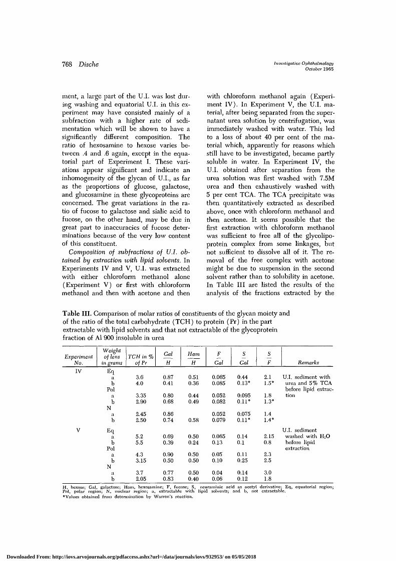
Composition of subfractions of U.I. ob-tained by extraction xoith lipid solvents. InExperiments IV and V, U.I. was extractedwith either chloroform methanol alone(Experiment V) or first with chloroformmethanol and then with acetone and then
with chloroform methanol again (Experi-ment IV). In Experiment V, the U.I. ma-terial, after being separated from the super-natant urea solution by centrifugation, wasimmediately washed with water. This ledto a loss of about 40 per cent of the ma-terial which, apparently for reasons whichstill have to be investigated, became partlysoluble in water. In Experiment IV, theU.I. obtained after separation from theurea solution was first washed with 7.5Murea and then exhaustively washed with5 per cent TCA. The TCA precipitate wasthen quantitatively extracted as describedabove, once with chloroform methanol andthen acetone. It seems possible that thefirst extraction with chloroform methanolwas sufficient to free all of the glycolipo-protein complex from some linkages, butnot sufficient to dissolve all of it. The re-moval of the free complex with acetonemight be due to suspension in the secondsolvent rather than to solubility in acetone.In Table III are listed the results of theanalysis of the fractions extracted by the
Table III. Comparison of molar ratios of constituents of the glycan moiety andof the ratio of the total carbohydrate (TCH) to protein (Pr) in the partextractable with lipid solvents and that not extractable of the glycoproteinfraction of Al 900 insoluble in urea
ExperimentNo.
Weightof lens
in grams
IV Eqab
Polab
Nab
V Eqab
Polab
Nab
TCH in %of Pr
3.64.0
3.352.90
2.452.50
5.25.5
4.33.15
3.72.05
Gal
H
0.870.41
0.800.68
0.860.74
0.690.39
0.900.50
0.770.83
Ham
H
0.510.36
0.440.49
0.58
0.500.24
0.500.50
0.500.40
F
Gal
0.0650.085
0.0520.082
0.0520.079
0.0650.13
0.050.10
0.040.06
S
Gal
0.440.13°
0.0950.11°
0.0750.11°
0.140.1
0.110.25
0.140.12
S
F
2.11.5°
1.81.3°
1.41.4°
2.150.8
2.32.5
3.01.8
Remarks
U.I. sediment withurea and 5% TCAbefore lipid extrac-tion
U.I. sedimentwashed with HjObefore lipidextraction
H, hexose; Gal, galactose; Ham, hexosamine; F, fucose; S, neuraminic acid as acetyl derivative; Eq, equatorial region;Pol, polar region; N, nuclear region; a, extractable with lipid solvents; and b, not extractable.°Values obtained from determination by Warren's reaction.
Downloaded From: http://iovs.arvojournals.org/pdfaccess.ashx?url=/data/journals/iovs/932953/ on 05/05/2018

Volume 4Number 5
Glycoproteins and glycolipoproteins of bovine lens 769
organic solvents and of the nonextractableresidues.
In Experiment V, the U.I. was obtainedin two subfractions by first centrifuging itssuspension in 7.5M urea at a speed of 3,600x g separating the coarse granular precipi-tate by centrifugation and recentrifugingthe supernatant for 30 minutes at 20,000x g to clarification. These two fractionswere extracted with chloroform methanoland were analyzed separately. To makepossible a comparison with the data fromExperiment IV, in which a somewhat dif-ferent extraction procedure was used, thedata in Experiment V in Table III wereobtained by recalculating the data fromU.I. 3,600 and U.I. 20,000 for the total U.I.fraction. Table III contains also the dataon the analysis of the glycan of the extrac-tion residue in both experiments. As canbe seen, there is essentially no significantdifference in the composition of the frac-tion extractable by organic solvents andthe nonextractable one, except for thelower ratio of galactose to total hexose inthe residue, which is particularly markedin the residue from the equatorial part. Aswill be seen below, all or one part of thisdifference is due to the presence of muchhigher amounts of glucose, particularly inthe residue from U.I. 3,600.
Comparison of the composition of twosubfractions of U.I. obtained by fraction-ated centrifugation. In Experiments IIIand V, U.I. was separated in two stages,first at 3,600 x g and then 20,000 x g for
30 minutes and obtained in form of twosediments, one of coarser, the other ofmore fluffy appearance. In Experiment V,both subfractions were, as mentionedabove, separately extracted exhaustivelywith H2O saturated Chlfrm. Meth., and thecomposition of the extracts and extractedresidue separately determined. The resultof the determinations of the constituents ofthe glycan in all these fractions are listedin Table IV, and the ratios of the hexosecomponents in the chloroform methanol ex-tracts and extraction residues illustratedby Figs. 1, 2, and 3. As mentioned in thepreceding paragraph, in Experiment V, thetwo subfractions were again subfraction-ated into a fraction soluble in chloroformmethanol and nonextractable residue, andboth these subfractions were separatelyanalyzed. As can be seen in both experi-ments, the subfraction 3,600 shows a verysignificantly lower proportion of galactosein the hexose fraction and a significantlylower hexosamine-hexose ratio than thefraction 20,000. Furthermore, it is apparentthat, in Experiment V, this difference inthe composition of the hexose componentresides with the residue which is insolublein chloroform methanol, as can be seenfrom Figs. 2 and 3. The paper chromatog-raphy of this residue in Experiment V, aswell as of the whole 3,600 subfraction inExperiment III, shows that this subfractionis characterized by a much higher contentof glucose than the whole subfraction20,000 or the chloroform methanol extract-
Table IV. Comparison of molar ratios of glycan constituents and of totalcarbohydrate (TCH) to protein (Pr) of the two urea-insolubleglycoprotein fractions sedimenting at different rates
ExperimentNo.
Illab
Vab
TCH in %ofPr
7.33.1
6.34.7
Gal
H
0.510.67
0.590.61
Ham
H
0.250.69
0.330.62
Ham
Gal
0.481.04
0.540.92
F
Gal
0.0650.11
0.070.10
S
Gal
0.080.21
0.110.18
S
F
1.21.95
1.61.85
H, hexose; Gal, galactose; Ham, hexosamine; F, fucose; S, neuraminic acid as acetyl derivative; a, sedimenting faster;and b, sedimenting slower.
Downloaded From: http://iovs.arvojournals.org/pdfaccess.ashx?url=/data/journals/iovs/932953/ on 05/05/2018

770 Dische Investigative OphthalmologyOctober 1965
Fig. 1. Chromatogram of the hexose componentsfrom the hydrolysate of the chloroform methanolextract of the 3,600 x g fraction of the albuminoidAl 900 of the equatorial region from ExperimentV. Spots from top to bottom, galactose, glucose,mannose, fucose; 1 and 3, standards; 2, sample.
able 3,600. The ratio of hexosamine togalactose, on the other hand, shows a muchsmaller difference between 3,600 and20,000. There are no significant differencesas far as other constituents are concerned.The subtraction 3,600 as a whole, and par-ticularly the chloroform methanol-insolu-ble part, is characterized by a significantlyhigher proportion of the glycan in theglycoprotein which reaches values between6 and 7 per cent.
The composition of glycans from thepostdialysis precipitate (P.D.P.). The analy-sis of the postdialysis precipitate was car-ried out in all five experiments, but the
Fig. 2. Chromatogram of the hexose componentsfrom the hydrolysate of the chloroform methanolextraction residue of the 3,600 x g and 20,000 x gfractions of the albuminoid Al 900 of the equa-torial region from Experiment V. Spots from topto bottom, galactose, glucose, mannose, fucose; 1,standard; 2, sample 3,600 x g; 3, sample 20,000x g; 4, standard.
separation of the chloroform methanol-extractable and not extractable carbohy-drate was carried out only in. ExperimentV. The results of the determination ofthese glycans are listed in Table V, andthe paper chromatograms of the hexoseportion are given in Fig. 4. It can be seenthat the glycan of this fraction is essentiallyof the same kind as that in the urea-in-soluble material, except that the ratio ofhexosamine to galactose appears to behigher, particularly in the polar and nu-clear regions.
Downloaded From: http://iovs.arvojournals.org/pdfaccess.ashx?url=/data/journals/iovs/932953/ on 05/05/2018

Volume 4Number 5
Glycoproteins and glycoUpoproteins of bovine lens 111
• i
tFig. 3. Chromatogram of the hexose componentsfrom the hyclrolysate of chloroform methanol ex-tracts from the 3,600 x g and P.D.P. fraction ofthe Al 900 albuminoid of the nuclear region fromExperiment V. Spots from top to bottom, galactose,glucose, mannose, fucose, I, standard; 2, 3,600 xg fraction; 3, P.D.P. fraction; 4, standard.
The composition of the glycan in theU.I. and P.D.P. of the Al 20,000 fraction.The glycans in the U.I. and P.D.P. of theAl 20,000 fraction were analyzed withoutfurther fraetionation in Experiments II, III,and IV. In Experiment V, U.I. was ex-tracted with chloroform methanol beforeanalysis. The results of the analysis of theglycans from Experiments II and V arelisted in Table VI. The values for U.I. inExperiment V were recalculated for thetotal fraction from the two subtractions. Alldata from Experiments III, IV, and V wereaveraged and the average data, as well asthe range of variations from one experi-ment to another, are given in the table. As
1 2Fig. 4. Chromatogram of the hexose componentsfrom the hydrolysate of the total P.D.P. fractionof the albuminoid Al 900 of the equatorial regionfrom Experiment III. Spots from top to bottom,galactose, glucose, mannose, fucose; J, standard;2, sample.
can be seen, the average values for thecomposition of this glycan do not showsignificant differences from those of thecorresponding fractions from Al 900. How-ever, the residue, not soluble in methanolchloroform, from U.I. of Experiment V,showed significantly lower values for thegalactose to hexose, and hexosamine tohexose ratios than the coiTesponding ex-tractable material. Fig. 5 shows the chro-matogram of hexoses of this nonextractablematerial which demonstrates the high con-tent of glucose in this fraction. The chro-matogram, in fact, resembles that in Fig. 3obtained from the nonextractable residueof the equatorial region of U.I. of Al 900.The presence of glucose expressed itself
Downloaded From: http://iovs.arvojournals.org/pdfaccess.ashx?url=/data/journals/iovs/932953/ on 05/05/2018

772 Dische Investigative OphthalmologyOctober 1965
Table V. Molar ratios of constituents of the glycan and the total carbohydrate(TCH) to protein (Pr) in the urea-soluble but water-insoluble glycoproteinalbuminoid complex from different regions of the lens
ExperimentNo.
IEqPolN
IIEqPolN
IIIEqPolN
IVEqPolN
Wet weighto
of lens1.15
1.15
2
2
TCH in %ofPr
2.00.61.5
1.050.480.55
0.750.20.18
0.730.140.09
Gal
H
0.820.800.69
0.850.820.80
0.770.820.81
0.750.800.80
Ham
H
0.680.690.57
0.680.640.75
0.730.940.83
0.780.480.64
F
Gal
0.760.100.05
0.090.100.05
0.0850.0650.07
0.060.070.08
S
Gal
0.170.130.16
0.180.130.14
0.06
0.060.070.09
S
F
2.61.83.2
2.21.52.8
0.941.11.15
H, hexose; Gal, galactose; Ham, hexosamine; F, fucose; S, neuraminic acid as acetyl derivative; Eq, equatorial region;Pol, polar region; and N, Nuclear region.
Table VI. Molar ratios of constituents of glycans of the U.I. and P.D.P.glycoprotein fraction and of its total carbohydrate (TCH) to protein (Pr)from Al 20,000 of different regions of the lens
Experiment No.
IIU.I. Eq
PolN
III, IV, V (combined)U.I. Eq
Pol
N
IIP.D.P. Eq
PolN
III, IV (combined)P.D.P. Eq
Pol
N
Wet weightof lens
1.15
2
1.15
2
TCH in %ofPr
3.62.851.40
2.9(2.65-3.35)
2.65(2.30-3.0)
3.25(2.40-4.05)
1.353.100.65
2.2(1.55-2.9)
1.15(0.7-1.6)
1.00(0.6-1.4)
Gal
H
0.690.700.86
0.62(0.57-0.65)
0.73(0.62-0.95)
0.81(0.71-0.93)
0.650.700.71
0.69(0.63-0.75)
0.76(0.68-0.86)
0.82(0.81-0.84)
Ham
H
0.530.670.55
0.49(0.44-0.54)
0.55(0.38-0.72)
0.62(0.60-0.65)
0.630.650.71
0.60
0.61
F
Gal
0.130.0750.055
0.10(0.09-0.12)
0.06(0.045-0.07)
0.075
0.050.0850.22
0.045(0.035-0.05)
0.04(0.02-0.06)
0.05(0.045-0.055)
S
Gal
0.230.130.105
0.195(0.14-0.27)
0.13(0.10-0.16)
0.13(0.10-0.19)
0.0750.150.105
0.155(0.13-0.18)
0.10(0.095-0.10)
0.105(0.10-0.115)
Eq, equatorial region; Pol, polar region; N, nuclear region; H, hexose; Gal, galactose; Hani, hexosamine; F, fucose;and S, neuraminic acid as acetyl derivative.
Downloaded From: http://iovs.arvojournals.org/pdfaccess.ashx?url=/data/journals/iovs/932953/ on 05/05/2018

Volume 4Number 5
Glycoproteins and glycolipoproteins of bovine lens 773
Fig. 5. Chromatogram of the hexose componentsfrom the hydrolysate of the chloroform methanolextraction of the residue of the 3,600 x g + 20,000x g fractions of the Al 20,000 of the polar regionfrom Experiment V. Spots from top to bottom,galactose, glucose, mannose, fucose; 1 and 3,standards; 2, sample.
also in the analytical determination ofhexoses of this fraction. The content incarbohydrate of U.I. from AI 20,000 alsodoes not differ significantly from thatfound in the corresponding fractions of Al900. The lower sedimentation rate of AI20,000, therefore, may be due to a highercontent in lipid.
Discussion
Our experiments show that the albumi-noid of the bovine lens consists of twofractions, one which represents about 7per cent of the total albuminoid protein inlenses of older animals and more than 10
per cent in younger lenses, is insoluble in7.5M urea. This fraction is found in thealbuminoid which sediments at 900 x g, aswell as in the albuminoid, which can bespun down only at 20,000 x g. The Al 900of the equatorial region can be split byfractionated centrifugation into a smaller,heavier fraction with about 4 to 5 per centcarbohydrate and a lighter one at about3 per cent carbohydrate. In the polar andthe nuclear regions, the heavier fractionsdo not significantly differ from the lightone. The albuminoid which goes into solu-tion at 7.5M urea also contains a protein -linked carbohydrate which, in most cases,comes out of solution when the urea isdialyzed out. This precipitated materialcontains much less carbohydrate, maxi-mally 1 per cent, and the content ofthe precipitating carbohydrate in percent of protein varies strongly from oneexperiment to another, but is significant-ly higher in lenses from younger ani-mals than those from older ones. Whenthe carbohydrate-containing albuminoidfractions are extracted with chloroformmethanol 2:1 saturated with water, about60 to 80 per cent of the carbohydrate-pro-tein complex is extracted. This carbohy-drate has the composition of a typicalsialofucohexosaminoglycan, and it is re-markable that the composition of the gly-can which is extracted by chloroformmethanol does not differ significantly in itscompositional pattern from that which isnot extractable, as far as the ratio of neu-raminic acid to galactose or hexosamine isconcerned. This ratio varies in differentfractions between .1 and .2. In eveiy case,therefore, it is completely different fromthe ratio which was found in glycolipidsfrom brain, red cell stromas, and kidney,where it is approximately 1:1. It is at pres-ent not possible to decide whether thecarbohydrate in the chlorofomi methanolextract is a real glycolipid linked to proteinby either covalent or weaker bonds orwhether it is a typical glycoprotein towhich attached lipidic groups impart thesolubility in chloroform methanol. Such
Downloaded From: http://iovs.arvojournals.org/pdfaccess.ashx?url=/data/journals/iovs/932953/ on 05/05/2018

774 Dische Investigative OphthalmologyOctober 1965
lipidic groups could be linked to the pro-tein either by hydrogen or hydrophobicbonds in the same way as are typical lipo-proteins of the serum. In this case, thewhole complex could be, therefore, desig-nated as a glycolipoprotein. The contentof lipids in this fraction from Al 900 can-not exceed more than 10 to 15 per centof the protein, as has been shown in Ex-periment V, by first weighing the dried ex-tracts from chloroform methanol from Al900 of all three regions of the lens andthen determining the nitrogen in these ex-tracts. In the rapidly sedimenting insolubleglycoprotein fraction from the equatorialpart, the material which is not extracted bychloroform methanol has a different com-position from the nonextractable materialfrom all the other glycoprotein fractionsin the lens insofar as it contains muchgreater amounts of glucose than was foundthroughout in the chloroform methanol-extractable fractions and in the nonextract-able residues from other glycoprotein frac-tions. In this subfraction, the ratio of hex-osamine to hexose is about one half of thatin all other fractions. This suggests thepossibility that the glucose may not bepart of the hexosaminoglycan, but mayform a separate polymer which may or maynot be fixed to the same protein as thehexosaminoglycan. The situation herecould be analogous to that found by Spiroand Spiro25 in the glycoprotein of thyro-globulin in which a chain consisting of 5mannose and 1 hexosamine apparently islinked to the protein which, in addition,carries a typical hexosaminoglycan con-taining galactose, mannose, and glucosa-mine in proportions remaining within therange found in most hexosaminoglycansof animal tissues. It must be noted that,in Experiments I and V, also a smallerfraction derived from the polar part ofthe lens, free of nuclei, which sedimentsonly at 20,000 x g, showed a much highercontent of glucose than all the other glyco-protein fractions, and this higher glucoseappears here also in the carbohydratewhich is not extractable by chloroform
methanol (Fig. 5). This fraction, in thisexperiment, is also characterized by themuch lower ratio of hexosamine to hexoseand appears, therefore, in every respect, tobe similar to the chloroform methanol-non-extractable, slow-sedimenting material fromthe equatorial region of Al 900. The failurein other experiments to obtain evidencefrom analytical data for the presence of apolymer rich in glucose and low in hexosa-mine in the polar region may be due to thefact that the separation of the two carbohy-drates by fractionated centrifugation is notcompletely reliable when the proportion ofone of the two fractions drops below a cer-tain level. It may well be, therefore, thatthe glucose-rich component is present insmaller relative concentrations also in thenuclear parts. The ubiquitous presence ofthis carbohydrate and its inadequate sepa-ration could explain the variations of theratios of galactose to hexose and of hex-osamine to galactose, observed in differentexperiments. The only moi-phological char-acteristic in common between the equa-torial region and the polar region appearsto be the presence of marked interdigita-tions of the surfaces between the adjacentlens fibers which are found on the one sidein the subcapsular region of the equatorialcortex and on the other side in the suturespresent in the polar region.20 The highercontent of the glucose polymer could be,therefore, characteristic for cell membranesor some intercellular material in these tworegions.
The presence of sialofucohexosaminogly-cans in the urea-insoluble part of the al-buminoid and its solubility in chloroformmethanol, which indicates the presence ofa lipidic component, shows a striking anal-ogy to the compositional pattern of thewater-insoluble part of the stromas fromred cells. This water-insoluble materialfrom the stromas was shown19'20 to containhexosaminoglycans which can be extractedwith chloroform methanol, but also a typi-cal glycoprotein with the glycan similar incomposition to a fraction extractable inchloroform methanol.21 The electron mi-
Downloaded From: http://iovs.arvojournals.org/pdfaccess.ashx?url=/data/journals/iovs/932953/ on 05/05/2018

Volume 4Number 5
Glycoproteins and glycolipoproteins of bovine lens 775
croscopy, furthermore, of cell surface mem-branes has, in recent years, demonstratedthat these cell membranes consist of alayer of lipids about 35 A wide covered onboth sides by two protein layers, each ofwhich has a diameter of about 20 A. Theseso-called unit membranes appear also tobe the main constituent of the endoplasmicreticulum and probably also of the nuclearmembrane. All these facts suggest that theurea-insoluble part of the albuminoid frac-tion might be nothing else but the plasmamembranes of the lenticular fibers. Thefact that the carbohydrate found in theurea-insoluble material is also present inthe postdialysis precipitate but forms herea much smaller part of the total complexthan in the urea-insoluble fraction suggeststhe possibility that the albuminoid proteinwithin the lens fibers is, in fact, partly ortotally arranged along the cytoplasmic sur-faces of the plasma membrane and keptin this position by weak bonds with theplasma membrane which are immediatelydisrupted by the mechanical forces usedduring homogenization. Depending on theimpact of these mechanical forces, whichcan vaiy from one experiment to another,greater or smaller proportions of theseplasma-membrane albuminoid complexeswould be disrupted so that the ratio of theurea-insoluble fraction and the postdialysisprecipitate will vary from one experimentto another, as indeed was the case in ourexperiments. The complex between theplasma membrane and albuminoid will dis-solve in 7.5M urea in which the albumi-noid protein itself dissolves and will comeout of solution after removal of the ureabecause of the insolubility of the plasmamembrane part of the complex, whereasthe albuminoid protein itself, once dis-aggragated by the urea remains in solutionand coprecipitates only with the plasmamembranes. This could explain why theamount of protein in the postdialysis pre-cipitate varied so much from one experi-ment to another.
To test this hypothesis, we can calculatethe amount of protein which we can expect
to be present in the two leaflets of theplasma membranes of the lens fibers andcompare it with the total amount of the pro-tein which is linked to the carbohydratepresent in the glycoprotein fraction of thelens. For this calculation, we assume thatonly the urea-insoluble material consists ofthe plasma membrane itself, whereas thecarbohydrate in the postdialysis precipitateis in a complex between these plasma mem-branes and the albuminoid protein itself.To calculate, therefore, the total amount ofthe protein present in the plasma membraneof lens fibers, we assume that all of thecarbohydrate in the postdialysis precipitatecomes from plasma membrane fragments,and we calculate the protein in the mem-brane fragments of the postdialysis pre-cipitate from the content of this precipitatein carbohydrate and the ratio of carbo-hydrate to protein found in the urea-in-soluble fraction which, we assume, con-sists of pure plasma membranes. Thiscalculation, of course, can only yield theorder of magnitude of the amounts of pro-tein present in the plasma membranes ofthe lens fibers. If the glycoproteins in theurea-insoluble material represent frag-ments of the plasma membrane, then theadhesion of albuminoid to these fragmentsin the postdialysis precipitate suggests thatthe albuminoid itself, at least in not-too-old animals, may be the equivalent of theso-called cortical region of the cytoplasmas is observed, for instance, in the fertilizedand unfertilized eggs of various animals.
The calculation of the order of magni-tude of the protein present in plasma mem-branes of lens fibers can be earned outwith any degree of accuracy only for thecortical layers of the lens. In our experi-ments, the combined equatorial and polarparts, approximately, represent about twothirds of the total weight of the lens. Ifwe assume that the lens is a sphere, a cor-tical shell corresponding to two thirds ofthe weight of a 2 gram lens would havea diameter of 2.5 mm. with an outer cir-cumference of 50 and an inner circumfer-ence of about 35 mm. If we assume that
Downloaded From: http://iovs.arvojournals.org/pdfaccess.ashx?url=/data/journals/iovs/932953/ on 05/05/2018
776 Dische Investigative OphthalmologyOctober 1965
lens fibers extend from the equator of thelens to the poles, they would have then alength between 9 and 12.5 mm. with anaverage of about 11 mm. To calculate thecross section and the circumference of thelens fibers, we can assume as diameters ofthe lens fibers those found in availableelectron micrographs20 to be diameters ofthe lens fibers. These show a cross sectionof about 20 [x2 and a circumference ofabout 25 fx. If, on the other hand, we as-sume that the diameter of the lens fibersis about the same as the diameter of thelens epithelia of the equatorial region, wecan assume that they represent cylindersof about 10 jx in diameter. The volume ofa lens fiber would, on this basis, be be-tween 2.75 and 8.8 105 ^3, and this wouldgive in a 1 gram cortex 1.15 to 3.6 106 fi-bers. The surface of the plasma membraneof a fiber would be 2.2 to 3.3 105 fx2, andthe volume of the two protein layers of theplasma membrane, each with a diameterof 2 mix, would be then 8.8 to 13.2 102 /*3
or 8.8 to 13.2 10"7 mm.3 Assuming that theprotein layer consists of proteins with apartial specific gravity of 1.25, this wouldrepresent a weight of the protein layers ofindividual lens fibers of approximately 10.4to 16.6 10"7 mg. The lower value corre-sponding to a number of 3.6 10° of lensfibers would then give the weight of theplasma membrane protein of 3.7 mg. in 1gram cortex; the higher, 1.9 mg. If theweight of the protein present in the glyco-protein of the lens is calculated under theassumption that the glycan in the post-dialysis precipitate is linked in the sameproportion to protein as in the urea-insolu-ble glycoprotein fraction, from the data inTable I it can be seen that for 2 gramlenses this amount varies between 2.5 and2.75 mg. per gram cortex. That corre-sponds about to the average value resultingfrom our calculation of the approximateamount of protein which has to be presentin the plasma membrane of the lens fibersin the cortex represented by the unit mem-branes shown by election microscopy. Thisagreement supports our assumption that
the glycolipoprotein insoluble in urea rep-resents fragments of the plasma membraneof the lens fibers, and the glycoprotein inthe postdialysis precipitate is a complex be-tween this glycolipoprotein and albumi-noid, or at least is the result of the aggre-gation of the albuminoid with the plasmamembrane fibers which takes place afterthe removal of the urea by dialysis. Furtherelectron microscopic study of the urea-in-soluble glycoprotein fraction will be car-ried out to test this hypothesis. If the totalamount of protein in the glycolipoproteinfraction is calculated in the same way forlenses from younger animals with a wetweight between 1.10 and 1.20 grams in-stead of 2 grams, the value for ExperimentII of Table I is 3.4 grams, significantlyhigher than those in the older lenses. Thisalso appears to be in agreement with theassumption that they represent plasmamembrane fragments, as in smaller lensesthe greater weight of the plasma mem-branes per gram lens, because of thegreater ratio of surface to volume, shouldbe expected.
This hypothesis is, furthermore, sup-ported by a comparison between the lensand the washed human red cells as far asthe chemical composition and the quanti-tative relations of the membrane materialto the wet weight of the whole cells areconcerned. Howe and associates19 isolatedfrom the stromas of human red cells, ob-tained by hemolysis at pH 5.5, a materialwhich, after being lyophilized and repeat-edly extracted with water, yielded a water-insoluble residue which represented about1 per cent of the wet weight of the washed,packed red cells. If we consider that thismaterial contained the lipid in addition toprotein and carbohydrate and that themass of the plasma membrane relative tothe whole cytoplasm must be greater inthe much smaller red cells than in the lensfibers, this value appears to be in excellentagreement with our calculated value forthe amount of membrane protein per gramlens cortex. From this stroma material,Howe and associates extracted, by a com-
Downloaded From: http://iovs.arvojournals.org/pdfaccess.ashx?url=/data/journals/iovs/932953/ on 05/05/2018

Volume 4Number 5
Glijcoproteins and glycolipoproteins of bovine lens 777
bination of an extraction with 50 per centphenol at 68° C, lyophilization of the waterphase and extraction with chloroformmethanol 2:1, a fraction which consistedof protein, lipid, and carbohydrate, andcontained acetylneuraminic acid, galactose,and hexosamine roughly in the proportionof 1:1:1. This material, therefore, containsthe same constituents as the material ex-tracted from the urea-insoluble fraction ofthe albuminoid. If we consider, further-more, that Eylar and MadofP1 succeededin isolating from stromas of human redcells a typical glycopeptide which did notcontain any lipid, but a glycan of approxi-mately the same composition as that of theglycolipid of the stroma, then the strikinganalogy in the compositional patterns ofthe carbohydrates from stromas and lensfibers becomes apparent. It must, of course,be noted that the composition of the gly-can in the glycolipid and glycopeptide ofthe stromas differs from that in the albumi-noid in that the ratio of sialic acid to galac-tose and hexosamine is very much higherand corresponds to that found in the gan-gliosides of the brain. This difference inthe composition of the glycan, however,appears biologically understandable as thered cells are not supposed to adhere toeach other, but rather to repel each other,which apparently requires a large concen-tration of negative charges on their sur-face. In fact, the removal of neuraminicacid by neuraminidase from the surface ofthe red cells leads immediately to the ag-gregation of these elements and to severeinterference with their normal physiologi-cal function. The compositional pattern ofthe glycan of the surface membrane ap-pears, therefore, to be directly related tothe contact relations of the respective cells.
Summary
Incubation of the albuminoid of bovinelenses with 7.5M urea at R.T. yields a frac-tion insoluble in 7.5M urea which repre-sents 7 to 10 per cent of the total albumin-oid. This fraction, can be dissolved by ex-traction with chloroform methanol into a
fraction soluble in this solvent and an in-soluble residue. Both these fractions do notdiffer essentially in the composition and inthe ratio of this carbohydrate to protein.They are characterized, furthermore by aveiy low ratio of sialic acid and galactosewhich differentiates them clearly from gan-gliosides as found in brain red cells and inkidney. The carbohydrate content and pro-portion of carbohydrate to protein is higherin the equatorial region than in the polarand nuclear regions. In the equatorial re-gion, in addition, one part of the urea-in-soluble fraction sedimenting in the centri-fuge at lower speed significantly differs inthe composition of its carbohydrate fromthe fraction sedimenting only at higherspeeds, insofar as the hexose componentin the last fraction contains a much higherproportion of glucose which does not ap-pear to be linked to hexosamine and is notextracted by chloroform methanol. Thehypothesis is set forth that this glycopro-tein and glycolipoprotein fraction insolublein urea represents isolated fragments of theplasma membrane of the lens fibers.
REFERENCES1. Dische, Z., Danilchenko, A., and Zelmenis,
G. Heteropolysaccharides of connective tissue,Ciba Foundation Symposium on the Chem-istry and Biology of the Mucopolysaccharides,1958, p. 116.
2. Dische, Z., and Robert, L.: The collagenlinked carbohydrate of the corneal stroma,Fed. Proc. 21: 172, 1962.
3. Dische, Z.: The glycans of the mammalianlens capsule—A model of basement mem-branes, in Small Blood Vessel Involvementin Diabetes Mellitus, The American Instituteof Biological Sciences, 1964, p. 201.
4. Holtfreter, J.: Significance of the cell mem-brane in embryonic processes, Ann. New YorkAcad. Sc. 49: 709, 1948.
5. Moscona, A.: The development in vitro ofchimeric aggregates of dissociated embryonicchick and mouse cells, Proc. Nat. Acad. Sc.43: 184, 1957.
6. Davson, H., and Danielli, J. F.: The permea-bility of natural membranes, ed. 2, Londonand New York, 1952, Cambridge UniversityPress, pp. 57-71.
7. Klenk, E., and Lempfrid, II.: The natureof the cell receptor for influenza virus.Ztschr. physiol. Chem. 307: 278, 1957.
Downloaded From: http://iovs.arvojournals.org/pdfaccess.ashx?url=/data/journals/iovs/932953/ on 05/05/2018

778 Dische Investigative OphthalmologyOctober 1965
8. Bogoch, S., and Quamina, A.: Abstracts ofthe thirteenth Annual Colloquium on Protidesof the Biological Fluids, Brugge, Belgium,April-May, 1965.
9. Hakamori, S., and Jeanloz, R. W.: Thechemical structure of glycolipid of tumortissue, Fed. Proc. 24: 231, 1965.
10. Dische, Z., Pallavicini, C , Kawasaki, H.,Smirnow, N., Cizek, L. J., and Chien, S.:Influence of the nature of the secretorystimulus on the composition of the carbo-hydrate moiety of glycoproteins of the sub-maxillary saliva, Arch. Biochem. & Biophys.97: 459, 1962.
11. Roseman, S.: Metabolism of the sialic acids,Sixth Internat. Congress of Biochemistry. VI.Carbohydrates, New York, 1964, Abst.
12. Bartholomew, B., Jourdian, G. W., and Rose-man, S.: Soluble sialyl-transferases from col-ostrum, Biochem. & Biophys. Res. Commun.10: 352, 1963.
13. Coulombre, J. L., and Coulombre, A. J.: Lensdevelopment: Fiber elongation and lens ori-entation, Science 142: 1489, 1963.
14. Dische, Z., Smirnow, N., and Zelmenis, G.:Glycoproteins of the lens in relation to ageand cataract formation. I. Glycoproteins ofthe bovine lens fibers, INVEST. OPHTH. 1: 646,1962.
15. Dische, Z., and Zelmenis, G.: Glycoproteinsof the lens in relation to age and cataractformation. II. Glycoproteins of lens fibers ofrabbit, INVEST. OPHTH. 2: 90, 1963.
16. Dische, Z., Zelmenis, G., and Larys, N.:Glycoproteins of the lens in relation to ageand cataract formation. III. Differences in
composition and distribution of the carbo-hydrate of glycoprotein of lens fibers indifferent regions of the lens, INVEST. OPHTH.2: 630, 1963.
17. Morner, K.: The proteins of the refractingmedia of the eye, Ztschr. physiol. Chem. 18:60, 1894.
18. Permutt, S., and Johnson, F. B.: Histochemicalstudies on the lens following radiation injury,Arch. Path. 55: 20, 1953.
19. Howe, C, Avrameas, S., de Vaux St. Cyr. C ,Grabar, P., and Lee, L. T.: Antigenic com-ponents of human erythrocytes, J. Immunol.91: 683, 1963.
20. Danon, D., Howe, C , and Lee, L. T.: Inter-action of polylysine with soluble componentsof human erythrocyte membranes. In press.
21. Eylar, E. H., and Madoff, M. A.: Isolationof a glycopeptide from the red cell mem-brane, Fed. Proc. 21: 402, 1962.
22. Thomann, H.: Amperometric determinationof sylfhydryl groups of animal lenses,Graefes Arch. Ophth. 160: 219, 1958.
23. Svennerholm, L.: Quantitative estimation ofsialic acids. II. A colorimetric resorcinol-hydrochloric acid method, Biochim. etbiophys. acta 24: 604, 1957.
24. Aminoff, D.: Methods for the quantitativeestimation of N-acetylneuraminic acid andtheir application to hydrolysates of sialo rau-coids, J. Biochem. 81: 384, 1961.
25. Spiro, R. G., and Spiro, M.: The carbohydrateof thyroglobulin, Fed. Proc. 22: 538, 1963.
26. Wanko, T., and Gavin, M. A.: Cell surfaces inthe crystalline lens, in Smelser, G. K., editor:The structure of the eye, New York, 1961,Academic Press, Inc.
Downloaded From: http://iovs.arvojournals.org/pdfaccess.ashx?url=/data/journals/iovs/932953/ on 05/05/2018