Mechanical Properties of Permanent Foaming Fixatives for ...

The fine structure produced in cells by primary fixatives2. Potassium dichromate
By JOHN R. BAKER
(From the Cytological Laboratory, Department of Zoology,University Museum, Oxford)
With 2 plates (figs, i and 2)
SummaryThe exocrine cells of the mouse pancreas were fixed in potassium dichromate solution,embedded in araldite or other suitable medium, and examined by electron microscopy.
Almost every part of these cells is seriously distorted or destroyed by this fixative.The ergastoplasm is generally unrecognizable, the mitochondria and zymogen granulesare seldom visible, and no sign of the plasma membrane, microvilli, or Golgi apparatusis seen. The contents of the nucleus are profoundly rearranged. It is seen to containa large, dark, irregularly shaped, finely granular object; the evidence suggests thatthis consists of coagulated histone. The sole constituent of the cell that is well fixedis the inner nuclear membrane.
The destructive properties of potassium dichromate are much mitigated when it ismixed in suitable proportions with osmium tetroxide or formaldehyde.
IntroductionPOTASSIUM dichromate was widely used as a fixative in the second half ofthe nineteenth century, chiefly in the form of 'Miiller's fluid'. Miiller him-self (18606) simply stated that his fluid consisted of an aqueous solution ofpotassium dichromate and sodium sulphate, without giving particulars as tothe concentrations of these salts. Subsequent authors used the dichromateat 2 to 2^%, and the sodium sulphate (probably Glauber's salt) at 1% (Frey,1871; Exner, 1878; Mojsvar, 1879). In the present paper Miiller's fluid mustbe taken to mean a 2^% aqueous solution of potassium dichromate with theaddition of 1 % of Glauber's salt. The latter is an 'indifferent' salt, not capableof exerting any fixative effect on tissues at the concentration at which it isused (Fischer, 1899), but possibly affecting fixation in the same way as sodiumchloride affects fixation by formaldehyde.
It is important to recognize that acidified potassium dichromate reactswith tissues in a very different way from the unacidified salt. The subjecthas attracted the attention of several authors, and has been comprehensivelyreviewed and investigated by Casselman (1955). It must suffice here tomention that when the pH falls below about 3-6, potassium dichromate pro-duces the same effects as a solution of chromium trioxide. Miiller himself(1860a) sometimes used a solution of potassium dichromate, sodium sulphate,and chromium trioxide, but this is not the fluid ordinarily known by Miiller's[Quart. J. micr. Sci., Vol. 106, pt. 1, pp. 15-21, 1965.]

16 Baker—Fine structure produced in cells by potassium dichrornate
name. The fine structure produced by chromium trioxide (and acidifiedpotassium dichromate) is under investigation by myself.
From the last decade of the nineteenth century onwards potassiumdichromate has been used chiefly in mixtures with other fixatives, or forPostchromirung (Benda, 1901) after preliminary treatment with other fixatives.In many of the mixtures (e.g. Zenker, 1894), acidification has made the saltact like chromium trioxide; but in others (e.g. Altmann, 1894; Orth, 1896;Helly, 1903; Regaud, 1910; Dalton, 1955) this is not so.
The present paper is concerned primarily with solutions of unacidifiedpotassium dichromate used alone or with the addition of sodium sulphate,but mention is also made of the effects of certain fixative mixtures.
Material and methodsThe test-object has been the same as that used before, in the investigation
of the fine structure produced by mercuric chloride (Baker, 1963); namely,the exocrine pancreatic cell of the house-mouse, Mus musculus. The reasonsfor the choice of this cell were given in the earlier paper. The pancreas wascut into small pieces with scissors or a razor blade.
Fixation. The fixatives used were i | and 2^% aqueous solutions ofpotassium dichromate, and Miiller's fluid. All three were used at roomtemperature. The period of fixation varied from 24 h to 20 days. Often thepieces of tissue were fixed for 24 h and then transferred to a 5% or a saturatedsolution of potassium dichromate maintained at 37° C, and left in this for 24 h.
Pieces of tissue were also fixed in a 1% aqueous solution of osmiumtetroxide at about 40 C, for comparison with those fixed in potassiumdichromate solution. It was found that cells of the chosen tissue fixed in thissimple solution appeared similar in fine structure to those that had been fixedin the buffered osmium tetroxide solution of Palade (1952). This confirmsthe results of Malhotra (1962).
Experiments on fixation with mixtures containing potassium dichromate,and on the postchroming of tissues that had been subjected to the action offixatives other than potassium dichromate, are mentioned on p. 20.
Washing out the fixative. In accordance with the usual practice in lightmicroscopy, pieces fixed with potassium dichromate or with mixtures con-taining this salt were washed (usually for about 5 h) in running water orrepeated changes of tap-water, and then rinsed with distilled water.
Embedding. After dehydration with ascending grades of ethanol, the pieceswere embedded in araldite, butyl methacrylate, or plexigum (usually the first-named).
Some of the pieces were passed through toluene into paraffin, in accordancewith the routine procedure of light microscopy. The cooled paraffin wasdissolved away with warm toluene, and this was replaced with absoluteethanol; the pieces were then embedded in araldite. The purpose was to findout whether embedding in paraffin causes any changes in the structure ofthe cell.

Baker—Fine structure produced in cells by potassium dicht-ornate 17
Sectioning. A Huxley ultramicrotome was used to produce sections showingsilver interference colours. The sections were mounted on carbon film overformvar.
Staining. Some of the sections were 'stained' with a 2% solution of uranylacetate in methanol, but unstained sections were also examined to make surethat the stain had not affected the structure.
Microscopy. Sections were examined with an Akashi TRS 50 electronmicroscope.
A small investigation was made by light microscopy, to clarify a problemthat had arisen in the course of the study by electron microscopy. (See p. 18.)
ResultsSince there is no method by which the fine structure of the living cell can
be established with certainty, the standard of comparison will be the structurerevealed after fixation by osmium tetroxide. The chief components of theexocrine pancreatic cell of the mouse, fixed by a 1% solution of osmiumtetroxide in distilled water, are shown in fig. r, A.
It makes little difference whether 1^ or 2^% potassium dichromate orMiiller's fluid is used as fixative, and essentially the same picture is producedwhether fixation is short or long. The use of a more concentrated fixativeat 370 C after initial fixation at room temperature does not produce anystriking modification in the image. A single description of the fine structureproduced by potassium dichromate will therefore suffice, though some smallexceptions to this will be mentioned.
Fig. 1, B has been chosen to represent the structure produced whenpotassium dichromate is used as fixative. The disruption of the cytoplasm isalmost complete. Nothing remains but a coagulum, with interspersed areasof various sizes, which appear to be empty spaces (s). The coagulum usuallyconsists of granules, mostly about 20 or 30 m/j, in diameter (figs. 1, B; 2, B),but their size cannot be measured accurately since they are not sharplydelimited, and they often merge with one another (fig. 2, D). There are alsolines in the micrographs, which must be taken to represent sections of mem-branes (fig. 1, B, m). The membranous element tends to be somewhat moreconspicuous when Mviller's fluid is used (fig. 2, c) instead of a simple solutionof potassium dichromate, and it sometimes happens that the cisternae of theergastoplasm are more or less intact in places, though nothing that can beinterpreted as a ribosome is seen.
As a general rule mitochondria are not preserved. Occasionally theysurvive and appear in micrographs as grey, sausage-shaped objects with veryindistinct traces of cristae. In the whole investigation no trace of the Golgiapparatus was ever seen in any preparation fixed with potassium dichromate.The zymogen granules have usually been dissolved away, and not even theirformer sites can be detected; but small remnants are sometimes seen in ovalspaces, or spaces that have probably contained them remain.

18 Baker—Fine structure produced in cells by potassium dichrornate
The plasma membrane does not survive, and the limits of the cells cannotbe determined. Microvilli are not seen.
The nucleus is considerably larger than in osmium preparations. It islimited by a very distinct single membrane, marked by arrows in figs, i, B and2, B to D. A double membrane, such as is seen in osmium preparations(fig. 2, A, E), never survives intact, but traces of a broken outer membranesometimes occur here and there. Since the outer membrane is a part of theergastoplasm, the disruption of the latter by potassium dichromate accountsfor its complete or almost complete absence. The striking difference betweenthe responses of the inner and outer nuclear membranes to the action ofpotassium dichromate is the most remarkable single fact that has emergedfrom this investigation.
The contents of the nucleus differ radically from those seen in osmiumpreparations. Very irregularly shaped dark masses (k in figs, i, B; 2, B, c) lieon a pale background. The dark masses consist of a close aggregation ofgranules, each roughly 20 m//. in diameter. The pale background often butnot always contains dark, sharply delimited granules, most of them ratherless than 40 m/x in diameter; lines radiate from these, probably representingsections of membranes. These granules are particularly clearly seen infig. 2, D at x. Their nature is obscure.
In an attempt to interpret the contents of the nucleus as seen in electronmicrographs, pieces of pancreas were fixed for 24 h in Miiller's fluid, embeddedin paraffin, sectioned, and treated with Feulgen's reagent and light green.One or more irregularly shaped masses were seen in each nucleus, stainedwith light green; they appeared not to extend to the periphery of the nucleus.The whole of the rest of the nucleus, right up to the nuclear membrane, wasfilled with a Feulgen-positive substance. It seems almost certain that thesubstance having an affinity for the green dye is the same as the dark, finelygranular substance seen in electron micrographs.
The results so far mentioned suggested the possibility that potassiumdichromate might not be a fixative at all: for such fixation as occurred mighthave been due to the ethanol used for dehydration. To test this possibility aversion of Miiller's fluid was prepared, in which the potassium dichromatewas replaced by an equimolar amount of a substance known to have no fixativeeffect, namely potassium chloride. Pieces of pancreas were left in this fluidfor 24 h and washed as usual. Sections were prepared as before for study bylight microscopy. They were treated with Feulgen's reagent. No signs ofnuclei were seen anywhere: the tissue was unrecognizable as cellular material
FIG. 1 (plate). Electron micrographs of parts of pancreatic exocrine cells of the mouse.A, fixed with osmium tetroxide (stained in bulk with uranyl nitrate and on the grid with
uranyl acetate; araldite). X 24,000.B, fixed with potassium dichromate (araldite; stained with uranyl acetate). X 24,000. The
arrow points to the inner nuclear membrane.er, ergastoplasm; g, Golgi apparatus; h, substance regarded as coagulated histone; m, mito-
chondrion; me, membrane; n, nucleus; s, space; x, unidentified granule; s, zymogen granule.

FIG. I
J. R. BAKKR

FIG. 2
J. R. BAKER

Baker—Fine structure produced in cells by potassium dichtornate 19
of any kind. Nothing was present except an extremely coarse reticulum,positive to Feulgen.
Since nucleoprotein is not coagulated by potassium dichromate, and DNAis readily dissolved by solutions of this salt (Fischer, 1899; Berg, 1905;Baker, 1958), the facts can only be understood on the assumption thatpotassium dichromate is primarily a fixative for the inner nuclear membrane.The Feulgen-positive material is retained within the nucleus, dispersedthroughout all parts of it except those occupied by the substance having anaffinity for light green, simply because the preservation of the inner nuclearmembrane prevents its escape.
It is known that potassium dichromate is a strong coagulant of histone(Pischinger, 1937). The evidence therefore suggests that the substance in thenucleus that has an affinity for light green and appears in electron micrographsas the dark, finely granular material, is coagulated histone.
Nucleoli cannot be recognized with certainty in electron micrographs ofpancreatic exocrine cells fixed with potassium dichromate. The three charac-teristic components of the nucleolus (Hay and Revel, 1962) are seen in osmiumpreparations, namely the dark nucleolonema, granules having the charactersof ribosomes, and pale intervening spaces (fig. 2, F) ; but nothing of this sortis seen after fixation with potassium dichromate. Roughly circular objectsof about the right size are often seen, but their fine structure suggests that theyare merely parts of the dark, finely granular substance.
Embedding in paraffin before araldite does not result in any characteristicchange in the fine structure of cells fixed by potassium dichromate. It followsthat cells fixed by this substance and studied by the routine processes of lightmicroscopy have the same fine structure as that revealed in sections that havebeen embedded in araldite, with or without previous embedding in paraffin.
The question next arose whether potassium dichromate is itself destructive,or whether the damage is caused by washing, dehydration, or embeddingafter the fixative has acted. To answer this, pieces of pancreas were trans-ferred to osmium tetroxide solution after fixation by potassium dichromateor Miiller's fluid, in order to stabilize the structure when the action of thedichromate was complete. Electron micrographs showed the usual appearance
FIG. 2 (plate). Electron micrographs of parts of pancreatic exocrine cells of the mouse.A, fixed with osmium tetroxide (stained with uranyl nitrate and acetate; araldite). X 24,000.B, fixed with potassium dichromate (araldite; stained with uranyl acetate). X 24,000.C, D, fixed with Muller's fluid (plexigum). X 18,000.E, fixed with osmium tetroxide (stained with uranyl nitrate and acetate; araldite). X 24,000.F, ditto. X 36,000.G, fixed with osmium tetroxide and transferred directly to potassium dichromate (araldite;
stained with uranyl acetate), x 24,000.The long arrows point to the inner nuclear membrane. The short arrows in F point to
filaments in the nucleus, arranged in pairs.er, ergastoplasm; h, substance identified as coagulated histone; n, nucleus; nl, nucleolus;
x, unidentified granule; z, zymogen granule.
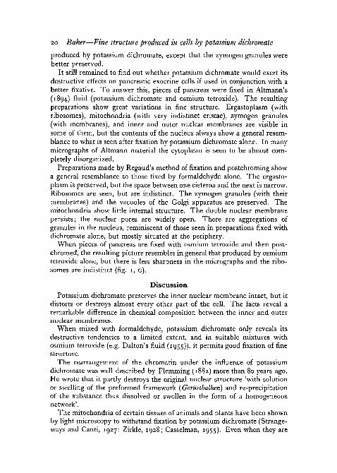
20 Baker—Fine structure produced in cells by potassium dichromate
produced by potassium dichromate, except that the zymogen granules werebetter preserved.
It still remained to find out whether potassium dichromate would exert itsdestructive effects on pancreatic exocrine cells if used in conjunction with abetter fixative. To answer this, pieces of pancreas were fixed in Altmann's(1894) fluid (potassium dichromate and osmium tetroxide). The resultingpreparations show great variations in fine structure. Ergastoplasm (withribosomes), mitochondria (with very indistinct cristae), zymogen granules(with membranes), and inner and outer nuclear membranes are visible insome of them, but the contents of the nucleus always show a general resem-blance to what is seen after fixation by potassium dichromate alone. In manymicrographs of Altmann material the cytoplasm is seen to be almost com-pletely disorganized.
Preparations made by Regaud's method of fixation and postchroming showa general resemblance to those fixed by formaldehyde alone. The ergasto-plasm is preserved, but the space between one cisterna and the next is narrow.Ribosomes are seen, but are indistinct. The zymogen granules (with theirmembranes) and the vacuoles of the Golgi apparatus are preserved. Themitochondria show little internal structure. The double nuclear membranepersists; the nuclear pores are widely open. There are aggregations ofgranules in the nucleus, reminiscent of those seen in preparations fixed withdichromate alone, but mostly situated at the periphery.
When pieces of pancreas are fixed with osmium tetroxide and then post-chromed, the resulting picture resembles in general that produced by osmiumtetroxide alone, but there is less sharpness in the micrographs and the ribo-somes are indistinct (fig. 1, G).
DiscussionPotassium dichromate preserves the inner nuclear membrane intact, but it
distorts or destroys almost every other part of the cell. The facts reveal aremarkable difference in chemical composition between the inner and outernuclear membranes.
When mixed with formaldehyde, potassium dichromate only reveals itsdestructive tendencies to a limited extent, and in suitable mixtures withosmium tetroxide (e.g. Dalton's fluid (1955)), it permits good fixation of finestructure.
The rearrangement of the chromatin under the influence of potassiumdichromate was well described by Flemming (1882) more than 80 years ago.He wrote that it partly destroys the original nuclear structure 'with solutionor swelling of the preformed framework (Geriistbalken) and re-precipitationof the substance thus dissolved or swollen in the form of a homogeneousnetwork'.
The mitochondria of certain tissues of animals and plants have been shownby light microscopy to withstand fixation by potassium dichromate (Strange-ways and Canti, 1927; Zirkle, 1928; Casselman, 1955). Even when they are

Baker—Fine structure produced in cells by potassium dichromate 21
not destroyed, however, there is a tendency for rod-shaped mitochondria tobecome ovoid or spherical.
It was already known from studies by light microscopy that the zymogengranules of the pancreas are not fixed by potassium dichromate alone, but arefixed by mixtures of this substance with formaldehyde (Levene and Feng,1962).
The fact that potassium dichromate has scarcely any fixative effect on thecells chosen for this investigation does not mean that it is necessarily a uselessingredient in fixative mixtures for light microscopy. Mixtures of potassiumdichromate with osmium tetroxide give preparations that are much morereadily stainable by anionic dyes than those fixed by osmium tetroxide alone.
It is a pleasure to acknowledge the skilful practical assistance of MissE. G. M. Collins and Mr. J. M. McCrae. The Akashi electron microscope andEdwards vacuum evaporator used in this investigation were granted to me bythe Wellcome Trustees, and the Huxley ultramicrotome by the GovernmentGrants Committee of the Royal Society.
ReferencesALTMANN, R., 1894. Die Elementarorganismen und ikre Bezielumgen zu den Zellen. Leipzig
(Veit).BAKER, J. R., 1958. Principles of biological microtechnique. London (Methuen).BAKER, J. R., 1963. Quart. J. micr. Sci., 104, 101.BENDA, C, 1901. Verh. Anat. Ges. (Bonn), 15, 155.BERG, W., 1905. Arch. mikr. Anat., 65, 298.CASSELMAN, W. G. B., 1955. Quart. J. micr. Sci., 96, 203.DALTON, A. J., 1955. Anat. Rec, 121, 281.EXNER, S., 1878. Leitfaden bei der mikroskopischen Untersuchung thierischer Geicebe. Leipzig
(Engelmann).FISCHER, A., 1899. Fixirung, Fdrbung und Bait des Protoplasmas. Jena (Fischer).FLEMMING, W., 1882. ZelhubstanZ, Kern und Zelltheilung. Leipzig (Vogel).FHEY, H., 1871. Das Mikroskop und die mikroskopische Technik. Leipzig (Engelmann).HAY, E. D., and REVEL, J. P., 1962. Fifth internat. Congr. Elekt. Micr. Philadelphia, 2,
p. 0-8.HELLY, K., 1903. Zeitschr. wiss. Mikr., 20, 413.LEVENE, C, and FENG, P., 1962. Quart. J. micr. Sci., 103, 461.MALHOTRA, S. K., 1962. Quart. J. micr. Sci., 103, 5.MOJSVAR, A. M. E. VON, 1879. Leitfaden bei soologisch-zootomischen Prapaririibungen fiir
Studierende. Leipzig (Engelmann).MOLLER, H., 18600. Verh. Physik.-med. Ges. Wiirzburg, 10, 138.MOLLER, H., 18606. Verh. Physik.-med. Ges. Wurzburg, 10, 179.ORTH, J., 1896. Berl. Klin. Woch., 33, 273.PALADE, G. E., 1952. J. exp. Med., 95, 285.PISCHINGER, A., 1937. Z. Zellforsch. mikr. Anat., 26, 249.REGAUD, C, 1910. Arch. d'Anat. micr., 11, 291.STRANGEWAYS, T. S. P., and CANTI, R. G., 1927. Quart. J. micr. Sci., 71, 1.ZENKER, K., 1894. Munch. Med. Woch., 41, 532.ZIRKLE, C, 1928. Protoplasma, 4, 201.