Effects of EMD liquid (Osteogain) on periodontal healing ...

(i)
FOR-i-_.-Ij i> I I i_j L i L! i . ........... ............. ..
Al,D A COPY MAY BE PLACED IN UJNIVEB&llY L. uBABI'.
nTHE EFFECTS OF USING ANTISEPSIS ON THE
HEALING OF DEHORNING WOUNDS//
IN CATTLE
DAVID OBADIAH/ KIHURANI
A THESIS SUBMITTED IN PART FULFILMENT FOR THE DEGREE OF MASTER OF SCIENCE IN VETERINARY
SURGERY IN THE UNIVERSITY OF NAIROBI
NOVEMBER - 1987
^ RS[ t t NA,R0B'

(ii)
This thesis presented for
DECLARATION
is my original work and has not a degree in any other University.
DAVID OBADIAH KIHURANI
been

(iii)
This thesis has been submitted for examination our approval as University Supervisors.
$
DEPARTMENT OF CLINICAL STUDIES
PROF. JAPHETH C. KIPTOON, B.V.Sc., M.Sc., Ph.1
with
DEPARTMENT OF CLINICAL STUDIES

(iv)
ABSTRACT .................. ................ (XV)
ACKNOWLEDGEMENTS .......................... (XIX)
LIST OF TABLES ............................ (VII)
LIST OF FIGURES ........................... (IX)
LIST OF APPENDICES ........................ (XI)
DEDICATION ................................ (XX)
INTRODUCTION .............................. 1
LITERATURE REVIEW ......................... 4Anatomy ............................... 4Anaesthesia ........................... 6Surgical Antisepsis ................... 8Methods of dehorning .................. 11Healing processes ..................... 13Factors affecting wound healing withreference to dehorning ................ 20Contamination and post surgical infection 23Organisms commonly causing infection ... 29Complications in dehorning .......... 30
MATERIALS AND METHODS ..................... 33Location .............................. 33Experimental animafs .................. 3 3
LIST OF CONTENTS
Page

(V)
Housing, feeding and routine treatments.. 34Experimental groups ...................... 36Experimental procedure .................. 39Group 1 and 2 cattle .................... 39Group 3 and 4 cattle .................... 42Group 5 cattle ........................ U3Post operative procedure for group 1-5.. 45Group 6 cattle ........................ 47Blood analysis ........................ lo
Isolation and identification of organisms £i
RESULTS ..................................... 61General observations .................... 61Time taken for antiseptic preparation anddehorning ................................ 71Microorganisms isolated in the studyof dehorning in cattle ................... 72Blood analysis .......................... 78Infection in the horn wounds (Sinusitis). 81The healing time and rate of woundhealing .................................. 88Appearance of the healed dehorning wound Comparing the Standard method and Cosmetic dehorning ................................ 93
DISCUSSION ........................... 96
LIST OF CONTENTS (Cont'd) Page
♦

(Vi)
CONCLUSIONS ................................ 108
REFERENCES ................................. 110
APPENDICES ................................ 118
LIST CONTENTS (Cont'dJ Page
♦
(vii)
Table No. 1
2
3
4
• **>
Experimental groups and animals ........................ 37
A comparison of the time takenfor the dehorning operationwhile observing differentdegrees of antisepsis........... 72
Number of times different microorganisms were isolated from cattle at various sources of sampling during the
LIST OF TABLES
Title Page
dehorning operation.......... 73
Comparison of the microorganisms isolated from theskin and hair around thebovine horn base, and thewound surfaces 48 hoursafter dehorning for thegroups 1 - 5 ................. 79
Incidence of Sinusitis in the study of dehorningin cattle 82

(viii)
»n
6 Microorganisms isolated in the horn wounds developing infection (Sinusitis), inorder of frequency............ 85
7 The level of Sinusitis in the weeks following dehorning ingroups 1 - 5 .................. 87
8 A comparison of the healing rate and healing time for dehorning wounds of cattlein the different groups..... 89
9 Breed distribution in relation to the different groups and the averagehealing time................ 91
LIST OF TABLES (Cont'd)
«yiahlp No. Title Page
«•
(ix)
The horn before dehorning. ... 41
The cross-section of the horn 41wound after dehorning........
The appearance of a healingwound 16 days after dehorning. 63
The horn wound 7 weeks after dehorning where the scab has fallen off before epithelial migration is complete........... 63
Epithelial migration from thewound margin to the center ofthe healing horn wound. Thescab comes off around theperiphery first as themigration takes places........ 66
Dehorning wound where the stump of the horn process was extending above the wound surfaces.A groove has begun to develop under the stump, as shown by the wooden stick, 10 weeks after dehorning............... 66
LIST OF FIGURES
Title Page
♦
7Figure
8
9
10
11
(:«)
TitleThe necrotic stump of the horn process continues to be removed and a crack has now developed on it's surface 11 weeks after dehorning.....................
The necrotic stump of the horn process has fallen off 12 weeks after dehorning...............
Cow dehorned without the hair being shaved around the horn wound and has subsequently developed sinusitis. Hair has become matted on the wound surface due to the discharge.
The healed wound after dehorning in the standard way.
The healed wound following Cosmetic dehorning............
LIST OF FIGURES (Cont'd)
Page
68
68
69
95
95
♦

(Xi)
APPENDIX No. 1
2
3
LIST OF APPENDICES
TitleA comparison of the average rectal temperature readings of the cattle in groups 1-5 that developed sinusitis with those that did not after dehorning...........
Response of Neutrophils after dehorning for groups 1-6................
Comparison of the average Absolute Neutrophil levels of the cattle in groups 1-5 that developed sinusitis with those that did not develop sinusitis after dehorning............
A comparison of the Packed Cell Volume (PCV) values in %, for groups 1-6 in the period after dehorning.
Page
119
121
122
123

(Xii)
•nftpppndix No. Title Page
5 A comparison of the Red6 3
Blood Cell count (xlO /mm )for groups 1-6 in theperiod after dehorning. ... 124
6 Total Protein values(in g/100 ml) for groups 1-6 in the period afterdehorning.................. 125
7-11 The incidence of sinusitisfor dehorning wounds of cattle in groups 1 to 6 with their corresponding
LIST OF APPENDICES (Cont'd)
healing times.............. 126-132
12 The healing times (inweeks) of dehorning wounds of cattle using different methods of antiseptic preparation................ 134
13 A comparison of the Breedsand the average healing times (weeks) of dehorning wounds of cattle in groups 1-6. 135
(xiii)
appendix No. Title Page14 The rates of healing of
dehorning wounds of the different cattle groups expressed as an average percentage reduction in the wound area with time (weeks) 136
15-19 Rate of healing, expressedas a percentage reduction in the wound area with time (weeks), for individual animals in groups 1-5......137-141
20 Analysis of variance forcomparison of the effect of variation of breeds (indigenous and exotic) and treatments (degree of antisepsis and location where the animals were housed) on the healing time of the horn wounds of
LIST OF APPENDICES (Cont'd)
cattle. 142
(xiv)
ABSTRACT
Dehorning of cattle is a common procedure worldwide, and it has been reported that it is often carried out without any antiseptic preparation of the skin around the horn prior to the operation. The aseptic technique is not considered practical or even necessary for many operative procedures performed on cattle (Heinze, 1970). The aseptic technique is, however, known to play an important role in the healing of wounds in general and not observing it results in the invasion of tissues by pathogenic bacteria which delay or prevent healing. The skin is also known to be a common source of contamination of wounds in animals. This project was therefore designed to investigate the effects of antiseptic preparation of the skin around the bovine horn on the rates of infection and healing of dehorning wounds. A comparison was also made between cosmetic dehorning, where the horn wound is sutured, with the standard method where the dehorning wound heals as an open wound.
Sixty horns of thirty cattle of mixed breeds and over one year of age were dehorned. Two groups of five cattle each were dehorned with no antiseptic preparation of the skin around the horn prior to the operation. One group was then housed in stalls in a building while the otfher was maintained on a farm

(xv)
under field conditions. Two other groups of five• ***cattle each were dehorned following complete antisepsis which included shaving the hair around the horns using a scalpel blade, washing the area with soap and water and applying surgical spirit.. One group was then housed in stalls and the other on a farm under field conditions. A fifth group of five cattle had partial antisepsis done prior to dehorning which involved trimming short the hair around the horns using scissors and washing the area with soap and water. This group was kept on a farm where healing took place. The last group of five cattle had cosmetic dehorning performed, and complete antiseptic preparation of the skin around the horns was done prior to the dehorning. These animals were housed in stalls after the dehorning and the healing of the wounds was observed. In all the dehorning operations the surgeon ensured his hands as well as the equipment used were clean to minimise the contamination from these sources.
The following parameters were studied: the timetaken for the antiseptic preparation and dehorning, which was measured using a stop watch; the types of microorganisms on the skin around the horn and the difference in this microbial population after antiseptic preparation; the incidence of infection of the horn wounds (Sinusitis) and the microorganisms

(xvi)
causing it. Samples for microbiological culture and identification were taken using a sterilised swab culturette, streaked on a blood agar plate and incubated at 37° C for 24 hours. Rectal temperatures and blood leucocyte levels were also assessed every week to determine whether the infection of the horn wounds was spreading to affect the rest of the animal’s body. The healing rate was determined by measuring the wound size each week following dehorning using Calipers. The healing time as well as the appearance of the horn wounds on healing were also studied.
More time was spent in observing complete antisepsis than partial antisepsis and no antisepsis in that order. Complete antiseptic preparation more effectively reduced the microbial flora on the skin around the horn as compared to partial antisepsis. The incidence of sinusitis was however similar for all the animals dehorned in the standard way irrespective of the method of antiseptic preparation or the place where the animals were housed during healing.
The microorganisms isolated most commonly in sinusitis in order of prevalence included Proteus vulgaris. Pseudomonas aeruginosa and Escherichia coli. which are common faecal contaminants of the environment of the animals. The rectal temperatures

(xvii)
occasionally rose above the physiological limit in the animals with sinusitis, but soon rexurn'Sa to the normal range. The blood leukocyte levels did not vary significantly from the normal physiological limits in all the animals, wixn or without sinusitis. Sinusitis, when it occurred, did not delay or prevent healing unless it was prolonged and epithelial cell migration had reached xne fronxai sinus opening.
The dehorning wounds where compiexe anxisepsis was observed had a shorter healing xime than xne ones where partial or no antisepsis was observed. The wounds where partial antisepsis was observed in turn healed faster than those where no anxisepsis was observed. Cosmetic dehorning xook a longer time xo perform than the standard method of dehorning but all the animals healed withoux developing sinusixis. These animals also healed fasxer as compared to the standard method (without suxuring the skin wound). In the standard method, incomplete wound contraction was observed resulting in a large epixheiiai scar and an irregular skin margin which was unattractive in appearance. In cosmetic dehorning, however, xhe skin edges fused well with little scar formaxion wixh an attractive postoperative appearance.
Antiseptic preparation, and more specifically shaving or trimming the hair around the horns prior to dehorning, were fecund to enhance the healing of

(xvii i)
the horn wounds. The application of antiseptics did not reduce the rate of infection of the horn wounds, or sinusitis, and this was most probably due to contamination from the environment. The sinusitis, when it occurred, did not spread to affect the general health of the animal. Cosmetic dehorning was more expensive and time consuming than dehorning without suturing the skin wound, but it had the shorter healing time, least infection rate and a more attractive postoperative appearance.
(xix)
ACKNOWLEDGEMENTSI am most grateful to my supervisors Dr. S.M.
Mbiuki and Prof. J.C. Kiptoon for their advice, guidance and support during x'ne course of this study.I am deeply indebted and thankful to Prof. J.C. Kiptoon, the previous Chairman, Clinical Studies Department, for allowing me to utilise facilities in the department for this study. I am also grateful to Dr. P. Roger Rowe, Director of Administration, International Laboratory for Research on Animal Diseases, for the use of equipment and facilities at the center that were not available at the time in the department.
I would like to express my thanks to Drs. S.D. Byagagaire, J.A.N. Mwangi and P.M.F. Mbit'ni for their invaluable assistance in surgery, and Dr. J.E.O. Rege for his unselfish help in statistical analysis.
My appreciation goes to Messrs J.K. Mania and J.M. Gitahi for the laboratory assistance, and Messrs E. Kiarie and G.K. Njane for the theatre assistance they rendered to me.
Thanks also go to the animal hospital attendants Messrs Kiplang’at and Kariuki for their help in handling and taking care of the experimental animals.
My sincere thanks and appreciation finally go to Dr. and Mrs. S. Mukolwe for their help in typing the final draft of this th%sis.

DEDICATION
I hereby dedicate this thesis to Agnes Wanjiru Kihurani and to my Mr. and Mrs. E.G. Kihurani.
my wife parents

1
INTRODUCTION
The surgical removal of the horns of cattle (dehorning) is a common and useful practice. It is indicated in cases of fracture of the horn; prevention of injury to neighbouring animals or humans; cosmetic reasons in cattle that are to be exhibited; and where abnormal growth of the horn has occurred.
Different degrees of antisepsis are employed before dehorning (Greenough, i974). Dehorning with a wire saw without shaving or washing around the base of the horn and observing no antisepsis is common (Greenough, 1974). The standard method however involves shaving around the base of the horn, cleaning and disinfecting the area before the horn is cut. In these methods a fly repeilant is applied on the skin wound (Greenough, 1974).
Cosmetic dehorning where the dehorning wound is sutured while observing strict antiseptic preparation has been reported (Heinze, 1970; Greenough, i974).
Post-operative wound infection can and does occur in all species, including man, despite the most stringent attention to antiseptic detail. Asepsis is never absolute and one weak link is the skin and hair coat of the patient. If it is impossible to render human skin free from*bacteria, it is obvious that

skin sterility of domestic animals is even less likely to be attained (Milne, i974). Kein’ze (iS70) has added that the aseptic technique is not practical or even necessary for many operative procedures performed on cattle.
However, the surgeon should strive to achieve asepsis in surgical treatment as the invasion of tissues by pathogenic bacteria and the subsequent infection delays or prevents wound healing. The delay can be affected by the virulence of the bacteria, the amount of contamination leading to infection, the degree of host resistance and the nature of the wound.
From the available literature there is no report of any study where different methods of antiseptic preparation before dehorning have been studied to find out their effects on the healing of dehorning wounds in cattle.
This project was therefore designed with the following objectives in mind:1. To determine whether antiseptic preparation
affects the healing rate of dehorning wounds.2. To determine the effects of antiseptic
preparation on the rate of infection of dehorning wounds.
3. To determine whether infection of the dehorning wounds affects the healing time and the general health o& the animal.

To compare cosmetic dehorning without suturing the skin wound.
with ̂ dehorning

-4-
literature REVIEW»nAnatomy
The horns (cornua) enclose the horn processes of the frontal bones except in polled breeds. They vary greatly in size, form and curvature. The base of the horn (basis cornus) has a thin edge which is continuous with the ordinary skin epithelium and is covered by a thin layer of soft horn. This epithelium at the base of the horn is specially modified to secrete the outer horn covering. At the base the horn is also encircled by rings, andtowards the apex the thickness of the horn increases until it becomes practically a solid mass. Theperiosteum is traversed by numerous blood vessels (Sisson, 1975 a).
The Frontal sinus may extend into the base of the horn for a distance of 10-20 cm depending on the breed and age of the cow (Greenough, 1974). A complete median septum seperates the right and left sides of the frontal sinus (Sisson, 1975 b). Thecavity is also divided into one major (caudal Frontalsinus) and one to four minor (rostral Frontal sinuses) compartments on each side of the median plane. The caudal Frontal sinus comprises thatportion of the sinus- lying caudal to the orbits and it is the one that extends into the horn processes of the Frontal bones (Sisson, 1975 b). The wail of the
♦
-5-
Frontal sinus is composed of compact bone and is lined by a mucoperiosteum which is continous wixn the mucous membrane lining the nasal cavity. The mucoperiosteum bears a pseudostratified columnar ciliated epithelium and also contains a few glands which are serous in nature (Hare, 1975).
The main innervation of the horn consists of Cornual branches of the Zygomaticotemporal nerve. This nerve is a branch of the Ophthalmic nerve which is one of the divisions of the Trigeminal nerve (Godinho and Getty, 1975). Greenough (1974) has described the course of the Cornual branch of x'ne Zygomaticotemporal nerve as running posteriorly under the Zygomatic ridge and fanning out into numerous branches about 3 cm from the base of the horn. Some innervation is also derived from the Cornual branches of the Infratrochlear nerve (Godinho and Getty, 1975). The Infratroclear nerve is also a branch of the Ophthalmic nerve which is a division of the Trigeminal nerve. In older cows and bulls the skin of the posterior aspect of the base of the horn may also be innervated by the first Cervical nerve (Evans, 1971).
The main blood supply to the horns is via the Cornual artery which is a branch of the External Carotid artery. The Cornual artery passes around the lateral aspect of the base of the horn, supplies the

- 6,-
horn and anastomoses across the caudal aspect of the frontal tuber with the artery of the opposite side (Goshal, 1975).
Anaesthes iaThe sedative Xylazine HC1 has been used very
successfully, either alone or in combination with a local anaesthetic, for a large number of brief surgical operations, including dehorning, in cattle. The dosage of Xylazine for cattle is 0.1 mg/Kg (Clemente, 1970) .
Regional anaesthesia by Cornual nerve block is the most commonly used method and is both simple and effective. Different authors have, however, described different methods of injection of the local anaesthetic. Hall and Clark (1983) as well as Evans (1971) have stated the site of injection as the upper third of the temporal ridge or lateral border of the frontal bone. This ridge extends as a sharp border between the lateral canthus of the eye and the horn base and can be readily palpated with the fingers. The needle is inserted about 2-3 cm from the base of the horn and immediately behind the ridge, to a depth °f 7-10 mm below the skin. 5 ml of 2% local anaesthetic (Lignocaine HC1) is injected for each born. Loss of sensation develops in from 10-15 roinutes and lasts about one hour. The needle should not be inserted too deeply^otherwise injection will

-7-
be made beneath the aponeurosis of the temporal muscle and the method will fail. In large a.nimals with well developed horns a second injection is made about 1 cm behind the first to block the posterior branch of the nerve.
Greenough (1974) has described the site for injection as 10 cm anterior to the horn just beneath the zygomatic ridge and at a depth of 3 cm, to block the cornual branch of the Zygomaticotemporal nerve. One injection of 5 ml local anaesthetic for each horn is usually successful. If this is unsuccessful, however, a second injection of 5 ml may be given subcutaneously on the rim of the orbit dorsal to the eye and near the medial canthus. The necessity for this latter injection has never been experienced, he notes .
Quin (1945) and Wallace (1980) have stated the point of injection as being midway between the base of the horn and the orbit close behind the lateral border of the frontal bone. A needle of 1.5 inches (3.8 cm) is used and inserted to about half its length in a downward and inward direction.
In older cows and bulls, skin sensation may be supplied from the first cervical nerve to the area 3ust posterior to the base of the horn. Infiltration °£ local anaesthetic in this area would then be Squired (Evans, 1971; Heath, 1984 ).

- 8 - »
Surgical antisepsis#•*«
Antisepsis, as originally developed by Lister, is one method used to prevent and conxroi infections. Its success depends upon the effective use of antiseptic agents which prevent or arrest the growth or action of microorganisms, either by inhibiting their activity or by destroying them. The preparations are applied to living tissue such as the skin of the surgical team and the area of operation on the patient (Altemeier, 1977).
A variety of antiseptics are commonly used: soap is a weak antiseptic. Non medicated soap will sterilise itself quickly after use, but as such is relatively ineffective in disinfecting the skin. It’s great value lies in its non-irritating detergent action to remove gross dirt, grease and oils, and surface cutaneous debris containing microorganisms, especially when washing is combined with mechanical friction. During pre-operative scrubbing, a preliminary cleaning of the nails is recommended followed by a 7 minute hand wash using soap, a good nylon bristle brush, and running warm water, not neglecting any area between the finger tips and a level above the elbows. The hands and arms are then dried with a sterile towel so that the following antiseptic solution will not be diluted and its effect weakened by water left on the skin. The

-9-
patient's skin of the operative area is also cleaned• **•by washing with soap, or a non-irritating detergent, and water immediately before the operation then an antiseptic is applied (Altemeier, 1977).
Ethyl alcohol (or Ethanol) in the proper concentration, is one of the best antiseptics for general use. The concentration is an important aspect and it is necessary to distinguish between percentage by volume and by weight. Strengths between 70% by weight (approximately 80% by volume when prepared at room temperature) and 92% by weight (Commercial alcohol, 95% by volume) are all about equally effective in reducing the bacterial flora of the skin. 70% by weight is recommended for routine use as it is less expensive, spreads evenly, wets efficiently and dries slowly. Timing is also important. Washing the hands and arms for 1 minute in 70% ethyl alcohol by weight has an antiseptic effect equivalent to 6 or 7 minutes of scrubbing. Washing for 3 minutes is approximately as effective as 20 minutes of scrubbing. Rubbing the skin with a sterile gauze or washcloth while using the alcohol enhances the degerming rate (Altemeier, 1977).
Chlorhexidine hydrochloride (Hibitane) has been used quite effectively as an antiseptic. It is reported to have high bacteriostastic and bactericidal activity against a variety of bacteria

-10-
and Is also effective against fungi. The activity is reduced to some extent by organic substances sHch as blood, pus and serum, but the effect is not great enough to prevent its usefulness (Grundy, 1977). Lowbury et_ a_l_ ( 1960) found that a 0.5% solution in 70% alcohol showed similar effectiveness in skin antisepsis as 1% iodine in 70% alcohol. Aqueous Chlorhexidine (0.5%) was however significantly less effective on its own. Topley e_t a_l_ (1975) have also cautioned that lower concentrations such as a 0.05% solution of Chlorhexidine, will not destroy the Pseudomonas species of bacteria and that these organisms may even survive in a 0.1% solution.
Iodine is rated highly as a skin antiseptic though it is often irritating to skin and other tissues. Lugol's solution which contains 5% iodine may even be dangerous if used on large areas of skin, causing burns or systemic symptoms of iodism. Iodine tincture is however an effective and reasonably safe skin antiseptic. A suitable preparation contains 1 or 2% iodine with an equal amount of potassium iodide in 70% ethyl alcohol (by weight). This solution spreads evenly, dries slowly, does not burn the skin and rarely causes discomfort to the patient (Altemeier, 1977).
Hydrogen peroxide is an oxidising agent andthough not strongly germicidal, it has the effect of
♦

~ii~
an antiseptic. It acts by changing the environment so that it becomes unsuitable for growth ox’anaerobic organisms (Altemeier, 1977).
Methods of dehorningThe aim of dehorning is to destroy completely or
remove the horn secreting tissue. To achieve this the horn is removed with about 1 cm of skin ax ix!s base to prevent regeneration of the horn (Greenoug'n, 1974).
The method of choice in adult caxtle is dependent on the surgeon's preference and xhe available instruments.
A stiff backed handsaw or electrical saw may be used. The electrical saw is quicker and sawing commences from the upper surface of the horn downward. Greenough, (1974) has stressed xhat the animal should be properly restrained or uninxentional injuries may be inflicted to the patient or surgeon.
Horn shears may be used although they are heavy and clumsy to use, but they quicken the operation. Greenough, (1974) has described them as the method of choice when the horns are small. To avoid breaking or splintering the horn, the blades are kept sharp and the animal’s head still.
Clemente, (1970) has described the use of an Angle-grinding machine whose grinding disc is

-12-
intended for working stone. The operation is carried out with the animal under general anaesthesia and lying down. The grinding disc cuts off the horns close to the skin of the head.
Dehorning wires are the most commonly used although the technique is very tiring to the operator. In this method the direction of the incision can be controlled once cutting begins, unlike the saws or the horn shears, and the heat generated by the wires reduces hemorrhage (Greenough, 1974 ) .
According to Greenough (1974) the wire saw can be used to cut the horns without the hair being clipped or any aseptic precautions taken. Fly repellants are used when necessary. For the methods described above, hemorrhage is controlled by grasping the bleeding vessel with a hemostat, twisting and pulling until it breaks. The severed end springs back beneath the skin and a clot forms (Greenough, 1974 ) .
Cosmetic dehorning is performed to minimise the possibility of infection within the Frontal sinus and when cattle are to be exhibited or kept for breeding. The technique is described by Heinze (1970) and he has stated that although the cornual nerve block is usually satisfactory, it is occassionally necessary to infiltrate the local anaesthetic adjacent to the

- 1 3 -
caudal border of the base of the horn for complete analgesia. The poll area is prepared fo’r aseptic surgery after which a skin incision is made downwards beginning 5 cm above the horn over the nuchal eminence. The incision is extended for an equal distance below the horn over the lateral border of the frontal bone. It should circumsribe the base of the horn at least 1 cm from it’s base to ensure removal of the horn-producing epithelium. The skin is then undermined, the edges reflected and the horns sawed off level with the skull. The cornual artery lying beneath the ventral skin incision is isolated and ligated before or after the horn is removed. The skin flaps then cover the exposed Frontal sinus and closure is done with simple interrupted sutures using non absorbable suture material. Interrupted tension sutures are placed together with skin sutures at 2.5 cm intervals. The tension sutures are removed in 5 days and the skin sutures after 10-14 days.
Healing ..proc e s s e §It is generally agreed that wound healing occurs
by first, second or third intention. First intention healing occurs when the wound is immediately cleaned, closed and there is minimal epithelization and formation of granulation tissue. Second intention healing takes place where granulation tissue must fill the base of the wound before epithelization can

-14-
be completed. Third intention healing occurs when the wound edges are approximated over a granulation tissue bed (Bojrab, 1982 a; Bojrab, 1982 b; Heinze, 1974; Peacock and Van Winkle, 1976; Frank, 1964).
In cosmetic dehorning the skin margins are approximated soon after the wound is created and healing can be said to occur by first intention. This is a clean controlled wound and healing occurs rapidly. In the standard method of dehorning where the wound is left to heal as an open wound, second intention healing occurs which is a more lengthy process.
Wound healing, occuring by either first or second intention, has four standard processes of inflammation, debridement, repair and maturation of the scar (Johnston, 1977). Injury, including every surgical procedure, is followed by inflammation which is characterised by a vascular and cellular response that protects the wound against excessive blood loss and invasion by foreign substances. Following the injury, small blood vessels adjacent to and within the wound become constricted and occluded. This process tends to limit bleeding. The effect however lasts for 5 -10 minutes and is followed by active vasodilation (Johnston, 1977; Peacock and Van Winkle, 1576). The injury also precipitates the release of chemicals believed to be important mediators of
*

-15-
inf lammatLon, such as histamine, bradykinin, serotonin, complement and lysosomal enzymes. These are responsible for the vascular dilation, the increased permeability of venules and the chemotoxic effect on leucocytes that follows (Bojrab,1982 a; Johnston,1977).
Following the vasodilation, blood flows into the wound and clots. Fibrinogen molecules from the blood quickly link up into interconnected strands of fibrin. At the surface, fibrin and other proteins in the blood serum dehydrate and form the scab. The scab is important in providing limited protection from external contamination, maintenance of internal homeostasis and a surface beneath which cell migration and movement of the wound edges can occur (Johnston, 1977). Concurrent with the initial vascular reactions, leucocytes in adjacent vessels adhere to the endothelium of the venules. As these vessels dilate they increase in permeability and there is leakage of plasma like fluid through the vessel walls. Leucocytes also begin to move through the vessel walls by a process called Diapedesis (Johnston, 1977; Peacock and Van Winkle, 1976).
The process of natural debridement begins about six hours after wounding. White blood cells migrate into the wound and remove and break down cellular debris, bacteria and other foreign material (Johnston, 1977). The numbers of neutrophils

-16-
increase rapidly in the first 24 hours and their primary function is the ingestion of microorganisms by phagocytosis (Bojrab, 1982 a; Johnston, 1977). In a clean wound such as the one made by the surgeon, the neutrophil has few bacteria to ingest and these cells fragment and die over the next 48 hours (Bojrab, 1982 a; Johnston, 1977). As the neutrophils degenerate, the outer membrane ruptures and lytic enzymes are released to attack the extracellular debris (Johnston, 1977; Heinze, 1974). Monocytes also begin to migrate to the site and on entering the wound they become macrophages. The macrophage population and activity increase between 24 and 72 hours and the cellular debris is phagocytised (Johnston, 1977; Bojrab, 1982 a). The duration of the debridement is dependent upon the amount of debris and the degree of contamination present (Stashak, 1984).
Repair processes begin soon after injury and proceed as fast as necrotic tissue, blood clots, debris and infection are removed from the injured area. In uncomplicated simple wounds, debris is usually removed by the third to fifth day and the process of fibroblast proliferation, capillary infiltration and epithelial proliferation and migration commence. These are an indication of tissue healing. The fibroblasts in a wound originate

-17-
from undifferentiated mesenchymal cells in,, nearby connective tissue. They migrate into the wound by advancing on the strands of the previously formed fibrin clot (Johnston, 1977).
New capillaries originate as bud-like structures on nearby vessels, penetrate the wound and grow into loops which ramify throughout the wound. The new tissue formed by the fibroblasts and the bud-like capillaries constitute what is referred to as granulation tissue (Johnston, 1977). The granulation tissue is important in the healing of open wounds for several reasons. It provides a surface for epithelial cells to migrate over, it is resistant to infection, the process of wound contraction is centered around it's development, and it carries fibroblasts responsible for collagen formation (Stashak, 1984).
Initially, fibroblasts manufacture and secrete the protein-polysaccharides and various glycoproteins that make up the ground substance in the healing tissue. About the fourth or fifth day, collagen synthesis commences, and as the fibroblasts deposit it, the fibrin network is broken down and the fibrin removed by the capillaries. Collagen synthesis is initially rapid and with it there is a rapid increase in the tensile strength of the wound between day 5 and 15 pos toperat i ve lyThereafter a balance is

-18-
reached until collagen synthesis ceases (Johnston, 1966; Stashak, 1984).
Epithelial regeneration begins by cell mobilisation and migration, and by cell division (mitosis) of the preexisting cells at the wound edges. As the migration proceeds, motion ceases abruptly if a mobile cell contacts another similar cell, a phenomenon known as contact inhibition. The movement is also controlled by the orientation of the collagen fibres and this is known as contact guidance (Johnston, 1977). Migratory activity begins about 3 to 5 days after the injury and the epithelium advances from each edge of the wound until it meets in the center of the wound with epithelium from the opposite margin. A bed of granulation tissue is however required for this epithelization to occur (Bojrab, 1982 a). If a scab is present theepithelial cells migrate underneath the scab and over the granulation tissue layer. The cells secrete a proteolytic enzyme which dissolves thebase of the scab and it falls off onceepithe 1ization is complete (Johnston, 1977). The factors that may arrest epithelization prematurelyinclude infection, excessive production ofgranulation tissue, repeated dressing changes,extreme hypothermia, and reduced oxygen tension(Stashak, 1984). The coverage of the wound by
*

-19-
migrating cells Is assisted by increased mitosis of basal cells in a zone near the cut edge. This occurs between 36 and 48 hours after wounding. When the epithelial cells have covered the wound they begin to differentiate and produce keratin (Johnston, 1977; Bojrab, 1982 a). The final process in wound healing is maturation of the scar. The number of fibroblasts decreases and thus the rate of collagen synthesis decreases and eventually balances the rate of collagen destruction. The latter is due to the secretion of collagenase by the proliferating epithelial cells, and those fibroblasts coming in contact with new epithelium are also induced to secrete collagenase (Peacock and Van Winkle, 1976). The collagen fibres present become thicker and denser and they develop a definate orientation related to normal tension on wound edges. It may take months to years for the wound to fully mature (Bojrab, 1982 a).
In the healing of an open wound the size of the scar is reduced in all cases by contraction of the wound. Wound contraction is the process whereby an open wound is reduced in size by the centripetal movement of the surrounding full thickness skin (Peacock and Van Winkle, 1976). The process of epithelization and wound contraction occur independently of one another, and as the skin margins draw toward one another they gradually obliterate the

newly formed epithelium. The skin movement is thought to be due to fibroblasts in the granulation tissue that develop characteristics typical of smooth muscle. The granulation tissue contracts pulling in the skin margins. Wound contraction is seen most in loose skinned areas but where there is insufficient mobility of skin (as surrounding a horn wound) the contraction is reduced and a wider scar is formed (Johnston, 1977).
Factors affecting wound healing with reference tot
dehorning.The patient’s condition is an important factor
in healing wounds. Young patients of normal weight on an adequate plane of nutrition and in good health heal most rapidly. Old patients heal more slowly because of a decreased ability to form granulation tissue, and they are also more susceptible to infection. Patients with hepatitis, renal and cardiac diseases also tend to exhibit a delay in wound healing (Stashak, 1984; Peacock and Van winkle, 1976).
Nutritional deficiencies also cause delays in wound healing. Protein deficiency for example delays wound healing by reducing the number and activity of fibroblasts in the granulation tissue. The collagen production is then reduced and the formation of mature collagen is slower, the tensile '

-21-
strength of the wound is decreased, and spontaneous wound disruption occurs more frequently. The effect of protein on wound healing can be correlated with the degree of deficiency. Wound neaiing is slowed when the serum protein levels fall to 6.0 grammes/decilitre. When the levels fail to 2.0 g/'di, wound healing is markedly inhibited (Stashak, iS64; Bojrab, 1982 a; Peacock and Van Winkle, i576).
Chronic hypovolemic anemia impairs wound healing. The reduced blood volume is thought to lead to reduced perfusion and hypoxia of tissues. Within limits, the reduced number of red blood cells present does not appear to be a factor in the reduced rate of wound healing (Bojrab, 1982 a; Stashak, 1964; Peacock and Van Winkle, 1976).
Wound healing depends on adequate local microcirculation to supply nutrients and oxygen. Tissue with high vascularity heals more rapidly than poorly vascularised tissue. Impairment of the microcirculation can occur from bandages applied too tightly, seroma formation, tight sutures and sutures that incorporate a large amount of tissue, local trauma or the use of local anaesthetics with vasoconstrictors. Adequate oxygen tension is required for cell migration and multiplication and protein collagen synthesis in the healing wound. Impairment of the blood flow and the subsequent

-22-
delivery of oxygen will therefore delay wouncl healing (Stashak, 1984; Bojrab, 1982 a).
Wound invasion by pathogenic bacteria and the subsequent infection delays or even prevents wound healing. The delay can be affected by the virulence of the bacteria, the amount of contamination leading to infection, the degree of host resistance and the nature of the wound (Bojrab, 1982 a). Foreignbodies, including organic material commonly found in the grossly contaminated wound, bone sequestra, suture material and glove powder promote infection by providing a protective surface area for bacteria to grow (Stashak, 1984). Hair is also a foreign body and must be removed (shaved) from around the wound to prevent secondary contamination and future irritation which would delay the healing process (Heggers and Jennings Jr., 1984; Heinze,. 1974 ). Infection delays healing by mechanically seperating the wound edges, reducing the vascular supply, and increasing the cellular response, which in turn results in prolongation of the debridement phase of healing. Some bacteria also produce toxins that cause cellular disruption and a delay in wound healing (Stashak, 1984; Peacock and Van Winkle, 1976; Bojrab, 1982 a).As stated by Peacock and Van Winkle (1976), the best way to prevent infection is to follow aseptic technique. #

-23-
Contamination and post surgical infectionDespite the use of the best technique, access of
pathogenic bacteria to the surgical wound is frequently possible (Milne, 1974). Clark (1960) has added that practically all surgical wounds are contaminated by bacteria by the time they are sutured. When aseptic techniques are followed the number of bacteria is small and infection seldom occurs.
The presence of a pathogen does not necessarily cause infection since over 50% of clean surgical wounds harbour such organisms after one hour of surgery. Consideration must be given to the physical status of the patient to explain why these pathogenic bacteria are prevented from exerting their sometimes lethal effects. In elective surgery, the ideal patient is one which is in good health and not suffering from any condition or defficiency state which could lead to a lowering of it's defence mechanism. The presence of any factor which prevents the body’s powers of resistance may enable pathogenic organisms to overwhelm the defense mechanism (Milne, 1974).
When infection occurs, staphylococci are usually the cause. Streptococci, Pseudomonaaes ana various gram-negative bacteria are involved less frequently (Clark, 1980).

-24-
Staphyloccoci have special properties that account for their frequent isolation fj’om wound infections. The organisms are widespread and their carriers include animals and humans, common sources being the nares and the skin. Virulent staphylococci are also able to resist lysis by leucocytes. After a surgical incision is made there is vasodilation in the area with increased blood flow to the wound site. Leucocytes enter the area and begin to phagocytise invading bacteria, especially staphylococci; this process is virtually complete in 3 hours. Phagocytosis of streptococci and gram-negative bacteria is less evident. After phagocytosis the bacteria are lysed by the leucocytes. Virulent staphylococci but not avirulent types can resisx lysis and eventually destroy the leucocytes. They thus are released again into the tissue usually infecting the wound (Clark, 1S80).
It is generally agreed that the main sources of bacterial contamination of wounds are derived from the patient, the surgical personnel, the equipment, utilised during the operation and the environment in which an operation is carried out (Milne, 1974; Davidson, et al, 1971 a; Davidson et al, i97i d ; Clark, 1980; Karobe, 1979).
The patient may act as a source of infection from the skin, nose, mouth, gastrointestinal, vaginal
«•

-25-
and urinary tracts. Any infected areas on the patient may also be sources of infection (Drake, et al. 1977; Kambe, 1979).
The common organisms present on the skin of both patients and surgical personnel may be divided into "transient" and "resident" flora (Price, 1938).
The transient flora are collected from extraneous sources, mainly by contact, and there is no limit to the varieties, pathogenic and non pathogenic, that may be present. These bacteria lie free on the skin or are loosely attached by grease and other fats along with dirt. Transient bacteria are removed with relative ease (as compared with resident flora) by shaving, washing with soap and water and the use of antiseptics such as ethyl alcohol (70% by weight), 1% iodine in alcohol and 0.5% Chlorhexidine in alcohol (Price, 1938; Price et. al. 1968; Lowbury et al. 1960; Altemeier, 1977).
The resident flora is a relatively stable population in size and composition, and this is due to opposing forces operating constantly to increase or decrease the flora. Increase is due chiefly to multiplication of the bacteria and only in small part to additions from extraneous sources. Decrease is due to friction, washing, death of bacteria, etc. (Price, 1938). The resident organisms are composed largely of staphylococci of little or low

-26-
pathogenicity, but some pathogenic bacteria are almost always present (Altemeier, 1977). The organisms are removed slowly by washing and they are less susceptible than transients to the action of antiseptics.
In addition to the resident flora there is a reservoir of bacteria hidden deeply in the skin but whose composition is similar. These bacteria probably come from the sebaceous ducts and glands. The superficial resident flora comes off in washings at a regular rate, whereas the deep bacteria begin to appear in washings in appreciable numbers only after 15 or more minutes of scrubbing. No antiseptic can be claimed to achieve "virtual disinfection" against resident flora, and it is not possible to sterilise skin without destroying it (Altemeier, 1977; Lowbury et al_, 1960 ) .
Clark (1980) has also stated that the factors that increase the probability of surgical wound infections include old age, debilitation and concomitant infections. The following factors that are related to surgery are also important in determining whether or not wound infection occurs: dead space and foreign bodies in tissue, the type of suture material, presence of hemoglobin and clots, the degree of tissue trauma and occurrence of shock

-27-
(Clark, 1980; Milne, 1974; Heggers and Jennings Jr, 1984).
The surgical personnel may also be a source of infection. Transfer of bacteria from the hand of a surgeon or other personnel in the surgical team to an operation wound is a cause of postoperative wound sepsis. The hands may retain skin flora following lax or inadequate preoperative washing or scrubbing, and it is also not possible to eliminate ail the resident microorganisms. When gloves are worn, rapid multiplication of the remaining skin bacteria occurs so that punctures and tears in the glove may result in the leakage of large numbers of organisms into the wound (Altemeier, 1977; Kambe, 1979).
Nasal secretion or exhaled air from attending personnel may also be a cause of postoperative complications (Kambe, 1979).
Though studies have shown that cultures of Staphylococci and other organisms obtained from humans can cause disease in animals, the latter are more resistant to bacteria of human origin than they are to bacteria of animal origin (Clark, i960).
Surgical equipment are also implicated as sources of infection. Surgical sutures can cause the development of persistent local infections (Kambe, 1979). The sutures impair the wound’s ability to resist infection, and their nature as foreign bodies

-28-
increases and prolongs the inflammatory reaction hence potentiating the development of infection (deHoll, et al, 1974; Kambe, 1979; Heggers and Jennings Jr, 1984).
Atmospheric contamination of sterile equipment can also cause a clean piece of surgery on a healthy patient to result in postoperative infection (Milne, 1974 ) .
The environment is also a source of infection. Despite stringent postoperative precautions, airborne contamination of all tissues near to or remote from the incision is a distinct probability (Milne, 1974).
Price, et. aJL. (1968 ) add that postoperative wound infection in large animals may arise primarily from atmospheric contamination of the wound rather than cutaneous bacteria escaping or surviving antiseptic action during presurgical preparation of the operative site. Contaminating organisms may be present in the air of the operating area, in washing water, furnishings and fixtures. Unnecessary and uncontrolled movement of the surgeon and operating team may also stir up dust leading to contamination by potentially pathogenic bacteria in the air (Milne, 1974; Kambe, 1979).
It can probably be said that all surgical wounds are contaminated with bacteria even when good aseptic procedures are used. Though a few thousand

bacteria may contaminate a surgical wound under good aseptic conditions, they will usually not cause clinical infections. It requires a large number of staphylococci, usually over 7 million, to cause infection in normal healthy animals (Clark, i960).
Organisms commonly causing infectionThe organisms isolated from the skin of small
animals are Staphylococcus aureus. Staphylococcus albus. Streptococcus spp, Escherichia coli. Pseudomonas spp. and Bacillus spp (Kambe, iS79).
The predominant resident microorganisms of the human skin are Corynebacterium spp, rropionibacterium spp, Staph. aureus, Staph. albus, Staphylococcus epidermidis. Peptococcus spp, Streptococcus viriaans■ Streptococcus faecalis, E. coli, Clostridium weichii, Proteus spp and Acinetobacter spp. (Jawetz et ai. 1978; Kambe, 1979).
The most frequently collected as potential contaminants from the environment are Staph, aureus. Staph, albus, E. coli, Proteus mirabilis. Klebsiella spp, Enterobacter spp, Pseudomonas spp and Bacillus spp (Drake et al, 1977; Kambe, 1979).
In their survey of several veterinary clinical laboratories, Heggers and Jennings Jr (1964) have indicated the following organisms as being present in Bovine wound infections, in order of frequency: Corynebacterium pyogenes (most common), oC hemolytic
-29-
- 3 0 -
Streptococci, E. coli. Proteus spp. , Gram negative anaerobes and Clostridium perfringens.
Fungi and yeasts are often present in skinfolds, and infection from patients , staff and pheenvironment has occurred with Candida spp,Aspergillus spp. and Torulopsis spp . (Jawetz et ai,1978; Kambe, 1979) .
The predominant organisms in the mouth and upperrespiratory tract include Staph. spp., Sprep. spp.and Neisseriae spp. (Jawetz et al, 1978).
Complications in dehorningSecondary hemorrhage may occur if the catpie
fight after dehorning or the wound is rubbed against a rough object. The dehorned animals are then inspected hourly for 6 hours after dehorning is completed to observe for this (Greenough, 1974).
Infection can be the most serious complication of dehorning. Occasionally a septicaemia develops but more often the infection remains localised and consists of a frontal or cornual sinusitis (Wallace, 1980). Hart (1949) has described two cases where the horn cores healed completely while an infection had set into the frontal sinus. The animals showed symptoms like dullness, reduced milk yield, anorexia, pyrexia, raising of the head and tilting it to the affected side. Percussion over the frontal sinus revealed a dull sound. Wallace (1980) has also

- 3 1 -
stated that sinusitis in cattle is commonly manifested by a slowly developing, unilateral enlargement of the skull above the orbit. Treatment consists of trephination, usually performed directly over the most prominent part of the swelling. The sinus is then thoroughly flushed with hydrogen peroxide and rinsed with saline solution or clean water. Administration of systemic antibiotics may be indicated if a fever accompanies the sinusitis. However, local antibacterial therapy with a solution, such as 2% Nitrofurazone in propylene glycol (Furacin solution) may be adequate (Wallace, I960). In some cases a drain, such as a Penrose latex drain may have to be left in the sinus for sometime and regular flushing done until the infection is controlled and healing by granulation is occurring (Wallace, i960).
When sinusitis occurs without closure of the horn wound, it is diagnosed when a mucopurulent discharge is seen to run from the sinus opening as the animal lowers it’s head. Unless other signs develop no treatment is necessary. Occassionaliy the sinus will become heavily infected and the ais'narge will be profuse and purulent. In these cases, regular irrigation of the sinus with a cleansing antiseptic fluid is advised (Greenough, 1974). It is essential that all the fluid be removed after the irrigation by tipping the head to one side.
♦

-32-
'Regeneration of the horn occurs if any secretory tissue is left following dehorning (Greenough, 1974). In most cases regrowth of horn is limited to the development of small, loosely attached scurs, which can be unsightly especially on pure-bred show cattle. Dehorning may have to be repeated to remove the scurs (Wallace, 1980 ) .
, In Cosmetic dehorning, suture dehiscence due to excess tension on the suture line may occur, but this is less common than in goats where the surface area of the horn base is large in comparison with thebovine (Bowen, 1977).

MATERIALS and methods
LocationThe experimental work took place at three
locations around the Faculty of Veterinary Medicine, University of Nairobi. The Faculty is located at Kabete which lies at approximately 1° 16! 5 and 36°44’ E, at an altitude of 1,932 metres above sea level. The three locations, which included the Veterinary clinic and two nearby farms, experience the same weather conditions.
Experimental animalsThirty cattle, fifteen males and fifteen females
were used for the experiments. They were aged between one and five years and were of mixed breeds.
Before the experimental work began all the animals underwent a thorough physical examination to determine their state of health. Rectal temperatures, pulse and heart rates, respiratory rates and ruminal movements were all measured. The various body systems were examined together with an assessment of the general body condition and appetite.
A preoperative blood sample was also taken from each animal and examined in the laboratory to determine the percentage number of leucocytes. The results of the blood analysis served as baseline values for subsequent post operative blood samples.

-34-
Blood smears were made on microscope slides, stained with 1:5 Giemsa stain and examined using a light microscope. The slides were checked for presence of parasites as evidence of subciinical infection that would have later resulted in clinical infection under stress of surgery.
Housing, feeding and routine treatmentsFifteen bulls kept at the Veterinary clinic were
housed in pairs or triplets, in stalls with concrete walls and floor. The roof was made of corrugated asbestos sheets. Each stall had at least one open window measuring 0.6 X 0.85 metres to provideadequate ventilation. The stalls were cleaned once every day.
Feeding took place twice daily and consisted of Rhodes grass (Chloris gavana) hay and wheat bran (Unga Ltd, Nairobi). Water was made available always and commercial salt licks (Maclik - Wellcome Ltd, Nairobi) were provided. Each animal was drenched with an anthelmintic containing Oxyciozaniae ana Levamisole^to remove any worms, and this was repeated approximately three times in the year.
Ten cows kept on one farm were raised as
£1. NILZAN - contains 3.0 w/v Oxyclozanide and i.5% w/v Levamisole HC1 B.P. (VET). - I.C.I. Ltd,
Britain.Great

-35-
breeding stock for beef. Rotational grazing onvarious paddocks on the farm containing naturalpastures was then practised. The animals returnedeach day to a paddock where they slept in the openwithout shelter. The experimental work was howeverdone during the dry season. Water was provided adlibitum in all the grazing paddocks including thenight paddock. Commercial salt licks were also giventwice a month. Drenching with an anthelmintic^was done three times in a year. The animals werealso sprayed on a spray race twice a week with a
2 _suitable acaricide, Chlorfenvinphos . following dehorning however spraying was withheld until a scab had covered the wound to prevent the acaricide from entering the frontal sinus and predisposing to sinusitis.
Five cows were kept on a second farm which was a dairy farm practising zero grazing. The animals were fed on the waste products of the brewing industry and chopped maize stalks ad libitum. Dairy meal (Unga Ltd, Nairobi) was also provided during milking. Water and commercial salt licks (Afya bora - Unga Ltd, Nairobi) were made available always. The animals were kept out in the open in a paddock measuring about 50 X 70 meters. The dehorned cows
2. SUPADIP^, Wellcome Ltd, Nairobi, Kenya.*

-36-*
were sprayed once a week with an acaricide using a hand spray, care being taken to avoid sp’raying pne horn wound.
Experimental groupsThe cattle were divided into six different,
groups of 5 animals each. The first five groups of animals were dehorned using the dehorning wire saw and the wounds left to heal as open wounds. The sixth group was dehorned using the cosmetic method of dehorning where the skin was sutured to cover the dehorning wound.
Group 1 cattle - no antisepsis, and kept in stalls in the Veterinary Clinic
This group consisted of 5 boran bulls (Table i). No antiseptic preparation was done and a wire saw was used to cut the horns without shaving or washing around the horns. The animals were then housed and fed in clean concrete stalls during the period of healing of the dehorning wounds.
Group 2 _cattle - no antisepsis and kept on a farmThis group consisted of 5 cows of both
indigeneous and exotic breeds (Table i) • Noantiseptic preparation was done, and the wire saw was used to cut the horns without shaving or washing around the horns. The animals were then kept out in the open on a farm during the healing period of the

Table 1: Experimental groups and animals
G r o u p D e g r e e of L o c a t i o n C a t t l e Num b e r Sexnum b e r a n t i s e p s i s b r e e d s in g r o u p
1 No V e t e r i n a r y B o r a n 5 Ma l ea n t i s e p s i s C l i n i c
2 N o F a r m B o r a n 2 F e m a l ea n t i s e p s i s B o r a n / c r o s s
C h a r o l a i s 1II
C h a r o l a i s 1II
H e r e f o r d 1II
3 C o m p l e t e V e t e r i n a r y G u e r n s e y 1 Ma l ea n t i s e p s i s C l i n i c A y r s h i r e 4 II
4 C o m p l e t e F a r m F r i e s i a n 4 F e m a l ea n t i s e p s i s G u e r n s e y 1
II
5 P a r t i a l Far m B oran 4 F e m a l ea n t i s e p s i s C h a r o l a i s 1
II
6 C o s m e t i c V e t e r i n a r y A y r s h i r e 4 M a l ed e h o r n i n g C l i n i c F r i e s i a n 1
II

-38-
wounds to observe their response in their natural habitat. •"
Group 3 cattle - complete antisepsis and kept in the stalls of the Veterinary Clinic
This group consisted of 5 bulls of exotic breeds (Table 1). The area around the horns was shaved, cleaned and disinfected before the wire saw was used to cut the horns. The animals were then housed in clean concrete stalls during the period of healing of the dehorning wounds.
Group— l.cattle - complete antisepsis and kept on a farm
In this group were 5 cows of the exotic breeds (Table 1). Each of the animals had the area around the horns shaved, cleaned and disinfected before the wire saw was used to cut the horns. The animals were then left on a farm and healing of the wounds observed.
Group__5__cattle - partial antisepsis and kept on afarm
This group also consisted of 5 cows of both indigenous and exotic breeds. The hair around the horns was trimmed short and the area washed before the wire saw was used to cut the horns. The animals were kept on a farm and the healing of the wounds observed.

-39-
Group__6__cattle__cosmetLc _dehorning and kept installs within the Veterinary Clinic
In this group were 5 bulls of exotic breeds (Table 1). Cosmetic dehorning was done while observing strict antiseptic preparation of shaving, cleaning and disinfecting the area of operation. The animals then recovered in clean stalls and healing of the wounds observed.
Experimental procedureThe cattle were restrained in a crush with the
aid of a head halter. Regional anaesthesia by cornual nerve block was then performed to desensitise the horn. Five millilitres of 2% Liaocaine KCiJ was injected midway between the base of the horn and the lateral canthus of the eyelids, at a depth of i-i.3 cm and just behind the lateral ridge of the frontal bone, for each horn. The effectiveness of theanaesthesia was checked for by pricking the area around the base of the horn with a needle and observing for a response.
Groups 1 and 2 cattleNo antisepsis was observed in these animals.
Following anaesthesia a sterilised cotton wool swab on a wooden stick (swab culturette) was applied along
R3. Xylocaine , Astra, Sweden*

the exact line of the proposed surgical’”' incision. This area was the skin and hair 1 cm below the base of the horn, around its circumference (Figure i). The swab was streaked on a blood agar plate which was incubated aerobically at 37° C for 24 hours, and the resultant microorganisms isolated and identified.
A horizontal incision was made with a scalpel blade 1 cm below the base of the horn on the lateral side, while a stop watch held by an assistant was started simultaneously to determine the time taken for the surgical operation. The incision ensured that when a dehorning wire saw was inserted there it would not slip upwards once sawing commenced. Sawing was done ensuring at least 1 cm of skin was removed together with the horn to prevent horn regeneration. Bleeding was controlled with the aid of gauze swabs and Mayo Oschner hemostats. The bleeding vessel was clamped, twisted and pulled, and the severed end retracted beneath the wound surface and a natural clot formed.
The other horn was removed using the same method following which the stop watch was stopped to mark the end of the actual surgery and the time in minutes recorded.
The wound surfaces (Figure 2) were then sampled using sterile swab culturettes for culture on blood agar plates. A combination of an antibiotic and fly

41
F igure 1 : The horn before dehornin
f ig u r e 2 : Cross-section oF the horn wound nft^r dehorning
V s Rt /c ^L DiariETeA.
*

- 4 2 -
repellant spray was applied on the wound surfaces.
Group 3 and 4 cattleComplete antisepsis was observed in these
animals. Following regional anaesthesia by cornualnerve block, a sterile swab culturette was appliedalong the line of the proposed surgical incision.This swab picked up bacteria that were likely tocontaminate the surgical wound from the skin andhair. The sample was streaked on a blood agar platewhich was incubated aerobically at 37° C for 24hours. Isolation and identification of the organismswas then done. A stop watch held by an assistantwas started as the poll area around the horns wasshaved with a scalpel blade, washed with surgical
5soap and water, and treated with surgical spirit(55% Ethanol, 12% Methanol, and 12% C'niorhexidine HC1). The stop watch was stopped while the poll area
4. TERRAMYCIN^ Spray - contains 4 g Oxytetracyciine hydrochloride ’ E.P. (2% w/v and 375 mg Gentian Violet E.P.). Pfizer Ltd.Sandwich, England.
5. HIBISCRUB^ - contains 20% v/v Hibitane Chlorhexidine Gluconate soln B.P. (equivalent to 4% w/v Chlorhexidine Gluconate) - I.C.I. Ltd, Alderly Park, Macclesfield, Cheshire,Great Britain
-4 3-
was dabbed dry with a sterile swab, arid anothersample for microbiological culture taken along theincision line by the assistant. This sample pickedup any bacteria left after cleansing. The surgeon inthe meantime prepared himself by scrubbing his hands
5with surgical soap and water and applying surgical spirit on them.
The stop watch was restarted as the surgeon made a horizontal incision with a scalpel blade i cm below the base of the horn on the lateral side. A wire saw, previously sterilised in an autoclave. was inserted into the incision and sawing done to remove the horn with 1 cm of skin attached to prevent horn regeneration. Bleeding was controlled with gauze swabs and Mayo Oschner hemostats.
The other horn was removed using the same method following which the stop watch was stopped to mark the end of the surgical operation and the time in minutes recorded. The wound surfaces were sampled using sterile swab culturettes for culture on blood agar plates. Terramycin spray was then applied on the horn wounds.
Group 5 cattlePartial antisepsis was observed in this group of
animals. A sterile swab culturette was first applied along the line of the proposed surgical incision to pick up bacteria likely to contaminate

- 4 4 -
the surgical wound from the skin and hair. The sample was used to streak a blood agar plate which was then incubated aerobically at 37° C for 24 hours. The resultant colonies were examined further to identify the bacteria.
A stop watch held by an assistant was started following which the poll area around the horns was trimmed of hair using a pair of Mayo scissors. After the hair was cut short to approximately 0.3 to 0.5 cm in height, the area was washed with surgical soap and water. The stop watch was stopped while the poll area was dabbed dry with a sterile swab, and another sample for microbiological culture taken along the proposed incision line. This sample picked up the bacteria left after washing. The preparation of the animal and the sample taking was done by a surgery assistant. In the meantime the surgeon prepared himself by scrubbing his hands with surgical soap and water, and applying surgical spirit on them.
The stop watch was restarted as a horizontal incision was made by the surgeon with a scalpel blade, 1 cm below the base of the horn on the lateralside. A wire saw was inserted into the incision andsawing done ensuring at least 1 cm of skin wasremoved together with the horn to prevent hornregeneration. Bleeding was controlled with the aidof gauze swabs and Mayo Oschner hemostats.

- 45 -
The other horn was removed using the same method following which the stop watch was stopped" to mark the end of the surgical operation and the time in minutes recorded.
The surgical wound was sampled using a sterile swab culturette for culture on a blood agar plate. Terramycin spray was then applied on the horn wounds.
Post operative procedure for groups 1-5Forty-eight hours after the operation the horn
wound surfaces were sampled with sterile swab culturettes and the microbial presence assessed by culture on blood agar plates and identification of organisms.
The rate of healing of the horn wounds wasdetermined by the change in size (area) of the woundwith time. The longest vertical and horizontaldiameters (Figure 2 ) were measured with calipersimmediately following the operation. The mean of thetwo diameters was then used to calculate the radius
2(r) and the area (7f r ) of the wound. The wound was measured every week and the percentage reduction in size calculated. This was assessed as the difference between the original area of the wound and the area each week, expressed as a percentage of the original area. This percentage reduction in size of the horn wound gave an estimate of the rate of the healing process.

- 46 -
The number of weeks taken for the wound to be completely covered by epithelium (i.e. the healing time) was also noted.
The rectal temperatures of the animals were taken once every day for the first week then once a week thereafter or whenever they showed signs of sinusitis developing. The colour of the ocular and vulval mucous membranes as well as the demeanour and appetite were also observed.
Blood samples were taken once every day for the first week following dehorning and then once a week thereafter. Sampling was discontinued when a scab had completely covered the opening into the frontal sinus (Figure 2) and the wound was healing without complications.
Complications such as secondary hemorrhage, sinusitis and horn regeneration were also observed
for. Sinusitis was diagnosed when a mucopurulent to purulent discharge was seen to run from the sinus opening. The number of horn wounds affected per group was noted as well as the onset and duration of the discharge. A swab of the discharge was taken to identify the causative organisms. When the discharge became profuse and purulent the horn wound was irrigated with Hydrogen peroxide and Lugoi:s iodine as an antiseptic, ensuring all the fluid drained out by tilting the head.

-1+7-
Group 6 cattleCosmetic dehorning was performed in this group
of animals. In addition to the original anaesthesia by Cornual nerve block, it was occassionaliy necessary to infiltrate the local anaesthetic caudal to the base of the horn to cater for variation in nerve supply to the area. A sterile swab culturette was then applied along the line of the proposed surgical incision, for microbiological culture.
A stop watch held by an assistant was then started while the poll area was prepared for aseptic surgery by a second surgery assistant. The area was shaved with a scalpel blade, washed with surgical soap and water, and treated with surgical spirit. The surgeon in the meantime also prepared himself for aseptic surgery by scrubbing his hands with surgical soap and a brush, washing, applying surgical spirit and wearing sterile gloves. The stop watch was stopped after the poll area had been prepared and the area was dabbed dry with a sterile swab. Anothersample for microbiological culture was then taken along the proposed incision line.
The stop watch was restarted as a skin incision was made starting 3 cm above the horn over the nuchal eminence to the base of the horn. The incision circumscribed the base of the horn to include at least 1 cm of skin at it’s base to ensure removal of all

-48-
modi fied horn producing epithelium. The incision was• **>extended for an equal distance (3 cm) below the horn
over the lateral border of the Frontal bone. The skin at the base of the horn was then freed from the horn by sharp dissection using a scalpel blade, and the horn sawed off level with the skull using -che dehorning wire saw. The adjacent skin was then undermined to free it from the subcutaneous tissue. The amount of undermining required was assessed by approximating the skin margins over the wound using Backhaus towel clamps. During undermining, care was taken not to damage the cartilage connecting the ear to the skull.
It was difficult to locate and ligate the Cornual artery prior to or after dehorning as described by Heinze (1970), but bleeding was sufficiently controlled using gauze swabs and Mayo Oschner hemostats to clamp the bleeding vessels.
The skin flaps were apposed using a nongabsorbable suture, nylon number 2, in a cruciate
pattern begining over the sinus opening which was the area of greatest tension. Any excess skin was trimmed off. The other horn was then excised and the skin margins sutured in a similar manner. The stop watch was stopped and the time recorded.
R6. ETHILON - Monofilament polyamide - n,thiconLtd. Bank Heatf avenue, Edinburgh, Scotland

- 4 9 -
A sterile swab culturette was applied along the suture line to obtain a sample for microbiological culture. A stent bandage using a rolled gauze pack was then laid over each suture line and held in place by skin sutures. The bandage assisted wound healing by first intention and prevented trauma to the suture line by the animal rubbing against objects.
The stent bandage was removed after 5 days andthe skin sutures after 14 days. In this group ofanimals healing was said to have occurred when theskin edges fused at the incision area. The durationof healing was measured in days, but for ease ofcomparison with the other groups this figure wasconverted into weeks.
The rectal temperatures were taken once every day for the first week then once a week thereafter to give an indication of any developing clinical infection. The demeanour, appetite and vulval or ocular mucous membranes were also observed every day. Blood samples were taken once every day for the first week then once a week until the wounds had healed.
Complications such as sinusitis and suture dehiscence were also observed for every day.
Blood analysisBlood samples, about 2 ml, were collected from
the Coccygeal artery dr Jugular vein once every day

-50-
for the first week then once a week thereafter to determine the behaviour of leucocytes as an indicator of infection. The total protein levels in the blood were also assessed to see the response in the healing process of the dehorning wounds. The red blood cell count and packed cell volume was monitored especially in cosmetic dehorning where there was sometimes a substantial loss of blood if blood vessels were cut.
•Blood for haematology was collected in E.D.T.A. 1
bottles and analysed immediately or within i2 hours from collection time. The analysis was done on the packed cell volume (PCV) in percentage; the total proteins in grams percent (gm %); erythrocyte (RBC) count in millions per cubic millimeters imm'3 j, and the total white blood cell (WBC) count in thousands per cubic millimeters.
The PCV and total plasma protein (T.P.) weremeasured from a small amount of blood spun in acapillary tube with a Microcapillary centrifuge0 at 12,000 r.p.m. for 5 minutes to separate blood cell
7. E.D.T.A. - Ethylene - diamine tetra-acetic acidsodium salt - Howse and McGeorge Ltd, P.0.Box 72030, Nairobi.
8. Microcapillary centrifuge - Model MBInternational equipment Co., Boston, Mass.(U.S.A.)
♦

-51-
from plasma. The PCV was then read from aMicrohematocrit reader^, and the T.P. from aRef ractometer^ . The RBC and WBC values were obtainedby the use of an electronic coulter counter* 11.
The differential leucocyte count was obtained bymaking a blood smear on a microscope slide andstaining it with 1:5 Giemsa stain. The smear was
12then examined with a light microscope at a X iOOO magnification under oil emersion. The different leucocytes were counted with a blood - cell calculator^.
Isolation and identification of organismsSamples for microbiological culture were
obtained using a sterilised cotton wool swab on a wooden stick. Isolation and identification was done for aerobic organisms only since the samples were
9. MICROHEMATOCRIT READER - Hawksley, England (i860)10. REFRACTOMETER - Atago SPR - T2 Japan11. COULTER electronics, Inc. 590 W. 20th St.,
Hialeah, Florida, 3301012. ERNST Leitz GMBH Wetzlar Typ. 020-441.003
(Germany)13. BLOOD cell calculator - The Marbel Blood
calculator Co. 30 W. Washington St., Chicago 2, ILL., U.S.A.

-52-
obtained from the skin or wound surfaces, and also from any discharges occuring during Sinusitis.
Blood agar plates were used to cult-ure the organisms. The plates were innoculated by streaking with the collecting swabs, following which they were incubated aerobically at 37° C for 24 hours. Colonies were examined and their morphology and haemolytic activity noted. They were then stained by the Gram’s method. Depending on the various types of colonies observed and their Gram reaction, cultures were selectively streaked on fresh blood agar plates and again incubated aerobically at 37° C for 24 hours.
Gram positive cocci were differentiated according to the arrangement of the cells i.e. in pairs, clusters or chains and then investigated further biochemically. Gram negative rod shaped organisms were innoculated on MacConkey agar, then incubated and lactose and non-lactose fermentation observed. These were biochemically investigated.
The biochemical tests included citrate utilisation, indole production, ability to grow in broth containing 7.5% potassium cyanide, reactions in triple sugar iron, methyl red reaction, Voges Proskauer test, nitrate reduction, gelatin liquefaction, catalase reaction, the presence of coagulase, oxidase test and motility (Cowan, i974).

-53-
Fermentation tests were also performed using lactose, maltose, xylose, inositol and sorbitol. fiedia were innoculated, incubated at 37° C for 24 hours and the results read according to Cowan (1974).
FungiFungi observed on blood agar plates were
transfered to two Sabouraud’s dextrose agar plates,-7one containing 5 X 10 mg Chloramphenicol per ml and
_ g4 X 10 mg Cyclohexidine per ml, and the other without. The fungi were identified on the basis oftheir macroscopic and microscopic morphology.
Colony morphologyThe following characteristics of colonial
morphology were taken into consideration: shape,size, Chromogenesis, opacity, elevation, surface and edge.
HaemolysisHaemolysis on blood agar was divided into three
categories. Alpha - haemolysis was indicated where the colony was surrounded by a zone of incomplete haemolysis. Beta haemolysis was shown by a clear zone of complete haemolysis. Gamma haemolysis was indicated by an absence of haemolysis either on the surface or within the agar.

- 5 4 -
Callular morphologyThis was characterised as to the shape, size and
presence of cellular structures such as metac'nromatic granules in Corvnebacterium species.
Catalase testThe slide method was used. One drop of distilled
water was put on a glass slide and a portion of the organism to be tested was picked up with a wire loop and mixed with the distilled water. 3% hydrogen peroxide was then added to the mixture and the presence of gas bubbles indicated a positive
reaction.
Coagulase testThis test is used to distinguish the pathogenic
Staphylococcus aureus from the non pathogenic Staphylococcus albus. Staph. aureus possess the enzyme coagulase which catalyses the coagulation of blood plasma. A loopful of rabbit plasma was placed near one end of a slide to act as the control.Another loopful of plasma was then placed near the opposite end of the slide and mixed with a colony of the test organism. The appearance of a gel-likeformation or clot in the plasma indicated thepresence of coagulase.

-55-
Urease testThe surface of a Urea agar slant was i*finocuiat.ed
with the test organism. It was then incubated aerobically at 37° C for 24 hours. Organisms capable of splitting urea released ammonia which in turn produced an alkaline medium. A red to pink colour developed in the agar indicating that urea had been hydrolysed.
Indole testThis test demonstrates the ability of certain
bacteria to convert the amino acid tryptophane intoindole. The test organism was innoculated into 5 mlof peptone water rich in tryptophane. The mixture was then incubated at 37° C for 24 hours. The presence of Indole was then tested for by thedevelopment of a red colour when paradimet'nyi- aminobenzaldehyde (Kovac’s reagent) in acid solution was added.
Citrate utilisation testSimmon’s citrate agar slants were innoculated
with the test organism and incubated at 37° C for 24 hours. The ability of the organism to utilisecitrate as the sole carbon and energy source was shown by the development of a blue colour and growth on the streak line. A negative reaction was shown by the persistence of the original green colour and the absence of growth. «.

-56-
Oxidase testSome bacteria possess oxidases that cata'Iyse the
transport of electrons between electron donors in the bacteria and the redox dye - tetramethyl-p-pnenyiene- diamine. An example is Pseudomonas species which give a positive reaction while all the enterobacteria give negative reactions. A 1 percent solution of tetramethyl-p-phenylene-diamine dihydrochloride was poured onto a blood agar plate containing cultures of the test organism. The solution was then decanted and the colonies of oxidase positive organisms rapidly developed a purple colour.
Gelatin liquefaction testSome microorganisms possess the proteolytic
enzyme gelatinase which is able to liquefy gelatin. A stab culture of the test organism was made into nutrient gelatin and this was then incubated at 37° C for 24 hours. The tube was then placed in arefrigerator at 4° C for 30 minutes. Persistence of the liquid state indicated that the gelatin had been liquefied.
Hydrogen sulphide production testProduction of hydrogen sulphide from sulphur
containing amino acids depends on the presence of desulphydrases in some microorganisms. Triple sugar iron agar was inoculated with the test organism and incubated at 37° C for*24 hours. The presence of

- 57 -
hydrogen sulphide was shown by the formation of a black precipitate.Nitrate reduction test
This is a test for the presence of the enzyme nitrate reductase which causes the reduction of nitrate to nitrite. The test organism was inoculated into nitrate broth containing 0.1% potassium nitrate and this was incubated aerobically at 37° C. The test reagent was then made by mixing 1.0 ml of aimethyi- alpha-naphthylamine. This was added to the nitrate broth culture and the development of a red or purple colour indicated the presence of nitrite, and hence the ability of the organism to reduce nitrate.
A negative result could have been due to nitrate reduction going past the nitrite stage to produce ammonia and gaseous nitrogen. Some zinc was then added to the medium to convert the ammonia to nitrite which then showed the positive result.
Potassium cyanide growth testThe ability of some organisms to grow in the
presence of potassium cyanide in a buffered peptone broth at a concentration of 1:13,300 was tested. The test organism was inoculated into the broth and incubated under aerobic conditions at 37° C for 24 hours. Positive results were indicated >̂y a cloudiness in the broth compared with the uninoculated broth. *

- 58 -
»ethyj red test and Voges Proskeuier test# n
Glucose phosphate and peptone water tubes were inoculated with the test organism and incubated aerobically for 24 hours at 37° C. About 5 drops of the methyl red reagent were added to 5 ml of the culture, mixed and read immediately. A positive reaction was indicated by a bright red colour while a negative reaction showed a yellow colour. After completion of the methyl red test, the same culture tube was used for the Voges Proskauer test, une mi of 40% potassium hydroxide and 3 ml of a 5% solution of alpha-naphthol in absolute ethanol were added. The culture was shaken at intervals to ensure maximum aeration. It was examined after 15 minutes and again after 1 hour. A positive reaction was indicated by the formation of a strong red colour.
Motility testThe hanging drop method was used to test for
motility. A drop of the culture in peptone water was placed in the centre of a glass slide. Motility was indicated by dart-like movements of the bacteria across the microscope field.
Fermentation testsThe carbohydrates used for the fermentation test
were maltose, xylose, inositol, sorbitol and lactose. A broth was prepared using peptone, sodium chloride,
- 5 9 -
dibasic potassium phosphate and phenol red, all dissolved in distilled water. A 10% solution of the carbohydrate to be tested was prepared and sterilised. To this was added 9 ml of the broth. One ml of the mixture was then pipetted into a sterile test tube containing an inverted Durham tube. The organism to be tested was then innoculatea into the test tube and incubated at 37° C under aerobic conditions for 24 hours. Fermentation of the carbohydrate was shown by acid production which was indicated by the pH indicator, and gas production within the inverted Durham tube.

- 61 -
RESULTSGeneral observations
Twenty four hours following dehorning, 7 out, of the 30 animals dehorned showed a serous to serosanguineous fluid oozing from the horn wound surfaces. This fluid exudation lasted one day and contrasted with the sinusitis that developed later in some animals which was of a longer duration, and mucopurulent in nature.
Two weeks after dehorning, 3 of the animals dehorned following complete antisepsis, showed marked thickening of the scab covering the wound surface in the form of a ridge near the skin margin (. Figure 3).
A flesh coloured (i.e., pale yellow to pink) membrane of tissue also developed from the sides of the opening into the frontal sinus, and moved to cover this opening. It was present in groups i, 2 and 3 where it began to develop at an average of 23, 27 and 22 days after dehorning respectively. The tissue layer took an average of 45, 15 and 22 days for groups 1, 2 and 3 respectively to completely cover the opening into the frontal sinus. This tissue layer later changed colour to dark brown or grey and became a part of the scab covering the wound surface (Figure 3). Underneath this scab, a bed of granulation tissue formed which was seen if the scab came off before epithelial migration had occurred.

Figure 3: The appearance of a healing wound 16 days after dehorning.
thickening of the scab to form a ridge
♦ Flesh coloured membrane of tissue
Figure 4: The horn wound 7 weeks after dehorningwhere the scab has fallen off before epithelial migration is complete.
G r a n u l a t i o n t i s s u e
Epithelia 1 cell layer
Part of the stump of the horn process. The rest has fallen offwith the scab.

-63-
FIGURE 3.
FIGURE 4
♦

- 64-
The granulation tissue filled the opening into the frontal sinus and allowed the migration of epi.tehelium over it but under the scab (Figure 4).
Epithelial migration occurred from the wound margins to the centre of the wound. As it did so the scab broke and pieces of it fell off around the periphery until finally the last remaining portion, usually at the centre of the wound, loosened and fell off (Figures 4 and 5). Unless the scab came off prematurely, by the time it fell off the epithelium layer would have covered the wound surface to complete the healing process. Wound contraction was minimal following dehorning but later occurred with the epithelial migration to reduce the size of the wounds.
Four animals had the stumps of the horn processes following dehorning, extending slightly above the wound surfaces. As epithelial migration could not occur over the stumps it was observed in two animals that a gradual deepening groove developed at the base of the projecting stump (Figure 6 and 7). The top part of the stump became necrotic and now formed part of the scab under which the migrating epithelium continued to move. The entire scab eventually came off breaking in portions as before to leave the epithelial layer (Figure 8). The other two animals showed the projecting stumps of the horn
*

Figure 5: Epithelial migration from the wound margin to the centre of the healing horn wound, and the scab comes off around the periphery first as the migration takes place .
Figure 6: Dehorning wound where the stump of thehorn process was extending above the wound surface. A groove has begun to develop under the stump, as shown by the wooden stick, 10 weeks after dehorning.
— stump of the horn process.
scab
Epithelial cell layer.
scab filling the opening into thefrontal sinus.

- 66-
FIGURE 5.

Figure 7: The Necrotic stump of the horn processcontinues to be removed and a crack has now developed on it's surface 11 weeks after dehorning.
Figure 8: The Necrotic stump of the horn process hasfallen off 12 weeks after dehorning.
______ ^ migrating Epithelial cell margin
scab filling the frontal sinusopening.

-68-
FIGURE 8.

-69-
processes collapsing inwards into the frontal sinus opening and breaking off in parts. The epithelial growth then continued across the wound surfaces. The process by which the horn stump became necrotic and eventually fell off took a period of about two weeks.
Occasionally in the animals dehorned without observing antisepsis (groups 1 and 2), the long hair surrounding the wound would become matted on the wound surface following sinusitis (Figure 9). This would sometimes cause dirt to stick onto the wound surface too.
Horn regeneration was seen in three animals. Two animals in group 3 showed partial horn regeneration or scurs i.e. on only a part of the wound surface, 12 and 14 weeks after dehorning. One animal in group 5 had complete horn regeneration of the left horn and by 23 weeks it had attained a height of approximately 2 cm. The wound had however appeared healed by 9 weeks.
Two of the animals in group 6 (Cosmetic dehorning) developed some wound dehiscence on the top right poll area 23 and 25 days post operatively. The sutures were removed on day 15. The wounds had however healed by day 36 and 32 respectively without the need for resuturing.
♦

- 70-
Figure 9: Cow dehorned without the hair being shavedaround the horn wound and has subsequently developed sinusitis. Hair has become matted on the wound surface due to the discharge (arrow).
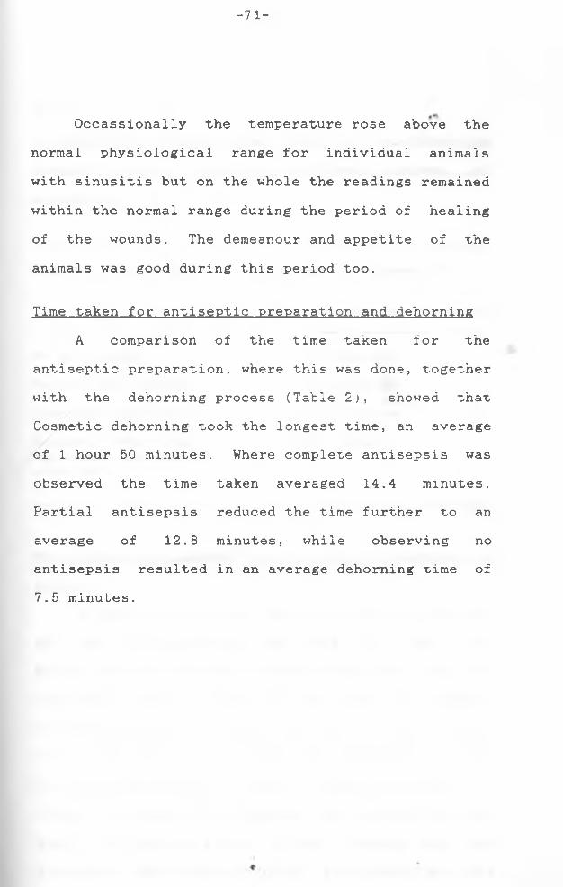
- 71 -
Occassionally the temperature rose above the normal physiological range for individual animals with sinusitis but on the whole the readings remained within the normal range during the period of healing of the wounds. The demeanour and appetite of the animals was good during this period too.
Time taken for antiseptic preparation and dehorningA comparison of the time taken for the
antiseptic preparation, where this was done, together with the dehorning process (Table 2;, showed that Cosmetic dehorning took the longest time, an average of 1 hour 50 minutes. Where complete antisepsis was observed the time taken averaged 14.4 minutes. Partial antisepsis reduced the time further to an average of 12.8 minutes, while observing no antisepsis resulted in an average dehorning time of7.5 minutes.

- 7 2 -
Table 2:A comparison__of__the__time__taken for the dehorningoperation__while___observing__different degrees ofantisepsis
Degree of antisepsis Average time taken for
entire dehorning operation
No antisepsisPartial antisepsisComplete antisepsisCosmetic dehorning (complete antisepsis observed)
7.5 minutes 12.6 minutes 14.4 minutes 110 minutes
Microorganisms isolated in the study 'of dehorning__incattle
A qualitative rather than quantitative analysis of the microorganisms was done, in that the microorganisms were isolated and identified from the different sampling sites without the actual numbers of each microorganism being counted. For this reason it is not possible to accurately compare x'ne groups on the basis of the antiseptic method used and it:s effect to reduce the microbial load around the horn base. The results given, (Table 3) however show the frequency with which different microorganisms were

Table 3: Number of times different microorganisms were isolated from cattle at varioussources of sampling during the dehorning operation.
G r o u p Source of samplesnum b e r M i c r o o r g a n i s m s
isolatedSkin and
h a i r at horn bas e
b e f o r e antis e p s i s
Skin and
h a i r a f t e r partial a n t i s e p s i s
Ski n a ftercomp l e t e
a n t i s e p s i s
Horn w o u n d s
i m m e d i a t e l y a f t e r deh o r n i n g
W o u n d s
48 h o u r s afterr::
dehorninc
Staphyl o c o c c u s aure u s 2 - - 1 1O t h e r staph., spp. 2 - - 1 0
1 Stre p t o c o c c u s faecalis 2 - - 0 2S t r e p t ococcus viri d a n s 4 - - 4 0O t h e r Strep, spp. 2 - - 0 5E s c h e r i c h i a coli 0 - - 0 1N u m b e r of samples 10 0 0 10 10Baci l l u s spp. 9 - - 5 10Staphyl o c o c c u s a lbus 3 - - 0 0O t h e r staph, spp. 6 - - 3 3
2 C o r y n e b a c t e r i u m spp. 1 - - 0 5Klebs i e l l a spp. 0 - - 0 1Strep, spp. 0 - - 0 1E. coli 0 - - 0 4

Table 3 contd.
G r o u pnum b e r
Sour c e of samples
\ M i c r o o r g a n i s m sisolated
Skin a n d h a i r at ho r n b a s e before antis e p s i s
Skin and h a i r a f t e r partial a n t i s e p s i s
Skin a f t e rc o m p l e t ea n t i s e p s i s
Ho r n w o u n d s i m m e d i a t e l y a f t e r d e h o r n i n g
W o u n d s 48 hour: a f t e r dehornir
2 Pse u d o m o n a s a e r u g i n o s a 0 - - 0 3P roteus vulg a r i s 0 - - 0 1
A s p e r g i l l u s sp. 1 - - 0 1
D e r m a t o p h i l u s sp. 1 - - 0 0
N u m b e r of samples 10 0 0 10 10
Staph, albus 6 - 0 2 7Staph, citreus 3 - 0 2 2O t h e r Staph, spp. 0 - 0 0 1
3 B a c i l l u s spp. 10 - 1 2 7C o r y n e b a c t e r i u m spp. 9 - 0 0 8
Strep, faecalis 2 - 0 2 1

Table 3 contd.
3 O t h e r Strep, spp. Ne i s s e r i a sp.
E. coli
Asp e r g i l l u s sp.
Num b e r of samples
4
534
10 0
00
0
0
10
0
1
0
0
10
53
1
1
10
Staph, alius 3 2 0 24 Staph, citreus 0 - 1 0 3
C o r y n e b a c t e r i u m spp. 1 - 0 0 0E. coli 0 - 1 0 GBacillus spp. 0 - 0 0 4A s p e r g i l l u s sp. 0 - 0 0 2N u m b e r of samples 10 0 10 1 0 10
B a c i l l u s spp. 10 7 - 0 05 C o r y n e b a c t e r i u m spp. 4 4 - 2 5
Staph, albus 6 5 - 4 5Staph, ci t r e u s 3 1 - 3 0O t h e r Staph, sp. 0 0 - 0 1Strep, spp. 2 0 - 0 2E. coli 0 2 - 0 1
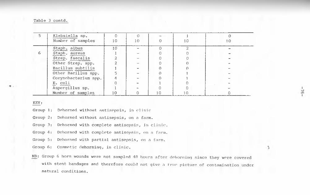
Table 3 contd.
5 K l e b s i e l l a sp. Num b e r of samples
010
010 0
110
010
Staph, albus 10 - 0 2 -6 Staph, aureus 1 - 0 0 -
Strep, faecalis 2 - 0 0 -
O t h e r Strep, spp. 2 - 0 0 -B a c i l l u s subtilis 1 - 0 0 -O t h e r Bacil l u s spp. 5 - 0 1 -
C o r y n e b a c t e r i u m spp. 4 - 0 1 -E. coli 0 - 1 0 -
A s p e r g i l l u s sp. 1 - 0 0 -Num b e r of samples 10 0 10 10 0
KEY:G r o u p 1: G r o u p 2: G r o u p 3: G r o u p 4: G r o u p 5:
G r o u p 6:
DehornedDeho r n e d
Dehor n e dDehor n e dD e h o r n e d
Cosmetic
w i t h o u t antisepsis, in clinic w i t h o u t antisepsis, on a farm, w i t h c o m p l e t e antisepsis, in clinic, with c o m p l e t e antisepsis, on a farm, with p a rtial antisepsis, on a farm,
dehorning, in clinic.
NB: G r o u p 6 horn w o u n d s we r e n o t sampled 48 h o u r s a f t e r d e h o r n i n g since they w e r e c o v e r e d
with stent b a n d a g e s and therefore could not g i v e a true p i c t u r e of c o n t a m i n a t i o n u n d e r n a tural conditions.

-77-
isolated at different sites for each group, and the decrease (or increase) in frequency gives an idea as to the effectiveness of the antiseptic method used.
In group 3, a wide range of microrganisms was isolated from the skin and hair before antisepsis, with Bacillus species appearing in all iO of the samples taken. After observing complete antisepsis only one sample from the skin had the Bacillus organism and none of the other microorganisms were isolated.
In group 4 where the dehorning was done on a farm, 7 out of 10 samples taken from the skin and hair before antisepsis showed no specific growth due to gross contamination. After observing complete antisepsis, 2 out of 10 samples showed the presence of Staphylococcus albus while Staphylococcus citreus and E. coli were isolated only once.
A number of the samples in group 3 showed various microorganisms on the horn wounds immediately after dehorning but in group 4 no bacteria were isolated from the 10 samples taken. Both groups however showed evidence of widespread contamination of the horn wounds 48 hours after dehorning.
In group 5 where partial antisepsis was observed, contamination was still evident after this, as illustrated by Bacillus species that were isolated in 7 out of 10 samples taken from the skin

-78-
and hair after partial antisepsis. Some of the samples from the horn wounds immediately after dehorning also showed evidence of bacterial growth and the number had increased by 48 hours after dehorning.
In groups 1 and 2 where no antisepsis was observed, bacteria were isolated from some of the samples from the horn wounds immediately after dehorning and 46 hours later, but not in significantly greater numbers than for the other groups.
A comparison of all the microorganisms isolated from the skin and hair around the bovine horn base with the wound surfaces 48 hours after dehorning for the groups 1 to 5 (Table 4) shows a similar distribution of microorganisms. Prominent isolates from both sites are the Staphlylococcus and Bacillus species.
Blood analysisTwenty four hours after dehorning, a rise in the
absolute neutrophil count was observed for the different groups. The rise did not correlate to the method of antisepsis or dehorning used but was a response to the injury. 48 hours after dehorning however the absolute neutrophil count had reduced to the normal physiological ranges for all the groups. Thereafter, until the <horn wounds had been sealed

Table 4: Comparison of microorganisms isolated from the skin and hair around the bovinehorn base, and the wound surfaces 48 hours after dehorning, for the groups 1-5.
Microorganisms Number of times isolatedskin and hair wound surface
1) Staphylococcus species 34 25a. Staph. albus 18 14b. Staph, citreus 6 5c. Staph, aureus 2 1d. Others 8 5
2) Bacillus species 29 293) Streptococcus species 16 16
a. Strep, faecalis 4 3b. Strept. viridans 4 -c. Others 8 13
4) Corynebacterium species 15 185) Neisseria species 5 36) Escherichia coJ.j 3 137) Pseudomonas aeruainosa - 38) Proteus vulaaris - 1

Table 4 contd.
Microorganisms Number of time isolatedskin and hair wound surface
9) Klebsiella species - 110) Aspergillus species 5 411) Dermatophilus species 1 -
Total nuumber of samples 50 50

-81-
with scab tissue, the levels remained wijjhin thephysiologic limits. Infection of the horn wounds (sinusitis) did not result in an increase or decrease in the leucocyte levels outside the physiologic limits.
The total protein levels did not vary significantly from the normal ranges during thehealing process for all the groups.
Despite substantial blood losses (.approx. ilitre) in the individual animals during cosmetic dehorning they were able to recover without any adverse effects. The packed cell volume and red blood cell counts did not change significantly. Infection in the horn wounds (sinusitis)
Sinusitis was a fairly common sequel ofdehorning irrespective of the method of antiseptic preparation prior to dehorning, or the location the cattle were kept in. Table 5 shows the incidence of sinusitis for the six groups. The number of horn wounds affected was similar, 5 or 6, for the groups i to 5, but in group 6 (Cosmetic dehorning) all the animals healed without developing infection of the wounds.
A variety of microorganisms were isolated from the horn wounds developing infection in groups i to 5: A total of 43 samples were taken and thecommonest organism isolated was Proteus vulgaris (i5

Table 5; Incidence of sinusitis in the study of dehorning in cattle.
Groupnumber
Location Number of horn wounds affected
Average onset of discharge
Average duration of discharge
Microorganisms isolated Number oftimesisolated
Pathogenicity
1 Clinic 6 18 days 25 days Staphylococcus aureus 1 Pathogenic* . Corynebacter. pyogenes 2 II
Pasteurella hemolytica 1 II
Pseudomonas aeruginosa 3 II
Proteus vulgaris 4 It
Escherichia coli 1 II
Streptococcus faecalis 1 II
Klebsiella spp. 3 Pathogenic2 Farm 5 12 days 15 days Streptococcus faecalis 1 II "t
Proteus vulgaris 4 II
Pseudomona aeruginosa 2 II
Staphylococcus aureus 2 II
Escherichia coli 2 •1Corynebacter. pyogenes 1 it

Table 5 continued.
Groupnumber
Location Number of horn wounds affected
Average onset of discharge
Average duration of discharge
Microorganisms isolated Number oftimesisolated
Pathogenicity
3 Clinic 6 20 days 7 days Corynebacter. pyogenes 4 PathogenicEscherichia coli 3 H
Bacillus spp. 2 Non-pathogenicPseudomonas aeruginosa 1 PathogenicStaphylococcus albus 1 Non-pathogenicKlebsiella spp. 1 PathogenicProteus vulgaris 1 I I
4 Farm 6 6 days 17 days Pseudomonas aeruginosa 2 PathogenicProteus vulgaris 4 I t
Klebsiella spp. 1 U
5 Farm 5 14 days 22 days Corynebacter. pyogenes 1 PathogenicPseudomonas aeruginosa 1 I I
Klebsiella spp. 3 I I
Escherichia coli 4 •1Streptococcus faecalis 1 I I
Proteus vulgaris 2 * l
6 Clinic Nil - - - - -
------------------------------------------------------------

- 84-
times). In contrast it was not isolated from the skin and hair around the horn base, and was only isolated once from the wound surfaces 48 hours after dehorning (Table 4). Pseudomonas aeruginosa and Escherichia coli were the next commonest organisms isolated in sinusitis, 9 times each.
Table 6 shows the relative distribution of the microorganisms. Staphylococcus albus, the most common species isolated from the skin and hair as well as the wound surfaces 48 hours after dehorning, was isolated only once from the horn wounds showing s i nus it is .

- 85 -
Table 6:Microorganisms isolated in the horn wounds developing infection (sinusitis) in order of frequency.
Microorganism Number of times isolated1. Proteus vulgaris 152. Pseudomonas aeruginosa S3. Escherichia coli (E. coli) 94. Corvnebacterium pyogenes 85. Klebsiella spp. 86. Staphylococcus aureus 37. Streptococcus faecalis 38. Bacillus spp. 29. Pasteurella hemolytica 110. Staphylococcus albus 1
Total number of samples taken = 43.NB Some discharges from the horn wounds that, went on
for prolonged periods ( more than 20 days), or whose colour and consistency changed were sampledmore than once.

-86-
Table 7 shows the level of sinusitis in thegroups 1 to 5 in the period following dehorning. Ourof a total of 18 cattle that developed sinusitis, thelevel of infection was highest during the second tofourth weeks with 14 (2nd week) then 16 (3rd and 4t'nweeks) animals showing the condition. The animalsthen began to recover without treatment except fortwo which were flushed with hydrogen peroxide andLugol’s iodine antiseptics. One of the two animals(No. 5 in group 3) showed a persistent sinusitis that
"t h.lasted upto the 16 week following dehorning despite the therapy. The horn wound healed in the iO1,11 week. Three other animals in the same group (Group 3) developed sinusitis with an average duration of 7 days (1 week) and the average healing time of the horn wounds for the group was 14 weeks excludinganimal no. 5.

-87-
Table 7.The level of sinusitis in the weeks following dehorning in groups 1 - 5 .
Period after Number of animalsdehorning with sinusitis
Week 1 8Week 2 14Week 3 16Week 4 16Week 5 9Week 6 6Week 7 4Week 8 3Week 9 3Week 10 2Week 11 2Week 12 2Week 13 1Week 14 1Week 15 1
rWeek 16 1Week 17 -
NumberTotal
of animals that developed number of Animals
sinusitis = 18 = 25.
*

-88-
The healing time and rate of wound healingTable 8 shows the rate at which the horn wounds
healed for five out of the six experimental groupsand the mean healing time for all six groups. Forthe groups 1-5 the rate of healing was expressed asthe percentage reduction in wound area with rime (inweeks). The initial average area of the horn woundsis given for each group, measured immediately afterdehorning. Group 6 had cosmetic dehorning performedwhere the skin is sutured over the horn wound, so therate of healing could not be obtained as for theother groups which had open wounds.
Group 1 had the largest initial average area of2the horn wounds at 47.0 cm . The wounds had reduced
in area by 30% at an average of 3 weeks, and by 60% and 90% at 8 and 16 weeks respectively. The wounds healed (100% reduction in wound size) at a mean healing time of 17.97 + 0.78 weeks. Following this same pattern the rates of healing and mean healing times of the other groups are given. Group 2 took the longest time to heal (23 weeks) and cattle in this group were dehorned with no antisepsis observed and maintained on a farm. The initial average areaof the horn wounds for the group was 38.7 cm^ which
2was smaller than for groups 1 (47.0 cm ) and 5 (39.82cm ) which healed in 18 and 16 weeks respectively.
Group 4 had the smallest initial average area of

Table 8; A comparison of the healing rate and healing time for dehorning wounds of cattle in different groups.
Group Group Group Group Group Group1 2 3 4 5 6
2Average area (cm ) ofwounds immediately after surgery 47.0 38.7 33.1 21.8 39.8 -
Time (weeks) at various %reductions in wound area:-
30% 3 4 3 2 2 -60% 8 13 8 5 790% 16 19 12 13 -100% 18 23 14 14 16
Mean healing time (weeks) 17.97+ 22.84 14.01+ 13.80+ 16.26+ 3.640.78 +1.19 0.80 0.42 2.33 +0.28
ioo01
KEY:-Group 1 - Dehorned without antisepsis, in clinic.Group 2 - Dehorned without antisepsis, on a farm.Group 3 - Dehorned with complete antisepsis, in clinic.
4. Dehorned with complete antisepsis on a farm5. Dehorned with partial antisepsis, on a farm.6. Cosmetic dehorning, in clinic.
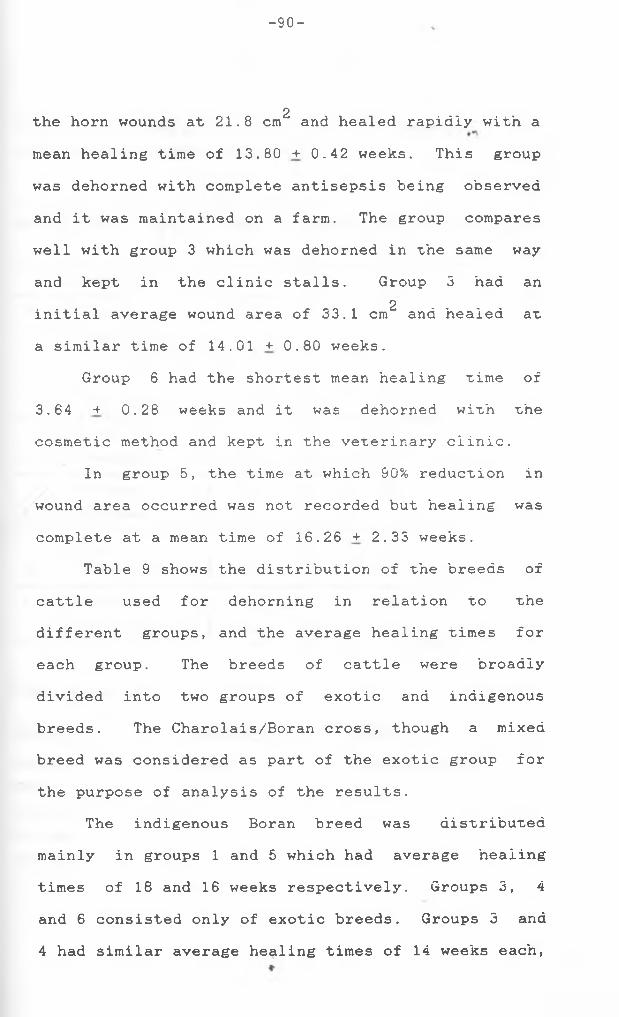
-90-
2the horn wounds at 21.8 cm and healed rapidly with amean healing time of 13.80+0.42 weeks. This groupwas dehorned with complete antisepsis being observedand it was maintained on a farm. The group compareswell with group 3 which was dehorned in the same wayand kept in the clinic stalls. Group 3 had an
2initial average wound area of 33.1 cm and healed at a similar time of 14.01 + 0.80 weeks.
Group 6 had the shortest mean healing time of 3.64 + 0.28 weeks and it was dehorned with thecosmetic method and kept in the veterinary clinic.
In group 5, the time at which 90% reduction in wound area occurred was not recorded but healing was complete at a mean time of 16.26 + 2.33 weeks.
Table 9 shows the distribution of the breeds of cattle used for dehorning in relation to the different groups, and the average healing times for each group. The breeds of cattle were broadly divided into two groups of exotic and indigenous breeds. The Charolais/Boran cross, though a mixed breed was considered as part of the exotic group for the purpose of analysis of the results.
The indigenous Boran breed was distributed mainly in groups 1 and 5 which had average healing times of 18 and 16 weeks respectively. Groups 3, 4and 6 consisted only of exotic breeds. Groups 3 and 4 had similar average healing times of 14 weeks each,

-91-
Table 9: Breed distribution in relation to the different groups and the average healing time.
Groups Cattle breeds Number in Average healing timegroup (weeks) for the grou;
1 Boran 5 182 Boran 2 23
Boran/charolaiscross
1
Charola,is 1Hereford 1Ayshire 3 14
3 Guernsey 1Friesian 4 14
4 Guernsey 1Boran 4 16
5 Charolais 16 Ayrshire 4 4
Friesian 1
Breed groups:
Exotic Indigenous MixedAyrshire Boran Boran/charolaisCharolais crossFriesianGuernseyHereford.

-92-
while group 6 (cosmetic dehorning) had a healingtime of 4 weeks. Group 2 had a mixture of indigenous (2 animals) and exotic (3 animals) breeds and the average healing time was 23 weeks.
Analysis of variance was done (see appendix 20) for comparison of the effect of the different breeds (indigenous and exotic) and the treatments (degree of antisepsis and the place where the animals werehoused) on the healing times of the horn wounds. The effect of the breeds on the healing rimes wassignificant (p < 0.05), showing that the differences in breeds influenced the healing time. The effect, of the treatments on the healing time was also significant (p <0.05 and 0.01) showing that thetreatment differences influenced the healing time.
The variation on the healing time due to the treatment differences was however greater than that due to the breed differences as shown by the probability values.
A pairwise comparison of the differenttreatments or groups was also done. Significant differences in the healing times were found between groups 1 and 3, 2 and 4, 2 and 5, 4 and 5 as well as 3 and 6. Groups 1 and 2 were combined and compared with the combined groups of 3 and 4 to compare the effects of no antisepsis with complete antisepsis. Significant differences in their healing times were found also. ♦

-93-
Appearance__of the healed dehorning wounds ’'comparingthe standard method and cosmetic dehorning
Figure 10 shows the appearance of the horn wound after dehorning in the standard way without suturing the skin. The photograph was taken approximately ia weeks after dehorning when the wound had healed, yet there was still a large scar with an irregular wound margin showing incomplete wound contraction, that was not attractive in appearance.
Figure 11 shows the appearance of the horn wound 3.5 weeks following Cosmetic dehorning. The wound edges had fused well with little scar formation resulting in a smooth outline of the poll and an attractive appearance.

Figure 10: The healed wound after dehorning in thestandard way.
Figure 11: The healed wound followingDehorning.
C o s m e t i c
-95-
FIGURE 10.
FIGURE 11.

-96-
DZSCUSSION
The dehorning wounds where complete antisepsis was observed healed significantly faster than those where partial antisepsis was observed. The latter wounds in turn healed faster than those where no antisepsis was observed. The difference in the healing time was thought to be due to the fact that the hair around the wounds was either shaved (in complete antisepsis) or trimmed (partial antisepsis). This is in agreement with Heggers and Jennings Jr(1984) and Heinze (1974) who have stated that hair as a foreign body causes irritation and secondary contamination of a wound therefore delaying the healing process. Shaving the hair therefore prevented the delay in healing as a result of this occurring, and by the time the hair grew again a protective scab had formed on the wound surface with epithelial regeneration taking place under the scab. Following an infection (sinusitis), when the long hair around the wound had not been shaved, it would sometimes be matted on the wound surface, further delaying healing due to mechanical separation of the wound margins.
The antiseptic used on the skin around the horn did not appear to affect the healing rate and time of the dehorning wound, which contrasts with Altemeier (1977) and Bojrab (*1982 a) who have said that

-97-
antiseptics are used to prevent andcontrol infections which cause a subsequent delay in wound healing. Surgical spirit used in complete antisepsis reduced the cutaneous flora but did not prevent infection. This was because it’s effect was shortlived due to evaporation, and the open wound was also subject to bacterial contamination from the environment.
The healing time was not dependent on the initial size of the horn wound. Similar sized wounds had markedly different healing times, and two groups treated in the same way though differing in the initial wound sizes, had similar healing times.
Cosmetic dehorning had a significantly shorter healing time than dehorning without suturing the skin wound. This is because healing in cosmetic dehorning occurs by first intention while in the standard method of dehorning second intention healing takes place. The latter is a slower process as a granulation tissue bed needs to form first before epithelization can be completed, and epithelial migration also takes place over a longer distance.
In the standard method of dehorning, some of the animals showed a serosanguineous fluid oozing from the horn wound surfaces 24 hours after dehorning. This was thought to be an inflammatory exudate and supports the findings of Bojrab (1982 a) who has

-98-
stated that by 8-12 hours after injury the process of inflammation has produced an exudate which contains plasma proteins, neutrophils, RBC and macrophages. The constituents of the fluid were however not determined.
The marked thickening of the scab in the form of a ridge near the skin margin, observed in some animals 2 weeks after dehorning, could have been caused by the process of wound contraction. Johnston (1977) has reported that in dogs significant wound contraction begins to be seen five to nine days after injury in an open wound. In experiments with rabbits and guinea pigs Peacock and Van Winkle (1976) have also described a marked bulging of the central granulation tissue over an open wound indicating that contraction was occurring within the wound contents. This wound contraction could then similarly have caused the scab on the wound surface to bulge out or appear thickened. When the scab eventually fell off, the epithelial cell layer underneath was level without any obvious bulging.
A flesh coloured (pale yellow to pink) membrane was also commonly seen developing from the sides of the frontal sinus opening of the horn wound at about the fourth week following dehorning. This was probably due to proliferation of the epithelial cells that line the wall of the frontal sinus as described by Hare (1975). The same- author has also described

-99-
the presence of a mucoperiosteum layer on the walls of the frontal sinus and this is probably wh£re the granulation tissue bed that filled the frontal sinus opening was derived from.
The necessity for granulation tissue to fill the frontal sinus opening before epithelization could take place supports the observations of Bojrab (1982 a) and Stashak (1984) who have said that in the full thickness open wound a granulating bed must be formed before epithelization can occur. A delay in the filling up of the frontal sinus opening with granulation tissue up to the level of the wound surface resulted in a delay in the epithelization process.
The horn stump projecting above the wound surface following dehorning delayed healing by preventing epithelization since this could not occur over the stump. The subsequent necrosis (Osteosis) of the projecting stump of bone may probably have been due to ischaemia. This is in agreement with Jubb e_t a_l ( 1985) who have stated that the ends of bone from which the periosteum is separated eg. in a fracture, undergo ischaemic necrosis due to disturbance of the local blood supply. The dead bone is then removed or demineralised and resorbed by osteoclasts. During dehorning the original blood supply of the projecting horn stump must have been
«■

-100-
severed and it underwent ischaemic necrosis. The osteoclasts then acted to remove the dead bone. Judd et al (1985) have reported that osteoclasts can erode bone at the rate of 2-3 iam per hour. This explains the progressive way in which the projecting horn stump was removed. Alternatively, the weakened, dead bone could be pushed inwards during wound contraction and eventually break off as was also abservea. The migrating epithelium then continues across the wound surface.
The dehorning wound healed by epitheiization followed subsequently by wound contraction. This supports the findings of Johnston (1977) and Peacock and Van Winkle (1976) who have said that the healing of open wounds occurs by the processes of epitheiization and contraction. The wound contraction was however delayed most probably due to the close fit of the skin onto the skull restricting movement. It has also been observed that in circular wounds the movement of skin across the wound bed is impeded and there is a 30% slower rate of contraction than in rectangular wounds (Johnston, 1977;. Peacock and van Winkle (1976) have stated that circular wounds contract incompletely or not at all because all edges of the wound become compressed by the force of contraction. In these experiments, incomplete contraction of the skin surrounding the open hornwounds was observed.

-101-
The incidence of sinusitis was similar for all the groups dehorned in the standard way despite the method of antisepsis used. The surgical spirit used in complete antisepsis reduced the number of bacteria isolated around the skin of the horn wound but, this did not prevent sinusitis developing. The sinusitis was most probably due to environmental contamination of the horn wound. This is supported by the findings of Price et al (1968) who have reported that postoperative wound infection in large animals (eg. bovines) may arise primarily from atmospheric contamination of the wound rather than from cutaneous bacteria escaping or surviving antiseptic action during presurgical preparation of the operative site.
The location where the animals were kept did not appear to influence the incidence of sinusitis as the number of horn wounds affected was similar for the groups. This suggests that in dry weather there is no advantage of keeping dehorned animals in buildings as compared to a farm with respect to the rate of infection of the horn wounds.
The cows dehorned by the cosmetic method did not develop sinusitis. This is in agreement with Keinze (1970) who has said that the method is used to minimise the possibility that the frontal sinuses will become infected. The covering of the frontal

-102-
sinuses with skin prevents atmospheric contamination and minimises sinusitis.
The two commonest organisms on the horn wounds 48 hours after dehorning were Bacillus species and Staphylococcus species (especially Staph. aibus') . These organisms have been stated by Kambe (i»75) as being the most frequently collected as potential contaminants from both the environment and the skin of the patient. He has also said that the risk of contamination from the environment is low when compared with the animal’s skin. The environmentconsidered in this case was that of the Small Animal Clinic building, while these experiments were conducted out in the open on a farm and in stalls with open windows. It is therefore likely that the environment played a more significant role in postoperative wound contamination in these experiments. No samples were however taken from the environment the animals were kept in so this cannot be confirmed.
The distribution of microorganisms on the skin and hair and on the horn wounds 48 hours afterdehorning was similar. The swabbing technique picked
#up mainly transient organisms from the skin and hair whose major source according to Jawetz et ai (i976) is exposure to and contact with the environment. It is therefore more probable that the microorganisms on

-103-
the wound surfaces mainly came by contact and exposure to the environment rather than by cutaneous bacteria moving or being carried by air onto the wound surface.
The most common organisms on the horn wounds 46 hours after dehorning (i.e. Staphylococcus species eg. albus. and Bacillus species) were not thecommonest causes of sinusitis. This is mos'C probably because of the low virulence of the microorganisms, and the local and systemic resistance of the host. This is in agreement with Howe (1964) who has reported that these factors contribute to the selective process, whereby the organisms cultured at closure of wounds in humans are not the ones causing a subsequent infection. The causal organism may also have been present initially in numbers too smaii to be picked up or gained access to the wound later.
Proteus vulgaris was isolated most frequently in the horn wounds that developed sinusitis followed by Pseudomonas aeruginosa. Escherichia coii.Corynebacterium pyogenes. and Klebsiella species. The first three and the last organism on the list have been described by Jawetz et a_l (i976) as occurring commonly in the intestinal tract flora and hence the faeces too. Contamination with faeces of the environment then most probably led to the contamination of the horn wounds and sinusitis.

-104-
The order of frequency given above of the microorganisms isolated in sinusitis differs to some extent from the findings of Heggers and Jennings Jr (1984) when describing the incidence of organisms in bovine wound infections. They have stated the order of frequency as Corvnebacterium pyogenes (most common), oC hemolytic Streptococci, Escherichia coii and Proteus species.
Most cases of sinusitis recovered by the T1,11 week following dehorning, without treatment. The earliest average healing time recorded from the groups dehorned in the standard way was i4 weeks (group 3 and 4), and in most of the animals the infection did not cause a delay in the healing process. This is because the frontal sinus opens in the middle of the horn wound and thus the discharge from within it does not prevent the movement of epithelium from the wound margins to the centre. Prolonged sinusitis, i.e., that which occurs up tothe expected healing time of the horn wound, causes a<delay in the healing due to mechanical seperation of
the migrating epithelial cell margins when they reach the frontal sinus opening. This is in agreement with Bojrab (1982 a) and Stashak (1984) who have said that mechanical seperation of wound margins in an infection causes delays in wound healing.
There was an occassional rise in the rectal

-105-
temperature of the animals with sinusitis, but this was not usually persistent and soon dropped to the normal physiological range given by Kelly (iS84). The blood leukocyte levels did not vary significantly from the normal physiological limits reported by Schalm et a l (1975), during the same period. This suggests that on the whole the infection of the horn wounds did not spread to affect the rest of the animal’s body. This finding supports that of Wallace (1980) who has said that occassionally a septicaemia develops but more often the infection remains localised and consists of a frontal sinusitis.
Infection of one horn wound without theinfection of the other on the same animal sometimesoccurred. When infection of both horn woundsoccurred, the microorganisms isolated also differedon occassion. This is because the frontal sinus isdivided into left and right sinuses by a completemedian septum (Sisson , 1975 b) and this thereforerestricts the spread of infection between the twosides.
In the standard method of dehorning (withoutsuturing the skin wound), observing completeantisepsis prior to dehorning almost doubled the average time taken for the entire dehorning operation as compared to observing no antisepsis. The time taken to observe antisepsis however varies depending
\

-106-
on the skill of the operator in shaving. Cosmetic dehorning took a much longer time than the ’"standard method as the skin around the base of the horn needed to be undermined to make it freely moveable, and then sutured. The standard method is therefore more practical if a large herd of cattle is to bedehorned, and it is also a cheaper method as there isno necessity for suture materials. A suggestion for a herd of cattle to be dehorned would be to trim the hair at the base of the horn using scissors prior to dehorning. Not much time is lost in this procedure and the healing of the horn wounds is enhanced.
The time taken for cosmetic dehorning can beconsiderably reduced to about 1 hour by the use of
Rthe sedative Xylazine HCL (Rompun ) at the dosage of0.1 mg/Kg intramuscularly. This eliminates struggling by the animal when the local anaesthetic is not completely effective, due to inaccurateadministration, or its effects wearing out.
In the standard method of dehorning where the horn wound was left to heal as an open wound, incomplete contraction of the skin of the circular wound resulted in an irregular skin margin. This finding agrees with that reported by Johnston (iS77), in experiments with rabbits, where a circular skin wound healed in a crumpled, unpredictable pattern. This, together with the large scar present in the

-107-
healed horn wound, made it’s postoperative appearance unattractive as compared to cosmetic dehorning where the wound edges fused well and the poll had a smooth outline. Wound dehiscence is a possibility in cosmetic dehorning after the sutures have been removed, but the wounds soon heal on their own without the need for resuturing. Cosmetic dehorning is therefore a better method in terms of the postoperative appearance of the animal. The rapid rate of healing and the low chances of sinusitis developing also make it a suitable method for individual, prized show cattle. This is in agreement with Greenough (1974) and Heinze (1970) who have recommended the method for small numbers of cattle to be exhibited when the postoperative appearance of the animal is important, and to minimise the possibility of sinusitis.
*

-108-
CONCLUSIONS.• **«
From the findings in this investigation someconclusions may be drawn:-1. That trimming the hair or shaving, irrespective
of the method of cleaning the skin around the horn prior to dehorning enhances the healing of dehorning wounds in cattle.
2. That antiseptic preparation of the skin around the bovine horn does not reduce the rate of infection of the wounds following dehorning, most probably due to contamination from the environment.
3. That sinusitis does not delay the healing ofdehorning wounds unless it is prolonged andepithelial cell migration has reached thefrontal sinus opening.
4. That infection of the horn wounds does not usually affect the general health of the animal.
5. That cosmetic dehorning, though more expensive and time consuming, enhances the healing, reduces the infection rate and has a more cosmetic postoperative appearance than dehorning without suturing the skin wound.
6. Generally, trimming of hair or shaving enhancesthe healing of dehorning wounds and not theapplication of antiseptics. Sinusitis is likelyto occur despite antiseptic preparation but this♦

-109-
sinusitis does not interfere with the general health of the animal.

- 110 -
REFERENCES
ALTEMEIER, W.G. (1977).Surgical Antiseptics. In "Pis infection,Sterilisation and Preservation, 2nd Ed., Lea and Febiger Co., Philadelphia, pp. 641-653.
BOJRAB, M.J. (1982 a)Wound Management.Mod. Vet. Pract. 63. (10): 791-796.
BOJRAB, M.J. (1982 b).Wound management.Mod. Vet. Pract. 63. ( 11): 867-874.
BOWEN, J.S. (1977).Dehorning the mature goat.J.A.V.M.A. 171 (12): 1249-1250.
CLARK, C.H. (1980).Prophylactic use of antibiotics in surgery. Mod. Vet. Pract. 6JL ( 1): 30-33.
CLEMENTE, C.H. (1970).R
Rompun as a basal anaesthetic for the serial dehorning of cattle under general anaesthesia with an angle grinding machine.Vet. Med. Rev. 3:196-198.

- 111 -
COWAN, S.T. (1974).Cowan and Steel's manual for the identiffbation
ndof Medical____ Bacter ia , 2 Ed., CambridgeUniversity Press, Cambridge. pp. 1-122,166-180.
DAVIDSON, A.I.G.; CLARK, C.; SMITH, G. (1971 a).Postoperative wound infection: A computeranalysis.Brit. J. Surg. 58.: 333-337 .
DAVIDSON, A.I.G.; SMITH, G.; SMYLIE, H.G. (1971 b).A bacteriological study of the immediate environment of a surgical wound.Brit. J. Surg. 58: 326-333.
DEHOLL, D.; RODEHEAVER, G.; EDGERTON, M.T.;EDLICH, R.F. (1974 ) .
Potentiation of infection by suture closure of dead space .Amer. J. Surg. 127: 716-720.
DRAKE, C.T.; GOLDMAN, E.; NICHOLS, R.L.; PIATRUSZKA, K.; NYHUS, M.L. (1977).
Environmental air and airborne infections.Ann. Surg. 185: 219-223.

- 112 -
EVANS, L.H. (1971).Regional analgesia in large animals'.”' In "Textbook o£ Veterinary Anaesthesia". The Williams and Wilkins Co.Baltimore, p.471.
FRANK, E.R. (1964).th
VETERINARY SURGERY. 7 Edition, BurgesPublishing Co. pp. 39-71.
GODINHO, H.P.; GETTY, R. (1975).Peripheral Nervous system. In "Sisson andGrossman's The______Anatomy oE the Domestic
thAnimals". 5 Edition. W.B. Saunders Co. Philadelphia, pp. 1084-1086.
GOSHAL, N.G. (1975).Ruminant heart and arteries. In "Sisson andGrossman's The Anatomy o£ the Domestic Animals". th5 Ed. W.B. Saunders Co. Philadelphia, pp. 967-968.
GREENOUGH, P.R. (1974).The Integumentary System. In "Textbook of Large Animal Surgery". The Williams and Walkins Co.Baltimore. pp. 199- 205

- 113-
GRUNDY, W.E. (1977).Antimicrobial preservatives in Pharmaceuticals.I n "Disinfection, Sterilisation and
ndPreservation". 2 Ed. Lea and Febiger Book Co., Philadelphia, p 764.
HALL, L.W.; CLARKE, K.W. (1983).th
Veterinary Anaesthesia. 8 Ed. Bailliere Tindall, London, p. 248.
HARE, W.C.D. (1975).General Osteology. In "Sisson and Grossman's
thThe Anatomy of the Domestic Animal". 5 Ed. W.B. Saunders Co. Philadelphia, p. 32.
HART, C.B. (1949).A complication arising from dishorning cattle. Vet. Rec. £1:218.
HEATH, R.B. ( 1984 ) .Local and Regional anaesthesia in ruminants. In "The Practice of Large Animal Surgery". Vol. 1. W.B. Saunders Co. Philadelphia, p. 200.
HEGGERS, J.P.; JENNINGS JR, P.B. (1984)Surgical methods in the management of wound infections. In "The Practice of Large animal Surgery". Vol. 1. W.B. Saunders Co.Philadelphia, pp . 143-146.
*

-114-
HEINZE, C.D. (1970).Surgical Procedures. In "Bovine Medicine’" and Surgery". First Edition. American Veterinary Publications, Illinois. pp. 798-804.
HEINZE, C.D. (1974).Wound healing and tissue repair. In "Textbook of Large Animal Surgery". The Williams and Wilkins Co. Baltimore. pp. 41-54.
HOWE, C.W. (1964).Bacterial flora of clean wounds and it's relation to subsequent sepsis.Amer. J. Surg. 107: 696-699.
JAWETZ, E.; MELNICK, J.L.; ADELBERG, E.A. (1978).thReview of Medical Microbiology, 13 Ed.
Lange Medical Publications, California, pp. 198-263.
JOHNSTON, D.E. (1977)The processes in wound healing.J.A.A.H.A., 11(3/4): 186-196.
JUBB, K.V.F.; KENNEDY, P.C.; PALMER, N. (1985)rd
Pathology of Domestic Animals. 3 Ed., Vol. 1, Academic Press Inc. Orlando, Florida, pp. 61-63.

-1 1 5 -
KAMBE, S. (1979).Wound Contamination in Veterinary Surgery. MSc Thesis. University o£ Nairobi, Kenya, pp. 7-15;67-131.
KELLY, W.R. (1984)rdVeterinary Clinical Diagnosis. 3 Ed.,
Bailliere Tindall, London, p. 27.
LOWBURY, E.J.L.; LILLY, H.A.; BULL, J.P. (1960). Disinfection of the skin of operation sites.Br. Med. J. 2: 1030-1044.
MILNE, F.J. (1974).General Surgical Conditions. In "Textbook of Large Animal Surgery. The Williams and Wilkins Co. Baltimore, pp. 8-34.
PEACOCK, E.E.; VAN WINKLE, W. (1976).nd
Wound Re pa i r. 2 Ed., W.B. Saunders Co.Philadelphia pp. 1-80.
PRICE, P.B. (1938).The Bacteriology of normal skin; A newquantitative test applied to a study of the bacterial flora and the disinfectant action of mechanical cleansing.J. Infect, dis. 63: 301-318.

-116-
PRICE, W.J.; BOWEN, J.M.; WOOLEY, R.E. (1968)Efficacy of Ethyl alcohol in reducing canine and bovine cutaneous bacterial flora.J.A.V.M.A. 152(7): 990-995.
QUIN, A.H. (1945).Painless dehorning with local anaesthesia.Vet. Rec. 57.: 452-453 .
SCHALM, O.W.; JAIN, N.C.; CARROLL, E.J. (1975).rd
Veterinary Haematology. 3 Ed., Lea and Febiger Co., Philadelphia, pp. 122-144.
SISSON, S. (1975 a).Common Integument. In "Sisson and Grossman's
thThe Anatomy of the Domestic Animals",5 Ed., W.B. Saunders Co., Philadelphia, pp. 1209-1210.
SISSON, S. (1975 b).Ruminant Osteology. In " Sisson and Grossman's
thThe Anatomy of the Domestic Animals ", 5 Ed. , W.B. Saunders Co., Philadelphia. pp. 772-773.
STASHAK, T.S. (1984).Wound healing. In "The Practice of Large Animal Surgery". Vol. 1, W.B. Saunders Co., Philadelphia. pp. 277-287.

-117-
TOPLEY, W.W.C.; WILSON, G.S.; MILES, A. (1975).The resistance of bacteria to Physical and Chemical agents. In "Principals of
thBacteriology, Virology and Immunity". 6 Ed., Edward Arnold Ltd, London, p. 165.
WALLACE, C.E. (1980).Dehorning. In "Bovine Medicine and Surgery", nd2 Ed., Vol. 2, American Veterinary Publications, Inc., Santa Barbara, California, pp. 1236-1241. *
*

- 118-
• -*
A P P E N D I C E S
♦

- 119-
A comparison of the average rectal temperature readings of the cattle in groups 1-5 that developed sinusitis with those that did not after dehorning.
Appendix 1:
Days after dehorning
Average temperature (°C) No. of animals showing sinusitisNo sinusitis Sinusitis
0 38.9 -1 39.0 -2 39.1 -3 38.8 -4 38.3 -5 38.5 -6 39.1 39.0 77 38.7 38.9 28 39.3 38.5 29 39.3 39.8* 410 39.2 39.1 311 38.2 38.9 112 38.8 39.1 213 38.6 39.0 814 38.6 39.1 215 38.7 39.2 416 38.1 39.1 217 38.6 39.7* 318 38.9 39.0 119 38.7 38.8 120 39.0 38.9 621 39.0 38.6 522 38.9 38.6 323 39.1 39.9* 1

- 120—
Appendix 1 continued
Days after dehorning
Average temperature (°C) No. of animalsNo sinusitis Sinusitis showing sinusitis
24 38.4 38.2 125 38.9 -26 - -
27 39.1 39.3 728 39.0 39.3 729 38.8 39.3 130 37.8 -31 38.0 -
32 - -
33 - -34 38.8 39.3 135 38.7 38.1 136 39.2 39.0 137 - 38.6 138 39.5* 39.9* 239 - -40 - -
41 38.7 -
42 39.3 38.5 243 39.2 -
Normal, physiological temperature range for adult cattle = 37.8 - 39.2° C (Average = 38.5° C) (Kelly, 1974).
* = indicates temperature values above the physiological range.
*

- 121-
Response of neutrophils after dehorning, for groups 1-6 (i.e. Absolute neutrophil count).
Appendix 2;
Period after Group numberdehorning
1 2 3 4 5 6Week 1Pre operativesample(control) 4336 2898 2156 2607 3201 19131 day 5039 5251 4264 4883 36842 days 4081 3838 2480 2775 2925 17843 days 3822 2716 3415 19784 days 4488 28495 days 2241Week 2 4445 3460 2617 1958 4171 1468Week 3 2410 2474 1984 2748 2718 1728Week 4 2002 2732 1547 - 2220 2611Week 5 - 2996 2175 2406 2359 -
Week 6 - 3294 1596 3402 2571 -Week 7 - 1754 — —
Physiological values for absolute neutrophil count = 600 - 4000. Neutrophils per mm^ of blood (Schalm et al., 1975).
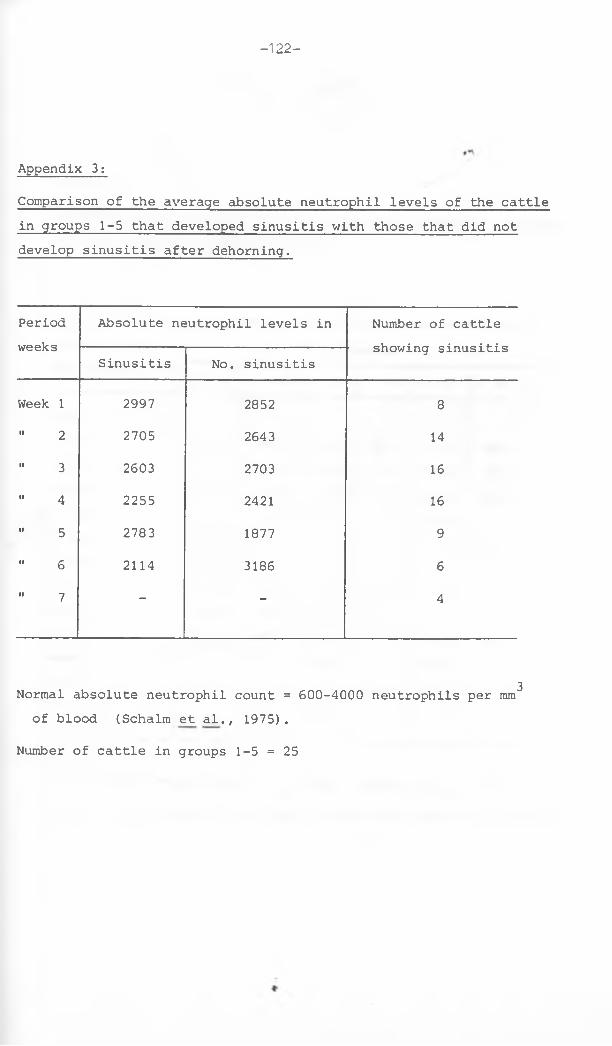
- 122-
Comparison of the average absolute neutrophil levels of the cattle in groups 1-5 that developed sinusitis with those that did not develop sinusitis after dehorning.
Appendix 3:
Periodweeks
Absolute neutrophil levels in Number of cattle
Sinusitis No. sinusitisshowing sinusitis
Week 1 2997 2852 8" 2 2705 2643 14" 3 2603 2703 16" 4 2255 2421 16" 5 2783 1877 9" 6 2114 3186 6" 7 “ - 4
Normal absolute neutrophil count = 600-4000 neutrophils per mm^ of blood (Schalm et al., 1975).
Number of cattle in groups 1-5 = 25
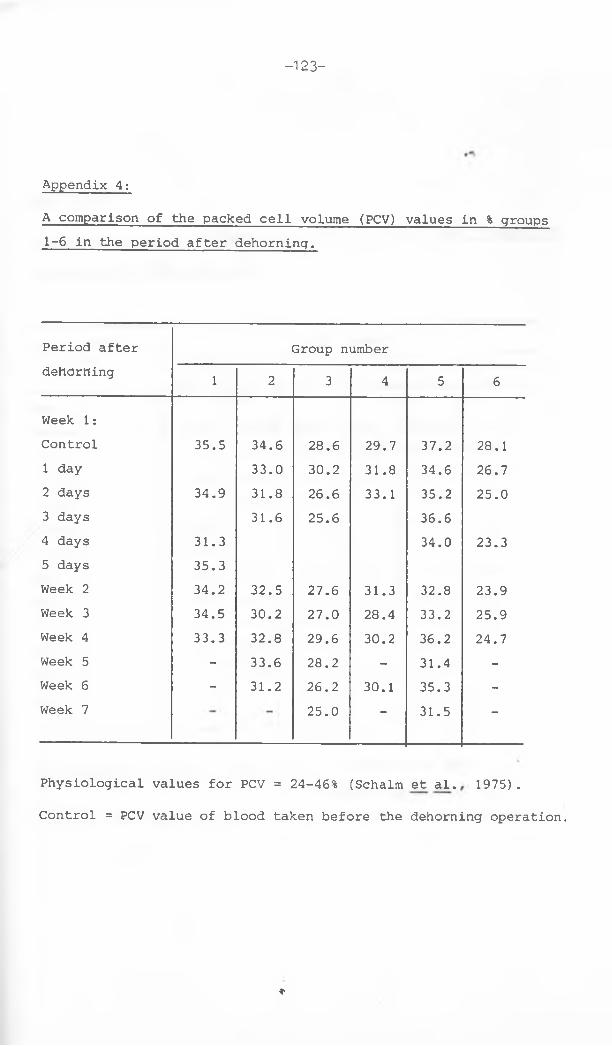
—123—
A comparison of the packed cell volume (PCV) values in % groups 1-6 in the period after dehorning.
Appendix 4:
Period after G r o u p n u m b e rdehdrhing 1 2 3 4 5 6
Week 1:Control 35.5 34.6 28.6 29.7 37.2 28.11 day 33.0 30.2 31.8 34.6 26.72 days 34.9 31.8 26.6 33.1 35.2 25.03 days 31.6 25.6 36.64 days 31.3 34.0 23.35 days 35.3Week 2 34.2 32.5 27.6 31.3 32.8 23.9Week 3 34.5 30.2 27.0 28.4 33.2 25.9Week 4 33.3 32.8 29.6 30.2 36.2 24.7Week 5 - 33.6 28.2 - 31.4 -Week 6 - 31.2 26.2 30.1 35.3 -Week 7 25.0 - 31.5 -
Physiological values for PCV = 24-46% (Schalm et al. 1975).C o n t r o l = P C V v a l u e of b l o o d t a k e n b e f o r e the d e h o r n i n g operation.
♦

-124-
6 3A comparison of the red blood cell count (xlO /mm ) for groups 1-6 in the period after dehorning,
Appendix 5;
Period after dehorning
Group number
1 2 3 4 5 6Week 1:Control 7,40 9.35 8.44 5.92 10,33 6,061 day 9.27 8.53 o
0yo 10.03 5.80
2 days 7.45 8.68 7,93 5,91 10,41 5.963 days 8,92 7.91 9.784 days 6,88 8,40 5.415 days 7,59Week 2 7,38 9,03 8.04 5,63 9.03 5.28Week 3 7.47 8.49 7.55 6.05 9.82 5.33Week 4 7,17 8.78 8.55 6.01 10.64 5,13Week 5 - 8.57 8.64 - 10,25 -Week 6 - 8.65 7.67 6,13 10.08 -Week 7 — — 7.61 — 9.54
Physiological values for RBC count = 5 . 0-10,0 (xlO /mm )(Schalm et al, 1975).
Control = RBC count in blood taken before the dehorning operation

-125-
Total p r o t e i n v a l u e s (in g/100 ml) for g r o u p s 1-6 in the p e r i o d after
dehorning.
Appendix 6;
Physiological values for total protein = 7.0-8„5g/100 ml (Schalm et al, 1975) Control= Total protein in blood taken just before the dehorning operation.

Appendix 7:
The incidence of sinusitis for dehorning wounds of cattle in group 1 with their corresponding healing times.
Group Bullnumber
Hornwound
Onset of discharge
Duration of discharge up to week:-
Microorganisms isolated Pathogenicity Healing time (weeks)
1 1 Left 2nd week 5 Staph, aureus Corynebacterium pyogenes Pasteurella hemolytica
PathogenicIIII
19.3
Right 1st week 3 Pseudomonas aeruginosa Proteus vulgaris
PathogenicII 20.3
2 Left 8th week 12 Proteus vulgaris Pseudomonas aeruginosa
PathogenicII 18.1
Right 2nd week 4 Proteus vulgaris Pseudomonas aeruginosa
PathogenicIt 19.4
3 Left No sinusitis 20.7Right No sinusitis o
c
oCN
4 Left No sinusitis 14 »3,X
Right No sinusitis 14.9Left 2nd week 7 Non specific - 16.0Right 3rd week 9 Corynebacterium pyogenes
E. coliStreptococcus faecalis Proteus vulgaris
PathogenicIIIIII
oc
VD
-126-

Appendix 8;The incidence of sinusitis and the corresponding healing times for dehorning wounds in group 2 cattle.
Group Cownumber
Hornwound
Onset of discharge
Duration discharge up to week
Microorganisms isolated Pathogenicity Healing time (weeks)
2
1 Left 1st week 2 Klebsiella species Streptococcus faecalis Proteus vulgaris Pseudomonas aeruginosa
PathogenicIIIIII
27.0
Right No sinusitis 27.0
2Left 1st week 4 Klebsiella sp.
Proteus vulgarisPathogenic
II21.0
Right No sinusitis 21.0

Appendix 8 contd.
Cownumber
Hornwound
Onset of discharge
Duration of discharge up to week:-
Microorganisms isolated Pathogenicity Healing time (weeks)
3
Left 1st week 4 Staphylococcus aureus E. coliProteus vulgaris
Pathogenic•1II
21.0
Right 5th week 6 E. coliKlebsiella sp.
PathogenicII
21.0
4Left No sinusiti s 17.3Right 1st week 4 Corynebacterium pyogenes Pathogenic
IIIIII
19.14Staphylococcus aureus
Pseudomonas aeruginosa Proteus vulgaris
5Left No sinusiti __________ s 27.0Right No sinusitis __________ I__ 27.0

Appendix 9;The incidence of sinusitis and the corresponding healing times for dehorning wounds in group 3 cattle.
Group Bullnumber
Hornwound
Onset of discharge
Duration of discharge up to week:-
Microorganisms isolated Pathogenicity Healing time(weeks)
3
1 Right 4th week 5 Corynebacter. pyogenes E. coli Bacillus sp.
PathogenicII
Non pathogenic
12.0
Left Nosinusitis
16.1
2
Left 2nd week 3 E. coli Pathogenic 15.0
Right 3rd week 4 Corynebacter. pyogenes Pseudomonas aeruginosa
PathogenicII
16.0s
3
Left Nosinusitis 15.0
Right Nosinusitis
15.0
-129-

Appendix 9 contd.Bull H o r n Onset of D u r a t i o n of M i c r o o r g a n i s m s isolated. P a t h o g e n i - H e a l i n gn u m b e r w o u n d di s c h a r g e up to w l e k cit y time(weeks)
4Left 3rd week 5 Corynebacterium pyogenes
Staphylococcus albusPathogenic Non pathogenic
11.0
Right 4th week 5 Klebsiella sp. Pathogenic 11.0
5Left 3rd week 16 Corynebacterium pyogenes
Bacillus sp.Proteus vulgaris E. coli
Pathogenic Non pathogenic Pathogenic
II
20.1
Right No sinusitis 29.0
NB: The right horn wound of bull no. 5 had a delay in the filling of the frontal sinus openingwith granulation tissue up to the level of the wound surface hence the prolonged healing time.
II•0
£ L-

Appendix 10:
The incidence of sinusitis and the corresponding healing times for dehorning wounds in group 4 cattle.
Group Cownumber
Hornwound
Onset of discharge
Duration of discharge up to week:-
Microorganisms isolated Pathogenicity Healing time (weeks)
. 1 Left No sinusitis 16.0Right No sinusitis 16-0
4 2 Left week 1 4 Klebsiella sp„ Pathogenic 13.0Right week 1 2 Pseudomonas aeruginosa
Proteus vulgarisPathogenic
I I
13-0
Left No sinusitis 13-03 Right 2nd week 4 Pseudomonas aeruginosa Pathogenic o
c
in4
Left week 1 3 Proteus vulgaris Pathogenic 13.64 Right Week 1 6 Proteus vulgaris Pathogenic 13.0
5 Left No sinusitis 13.0Right 2nd week 3 Proteus vulgaris 13.0

Appendix 11:
The incidence of sinusitis and the healing time for dehorning wounds in groups 5 and 6.
Group Cownumber
Hornwound
Onset of discharge
Duration of discharge up to weeks:-
Microorganisms isolated Pathogenicity Healing Time (weeks)
5
1
* •
left 3rd week. 6 Proteus vulgaris E. coli
Pathogenic 27.1
right No sinusitis 30.1
2 left No sinusitis 9.3
right No sinusitis 9.3
3 left 2nd week 4 E. coliProteus vulgaris
PathogenicII 19.0 *
right 2nd week 4 E. coliKlebsiella spp.
PathogenicII 19.0
■ZZ l
-

Appendix 11 continued
Group Cownumber
*. t ■
i Horn wound
Onset of discharge
Duration of discharge up to week:
Microorganisms isolated Pathogenicity Healing Time (weeks)
5 4 left 1st week 7 Corynebacterium pyogenes Pseudomonas aeruginosa Klebsiella sp.
PathogenicIIII
12.3
right 3rd week 6 E. coliStreptococcus faecalis Klebsiella sp.
PathogenicIIII
12.3
4
left No sinusitis 12.1
5 right No sinusitis 12.1
6 1-5 All healed without developing sinusitis Average g = 3.6
-133-

-1 34-
flppendix 12;The healing times (weeks ) of dehorning wounds of cattle using different methods of antiseptic preparation,
Groups Hornwounds
Animals Average healing time1 2 3 4 5
1 Left 19.3 18,1 - 20,7. 14,3 16,0 17.9Right 20.3 19.4 20,0 14.9 16.0
2 Left 27.0 21.0 21,0 17,3 27,0 22.8Right 27,0 21.0 21.0 19.1 27,0
3 Left 16,1 15,0 15.0 11.0 20.1 14,0Right 12,0 16,0 15.9 11.0 29.0
4 Left 16,0 13.0 13.0 13.0 13,0 13.8Right 16.0 13.0 15.0 13,0 13,0
5 Left 27.1 9.3 19.0 12.3 12.1 16.3Right 30.1 9,3 19.0 12.3 12.1
6 Left 3.7 4.1 3.4 2,7 3.3 3.6Right 4.8 4.1 5.1 2.3 2.9
Key to groups:1 = Dehorned with no antisepsis, in veterinary clinic2 = Dehorned with no antisepsis, on a farm3 = Dehorned with complete antisepsis, in clinic4 = Dehorned with complete antisepsis, on a farm5 = Dehorned with partial antisepsis, on a farm6 = Cosmetic dehorning, in clinic
*NB: Animal No, 5 of group 3 was excluded in the calculation of the average healing time for the group. The left horn wound had a persistent sinusitis up to week 16, while the right horn wound had a delay in the frontal sinus opening filling with granulation tissue.

- 135-
A comparison of the breeds and the average healing times (weeks)
of dehorning wounds of cattle in groups 1-6.
Appendix 13:
Group Animalsnumbe:c 1 2 3 4 51 Breed Boran Boran Boran Boran Boran
HealingTime(H.T.) 19.8 18.8 20.4 14.6 16.0
2 Boran Boran Boran Charolais C/b cross HerefordH.T. 27.0 21.0 21.0 18.2 27.0
3 Breed Guernsey Ayrshire Ayrshire Ayrshire AyrshireH.T. 14.1 15.5 15.5 11.0 24.6
4 Breed Friesian Friesian Guernsey Friesian FriesianH.T. 16.0 13.0 14.0 13.0 13.0
5 Breed Boran Charolai:i \Boran Boran BoranH.T. 28.6 .9.3 19.0 12.3 12.1
6 Breed Ayrshire Friesian Ayrshire Ayrshire AyrshireH.T. 4.3 4.1 4.3 2.5 3.1
C / B = C h a r o l a i s / B o r a n cross
«■
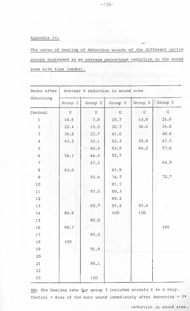
- 136—
•nThe rates of healing of dehorning wounds of the different cattle groups expressed as an average percentage reduction in the wound area with time (weeks).
Appendix 14:
Weeks after dehorning
Average % reduction in wound area
Group 1 Group 2 Group 3 Group 4 i Group 5Control 0 0 0 0 0
1 14.6 7.8 19.7 13.8 21.62 23.4 15.5 30.7 36.6 34.83 34.8 22.7 41.0 46.64 43.2 32.1 52.3 59.8 47.55 40.4 53.9 64.0 57.66 54.1 44.4 55.77 47.2 64.98 63.0 65.99 55.4 74.7 72.710 81.711 57.0 89.312 89.213 65.7 91.4 95.414 84.8 100 10015 80.816 90.7 10017 85.218 10019 91.82021 95.12223 100
NB: The healing rate ^pr group 3 includes animals 1 to 4 only.Control = Area of the horn wound immediately after dehorning - 0%
reduction in wound area.

-137-
Rate of healing expressed as a percentage reduction in the wound area” •**>with time (weeks) for group 1 cattle,
Appendix 15;
Weeks after dehorning Percentage (%) reduction in wound area
Animal 1 Animal 2 Animal 3 Animal 4 Animal 5Control 0 0 0 0 0
1 5„ 7 21.0 19.6 12.9 14.12 21.3 19.8 27.6 14.8 33.53 31.5 38.8 49.0 26.5 28.54 31.9 45.5 45.5 44.6 48.85 45.96 56.6 56.1 47.2 56.57 49.58 55.7 57.6 68.4 70.49 55.310 61.0 60.011 67.212 70.3 89.713 96.214 75.0 74.3 97.515 77.4 10016 85.9 00 <T
\o t
o 10017 81.21819 10020 10021 100

-138-
Rate of healing expressed as a percentage reduction in the wound area with time (weeks) for group 2 cattle. t*'
Appendix 16;
Weeks after dehorning
Percentage (%) reduction in wound area
Animal 1 Animal 2 Animal 3 Animal 4 Animal 5
Control 0 0 0 0 01 0 5.5 8.7 10.7 14.32 4„9 13.3 20.4 30.2 8.73 14.2 11.5 23.8 48.3 16.04 32.6 22.1 35.5 49.3 20.95 39.4 31.1 45.4 59.3 27.06 37.5 27.8 53.5 68.2 35.07 38.4 33.6 47.3 75.7 41.189 45.2 46.1 52.5 93.6 39.61011 45.2 45.9 54.4 97.3 42.41213 55.6 57.5 66.4 98.3 50.71415 67.1 86.0 84.1 98.8 68.11617 71.8 87.8 91.4 99.4 75.61819 88.1 97.2 94.9 100 82.92021 95.6 100 10.0 84.92223 95.7 88.32425 98.5 93.12627 100 100
♦

-139-
Appendix 17;
Rate of healing expressed as a percentage reduction in the wound area with time (weeks) for group 3 cattle.
Weeks after dehorning % reductioin in wound area
Animal 1 Animal 2 Animal 3 Animal 4 Animal 5
Control 0 0 0 0 0
1 16„ 1 18.3 21.4 23.3 15.12 2 3 c 0 36 c 9 32.5 30.4 2 1 . 63 3 4 c 4 39.0 40.0 50.8 37.34 5106 50. 3 53.6 54.0 38.45 52„ 7 55.0 55.0 53.0 39.56 5106 56.0 59.57 62.2 40.58 55c 3 62.1 76.5 6 9 . 9 40.59 71 c6 6 8 . 2 8 6 . 5 72.5 41.6
10 8 5 . 0 82.2 87.0 72.5 4 8 . 7
11 8 9 . 9 81 c4 8 6 . 1 100 5 3 . 9
12 9 8 . 5 81.5 87.6 61.113 86.4 93.7 6 3 . 3
14 92.1 92.8 69.615 100 96.8 97.5 70.216 100 100 74.817 80.418 85.519 86.420 9 0 . 32122232425 100
*

- 140-
Appendix 18: ,nRate of healing expressed as a percentage reduction in the wound area with time (weeks) for group 4 cattle.
Weeks after % reduction in wound areadehorning Animal 1 Animal 2 Animal 3 Animal 4 Animal 5
Control 0 0 0 0 0
1 16.7 0 27.2 13.6 11.5
2 34.0 30.5 41.9 43.0 33.8
34 47.9 55.8 71.4 62.3 61.8
5 49.6 62.5 67.8 72.4 67.7
678910111213 80.4 100 96.6 100 100
14 100
1516 100

-141
Rate of healing expressed as a percentage reduction in the wound area with time (weeks) for group 5 cattle.
Appendix 19:
Weeks after % reduction in wound areadehorning Animal 1 Animal 2 Animal 3 Animal 4 Animal 5Control 0 0 0 0 0
1 12.9 26.6 20.5 19.9 28.02 25.9 39.5 33.0 37.9 37.53 37.6 52.6 53.7 40.0 49.34 41.7 55.9 52.9 39.75 45.9 69.2 57.66 49.7 67.87 42.9 87.6 64.38 52.1 90.09 50.3 67.810 100 78.6 92.911 56.3 75.31213 68.5 80.9 100 1001415 74.0 89.71617 75.3 95.71819 84.1 1002021 86.223 88.625 92.027 96.929 100
♦

Appendix 20:
Analysis of Variance for comparison of the effect of variation o£ breeds (Indigenous and Exotic) and treatments .(Degree of antisepsis and location where the animals were housed)on the healing time of the horn wounds of cattle.
Parameter Source of variation
Degrees of freedom
Sum of squares
Mean sum of squares
F-value Probability Significance
Healingtime
Breed 1 86.12 86.12 6.85 0.0117 P<0.05 (Significant)Treatments 5 1459.99 292.00 23.21 0.0000 P<0.0001 (Significant)Error 51 641.60 12.58Totalobservations
57 15425.16 270.62 21.51
Sum of squares due to the whole model = 14783.56i.e. Total observations sum of squares (minus) - Error sum of squares