The Effects of Pharmacological and Lentivirus-Induced Immune Suppression on Orbivirus
11
JOURNAL OF VIROLOGY, 0022-538X/98/$04.0010 July 1998, p. 5599–5609 Vol. 72, No. 7 Copyright © 1998, American Society for Microbiology. All Rights Reserved. The Effects of Pharmacological and Lentivirus-Induced Immune Suppression on Orbivirus Pathogenesis: Assessment of Virus Burden in Blood Monocytes and Tissues by Reverse Transcription-In Situ PCR SCOTT J. BRODIE, 1,2 * WILLIAM C. WILSON, 2 PATRICIA M. O’HEARN, 1 DAVID MUTHUI, 1 KURT DIEM, 1 AND LEONARD D. PEARSON 3 Virology Division/Retrovirology Laboratory, University of Washington School of Medicine, Seattle, Washington 98144 1 ; Arthropod-Borne Animal Disease Research Laboratory, Agricultural Research Service, U.S. Department of Agriculture, Laramie, Wyoming 82071 2 ; and Department of Microbiology, College of Veterinary Medicine and Biomedical Sciences, Colorado State University, Fort Collins, Colorado 80523 3 Received 28 January 1998/Accepted 31 March 1998 We investigated the effects of pharmacological and lentivirus-induced immunosuppression on bluetongue virus (BTV) pathogenesis as a mechanism for virus persistence and induction of clinical disease. Immuno- logically normal and immunosuppressed sheep were infected subcutaneously with BTV serotype 3 (BTV-3), a foreign isolate with unknown pathogenicity in North American livestock, and with North American serotype 11 (BTV-11). Erythrocyte-associated BTV RNA was detected earlier and at greater concentrations in sheep treated with immunosuppressive drugs. Similarly, viral RNA and infectious virus were detected in blood monocytes earlier and at higher frequency in immunosuppressed animals: as many as 1 in 970 monocytes revealed BTV RNA at peak viremia, compared to <1 in 10 5 monocytes from immunocompetent sheep. Animals infected with BTV-3 had a higher virus burden in monocytes and lesions of greater severity than those infected with BTV-11. BTV RNA was detected by in situ hybridization in vascular endothelial cells and cells of monocyte lineage, but only in tissues from immunocompromised animals, and was most abundant in animals infected with BTV-3. In contrast, reverse transcription-in situ PCR showed BTV RNA from both viral serotypes in high numbers of tissue leukocytes and vascular endothelial cells from both immunosuppressed and, to a lesser extent, immu- nocompetent animals. Collectively, these findings show that BTV infection is widely distributed during acute infection but replication is highly restricted in animals with normal immunity. These findings also suggest that in addition to virulence factors that define viral serotypes, immunosuppression could play a role in the natural history of orbivirus infection, allowing for higher virus burden, increased virus persistence, and greater potential for acquisition of virus by the arthropod vector. Bluetongue virus (BTV) is the causative agent of blue- tongue, a noncontagious, often sporadic disease of domestic and wild ruminants (3). BTV represents 1 of 13 serogroups in the genus Orbivirus, family Reoviridae. All members of this genus replicate in the cytoplasm of infected cells (12) and have a double-layered protein capsid consisting of seven polypep- tides, each of which is encoded by one of 10 double-stranded RNA viral segments (12). In temperate climates, outbreaks of BTV occur seasonally in association with the arthropod vector of the Culicoides species (51). The outcome of infection varies between animals, ranging from subclinical or mild disease to acute and fatal disease. Acute disease, as seen in sheep and some wild ruminants, is characterized by inflammation, hem- orrhage, and/or necrosis of mucosal surfaces in the oronasal and alimentary systems (6). Animals that survive acute infec- tion may develop chronic dermatitis and vesicular and erosive lesions at interdigital and mucosal surfaces. Prolonged viremia occurs in many BTV-infected animals and persists despite a rapid and specific humoral (35, 48) and cellular (30) immune response. Although BTV is ultimately eliminated, mechanisms allowing for prolonged infection are poorly understood. Unlike most single-stranded RNA viruses, orbiviruses are genetically and antigenically stable throughout infection (14, 18, 19, 29). Genetic recombination can occur via BTV gene segment reassortment; however, point mutations (viral escape mutants) do not arise in vivo, at least at the high frequency noted with many nonsegmented single-stranded RNA viruses. Still, there is strong evidence for pathogenetic differences in BTV isolates (45); however, it is not clear if these differences result from variations in the virus or if host factors that determine susceptibility to infection ultimately determine the outcome of infection. BTV binds the surface of erythrocytes, and cell-associated viral RNA can be detected for several months following infec- tion, usually long after infectious virus has been eliminated (36). Virus and viral antigens are internalized within erythro- cyte vesicles and concealed from the immune response. This likely contributes to virus persistence but does not explain the wide variation in the host response to infection. Recently, massive covert infection with epizootic hemorrhagic disease virus, an orbivirus and close relative of BTV, was shown to pre- cede virus-specific immunity and to facilitate rapid disease pro- gression (11). Animals with a high viral burden that did not succumb to infection had weakened immune responses and ulti- mately developed severe diseases. Deficiencies in BTV immunity could explain the differential pathogenesis of BTV, including why prolonged viremia is observed in some animals and not others. * Corresponding author. Mailing address: University of Washington School of Medicine, Department of Laboratory Medicine, Vaccine/ Virology Division, Room T293X, Seattle, WA 98195. Phone: (206) 685-6894. Fax: (206) 685-3639. E-mail: [email protected]. 5599 Downloaded from https://journals.asm.org/journal/jvi on 05 February 2022 by 176.111.114.217.
Transcript of The Effects of Pharmacological and Lentivirus-Induced Immune Suppression on Orbivirus

JOURNAL OF VIROLOGY,0022-538X/98/$04.0010
July 1998, p. 5599–5609 Vol. 72, No. 7
Copyright © 1998, American Society for Microbiology. All Rights Reserved.
The Effects of Pharmacological and Lentivirus-Induced ImmuneSuppression on Orbivirus Pathogenesis: Assessment of Virus
Burden in Blood Monocytes and Tissues by ReverseTranscription-In Situ PCR
SCOTT J. BRODIE,1,2* WILLIAM C. WILSON,2 PATRICIA M. O’HEARN,1 DAVID MUTHUI,1
KURT DIEM,1 AND LEONARD D. PEARSON3
Virology Division/Retrovirology Laboratory, University of Washington School of Medicine, Seattle, Washington 981441;Arthropod-Borne Animal Disease Research Laboratory, Agricultural Research Service, U.S. Department
of Agriculture, Laramie, Wyoming 820712; and Department of Microbiology, College of VeterinaryMedicine and Biomedical Sciences, Colorado State University, Fort Collins, Colorado 805233
Received 28 January 1998/Accepted 31 March 1998
We investigated the effects of pharmacological and lentivirus-induced immunosuppression on bluetonguevirus (BTV) pathogenesis as a mechanism for virus persistence and induction of clinical disease. Immuno-logically normal and immunosuppressed sheep were infected subcutaneously with BTV serotype 3 (BTV-3), aforeign isolate with unknown pathogenicity in North American livestock, and with North American serotype 11(BTV-11). Erythrocyte-associated BTV RNA was detected earlier and at greater concentrations in sheep treatedwith immunosuppressive drugs. Similarly, viral RNA and infectious virus were detected in blood monocytesearlier and at higher frequency in immunosuppressed animals: as many as 1 in 970 monocytes revealed BTVRNA at peak viremia, compared to <1 in 105 monocytes from immunocompetent sheep. Animals infected withBTV-3 had a higher virus burden in monocytes and lesions of greater severity than those infected with BTV-11.BTV RNA was detected by in situ hybridization in vascular endothelial cells and cells of monocyte lineage, butonly in tissues from immunocompromised animals, and was most abundant in animals infected with BTV-3.In contrast, reverse transcription-in situ PCR showed BTV RNA from both viral serotypes in high numbers oftissue leukocytes and vascular endothelial cells from both immunosuppressed and, to a lesser extent, immu-nocompetent animals. Collectively, these findings show that BTV infection is widely distributed during acuteinfection but replication is highly restricted in animals with normal immunity. These findings also suggest thatin addition to virulence factors that define viral serotypes, immunosuppression could play a role in the naturalhistory of orbivirus infection, allowing for higher virus burden, increased virus persistence, and greaterpotential for acquisition of virus by the arthropod vector.
Bluetongue virus (BTV) is the causative agent of blue-tongue, a noncontagious, often sporadic disease of domesticand wild ruminants (3). BTV represents 1 of 13 serogroups inthe genus Orbivirus, family Reoviridae. All members of thisgenus replicate in the cytoplasm of infected cells (12) and havea double-layered protein capsid consisting of seven polypep-tides, each of which is encoded by one of 10 double-strandedRNA viral segments (12). In temperate climates, outbreaks ofBTV occur seasonally in association with the arthropod vectorof the Culicoides species (51). The outcome of infection variesbetween animals, ranging from subclinical or mild disease toacute and fatal disease. Acute disease, as seen in sheep andsome wild ruminants, is characterized by inflammation, hem-orrhage, and/or necrosis of mucosal surfaces in the oronasaland alimentary systems (6). Animals that survive acute infec-tion may develop chronic dermatitis and vesicular and erosivelesions at interdigital and mucosal surfaces.
Prolonged viremia occurs in many BTV-infected animalsand persists despite a rapid and specific humoral (35, 48) andcellular (30) immune response. Although BTV is ultimatelyeliminated, mechanisms allowing for prolonged infection are
poorly understood. Unlike most single-stranded RNA viruses,orbiviruses are genetically and antigenically stable throughoutinfection (14, 18, 19, 29). Genetic recombination can occur viaBTV gene segment reassortment; however, point mutations(viral escape mutants) do not arise in vivo, at least at the highfrequency noted with many nonsegmented single-strandedRNA viruses. Still, there is strong evidence for pathogeneticdifferences in BTV isolates (45); however, it is not clear if thesedifferences result from variations in the virus or if host factorsthat determine susceptibility to infection ultimately determinethe outcome of infection.
BTV binds the surface of erythrocytes, and cell-associatedviral RNA can be detected for several months following infec-tion, usually long after infectious virus has been eliminated(36). Virus and viral antigens are internalized within erythro-cyte vesicles and concealed from the immune response. Thislikely contributes to virus persistence but does not explain thewide variation in the host response to infection. Recently,massive covert infection with epizootic hemorrhagic diseasevirus, an orbivirus and close relative of BTV, was shown to pre-cede virus-specific immunity and to facilitate rapid disease pro-gression (11). Animals with a high viral burden that did notsuccumb to infection had weakened immune responses and ulti-mately developed severe diseases. Deficiencies in BTV immunitycould explain the differential pathogenesis of BTV, including whyprolonged viremia is observed in some animals and not others.
* Corresponding author. Mailing address: University of WashingtonSchool of Medicine, Department of Laboratory Medicine, Vaccine/Virology Division, Room T293X, Seattle, WA 98195. Phone: (206)685-6894. Fax: (206) 685-3639. E-mail: [email protected].
5599
Dow
nloa
ded
from
http
s://j
ourn
als.
asm
.org
/jour
nal/j
vi o
n 05
Feb
ruar
y 20
22 b
y 17
6.11
1.11
4.21
7.

Impairment of the immune system by drugs (33, 40), envi-ronmental contaminants (20, 33), or UV radiation (27) or bypathogens such as human immunodeficiency virus (43) andmeasles virus (50, 52) are just a few examples of conditions thatare known to contribute to the severity of the disease state. Inthis study, we investigated the effects of pharmacological andretrovirus-induced immunosuppression in sheep as a mecha-nism of orbivirus persistence and disease induction. We dem-onstrated massive covert infection of vascular endothelium andcells of monocyte lineage and propose that widespread dissem-ination of virus during early stages of infection is augmented byimmunosuppression and provides another mechanism for viruspersistence and acute disease.
MATERIALS AND METHODS
Experimental design. Age (4.1 6 0.2 years)- and breed (Rambouillet)-matched BTV-seronegative ewes were segregated into three groups (Table 1)and housed separately in barrier-maintained biocontainment level 3 facilities for2 weeks prior to treatment and/or infection. Animals in group 1 (n 5 8) werehealthy and had immune responses, as described below, within normal ranges.These animals received immunosuppressive doses of dexamethasone (40 mgintramuscullarly; Phoenix, St. Joseph, Mo.), azothioprine (Immuran; 200 mgorally; Glaxo Wellcome, Research Triangle Park, N.C.), and cyclophosphamide(Cytoxan; 50 mg/m2 orally; Bristol-Meyers Squibb, Princeton, N.J.) for 8 weeksprior to experimental BTV infection. Drug dosages for dexamethasone andcyclophosphamide were estimated based on veterinary formulary indexes forsheep and/or cattle. The dose of azothioprine was derived from that used forhumans and adjusted for body surface area. It is important to note that thesecategories of drugs are not commonly used in food animals, and rumen metab-olism may affect their absorption. During this time, animals were monitored for
clinical and laboratory indicators of drug-related toxicities that could be confusedwith BTV-mediated disease, including erythrocyte and platelet counts. Animalsin group 2 (n 5 8) were seropositive for ovine lentivirus (OvLV), and all showedclassic signs of chronic lentivirus infection, including severe emaciation andinterstitial pneumonia (7, 9, 10). Animals in group 3 (n 5 8) were clinicallynormal and seronegative for OvLV, and they were not treated prior to experi-mental BTV infection. Half of the animals in each group were infected bysubcutaneous inoculation with BTV (105 50% tissue culture infective doses[TCID50]) serotype 3 (BTV-3), a Central American isolate with unknown patho-genicity in North American livestock. The remaining animals were inoculatedwith BTV serotype 11 (BTV-11), a serotype common to North America and anisolate obtained during an outbreak in Colorado (44, 46).
BTV infection in animals was determined by a rise in serum antibody and bydetection of erythrocyte-associated viral RNA by using nested reverse transcrip-tion (RT)-PCR (14). Animals were monitored for fever and clinical signs of BTVinfection which included edema, cyanosis, congestion, and/or icterus of mucousmembranes, as well as hemorrhage from oral, nasal, rectal, and urogenital cav-ities (signs associated with BTV-induced vasculitis). Those with severe clinicalsigns were euthanatized by intravenous overdose of pentobarbital (100 mg/kg ofbody weight). The remainder of animals were sacrificed at peak pyrexia and/orviremia or allowed to resolve clinical signs and then assessed for duration of viruspersistence. All animals were kept in accordance with the guidelines prepared bythe committee on the Care and Use of Laboratory Animals, National ResearchCouncil.
Clinical samples. Tissue biopsy samples, including oral mucocutaneous skin(obtained by punch biopsy), bone marrow from the iliac crest (obtained byneedle aspiration), prescapular lymph node (obtained by tru-cut biopsy), andperipheral blood were collected at 2-day intervals for the first 2 weeks followinginfection and every 4 days thereafter until death. All procedures were performedunder sedation and local anesthesia. Tissues were also collected at the time ofdeath and included peripheral blood, skin (coronary band and oral mucocuta-neous juncture), skeletal muscle (tongue), right frontal cerebral cortex, midbrain,spinal cord (second cervical vertebra), right caudal lung lobe (including pulmo-nary artery), trachea, tonsil, heart, spleen, kidney, liver, urinary bladder, lymphnodes (submandibular, mediastinal, prescapular, supramammary, and mesenter-ic), rumen, abomasum, and small and large bowel. Citrated blood samples wereobtained from all animals prior to inoculation and following infection, andplasma was separated from cells by centrifugation and stored frozen at 270°C.Mononuclear leukocytes, which included purified monocytes (discussed below),and erythrocytes were added to an equal volume of buffered lactose peptone (50mM Na2PO4, 10 mM NaH2PO4, 0.2% peptone, 10% lactose) and stored at 4°Cfor preservation of virus infectivity. Tissues were fixed for 48 h in 4% deionizedparaformaldehyde. Paraffin-embedded sections were examined for viral RNA byin situ hybridization and RT-in situ PCR. Tissues were also snap-frozen in OCT(optical cutting temperature) compound (Miles Inc., Elkhart, Ind.) for detectionof viral antigens by immunohistochemistry.
Measurements of immunosuppression. A variety of tests were used collec-tively to assess lentivirus- and drug-induced immunosuppression, including (i)complete blood cell counts, (ii) plasma immunoglobulin G (IgG) and IgM con-centrations, (iii) peripheral blood lymphocyte (PBL) proliferative response tophytohemagglutinin (PHA-P; Sigma Chemical, St. Louis, Mo.), (iv) PBL CD4/CD8 ratios, and (v) quantitative expression of interleukin-2 receptor (IL-2R)(CD25) and major histocompatibility class II (MHC-II) DR, DQ antigens inPHA-stimulated PBL. Immunological and hematological tests were performedin all animals (groups 1 to 3) prior to BTV infection (Table 1) and at 8 weeksfollowing drug treatment (group 1). Plasma immunoglobulin concentrationswere determined by radial immunodiffusion using commercial kits (Bethyl Lab-oratories, Montgomery, Tex.). Peripheral blood mononuclear cells were sepa-rated on Histopaque (specific gravity, 1.077; Sigma), and adherent cells weredepleted by adhesion to fibronectin-coated (20 mg/ml; Gibco, Grand Island,N.Y.) plastic surfaces (8). Greater than 94% of nonadherent cells were shown tobe PBL based on reactivity with monoclonal antibodies (MAbs) to T cells (cloneSBU-T1; University of Melbourne, Victoria, Australia) and B cells (clone B-B2;Veterinary Medical Research and Development [VMRD], Pullman, Wash.).Conversely, adherent cells were .96% CD14 positive (clone M; VMRD) andwere designated monocytes. PBL (106/ml) were stimulated with PHA (5 mg/ml)for 24 and 48 h, and IL-2R (clone A5/IL-2R; VMRD) and MHC-II (clone 28.1;VMRD) expression was determined by flow cytometry (EPICS V; Coulter Corp.,Hialeah, Fla.). The peripheral blood CD4/CD8 ratio was determined similarly,using MAbs to the ovine CD4 (SBU-T4 clones 44.38 and 44.97; University ofMelbourne) and CD8 (SBU-T8 clone 38.65; University of Melbourne) humanhomologs. Lastly, lymphocyte proliferation in response to PHA stimulation wasassessed by [59-3H]thymidine (3HT; Amersham Corp., Arlington Heights, Ill.)incorporation and as described previously (25). Briefly, 2 3 106 PHA-stimulated(24 and 48 h of stimulation) and unstimulated PBL were incubated with 1 mCi of3HT (18 h, 37°C, 5% CO2). Cell proliferation was a measurement of 3HT uptakeand was assessed by liquid scintillation beta particle emission (Packard Instru-ment Co., Grove, Ill.).
Viruses. Animals were infected by subcutaneous injection with 105 TCID50 ofBTV-3 (Central American strain) or BTV-11 (Colorado front-range strain) (Ta-ble 1). BTV-3 is exogenous to North America but found commonly in other partsof the world and for purposes of this study was isolated from Culicoides insignis
TABLE 1. Experimental groups
Group Animalno.
Virusinoculuma
Antibody titerb on day:
6 8 10 12
I (pharmacologicallyimmunosuppressed) 16 BTV-3 — 1:1 1:100 1:1,000
18 BTV-3 — 1:1 1:10 1:1,00019 BTV-3 — — 1:10 1:10024 BTV-3 1:1 1:10 1:1,000 .1:1,00020 BTV-11 — 1:1 1:100 .1:1,00023 BTV-11 — — — 1:10028 BTV-11 — 1:1 1:100 1:1,00029 BTV-11 — — 1:100 1:1,000
II (lentivirus inducedimmunosuppression) 22 BTV-3 — 1:1 1:10 1:10
525 BTV-3 1:1 1:10 1:1,000 .1:1,0001416 BTV-3 — — 1:10 .1:1,0009336 BTV-3 1:1 1:100 1:100 .1:1,000
14 BTV-11 — 1:1 1:10 1:100487 BTV-11 — 1:1 1:100 1:1,000
3678 BTV-11 — 1:100 1:1,000 .1:1,0006858 BTV-11 — 1:10 1:10 1:100
III (immunocompetent,clinically normal) 13 BTV-3 1:1 — 1:10 1:100
17 BTV-3 — 1:100 1:100 1:10025 BTV-3 — 1:1 1:100 .1:1,00027 BTV-3 — 1:1 1:10 1:10021 BTV-11 — 1:10 1:1,000 .1:1,00026 BTV-11 1:1 1:10 1:100 1:10030 BTV-11 1:1 1:100 1:100 .1:1,00031 BTV-11 — — 1:100 1:1,000
a Animals were infected by subcutaneous injection of 105 TCID50 of BTV-3and BTV-11.
b Serial 10-fold dilutions of sera were analyzed for antibody to the BTV innercapsid antigen VP7. Results are presented as the maximal titer to which antibodycould be detected by ELISA. —, antibody not detectable. No antibody wasdetectable in any animal on day 4.
5600 BRODIE ET AL. J. VIROL.
Dow
nloa
ded
from
http
s://j
ourn
als.
asm
.org
/jour
nal/j
vi o
n 05
Feb
ruar
y 20
22 b
y 17
6.11
1.11
4.21
7.

in Central America (41). Unlike BTV-11, which was isolated from sheep duringa disease outbreak in North America, the pathogenesis of BTV-3 in NorthAmerican livestock is not known. The virus inocula were prepared in babyhamster kidney (BHK) cells, and first-passage cell culture supernatants wereused for animal and cell culture inoculation. The concentration of viruses usedfor animal inoculations was based on previous animal infection studies (46).
Animals with classical signs of lentivirus infection were shown to harbor OvLVby methods described previously, including virus isolation (8) and in situ hybrid-ization (9, 10). To assess the effects of lentivirus infection on BTV replication,monocytes were infected in vitro with OvLV strain 85/34 (37) and then latercoinfected with BTV-3 or BTV-11. All animal inoculations and laboratory pro-cedures utilizing foreign viruses were performed in a biocontainment level 3facility.
Plasma antibody. Plasma was assessed for BTV and OvLV antibodies byindirect enzyme-linked immunosorbent assay (ELISA) (Veterinary DiagnosticTechnology, Inc., Wheat Ridge, Colo.). In addition, serial twofold dilutions ofthawed plasma starting with a 1:100 dilution were used to estimate antiviralantibody concentrations to BTV-3 and BTV-11 nonstructural (NS1, NS2, andNS3) and major structural (VP3, VP7, VP2, and VP5) proteins by immunoblotanalysis using methods described previously (8). Antibody titers were expressedas the reciprocal of the highest dilution of a sample which revealed the specificband of the viral polypeptide.
In vitro analysis of viral coinfection. Peripheral blood was obtained fromretrovirus- and orbivirus-free sheep (21-95, 22-95, and 23-95). Monocytes wereisolated and purified as described earlier and applied at 106 cells per well infibronectin-coated eight-quadrant chamber slides. The cells were treated withphorbol 12-myristate 13-acetate (1 ng/ml; Sigma) and then maintained in RPMI1640 medium (Gibco) supplemented with 100 U of penicillin, 100 mg of strep-tomycin, and 0.25 mg of amphotericin B per ml, 2 mM L-glutamine, 25 mMHEPES buffer, 5 mM 2-mercaptoethanol, 10% fetal calf serum, and 10% con-ditioned medium (F0165 monocyte/macrophage maintenance medium; PanData Systems, Rockville, Md.). Under these conditions, monocyte-derived mac-rophages were shown to maintain viability for at least 21 days.
Approximately 20 ng of viral capsid antigen (OvLV at a multiplicity of infec-tion [MOI] of ;0.02) derived from OvLV strain 85/34 was applied to cultures of106 ovine monocytes. After 2 h of absorption, the cultures were washed andincubated for another 48 h, and infection was verified by immunocytochemistryas described previously (7, 10). Monocyte cultures were then inoculated with 105
TCID50 of BTV-3 or BTV-11. Following 2 h of incubation with BTV, the cultureswere washed, and virus infection and replication were assessed at 24-h intervalsby end-point RT-PCR (11) and antigen capture ELISA (37, 39), respectively.The lower limits of detection were 10 virus copies per 50-ml sample by RT-PCRand 0.3 ng of viral capsid antigen per ml by capture ELISA.
Gross and microscopic pathology. Animals were euthanatized at the height ofclinical disease and/or peak viremia, and gross lesions were identified. Paraform-aldehyde-fixed, paraffin-embedded tissues were sectioned (5 mm) by routinemethods, stained with hematoxylin and eosin, and examined by incident lightmicroscopy.
Virus burden in blood monocytes and tissues. Experiments were performed toestimate the virus burden in peripheral blood monocytes and selected tissuesfrom sheep naturally infected with OvLV (group 2) and those experimentallyinfected with BTV-3 and BTV-11 (Table 1, groups 1 to 3).
(i) Virus isolation. Isolation of BTV from peripheral blood erythrocytes andmonocytes and from biopsy tissues of bone marrow, skin, and prescapular lymphnode at sequential time points following infection was attempted. Tissues werealso collected at postmortem for virus isolation and included brain, cervicalspinal cord, heart, lung, mediastinal lymph node, spleen, kidney, liver, rumen,and small intestine. Monolayer cultures (25-cm2 tissue culture flasks) of BHKcells were inoculated with lysates of bone marrow and erythrocytes (107 cells),solid tissues (0.5 cm3), or serial fourfold dilutions of monocytes (starting at 8 3106 cells). The cultures were maintained in RPMI 1640 medium as describedearlier. The monolayers were observed daily for cytopathic effects, passaged at6-day intervals, and maintained for a minimum of 18 days or until cytopathiceffects were observed. The cells were then harvested (1% trypsin and sodiumEDTA) and cytocentrifuged to glass microscope slides (Superfrost Plus; FisherScientific, Pittsburgh, Pa.). The slides were treated with 100% acetone for 10 minat 4°C, air dried, and then reacted with digoxigenin (DIG)-labeled RNA probesto BTV-10 as described previously (11).
(ii) Solution-based RT-PCR. Serial fourfold dilutions of peripheral blooderythrocytes and monocytes (starting with 8.0 3 106 cells in triplicate) wereprepared as lysates by incubating the diluted cells in 20 mg of proteinase Kfollowed by 13 PCR buffer containing 0.45% Tween 20. Viral RNA was ex-tracted with phenol-chloroform-isoamyl alcohol (25:24:1; United States Bio-chemical, Cleveland, Ohio), and nested RT-PCR was performed as describedpreviously (11). Briefly, double-stranded viral RNA was denatured by using 50%formamide (United States Biochemical) and heat (70°C, 10 min). Sample RNAwas added to the RT reaction mix (RT-PCR kit; Perkin-Elmer, Norwalk, Conn.)containing both the antisense (BT10N1-1164C)- and sense (BT10N1-107)-strandprimers (see below) and then incubated at 42°C for 1 h followed by 95°C for 5min. The oligonucleotide primers used in the RT reaction and PCR were de-signed based on conserved sequence within the BTV gene segment 6 (49). Theconditions of the first PCR were 95°C for 0.5 min, 52°C for 1 min, 72°C for 2 min,
and 5 min in the final cycle, with a total of 40 cycles. The second PCR consistedof 25 cycles under the same conditions of the first PCR. Cloned (pGEM vectorkit; Promega, Madison, Wis.), and purified cDNA from gene segment 6 ofBTV-10 was used as a positive control. Additional negative controls consisted ofsterile water, erythrocyte lysates from sheep negative for BTV by serology, andplasmid vector DNA. Amplification products were resolved by electrophoresis in2% agarose gels, blotted to Gene-Screen Plus nylon membranes (DuPont NEN,Wilmington, Del.), and hybridized with BTV-specific DIG-11-dUTP-labeledDNA probes designed to be internal to the amplification product. BTV-10-infected and uninfected BHK cells and DIG-labeled plasmid DNA (pGEM; 1kb) were used as controls for all hybridization reactions.
(iii) Immunochemistry. MAbs to conserved epitopes to capsid proteins ofBTV VP7 (50 mg/ml; 10D4.90, IgG1) and OvLV p25 (50 mg/ml; 3F, IgG1) werereacted with chamber slide preparations of monocytes and snap-frozen cryostat-sectioned tissues. The procedures utilized an avidin-biotin-peroxidase complextechnique with diaminobenzidine as the chromogen (ABC Vectastain kit; VectorLaboratories, Burlingame, Calif.), performed by methods described previously(7, 8). Irrelevant MAbs (IgG1) to bovine herpesvirus 1 glycoprotein, BTV-11-infected and uninfected BHK cells, OvLV strain 85/34-infected and uninfectedgoat synovial cells, and a variety of tissues from sheep that were negative for BTVand OvLV by serology and PCR were used as controls.
(iv) In situ hybridization. A full-length copy of the coding region of genesegment 6 from BTV-10 (1,769 bp; GenBank accession no. Y00422) was clonedinto a transcription vector under the control of T7 and SP6 RNA polymerasepromoters (PCR II; Invitrogen, San Diego, Calif.). The construct was linearizedand used as a template for transcription using T7 and SP6 polymerase andDIG-labeled ribonucleotides (DIG RNA labeling mix; Boehringer Mannheim,Indianapolis, Ind.). The resulting transcripts were precipitated with ethanol andquantified spectrophotometrically. The specificity and working concentrations ofthe final products were determined by dot blot analysis (11). Probes derived fromBTV-10 were shown to react with BTV-3 and BTV-11 with high affinity. Exper-iments were also performed to evaluate the sensitivity of BTV-specific RNAprobes. Briefly, BHK cells were infected with BTV-10 at MOIs of 0.1, 1, and 10for 1, 4, 12, and 24 h. Cytocentrifuged preparations of fixed (Permeafix) cellscontaining known copy numbers of virus were then hybridized to BTV-specificriboprobes. Using this technique, cells containing $20 virus copies could bedetected. In addition, full-length cDNA from the OvLV p25gag (visna virus strain1514) was labeled with 35S by random priming (Boehringer Mannheim). Probeconstruction and hybridization conditions, including controls, were as describedpreviously (9, 10). OvLV probe sensitivity approximated 20 virus copies.
Tissue sections were deparaffinized, digested with proteinase K (20 mg/ml; 60min at 37°C; Sigma), rinsed in diethyl pyrocarbonate-treated water, coverslipped,and then heated to 70°C, and BTV RNA probes (sense and antisense) wereapplied at a final concentration of 5 ng in a solution containing hybridization mixand formamide (1:1). Hybridization was performed overnight at 42°C in a hu-midified chamber. Following incubation, the slides were rinsed and blocked witha commercial reagent (Boehringer Mannheim) supplemented with 5% horse and5% sheep serum. Alkaline phosphatase-conjugated anti-DIG antibody (Boehr-inger Mannheim) was diluted 1:100 (1,500 mU/ml) in phosphate-buffered salineand applied to tissues for 30 min at room temperature. The slides were thenreacted with nitroblue tetrazolium-5-bromo-4-chloro-3-indolylphosphate sub-strate for 10 to 30 min. The presence of viral nucleic acid was indicated by apurple cell-associated precipitate. In addition, combined in situ hybridization forOvLV, using 35S-labeled DNA probes, and immunohistochemistry for ovinemonocytes/macrophages (CD14; VMRD) was performed on selected tissues asdescribed previously (9, 10). Cytospin preparations of BTV-10-infected anduninfected cultured BHK cells, OvLV-infected and uninfected goat synovialmembrane cells, and tissue from sheep that were negative for BTV and OvLV byPCR were used as controls. DIG-labeled plasmid DNA and antisense RNAprobes from epizootic hemorrhagic disease virus serotype 2 (gene segment 6)were used as negative controls for all hybridizations.
(v) PCR-driven RT In Situ hybridization. Following deparaffinization, tissuesections were rehydrated, washed in diethyl pyrocarbonate-treated water, andtreated overnight at 37°C in a RNase-free DNase I solution (Boehringer Mann-heim). The sections were then treated with the RT mix according to the manu-facturer’s recommendations (RT-PCR kit; Perkin-Elmer). Conditions for in situPCR were similar for both BTV and OvLV. Briefly, a solution containing 103PCR buffer (50 mM KCl, 10 mM Tris HCl [pH 8.3]), 4 mM MgCl2, 0.01%gelatin, 200 mM deoxynucleoside triphosphates, 50 pM each primer, and Taqpolymerase (0.15 U/ml) was made. The PCR primers were specific for BTV genesegment 6 (BT10N1-107, 59-39 [TCACCACAATGGACTTGCAGTCAC];BT10N1-1164C, 39-59 [CGACGCCGGTACAGAGTCTACA]) and OvLVp25gag gene (VVgpr.i3, 59-39 [ACATAGGGAAGTTTACCCTATTGTG];VVgpr.i4, 39-59 [TCTATTCCCAGGCATCATAACCAATAC]). Both resultedin amplification products of about 300 bp. The PCR mixture was then added totissue sections in volumes that ranged from 40 to 60 ml, depending on the size ofthe section, and coverslipped. Coverslips were anchored with nail polish, andedges were covered with mineral oil to prevent evaporation. The slides were thenplaced directly on the aluminum block of a thermocycler (Omnigene modelHB-OS-BB; Hybaid, Woodbridge, N.J.). After 30 cycles, each consisting ofdenaturation at 94°C for 1 min and annealing at 55°C for 2 min, followed bypolymerization for 2 min at 72°C, the slides were removed and treated for 5 min
VOL. 72, 1998 EFFECTS OF IMMUNOSUPPRESSION ON ORBIVIRUS PATHOGENESIS 5601
Dow
nloa
ded
from
http
s://j
ourn
als.
asm
.org
/jour
nal/j
vi o
n 05
Feb
ruar
y 20
22 b
y 17
6.11
1.11
4.21
7.

with xylenes to remove mineral oil and for 5 min in 100% ethanol and then airdried. Amplified DNA was detected by hybridization with DIG-labeled RNAprobes specific for full-length BTV gene segment 6 or DIG-labeled DNA probesspecific for full-length OvLV p25gag (as for in situ hybridization). Both probeswere internal to the amplification product, and the BTV RNA probe was tran-scribed in sense orientation. The remainder of the procedure, including controls,was as described for in situ hybridization.
Data analysis. Data are presented as the mean 6 standard error of the mean.Differences between two independent groups were evaluated by using the Mann-Whitney U test. Differences between more than two independent groups wereevaluated by using the Kruskal-Wallis rank-sum test. Statistical analysis wasperformed with StatView 4.0 (Abacus Concepts, Berkeley, Calif.) software and aMacintosh Centris 650 computer. P values of ,0.05 were considered significant.
RESULTSMeasurements of immunosuppression. Treatment groups
are identified in Table 1. Animals treated with immunosup-pressive drugs demonstrated minor elevations in peripheralblood neutrophils ([9.6 6 1.4] 3 103/ml), a slight decrease inlymphocytes ([1.6 6 0.6] 3 103/ml), and lowered serum cortisolconcentrations (1.2 6 0.4 mg of baseline cortisol/dl) within 2months following drug treatment (all values were less than thereported standard mean). The leukogram and biochemicalprofiles of lentivirus-infected sheep, including serum cortisol,were within normal limits, although there was an inversion ofthe normal CD4 to CD8 ratio (22.3% CD4/43.6% CD8) inthree (525, 9336, and 3678) of six OvLV-seropositive sheep.Both drug-treated and lentivirus-infected animals had de-pressed lymphocyte proliferative responses to PHA (1,593 6304 cpm) compared to untreated animals (45,314 6 1,230)(P , 0.001). Similarly, immunosuppressed animals showed re-duced expression of the cell surface antigens IL-2R (23.4 65.3) and MHC-II (54.8 6 9.7) compared to uninfected animals(38.5 6 6.9 and 74.6 6 11.2, respectively) (P , 0.05). PlasmaIgM and IgG concentrations were similar between all groups.
Characterization of chronic-productive OvLV infection. Inaddition to inverted CD4/CD8 ratios and depressed lympho-cyte proliferative responses, all OvLV-seropositive animalshad clinical signs of emaciation, lymphadenopathy, and respi-ratory distress. Postmortem histopathological findings in-cluded widespread lymphocytic infiltrative and proliferativelesions: lesions characteristic of chronic OvLV infection andsuggestive of immune dysfunction (9). OvLV RNA localized in
high copy number to CD14-bearing cells (monocytes/macro-phages) within the pulmonary interstitium (Fig. 1A) and withinthe prescapular and mediastinal lymph nodes. In situ PCRrevealed OvLV DNA in high numbers of monocytes/macro-phages (CD14), which were found in greatest density withinthe pulmonary interstitium and proximal to lymphoid aggre-gates (Fig. 1B).
The prescapular lymph node was biopsied at sequential timepoints and examined histologically for the effects of concurrentOvLV infection on BTV replication and disease severity insitu. Although both OvLV and BTV infect monocytes, therewas no evidence that OvLV and BTV colocalized to mononu-clear cells in situ. Similarly, when primary monocyte cultureswere coinfected with OvLV and BTV, there was no evidence ofinterference with the replication of either virus as assessed bycapture ELISA and no evidence of viral coinfection by com-bining immunocytochemistry with in situ hybridization (datanot shown).
Effects of immunosuppression on BTV pathogenesis. Pe-ripheral blood and biopsy tissues were collected at sequentialtime points following BTV infection and then assessed for viralnucleic acid, viral proteins, and infectious virus. Because wefound no difference when comparing the input virus (BTV-3and BTV-11) and concentrations of BTV RNA in erythrocytesand monocytes (P , 0.05), results are presented as a summa-tion of both virus serotypes within the specific treatment group(Fig. 2 and 4). In contrast, there were differences in BTV-3 andBTV-11 replication in monocytes; thus, these data are pre-sented independently (Fig. 5).
Kinetics of infection in sequential blood and biopsy sam-ples. The times from subcutaneous inoculation to first detec-tion of virus in peripheral blood, and concentrations of virus inblood, were compared between different treatment groups.The presence of erythrocyte-associated viral RNA (Fig. 2) anderythrocyte-associated infectious virus (Fig. 3) was deter-mined. Because viral RNA was found in peripheral blood, inmost cases within 24 h of inoculation, initial detection was notdependent on amplification within regional lymph nodes. Al-though erythrocyte-associated BTV RNA was rapidly detectedin the blood of animals in all experimental groups, viral RNAwas detected at highest concentrations in animals that received
FIG. 1. Localization of OvLV in chronic lymphocytic inflammatory lesions (lung; bars 5 100 mm). (A) Viral transcripts localize to interstitial macrophages (arrows)adjacent to lymphoid aggregates (f) in lesions of lymphocytic interstitial pneumonia (animal 9336, in situ hybridization [35S-DNA]); (B) cellular localization of OvLVproviral DNA by in situ PCR (arrows) to areas adjacent to lesions of proliferative lymphocytic hyperplasia (f) (animal 525, anti-DIG–alkaline phosphatase).
5602 BRODIE ET AL. J. VIROL.
Dow
nloa
ded
from
http
s://j
ourn
als.
asm
.org
/jour
nal/j
vi o
n 05
Feb
ruar
y 20
22 b
y 17
6.11
1.11
4.21
7.

immunosuppressive drugs (Fig. 2; P , 0.05). Infectious viruswas also bound to erythrocytes but was not detected for at least2 to 3 days following infection and was most abundant inanimals that had been treated with immunosuppressive drugsor had chronic OvLV infection (Fig. 3).
Peripheral blood monocytes were isolated at sequential timepoints following infection and assessed for BTV RNA by RT-PCR (Fig. 4), and infectious virus was analyzed by cocultiva-tion with an indicator cell line (Fig. 3 and 5). The kinetics ofBTV infection and virus burden in monocytes was comparedbetween treatment groups and between animals infected withBTV-3 (Fig. 5A) and BTV-11 (Fig. 5B). Monocyte-associatedBTV was detected earlier and in higher cell numbers (at alltime points) in immunosuppressed animals. As many as 1 in970 monocytes revealed either BTV RNA (Fig. 4) or infectiousvirus (Fig. 5) at peak viremia (6 to 8 days following infection),compared to #1 in 105 monocytes from untreated clinicallynormal sheep (P , 0.01). Immunosuppressed animals thatwere infected with BTV-3 had higher numbers of monocytesyielding infectious virus (Fig. 5A) at all time points compared
to those infected with BTV-11 (Fig. 5B) (P , 0.05). Activatedlymphocytes have also been reported as target cells for BTV(24), although our preliminary studies did not confirm thesefindings. BTV RNA was rarely detected by PCR in cultured
FIG. 2. Detection of peripheral blood erythrocyte-associated BTV RNA fol-lowing experimental BTV infection. Viral RNA was extracted from endpointdilutions of peripheral blood erythrocytes that were collected at sequential timepoints and then assessed for cell-associated BTV RNA by RT-PCR. Erythrocyte-associated viral nucleic acid was detected in most animals from all treatmentgroups within 24 h following subcutaneous inoculation. The numbers of eryth-rocytes yielding viral RNA were significantly less at all time points in animals thathad received immunosuppressive drugs (P , 0.05), suggesting that these animalshad higher virus concentrations in blood. Animals were organized by treatmentgroups, and results represent mean 6 standard error of the mean of combinedBTV serotypes.
FIG. 3. Time at which infectious BTV was first recovered from clinical sam-ples. Peripheral blood erythrocytes (RBC), blood monocytes, and biopsy tissueswere collected at 2-day intervals following experimental BTV infection andcocultivated with BHK cells as an indicator cell line. Infection was determined incytospin preparations of BHK cells by in situ hybridization to DIG-labeledBTV-specific riboprobes. Animals were organized by treatment groups, andresults represent mean 6 standard error of the mean of combined BTV sero-types.
FIG. 4. Quantification of BTV RNA in peripheral blood monocytes. ViralRNA was extracted from endpoint dilutions of CD141 monocytes collected atsequential time points following experimental infection and then assessed byRT-PCR. The numbers of monocytes yielding BTV RNA were significantly less(P , 0.05) at all time points in animals that had received immunosuppressivedrugs and at most time points in animals with concurrent lentivirus infection.BTV burden declined sharply around the time virus-specific antibody was firstdetected (*). Animals were organized by treatment groups and results representmean 6 standard error of the mean of combined BTV serotypes.
FIG. 5. Quantification of infectious BTV in peripheral blood monocytes ofanimals infected with BTV-3 (A) and BTV-11 (B). PBL expressing the CD141
surface antigen were isolated, quantified by limiting dilution, and then coculti-vated with an indicator cell line. Infection was first determined by in situ hybrid-ization with DIG-labeled BTV RNA to cytospin preparations of BHK cells. Ittook lesser numbers of monocytes to recover infectious BTV from animalstreated with immunosuppressive drugs and those with concurrent lentivirus in-fection compared to untreated animals (P , 0.05). Immunosuppression resultedin an increased virus burden at all time points and occurred with both BTVserotypes. However, immunocompromised animals that were infected withBTV-3 maintained a higher virus burden throughout the course of infection.Asterisks indicate when BTV-specific antibody was first detected in plasma.Results represent mean 6 standard error of the mean.
VOL. 72, 1998 EFFECTS OF IMMUNOSUPPRESSION ON ORBIVIRUS PATHOGENESIS 5603
Dow
nloa
ded
from
http
s://j
ourn
als.
asm
.org
/jour
nal/j
vi o
n 05
Feb
ruar
y 20
22 b
y 17
6.11
1.11
4.21
7.

lymphocytes (cultures with .99% CD21 cells), and infectiousvirus was never isolated from this cell population in assaysusing the same indicator cell line as for monocytes.
The kinetics of BTV infection, including time from infectionto first detection in target tissues, virus replication, and tissuedistribution of virus, were investigated in biopsy samples frombone marrow, lymph node, and skin collected at 2-day intervalsfollowing infection (Fig. 3). Erythrocytes act as carrier cells forBTV, and they were the first cell type from which infectiousvirus was isolated (presumably by attachment to the cell sur-face). Virus was next detected in peripheral blood monocytes,followed by prescapular lymph node and bone marrow, andthen much later in skin. There were no differences betweenanimals infected with BTV-3 and BTV-11 in the ability of thevirus to target specific tissues (P . 0.05) and no differencesbetween treatment groups with respect to isolation of virusfrom erythrocytes and monocytes (P . 0.05). However, therewere differences between treatment groups in the ability toisolate infectious virus from bone marrow, lymph node, andskin. Virus was detected earlier in immunosuppressed animals(both groups 1 and 2) than in those left untreated. Interest-ingly, BTV was never detected in the skin of untreated (im-munocompetent) animals.
Serological and pathological features. Plasma antibody toBTV could be detected as early as 8 days following infection inmost animals. The concentration of specific antibody and an-tibody reactivity to specific viral proteins did not vary betweentreatment groups or between animals infected with differentBTV serotypes (P . 0.05 [Table 1]). Clinical signs were gen-erally mild and sporadic and included lethargy, fever, andlameness. Gross pathological changes included widespreadedema and erythema of mucous membranes and surroundingskin (mouth, anus, and urogenital tract), findings suggestive ofacute vascular crises. Although gross postmortem lesions weregenerally mild and nonspecific, several animals (19, 24, 525,3678, and 9336) showed mild petechial hemorrhages on thepleural surface of the lung, base of the pulmonary artery,and/or serosal surface of the rumen and reticulum, lesionscharacteristic of acute orbivirus infection. In contrast, histo-logical lesions were common (Fig. 6) and were most severe inanimals that had received immunosuppressive therapy and insheep infected with BTV-3. We identified a wide variety ofmicroscopic lesions, including acute endothelial hypertrophyassociated with vascular stasis (Fig. 6A; most infected animals),pulmonary interstitial congestion and intra-alveolar hemor-rhage (Fig. 6B; sheep 16, 24, 525, 1416, and 3678), and hem-orrhage into the myocardium (Fig. 6C; sheep 16, 24, 525, and9336) in association with vascular thrombosis and necrosis(Fig. 6D). Multifocal hemorrhage was also present in mostlymph nodes (Fig. 6E; most immunosuppressed animals) andwas found throughout the central nervous system (CNS), in-cluding the cervical spinal cord, midbrain, and cerebrum (Fig.6F; sheep 18, 24, 525, 1416, 3678, and 9336). Surprisingly, noneof the animals with CNS lesions showed gross neurologicaldeficits. Hepatic (Fig. 6G) and renal (Fig. 6H) hemorrhageand/or congestion were found in most animals in all treatmentgroups. Again, signs of hepatic or renal failure were not ob-served. Of the few animals that were allowed to recover fromacute infection, four (22, 24, 1416, and 9336) of eight hadchronic inflammatory lesions in the myocardium (Fig. 7A)and/or skin at mucocutaneous junctures (Fig. 7B), and all fourwere immunosuppressed.
Localization of BTV proteins and transcripts. Cells andtissues from which BTV was isolated by cocultivation did notexpress the VP7 capsid antigen by immunochemistry. Fromthese same samples, cell-associated viral RNA was rarely de-
tected by in situ hybridization and, when present, was foundonly in endothelial and mononuclear cells within lymphoidtissues (e.g., spleen, lymph nodes, palantine tonsil, and Peyer’spatches) and only from animals with concurrent OvLV infec-tion or those that had received immunosuppressive drugs. Fur-thermore, tissues from animals containing detectable viralRNA (i.e., $20 virus copies) were all infected with BTV-3.Interestingly, monocyte-derived macrophages expressed highlevels of VP7 capsid antigen (Fig. 8A) following 48 h in culture.Collectively, these findings suggest that viral gene expression isrestricted in the host, including animals with measurable im-munosuppression.
Localization of BTV RNA by RT in situ PCR. In contrast toin situ hybridization, viral RNA was detected by RT-in situPCR in vast numbers of vascular endothelium, particularly inthin-walled vessels, in the myocardium (Fig. 8B), pericardialsac, lung, CNS (cerebrum, midbrain, and spinal cord) and mostlymphoid tissues from both immunosuppressed and, to a lesserextent, clinically normal animals. Infection of endothelium wasfrequently associated with hemorrhage attributable to micro-vascular degeneration. Viral transcripts were also localizedwithin the cytoplasm of mononuclear leukocytes with morpho-logical characteristics of monocytes/macrophages (Fig. 8C, in-set) within the submandibular lymph node (Fig. 8C), palantinetonsil (Fig. 8D), spleen (Fig. 8E), Peyer’s patches of the ileum(Fig. 8F), pulmonary interstitium (Fig. 8G), and bone marrow(Fig. 8H) and to cells lining hair follicles in areas of chronicinflammation, ulceration, and vesicle formation (Fig. 7B, in-set). Viral RNA was also detected, although infrequently, inhistologically normal tissues in association with vascular endo-thelium and/or resident mononuclear leukocytes.
Staining was predominantly extranuclear (Fig. 8C, inset), aswould be expected with an RNA virus thought to have only acytoplasmic replication cycle. Omitting PCR resulted in amuch reduced or absent hybridization signal. Misprimingevents, nonspecific hybridizations, and other potential causesof false reactions were discounted, because control samplesgave predicted results, including cytoplasmic localization ofviral RNA. Histologic sections from all tissue samples thatwere positive by RT-in situ PCR were subjected to protease-digestion prior to RNA extraction, and the presence of BTVRNA was confirmed by solution-based RT-PCR. Experimentsusing BHK cell culture infected with a specific MOI of BTV-3and -11 showed that cells containing $20 genomic copies couldbe detected by in situ hybridization, and as few as one viruscopy was detected by RT-in situ PCR. Collectively, these re-sults show that RT-in situ PCR was specific for BTV and thatvirus burden was generally very low (i.e., ,20 virus copies percell), yet infection was widespread and high numbers of indi-vidual cells were infected.
DISCUSSION
Because of the wide range in individual response to BTVinfection, we investigated the effects of immunosuppression onvirus pathogenesis in breed- and age-matched sheep experi-mentally infected with antigenically distinct BTV isolates.Erythrocyte-associated BTV RNA was detected by solution-based PCR usually within 24 h following infection, several daysbefore infectious virus was first isolated, and remained at highlevels for the duration of the study. The association of BTVwith erythrocytes does not progress beyond virus adsorption.Instead, virions persist within invaginations of the cell mem-brane (36, 49). Presumably because of this virus-cell associa-tion, viremia (35 to 42 days) and antigenemia (160 days) areprolonged (2, 31). In contrast, peripheral blood monocytes can
5604 BRODIE ET AL. J. VIROL.
Dow
nloa
ded
from
http
s://j
ourn
als.
asm
.org
/jour
nal/j
vi o
n 05
Feb
ruar
y 20
22 b
y 17
6.11
1.11
4.21
7.

FIG. 6. Representative histopathologic lesions in sheep experimentally infected with BTV following immunosuppressive therapy. (A and B, lung) Arteriole withfibrin thrombus (l) and hypertropy of vascular endothelium (sheep 16, BTV-3) (A) and interstitial congestion and intra-alveolar (a) hemorrhage (sheep 24, BTV-3) (B).(C and D, heart) Intrafascicular myocardial hemorrhage (sheep 24, BTV-3) (C) and myocardial infarction associated with vascular thrombosis (l) and necrosis of thevessel wall (w) and surrounding cardiac muscle (sheep 24, BTV-3) (D). (E) Subcapsular (c) hemorrhage (arrow) of submandibular lymph node (sheep 20, BTV-11).(F) Hemorrhagic foci in cerebral cortex (sheep 18, BTV-3). (G) Severe hepatic congestion (sheep 23, BTV-11). (H) Severe intertubular hemorrage and renal tubularnecrosis (sheep 18, BTV-3). Hematoxylin and eosin; bars 5 100 mm.
VOL. 72, 1998 EFFECTS OF IMMUNOSUPPRESSION ON ORBIVIRUS PATHOGENESIS 5605
Dow
nloa
ded
from
http
s://j
ourn
als.
asm
.org
/jour
nal/j
vi o
n 05
Feb
ruar
y 20
22 b
y 17
6.11
1.11
4.21
7.

be infected with BTV, as shown both in vitro (1, 24, 55) and invivo (2, 17). However, the extent of infection in immunocom-petent animals is thought to be minimal: usually less than 1 in105 peripheral blood mononuclear cells harbors infectiousBTV (17). Thus, the significance of monocyte infection in thepathogenesis of BTV is still largely unknown. Our resultsshowed that monocytes are permissive to BTV infection, andanimals with impaired immunity demonstrated significantlyhigher monocyte-associated virus burdens for a longer periodof time. These findings suggest that immunosuppression couldplay a role in the natural history of orbivirus infection, allowingfor increased virus persistence. Interestingly, differences werenot observed when viral serotypes and RNA concentrations inmonocytes were compared; yet BTV-3 replicated to a signifi-cantly higher titer in primary monocytes than did BTV-11. Thisfinding suggests that both serotypes can enter monocytes butdiffer in capacity to replicate. Phenotypic differences in viralstrains are common. Lentiviruses with “rapid-high” and “slow-low” growth potential have been described (37).
After an initial burst of virus replication in monocytes, rep-lication ensued in regional lymph nodes, followed by releaseand systemic spread of virus to resident mononuclear leuko-cytes in the lung, CNS, and lymphoid and hematogenous tis-sues and to vascular endothelium in a wide variety of tissues.The profound differences in disease expression observed indifferent ruminant species following orbivirus infection arethought to be related to the extent of virus infection of endo-thelial cells (16, 35). Our findings support these earlier studiesby demonstrating massive covert infection of vascular endo-thelium and mononuclear leukocytes, particularly in animalswith impaired immunity. Widespread dissemination of BTVduring early stages of infection may also provide a mechanismfor accelerated disease and increased viral persistence by eva-sion of host immunity. Furthermore, because there was a dif-ference between immunosuppressed and untreated animals inerythrocyte- and monocyte-associated virus burden and be-cause these differences could not be accounted for by antigen-specific immune mechanisms, nonspecific mechanisms of im-munity are likely to be important in controlling BTV in earlyinfection. BTV infection of endothelium results in the rapidrelease of a variety of cytokines including gamma interferon(16, 26). Drug (33, 40)- and retrovirus (28)-induced immuno-suppression is known to down-regulate cytokine gene expres-sion and, in turn, may contribute to BTV persistence and/orpathogenesis.
Severe fluctuations in temperature, UV radiation, prolonged
transportation, and overcrowding are known to have an ad-verse affect on the ruminant immune system (42) by impedingthe production of arachidonic acid and synthesis of leukotri-enes and prostaglandins. Exogenous corticosteroids such asdexamethasone block cell membrane-associated phospho-lipase A2 and prevent the production of arachidonic acid,which in turn affects leukopoiesis and leukocyte circulation.Alternatively, azothioprine is a purine analog that is quicklymetabolized to the toxic derivative, 6-mercaptopurine. Its ac-tion affects predominantly T cells, although its onset of actionis slow, often requiring greater than 1 month to take effect. Incontrast, cyclophosphamide is a powerful alkylating agent. Itcauses rapid and severe immunosuppression by preferentiallysuppressing B cells. When these drugs are used in combination,as in the present study, a significant state of immunosuppres-sion can be achieved. Animals treated with immunosuppressivedrugs or those with chronic lentivirus infection, including thosereceiving high doses of cyclophosphamide, were shown to havenormal antibody responses to BTV; however, most showeddecreased lymphocyte proliferative responses to mitogens, de-creased expression of cell surface antigens associated with cel-lular activation (MHC-II and IL-2R), and/or inverted CD4/CD8 lymphocyte ratios, all indicators of cellular immunedysfunction. Immunosuppression of mice by cyclophospha-mide was shown to change the course of herpes simplex virusinfection. Treatment affected dendritic cells and T cells, whichnormally prevent viral spread to the pancreas, CNS, and lym-phoreticular organs (5). Similarly, immunosuppressed mice in-fected with murine herpesvirus had 2- to 3.5-times-higher viralburdens, and greater tissue distribution of virus-infected cells,and virus was recovered for longer periods of time (40).
OvLV infection is common in North America and has beenassociated with immune dysfunction (7, 9, 10). Infection hasbeen reported to result in inverted CD4/CD8 T-lymphocyteratios (32), decreased numbers of mature plasma cells withinhyperplastic lymph nodes (21), decreased lymphocyte-gener-ated IL-2 (22), decreased concanavalin A-induced suppressorcell activity (23), depressed cutaneous delayed-type hypersen-sitivity responses (47), decreased responses of PBL and bron-choalveolar lavage cells to mitogens (4), and increased pulmo-nary opportunistic infections (7). Although there was noevidence that OvLV altered BTV replication in blood mono-cytes infected in vitro, animals with chronic OvLV infectionshowed weakened cellular immunity and, in addition to havinghigher BTV burdens in monocytes and higher numbers ofBTV-infected cells in tissues, had a wider range of BTV-asso-
FIG. 7. Chronic inflammation of the myocardium (A; sheep 24, BTV-3) and skin of the lower lip (B; sheep 29, BTV-11) 30 days following experimental infectionwith BTV-3. The inset in panel B shows BTV RNA localized to cells lining hair follicles (f) (anti-DIG–alkaline phosphatase). Hematoxylin and eosin; bars 5 100 mm.
5606 BRODIE ET AL. J. VIROL.
Dow
nloa
ded
from
http
s://j
ourn
als.
asm
.org
/jour
nal/j
vi o
n 05
Feb
ruar
y 20
22 b
y 17
6.11
1.11
4.21
7.
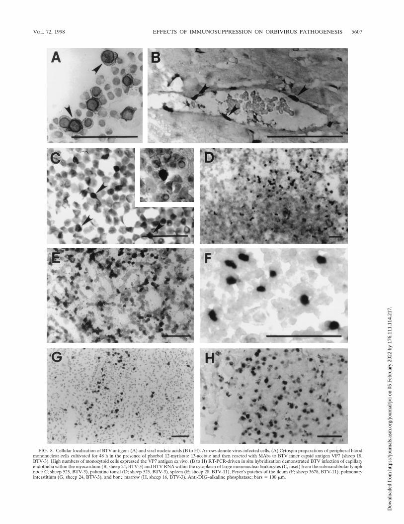
FIG. 8. Cellular localization of BTV antigens (A) and viral nucleic acids (B to H). Arrows denote virus-infected cells. (A) Cytospin preparations of peripheral bloodmononuclear cells cultivated for 48 h in the presence of phorbol 12-myristate 13-acetate and then reacted with MAbs to BTV inner capsid antigen VP7 (sheep 18,BTV-3). High numbers of monocytoid cells expressed the VP7 antigen ex vivo. (B to H) RT-PCR-driven in situ hybridization demonstrated BTV infection of capillaryendothelia within the myocardium (B; sheep 24, BTV-3) and BTV RNA within the cytoplasm of large mononuclear leukocytes (C, inset) from the submandibular lymphnode C; sheep 525, BTV-3), palantine tonsil (D; sheep 525, BTV-3), spleen (E; sheep 28, BTV-11), Peyer’s patches of the ileum (F; sheep 3678, BTV-11), pulmonaryinterstitium (G, sheep 24, BTV-3), and bone marrow (H, sheep 16, BTV-3). Anti-DIG–alkaline phosphatase; bars 5 100 mm.
VOL. 72, 1998 EFFECTS OF IMMUNOSUPPRESSION ON ORBIVIRUS PATHOGENESIS 5607
Dow
nloa
ded
from
http
s://j
ourn
als.
asm
.org
/jour
nal/j
vi o
n 05
Feb
ruar
y 20
22 b
y 17
6.11
1.11
4.21
7.

ciated lesions. Proliferative and infiltrative lesions associatedwith chronic OvLV infection may provide the optimal micro-environment for BTV replication and dissemination. In addi-tion to their immunosuppressive properties, lentiviruses areknown to have bidirectional interaction with other viruses (28).They can interact and alter and/or accelerate the diseasecourse. For instance, HIV up-regulates herpesvirus genomeexpression and promotes transmissibility. This has been dem-onstrated in vitro through experiments showing transactiva-tion, CD4 up-regulation, Fc receptor induction, pseudotypeformation, cytokine production, and antigen presentation (15,28, 34, 38).
Vascular degeneration and hemorrhage were present in awide variety of tissues, mostly from animals treated with im-munosuppressive drugs or with concurrent OvLV infection.The lesions were acute (lacked evidence of inflammation or areparative process) and nonexpansive and were found exclu-sively in immunosuppressed animals, mostly those infectedwith BTV-3. Studies in mice have shown that intracranial BTVinfection typically results in cerebral hemorrhage and necrosis(53). Others have shown that neurovirulence is attributed togenomic differences in viral serotypes (13) and the ability ofviruses to gain access to the CNS (54). We observed highnumbers of vascular endothelial cells in the CNS of immuno-compromised animals to be infected with BTV-3, yet viral geneexpression remained low. Furthermore, there was no evidenceof neuronal or glial cell infection. These findings suggest thatBTV-3 may have an increased tropism for the CNS, at leastunder circumstances of impaired immunity. In a related study,we have shown that deer infected with epizootic hemorrhagicdisease virus, an orbivirus closely related to BTV, had signifi-cant CNS involvement manifest by widespread cerebral hem-orrhage and massive infection of vascular endothelium (11).
Our results show that widespread dissemination of BTVoccurs during early stages of infection and is heightened inanimals with impaired immunity. This may provide a mecha-nism for accelerated disease and, for animals that survive,increased viral persistence. These findings also suggest that inaddition to virulence factors that define viral serotypes, immu-nosuppression could play a role in the natural history of orbi-virus infection, allowing for higher virus burden, increasedvirus persistence, accelerated disease, and greater potential foracquisition of virus by the arthropod vector.
ACKNOWLEDGMENTS
We thank Katherine D. Bardsley and Tamarind Keating for techni-cal assistance and manuscript preparation, Walter J. Tabachnick forhelpful discussions and manuscript critique, Lee H. Thompson forproviding viruses, and Edward McGriff, Leonard Santistevan, and Dar-lene Tabachnick for animal care. We also acknowledge the WyomingState Veterinary Laboratory for hematological and clinical chemistryanalyses.
This study was supported by funds from the USDA, AgriculturalResearch Service, the Colorado State University Experimental Station(1-56271), and the Public Health Service (AI36613).
REFERENCES
1. Barratt-Boyes, S. M., P. V. Rossitto, J. L. Stott, and N. J. MacLachlan. 1992.Flow cytometric analysis of in vitro bluetongue virus infection of bovineblood mononuclear cells. J. Gen. Virol. 73:1953–1960.
2. Barratt-Boyes, S. M., and N. J. MacLachlan. 1994. Dynamics of viral spreadin bluetongue virus infected calves. Vet. Microbiol. 40:361–371.
3. Barratt-Boyes, S. M., and N. J. MacLachlan. 1995. Pathogenesis of blue-tongue virus infection of cattle. J. Am. Vet. Med. Assoc. 9:1322–1329.
4. Begara, I., L. Juj’an, D. S. Collie, H. R. Miller, and N. J. Watt. 1995. In vitroresponse of lymphocytes from bronchoalveolar lavage fluid and peripheral
blood to mitogen stimulation during natural maedi-visna virus infection. Vet.Immunol. Immunopathol. 49:75–88.
5. Berkowitz-Balshayi, C., A. Rosen-Wolff, G. Darai, and Y. Becker. 1995.Immunosuppression of mice by cyclophosphamide (CyP) and cyclosporin A(CsA) changes the course of HSV-1 infection and the distribution of viralDNA in target organs. In Vivo 9:155–162.
6. Brewer, A. W., and N. J. MacLachlan. 1994. The pathogenesis of bluetonguevirus infection of bovine blood cells in vitro: ultrastructural characterization.Arch. Virol. 136:287–298.
7. Brodie, S. J., K. A. Marcom, L. D. Pearson, B. C. Anderson, A. de laConcha-Bermejillo, J. A. Ellis, and J. C. DeMartini. 1992. The effects ofvirus load in the pathogenesis of lentivirus-induced lymphoid interstitialpneumonia. J. Infect. Dis. 166:531–541.
8. Brodie, S. J., L. D. Pearson, G. D. Snowder, and J. C. DeMartini. 1993.Host-virus interaction as defined by amplification of viral DNA and serologyin lentivirus infected sheep. Arch. Virol. 130:413–428.
9. Brodie, S. J., L. D. Pearson, M. C. Zink, H. M. Bickle, B. C. Anderson, K. A.Marcom, and J. C. DeMartini. 1995. Ovine lentivirus expression and disease:virus replication, but not entry, is restricted to macrophages of specifictissues. Am. J. Pathol. 146:1–13.
10. Brodie, S. J., H. M. Bickle, and J. C. DeMartini. 1995. Ovine lentivirus-associated subclinical encephalomyelitis: virological markers in cerebrospi-nal fluid are predictive of central nervous system disease. Clin. Immunol.Immunopathol. 77:14–18.
11. Brodie, S. J., K. D. Bardsley, J. O. Mecham, K. Diem, S. E. Norelius, andW. C. Wilson. Epizootic hemorrhagic disease: analysis of tissues by amplifi-cation and in situ hybridization reveals widespread orbivirus infection at lowcopy number. J. Virol. 72:3863–3871.
12. Calisher, C. H. 1995. Medically important arboviruses of the United Statesand Canada. Clin. Microbiol. Rev. 7:89–116.
13. Carr, M. A., C. C. de Mattos, C. A. de Mattos, and B. I. Osburn. 1994.Association of bluetongue virus gene segment 5 with neuroinvasiveness.J. Virol. 68:1255–1257.
14. Cheney, I. W., M. Yamakawa, P. Roy, J. O. Mecham, and W. C. Wilson. 1996.Molecular characterization of the segment 2 gene of epizootic hemorrhagicdisease virus serotype 2: gene sequence and genetic diversity. Virology 224:555–560.
15. Clouse, K. A., P. B. Robbins, B. Fernie, J. M. Ostrove, and A. S. Fauci. 1989.Viral antigen stimulation of the production of human monokines capable ofregulating HIV-1 expression. J. Immunol. 143:470–475.
16. Coen, M. L., J. A. Ellis, D. T. O’Toole, and W. C. Wilson. 1991. Cytokinemodulation of the interaction between bluetongue virus and endothelial cellsin vitro. Vet. Pathol. 28:524–532.
17. de la Concha-Bermejillo, A., C. E. Schore, C. A. Dangler, C. C. de Mattos,C. A. de Mattos, and B. I. Osburn. 1992. Comparison of slot blot nucleic acidhybridization, immunofluorescence, and virus isolation techniques to detectbluetongue virus in blood mononuclear cells from cattle with experimentallyinduced infection. Am. J. Vet. Res. 53:2245–2250.
18. de Mattos, C. A., C. C. P. de Mattos, B. I. Osburn, and N. J. Maclachlan.1994. Heterogeneity of the L2 gene of field isolates of bluetongue virusserotype 17 from the San Joaquin Valley of California. Virus Res. 31:67–87.
19. de Mattos, C. C. P., C. A. de Mattos, B. I. Osburn, and N. J. MacLachlan.1994. Evolution of the L2 gene of strains of bluetongue virus serotype 10isolated in California. Virology 201:173–177.
20. de Swart, R. L., P. S. Ross, H. H. Timmerman, H. W. Vos, P. J. Reijnders,J. G. Vos, and A. D. Osterhaus. 1995. Impaired cellular immune response inharbour seals (Phoca vitulina) feeding on environmentally contaminatedherring. Clin. Exp. Immunol. 101:480–486.
21. Ellis, J. A., and J. C. DeMartini. 1985. Immunomorphologic and morpho-metric changes in pulmonary lymph nodes of sheep with progressive pneu-monia. Vet. Pathol. 22:32–41.
22. Ellis, J. A., and J. C. DeMartini. 1985. Partial purification and assay innormal sheep and sheep with ovine progressive pneumonia. Vet. Immunol.Immunopathol. 8:15–25.
23. Ellis, J. A., and J. C. DeMartini. 1985. Evidence of decreased concanavalinA induced suppressor cell activity in the peripheral blood and pulmonarylymph nodes of sheep with ovine progressive pneumonia. Vet. Immunol.Immunopathol. 8:93–106.
24. Ellis, J. A., M. L. Coen, N. J. MacLachlan, W. C. Wilson, E. S. Williams, andA. J. Leudke. 1993. Prevalence of bluetongue virus expression in leukocytesfrom experimentally infected ruminants. Am. J. Vet. Res. 54:1452–1456.
25. Fiscus, S. A., J. C. DeMartini, and L. D. Pearson. 1982. Mitogen-inducedblastogenesis of peripheral blood and efferent lymph lymphocytes fromsheep. Am. J. Vet. Res. 43:629–632.
26. Foster, N. M., A. J. Luedke, I. M. Parsonson, and T. E. Walton. 1991.Temporal relationships of viremia, interferon activity, and antibody re-sponses of sheep infected with several bluetongue virus strains. Am. J. Vet.Res. 52:192–196.
27. Garssen, J., W. Goettsch, F. de Gruijl, W. Slob, and H. van Loveren. 1996.Risk assessment of UVB effects on resistance to infectious diseases. Photo-chem. Photobiol. 64:269–274.
5608 BRODIE ET AL. J. VIROL.
Dow
nloa
ded
from
http
s://j
ourn
als.
asm
.org
/jour
nal/j
vi o
n 05
Feb
ruar
y 20
22 b
y 17
6.11
1.11
4.21
7.

28. Griffiths, P. D. 1996. Herpesviruses and AIDS. J. Antimicrob. Chemother.37(Suppl. B):87–95.
29. Holland, J. J., K. Spindler, F. Horodyski, E. Grabau, S. Nichol, and S.VandePol. 1982. Rapid evolution of RNA genomes. Science 215:1577–1585.
30. Jeggo, M. H., R. C. Wardley, and J. Brownlie. 1984. A study of the role ofcell-mediated immunity in bluetongue virus infection in sheep, using cellularadoptive transfer techniques. Immunology 52:403–410.
31. Katz, J., D. Alstad, G. Gustafson, and J. Evermann. 1994. Diagnostic anal-ysis of the prolonged bluetongue virus RNA presence found in the blood ofnaturally infected cattle and experimentally infected sheep. J. Vet. Diagn.Invest. 6:139–142.
32. Kennedy-Stoskopf, S., C. M. Zink, and O. Narayan. 1989. Pathogenesis ofovine lentivirus-induced arthritis: phenotypic evaluation of T lymphocytes insynovial fluid, synovium, and peripheral circulation. Clin. Immunol. Immu-nopathol. 52:323–330.
33. Kinlen, L. 1992. Immunosuppressive therapy and acquired immunologicaldisorders. Cancer Res. 52(Suppl. 19):5474–5476.
34. Lusso, P., A. De Maria, M. Malnati, F. Lori, S. E. DeRocco, M. Baseler, andR. C. Gallo. 1991. Induction of CD4 and susceptibility to HIV-1, infection ofhuman CD81 T lymphocytes by human herpesvirus 6. Nature 349:533–535.
35. MacLachlan, N. J., G. Jagels, P. V. Rossitto, P. F. Moore, and H. W.Heidner. 1990. The pathogenesis of experimental bluetongue virus infectionof calves. Vet. Pathol. 27:223–229.
36. MacLachlan, N. J., R. A. Nunamaker, J. B. Katz, M. M. Sawyer, G. Y. Akita,B. I. Osburn, and W. J. Tabachnick. 1994. Detection of bluetongue virus inthe blood of inoculated calves: comparison of virus isolation, PCR assay, andin vitro feeding of Culicoides variipennis. Arch. Virol. 136:1–8.
37. Marcom, K. A., S. J. Brodie, L. D. Pearson, and J. C. DeMartini. 1992.Analysis of ovine lentivirus infectivity and replication using a focal immu-noassay and an antigen-capture ELISA. J. Clin. Microbiol. 30:2852–2858.
38. McKeating, J. A., P. D. Griffiths, and R. A. Weiss. 1990. HIV susceptibilityconferred to human fibroblasts by cytomegalovirus-induced Fc receptor.Nature 343:659–661.
39. Mecham, J. O. 1993. Detection of bluetongue virus from blood of infectedsheep by use of an antigen-capture enzyme-linked immunosorbent assayafter amplification of the virus in cell culture. Am. J. Vet. Res. 54:370–372.
40. Mistr’ikov’a, J., D. Furd’ikov’a, I. Oravcova, and J. Rajc’ani. 1996. Effect ofimmunosuppression on Balb/c mice infected with murine herpesvirus. ActaVirol. 40:41–44.
41. Mo, C. L., L. H. Thompson, E. J. Homan, M. T. Oviedo, E. C. Greiner, J.Gonzalez, and M. R. Saenz. 1994. Bluetongue virus isolations from vectors
and ruminants in Central America and the Caribbean. Am. J. Vet. Res.55:211–215.
42. Morrison, W. I. 1986. The ruminant immune system in health and disease.Cambridge University Press, New York, N.Y.
43. Nelson, J. A., P. Ghazal, and C. A. Wiley. 1990. Role of opportunistic viralinfections in AIDS. AIDS 4:1–10.
44. Parsonson, I. M. 1990. Pathology and pathogenesis of bluetongue infections.Curr. Top. Microbiol. Immunol. 162:119–141.
45. Parsonson, I. M. 1992. Overview of bluetongue virus infection of sheep, p.713–724. In T. E. Walton and B. I. Osburn (ed.), Bluetongue, African horsesickness, and related orbiviruses. CRC Press, Boca Raton, Fla.
46. Parsonson, I. M., L. H. Thompson, and T. E. Walton. 1994. Experimentallyinduced infection with bluetongue virus serotype 11 in cows. Am. J. Vet. Res.55:1529–1534.
47. Pyrah, I. T., and N. J. Watt. 1996. Immunohistochemical study of the de-pressed cutaneous DTH response in sheep naturally infected with an ovinelentivirus (Maedi-Visna virus). Clin. Exp. Immunol. 104:32–36.
48. Richards, R. G., N. J. MacLachlan, H. W. Heidner, and F. J. Fuller. 1988.Comparison of virologic and serologic responses of lambs and calves infectedwith bluetongue virus serotype 10. Vet. Microbiol. 18:233–242.
49. Shad, G., W. C. Wilson, J. O. Mecham, and J. F. Evermann. 1997. Blue-tongue virus detection: a safer reverse-transcriptase polymerase chain reac-tion for prediction of viremia in sheep. J. Vet. Diagn. Invest. 9:118–124.
50. Starr, S. E. 1996. Novel mechanism of immunosuppression after measles.Lancet 348:1257–1258.
51. Tabachnick, W. J. 1996. Culicoides variipennis and bluetongue-virus epide-miology in the United States. Annu. Rev. Entomol. 41:23–43.
52. Tishon, A., M. Manchester, F. Scheiflinger, and M. B. Oldstone. 1996. Amodel of measles virus-induced immunosuppression: enhanced susceptibilityon neonatal human PBLs. Nat. Med. 2:1250–1254.
53. Venter, E. H., J. J. van der Lugt, and G. H. Gerdes. 1993. Detection ofbluetongue virus RNA in cell cultures and in the central nervous system ofexperimentally infected mice using in situ hybridization. Onderstepoort J.Vet. Res. 60:39–45.
54. Waldvogel, A. S., J. L. Stott, K. R. E. Squire, and B. I. Osburn. 1986.Strain-dependent virulence characteristics of bluetongue virus serotype 11.J. Gen. Virol. 67:765–769.
55. Whetter, L. E., N. J. MacLachlan, D. H. Gebhard, H. W. Heidner, and P. F.Moore. 1989. Bluetongue virus infection of bovine monocytes. J. Gen. Virol.70:1663–1676.
VOL. 72, 1998 EFFECTS OF IMMUNOSUPPRESSION ON ORBIVIRUS PATHOGENESIS 5609
Dow
nloa
ded
from
http
s://j
ourn
als.
asm
.org
/jour
nal/j
vi o
n 05
Feb
ruar
y 20
22 b
y 17
6.11
1.11
4.21
7.