PURINERGIC MECHANISMS AS POTENTIAL PHARMACOLOGICAL … · PHARMACOLOGICAL TARGETS FOR ENTERIC...
167
CÁTIA ANDREIA RODRIGUES VIEIRA PURINERGIC MECHANISMS AS POTENTIAL PHARMACOLOGICAL TARGETS FOR ENTERIC INFLAMMATORY MOTILITY DISORDERS Tese de Candidatura ao grau de Doutor em Ciências Biomédicas, submetida ao Instituto de Ciências Biomédicas Abel Salazar da Universidade do Porto. Orientador – Professor Doutor Paulo Correia-de-Sá Categoria – Professor Catedrático Afiliação – Instituto de Ciências Biomédicas Abel Salazar da Universidade do Porto.
Transcript of PURINERGIC MECHANISMS AS POTENTIAL PHARMACOLOGICAL … · PHARMACOLOGICAL TARGETS FOR ENTERIC...

CÁTIA ANDREIA RODRIGUES VIEIRA
PURINERGIC MECHANISMS AS POTENTIAL
PHARMACOLOGICAL TARGETS FOR ENTERIC
INFLAMMATORY MOTILITY DISORDERS
Tese de Candidatura ao grau de Doutor em
Ciências Biomédicas, submetida ao Instituto de
Ciências Biomédicas Abel Salazar da Universidade
do Porto.
Orientador – Professor Doutor Paulo Correia-de-Sá
Categoria – Professor Catedrático
Afiliação – Instituto de Ciências Biomédicas Abel
Salazar da Universidade do Porto.

CÁTIA ANDREIA RODRIGUES VIEIRA
PURINERGIC MECHANISMS AS POTENTIAL
PHARMACOLOGICAL TARGETS FOR ENTERIC
INFLAMMATORY MOTILITY DISORDERS
Dissertation in fulfilment of the requirements for the
PhD degree in Biomedical Sciences, submitted to
Instituto de Ciências Biomédicas Abel Salazar of
the University of Porto
Supervisor – Professor Paulo Correia-de-Sá
Category – Full Professor
Affiliation – Instituto de Ciências Biomédicas Abel
Salazar of the University of Porto

This research was partially supported by Fundação para a Ciência e a Tecnologia
(FCT,FEDER funding) (projects: PTDC/SAU-OSM/73576/2006,
EEQ/1168/SAU/2005, REEQ/1264/SAU/2005, PEst-OE/SAU/UI0215/201 and PEst-
OE/SAU/UI0215/2014) and by University of Porto / Caixa Geral de Depósitos
(Investigação Científica na Pré-Graduação).
The author was in receipt of a PhD Studentship from FCT (POPH – QREN/FSE
funding, SFRH/BD/79091/2011).

ACKNOWLEDGEMENTS
My acknowledgements goes to Instituto de Ciências Biomédicas Abel Salazar,
to University of Porto and to Foundation for Science and Technology (FCT).
I would like to make a very special acknowlegment to Professor Paulo Correia-
de-Sá for the opportunity to integrate and belong to his group, has now gone a few
years. To him, I owe my progress and growth in science. Thanks for his technical
and scientific orientation, for the permanent disponibility and by his wise teachings.
I wish to thank him, because he believed in me and in my scientific skills.
I would like to thanks all the persons that had worked with me and had a direct
collaboration in this work. A special thanks to Dr.ª Teresa Magalhães-Cardoso, to
Professor Fátima Ferreirinha, to Dr.ª Isabel Silva and to Dr.ª Ana Sofia Dias. I
would like to thank also to Professor Jean Sévigny for his collaboration in this
work.
I would like to make a special acknowlegment to Dr.ª Alexandrina Timóteo, to
Professor Graça Lobo and to Dr.ª Isabel Silva for their support, in the good and in
the bad moments. Thanks a lot for their invaluable friendship.
I would like to thank to the group working at the Laboratory of Pharmacology,
for all the moments that shared with me in the last years. A special thanks goes to
Professor Laura Oliveira, to Professor Patrícia Sousa, to Professor Adelina, to
Professor Miguel Faria, to Professor Miguel Cordeiro, to Professor Margarida
Duarte-Araújo, to Dr.ª Mariana Certal, to Dr.ª Aurora Barbosa and to Dr. Bruno
Bragança. I would like also to thank to the younger elements, Dr.ª Adriana, Dr.ª
Mafalda and to Dr.ª Salomé to their sympathy. I also thank to Mrs Belmira, Helena
Costa e Silva and Suzete Liça for their friendship and technical assistance.
I would like to thank to the persons that are not anymore in the group, but that
shared with me very good moments in the time that they were part of it. Thanks to
Doctor Bernardo Noronha-de-Matos, to Doctor Ana Rita Pinheiro, to Dr.ª Patrícia
Marques, to Dr.ª Cristina Eusébio Mendes, to Dr.ª Cristina Costa, to Dr. Diogo
Paramos, to Dr.ª Silvia Marques, to Dr.ª Nádia Monteiro and to Dr.ª Sónia Gomes.

My very special thanks go to my Family, in special to my parents and to my
sister and brother, for all their support (always unconditional), for all the help and
for the words of encouragement in all moments. Despite the distance they are
always close.
I would like to make my special acknowledgment to my second family, the
family that adopted me here in Porto.Thanks a lot to Sr. Carlos, D. Rosário, to
Rosarinho, to Pedro and to my little princess Leonor to all the moments that share
all days with me.
Finally, and as it should be, I have to make a very special thanks to my sweet
love for all his care, friendship and support in any and all the moments, even in the
lazy moments on the couch. Thanks for being everyday there, sharing his life with
me.

TABLE OF CONTENTS
I. ABBREVIATIONS ------------------------------------------------------------------------------ 9 II. RESUMO --------------------------------------------------------------------------------------- 13 III. ABSTRACT ------------------------------------------------------------------------------------ 17 IV. INTRODUCTION ----------------------------------------------------------------------------- 21
1. Autonomic nervous system ------------------------------------------------------------------------------------ 21 2. Enteric nervous system ----------------------------------------------------------------------------------------- 22
2.1. Enteric neurons -------------------------------------------------------------------------------------------- 24 2.1.1. Morphology of enteric neurons ------------------------------------------------------------------ 24 2.1.2. Electrophysiological properties of enteric neurons------------------------------------------ 25 2.1.3. Neurochemical code of enteric neurons ------------------------------------------------------- 25 2.1.4. Function of enteric neurons ---------------------------------------------------------------------- 25
2.1.4.1. Sensory neurons --------------------------------------------------------------------------------- 26 2.1.4.2. Motor neurons ------------------------------------------------------------------------------------ 27 2.1.4.3. Interneurons -------------------------------------------------------------------------------------- 28 2.1.4.4. Secretomotor and vasomotor neurons ----------------------------------------------------- 28 2.1.4.5. Intestinofugal neurons -------------------------------------------------------------------------- 28
2.1.5. Enteric neurons in the longitudinal muscle-myenteric plexus (LM-MP) ---------------- 29 2.2. Enteric Glial Cells ------------------------------------------------------------------------------------------ 30 2.3. Interstitial cells of Cajal ----------------------------------------------------------------------------------- 33 2.4. Fibroblast like cells (FLC)-------------------------------------------------------------------------------- 34 2.5. Smooth muscle cells -------------------------------------------------------------------------------------- 35
3. Purinergic Transmission ---------------------------------------------------------------------------------------- 36 3.1. Ectonucleotidases ----------------------------------------------------------------------------------------- 38 3.2. Purinergic receptors -------------------------------------------------------------------------------------- 41
3.2.1. Adenosine Receptors (P1 receptors) ---------------------------------------------------------- 42 3.2.1.1. Adenosine ----------------------------------------------------------------------------------------- 43 3.2.1.2. Adenosine Transport---------------------------------------------------------------------------- 45
3.2.2. P2 Receptors----------------------------------------------------------------------------------------- 46 3.2.2.1. P2X Receptors ----------------------------------------------------------------------------------- 47 3.2.2.2. P2Y Receptors ----------------------------------------------------------------------------------- 48
4. Cholinergic Transmission -------------------------------------------------------------------------------------- 49 5. Purinergic and cholinergic transmission in the Myenteric Plexus ------------------------------------ 50
5.1. P1 Receptors ----------------------------------------------------------------------------------------------- 51 5.2. P2X Receptors --------------------------------------------------------------------------------------------- 52 5.3. P2Y Receptors --------------------------------------------------------------------------------------------- 54 5.4. Nicotinic and Muscarinic Receptors ------------------------------------------------------------------- 56
6. Inflammatory bowel diseases --------------------------------------------------------------------------------- 58 6.1. Ileitis ---------------------------------------------------------------------------------------------------------- 60 6.2. Animal models of inflammatory bowel diseases --------------------------------------------------- 60
6.2.1. TNBS-Induced ileitis -------------------------------------------------------------------------------- 61
V. GOALS ------------------------------------------------------------------------------------------ 65 VI. ORIGINAL RESEARCH PAPERS ------------------------------------------------------- 66
PAPER 1 ------------------------------------------------------------------------------------------------------------------ 67 ABSTRACT -------------------------------------------------------------------------------------------------------------- 68 INTRODUCTION -------------------------------------------------------------------------------------------------------- 69

MATERIAL AND METHODS -------------------------------------------------------------------------------------------- 72 RESULTS --------------------------------------------------------------------------------------------------------------- 79 DISCUSSION AND CONCLUSIONS ----------------------------------------------------------------------------------- 96
PAPER 2 ---------------------------------------------------------------------------------------------------------------- 104 ABSTRACT ------------------------------------------------------------------------------------------------------------ 105 INTRODUCTION ------------------------------------------------------------------------------------------------------ 106 MATERIAL AND METHODS ------------------------------------------------------------------------------------------ 109 RESULTS ------------------------------------------------------------------------------------------------------------- 115 DISCUSSION AND CONCLUSIONS --------------------------------------------------------------------------------- 126
VII. CONCLUDING REMARKS --------------------------------------------------------------- 137 VIII. REFERENCES ------------------------------------------------------------------------------- 141

ABBREVIATIONS
9 PhD Thesis: Cátia Andreia Rodrigues Vieira
I. ABBREVIATIONS
ABC proteins, ATP-binding cassette transporters
ACh, acetylcholine
ADA, adenosine deaminase
ADO, adenosine
ADP, adenosine diphosphate
AHP, prolonged after-hyperpolarization
AIN, ascending interneuron
AK, adenosine kinase
AMP, adenosine monophosphate
ANO-1, anoctamine 1
ANS, autonomic nervous system
AP, alkaline phosphatases
ATP, adenosine triphosphate
BDNF, brain-derived neurotrophic factor
Calb, calbindin
cAMP, cyclic adenosine monophosphate
CFTR, cystic fibrosis transmembrane conductance regular
CGRP, calcitonin gene related peptide
ChAT, choline acetyltransferase
C-Kit, tyrosine kinase receptor
CM, circular muscle
CNS, central nervous system
CNT, concentrative nucleoside transporters
CO, carbon monoxide
DAG, diacylglycerol
DIN, descending interneurons
DMPP, dimethylphenylpiperazinium
DMSO, dimethyl sulfoxide
DPCPX, 1,3-dipropyl-8-cyclopentylxanthine
DSS, dextran sodium sulphate
DYN, dynorphin
ABBREVIATIONS
10 PhD Thesis: Cátia Andreia Rodrigues Vieira
EFS, electrical field stimulation
EMN, excitatory motor neuron
ENK, enkephalins
E-NPP, ecto-nucleotide pyrophosphatases/phosphodiesterases
ENS, enteric nervous system
ENT, equilibrative nucleoside transporters
E-NTPDase, ecto-nucleoside triphosphate diphosphohydrolase
EPAN, extrinsic primary afferent neuron
ER, endoplasmic reticulum
fEPSPs, fast excitatory post-synaptic potentials
FLC, fibroblast-like cell
GABA, gamma-aminobutyric acid
GFAP, intermediate filament glial fibrillary acidic protein
GI, gastrointestinal
GPCR, G protein-coupled receptors
GPI, glycosylphosphatidylinositol
GRP, gastrin releasing peptide
HO1, heme oxygenase-1
HPLC, high performance liquid chromatography
HX, hypoxanthine
IBD, inflammatory bowel disease
ICC, interstitial cells of Cajal
IFN, intestinofugal neuron
IFN-γ, interferon γ
IL, interleukin
IMN, inhibitory motor neuron
INO, inosine
iNOS, inducible nitric oxide synthase
IP3, inositol triphosphate
IPAN, intrinsic primary afferent neuron
KO, knockout
LGIC, ligand-gated ionotropic channel
LM-MP, longitudinal muscle-myenteric plexus

ABBREVIATIONS
11 PhD Thesis: Cátia Andreia Rodrigues Vieira
mAChR, muscarinic receptors of acetylcholine
Min, minutes
NA, noradrenaline
nAChR, nicotinic receptors of acetylcholine
NBTI, S-(4-Nitrobenzyl)-6-thioinosine
NGF, nerve grow factor
nNOS, nitric oxide synthase
NO, nitric oxide
NOS, nitric oxide synthetase
NT, nucleoside transporter
NT-3, neurotrophin-3
NT-4, neurotrophin-4
NYP, neuropeptide Y
PBS, phosphate saline buffer
PDGFR-α, a platelet derived growth factor receptor α
PGP 9.5, protein gene product 9.5
PLC, phospholipase C
PLP, periodate-lysine-paraformaldehyde
PM-ML, plexo mioentérico-músculo longitudinal
PVG, sympathetic prevertebral ganglia
ROS, reactive species
S100, calcium-binding protein
SAH, S-adenosyl-L-homocysteine hydrolase
SLC, solute carrier transporters
SM, submucosal plexus
SMC, smooth muscle cells
SMN, secretomotor/ vasomotor neuron
Sox 10, transcription factor SRY box-containing gene 10
SP, substance P
SNS, sympathetic nervous system
TNBS, 2,4,6-trinitrobenzenesulfonic acid.
TNF, tumour necrosis factor
TRC, T cell receptor

ABBREVIATIONS
12 PhD Thesis: Cátia Andreia Rodrigues Vieira
TTX, tetrodotoxin
UDP, uridine diphosphate
UTP, uridine triphosphate
UV, ultraviolet
VAChT, ACh vesicular transporter
VIP, vasoactive intestinal polypeptide
VNUT, vesicular nucleotide transporter
5-HT, 5-hydroxytryptamine
RESUMO
13 PhD Thesis: Cátia Andreia Rodrigues Vieira
II. RESUMO
As purinas parecem desempenhar um papel fisiológico na regulação da
motilidade intestinal, podendo afectar a actividade dos nervos entéricos, das fibras
musculares, das células entéricas gliais, das células intersticiais de Cajal (ICC) e
das células do tipo fibroblasto (FLC). A inflamação do intestino provoca alterações
estruturais e funcionais mais ou menos duradoiras no sistema nervoso entérico
(SNE) e está associada à elevação dos níveis extracelulares das purinas,
nomeadamente ATP e adenosina. Partindo de estudos anteriores controversos
olhando para o controlo da inflamação pelas purinas, este projecto foi desenhado
para definir o papel da cascata purinérgica no controlo da actividade do SNE após
uma situação de inflamação aguda. O propósito do trabalho foi apontar possíveis
alvos farmacológicos para o tratamento das doenças inflamatórias intestinais.
Neste contexto, foram gerados modelos animais portadores de ileíte induzida
através da instilação intraluminal de TNBS na ratazana e os mesmos foram
utilizados para fins experimentais sete dias após a indução – modelo de ileíte pós-
inflamatória. Todas as experiências foram realizadas em preparações de plexo
mioentérico-músculo longitudinal (PM-ML) de íleo destes animais.
Neste modelo animal, colocou-se a hipótese da reacção inflamatória induzir
alterações na densidade da população celular residente (para além da infiltração
por células inflamatórias) capaz de gerar um desequilíbrio na libertação de
purinas e, consequentemente, na libertação de ACh[3H] – o neurotransmissor
mais relevante no controlo da motilidade intestinal. Para responder a essa
questão, foram usadas preparações de PM-ML do íleo de ratazana marcadas com
anticorpos específicos para cada um dos componentes celulares envolvidos. Tal
como seria de esperar, verificou-se um aumento significativo do número de
células inflamatórias marcadas com CD11B/Ox42 localizadas na região adjacente
aos gânglios mioentéricos, poupando o seu interior. A reacção inflamatória fez-se
acompanhar de um aumento da densidade das células gliais, marcadas
positivamente com GFAP ou S100β, e de uma perda de células intersticiais
(ICC/FLC) imunoreactivas contra a vimentina e anoctamina-1 (Ano-1). Tal como
RESUMO
14 PhD Thesis: Cátia Andreia Rodrigues Vieira
constatado por outros autores em modelos experimentais de ileíte pós-
inflamatória, não se observaram alterações significativas na densidade e na
distribuição de dois marcadores neuronais específicos, PGP9.5 e o NF200,
contrariamente aos resultados obtidos em modelos mais severos de colite
induzida pelo TNBS, onde a perda neuronal foi evidente.
Seguidamente exploraram-se as alterações funcionais subjacentes às
modificações estruturais encontradas no modelo de ileíte pós-inflamatória. A
estimulação das preparações de PM-ML do íleo de ratazanas submetidas ao
tratamento com TNBS libertaram menor quantidade de ACh[3H]. Contudo, esta
diminuição da transmissão colinérgica não foi acompanhada por uma perda
significativa da imunoreactividade para o transportador vesicular de ACh, VaChT.
Em animais controlo, a libertação de ACh resulta da exocitose vesicular em
resposta à despolarização nervosa, sensível à TTX. No entanto, no tecido sujeito
a inflamação a libertação de ACh para além de se encontrar significativamente
reduzida, é também, menos sensível ao bloqueio pela TTX. A hipótese de que a
libertação de ACh nos neurónios inflamados se pode dever à acção de um
gliotransmissor excitatório capaz de activar directamente os terminais nervosos na
ausência de potencial de acção foi comprovada através da inibição pelo
fluoroacetato de sódio. Trata-se de uma toxina que afecta o metabolismo das
células gliais entéricas e que reduziu significativamente os níveis de ATP
libertados pelo plexo mioentérico do íleo de ratazana após tratamento pelo TNBS.
O efeito inibitório do fluoroacetato de sódio sobre a libertação de ACh[3H]
nas preparações inflamadas pelo TNBS foi reproduzido pelo antagonista selectivo
dos receptores P2X7, A438079, e pelo o inibidor dos hemicanais contendo
panexina-1, carbenoxolona. O mesmo efeito não foi observado quando se
preveniu a actividade das células intersticiais ICC/FLC através da inibição das
correntes Cav3 (do tipo T) com mibefradil. Estes resultados sugerem que o ATP
parece ser o gliotransmissor excitatório responsável pela activação directa dos
terminais mioentéricos através da activação de receptores ionotrópicos do tipo
RESUMO
15 PhD Thesis: Cátia Andreia Rodrigues Vieira
P2X2 e P2X2/3 e que a sua acumulação é dependente da actividade do complexo
P2X7/ panexina-1.
Contrariamente ao aumento da quantidade de ATP libertado pelo íleo de
ratazanas tratadas com TNBS observou-se uma redução significativa dos níveis
extracelulares do seu metabolito, adenosina, bem como da neuromodulação
colinérgica mediada pela activação preferencial de receptores facilitatórios de
subtipo A2A localizados nos terminais nervosos colinérgicos. Os resultados
mostram que a disparidade entre o aumento da libertação de ATP e o défice de
formação de adenosina na ileíte pós-inflamatória pode ser atribuída, pelo menos
em parte, à inibição anterógrada da ecto-5'-nucleotidase/CD73 (a enzima que
converte AMP em adenosina) devido à acumulação de elevados níveis de ATP e
ADP na sinapse mioentérica. Esta acumulação deve-se a uma redistribuição da
localização da NTPDase2 (uma ATPase) da região ganglionar para a junção
neuromuscular, sem afectar a densidade da NTPDase3 no plexo mioentérico dos
animais tratados com TNBS, facto que condiciona a formação de ADP a partir do
ATP naquela região intestinal. O défice de acumulação extracelular de adenosina
na ileíte pós-inflamatória pode, ainda, dever-se ao aumento da sua inactivação
em inosina pela ADA (frações membranar e solúvel). Existe um terceiro
mecanismo passível de ser responsabilizado pela redução da neuromodulação
adenosinérgica no plexo mioentérico inflamado, que deriva da redução
significativa da densidade da população de células intersticiais ICC/FLC. De facto,
a inibição da actividade das ICC/FLC através do bloqueio dos canais de Cav3 (do
tipo T) característicos destas células, reduz consideravelmente os níveis de
adenosina libertados pelo plexo mioentérico, tanto em animais controlo como
injectados com TNBS, situação que contrasta com a ausência de efeito da
gliotoxina, fluoroacetato de sódio. Sabe-se que as células intersticiais libertam
quantidades significativas de adenosina por intermédio do transportador
equilibrativo de nucleósidos sensível ao dipiridamol, mediante a activação de
receptores muscarínicos do subtipo M3. Este mecanismo pode estar deficitário no
intestino inflamado, já que a libertação de ACh se encontra atenuada e que a
densidade de células intersticiais ICC/FLC também está diminuída, situação que

RESUMO
16 PhD Thesis: Cátia Andreia Rodrigues Vieira
culmina no bloqueio do mecanismo de retroalimentação positivo operado pela
activação de receptores facilitatórios A2A nas terminações nervosas colinérgicas.
Em conclusão, os resultados mostram que a transmissão colinérgica está
significativamente afectada no modelo de ileíte pós-inflamatória e que esse
distúrbio pode resultar da existência de um desequilíbrio entre a neuromodulação
adenosinérgica, influenciada pela actividade das células intersticiais ICC/FLC
predominante em situações fisiológicas, e a excessiva activação dos mecanismos
mediados pelo ATP resultantes da proliferação glial promovida pelo processo
inflamatório. Se por um lado, a libertação de ATP através da actividade do
complexo P2X7/panexina-1 pode levar à activação de receptores ionotrópicos dos
subtipos P2X2 e P2X2/3 nos terminais nervosos colinérgicos mioentéricos
favorecendo a libertação directa do neurotransmissor para manter o tónus
colinérgico, por outro lado a metabolização do ATP em ADP, sem a consequente
formação de adenosina no íleo inflamado, pode quebrar a facilitação da libertação
de ACh operada pela activação sucessiva dos receptores facilitatórios M3 e A2A
localizados, respectivamente, nas células intersticiais e nos terminais nervosos
colinérgicos. Assim, os resultados deste trabalho sugerem que a manipulação da
libertação diferencial das purinas, a intervenção sobre as suas vias de inactivação
e a activação de certos subtipos de receptores purinérgicos podem constituir
alvos terapêuticos interessantes para o tratamento das doenças inflamatórias
intestinais.

ABSTRACT
17 PhD Thesis: Cátia Andreia Rodrigues Vieira
III. ABSTRACT
Purines seem to play a physiological role in the regulation of gut motility by
affecting the activity of enteric nerves, muscle fibers, enteric glial cells, interstitial
cells of Cajal (ICC) and fibroblast cells (FLC). Intestinal inflammation may cause
long-term structural and functional changes in the enteric nervous system (ENS),
which might be related to increased levels of extracellular purines, namely ATP
and adenosine. Taking into account controversies in the literature regarding the
control of enteric inflammation by purines, this project was designed to investigate
the role of the purinergic cascade in the control of SNE activity following acute
inflammation. Our aim was to identify putative molecular targets for the
pharmacological treatment of inflammatory enteric disorders. To achieve this goal,
we used an animal model of postinflammatory ileitis due to intraluminal instillation
of TNBS in the rat; experimental animals were used seven days after surgery. All
experiments were performed in longitudinal muscle-myenteric plexus (LM-MP)
preparations of rat ileum.
In this animal model, we hypothesized that inflammation may cause
relevant changes in the density of resident cell populations (beyond the typical
inflammatory infiltrates), which may unbalance the release of purines and,
consequently, the release of [3H]ACh – the most important neurotransmitter in the
control of gut motility. To answer this question, LM-MP preparations were stained
with specific antibodies directed against each specific cell type. As expected, we
observed a significant increase in the number of inflammatory cells stained with
CD11b/OX42, which were localized adjacent to myenteric ganglia, without
penetrating this structure. The inflammatory insult, cause a significant increase in
the density of enteric glial cells, exhibiting positive immunoreactivity against GFAP
and S100β, and a substantial loss of interstitial cells ICC/FLC expressing vimentin
and anoctamine-1 (Ano-1). In agreement with the results obtained by other
authors using the postinflammatory ileitis rat model, we failed to detect any
changes in the density and distribution of two specific neuronal markers, PGP9.5
ABSTRACT
18 PhD Thesis: Cátia Andreia Rodrigues Vieira
and NF200. These findings are in contrast with those obtained in more severe
TNBS-induced colitis where neuronal loss is more evident.
Next, we investigated functional abnormalities reflected in the enteric
nervous system that could be motivated by the structural changes detected in the
postinflammatory ileitis rat model. Stimulation of the myenteric plexus of the ileum
of rats treated with TNBS released significantly smaller amounts of [3H]ACh than
their control littermates. However, the reduction in the cholinergic tone was not
accompanied by changes in the immunoreactivity against the ACh vesicular
transporter, VAChT. In control animals, ACh release results from vesicle
exocytosis of depolarized cholinergic nerves sensitive to TTX. Conversely, ACh
release from inflamed myenteric nerve terminals was less sensitive to action
potential blockade with TTX, indicating that it might occur through direct activation
of axon terminals by an excitatory mediator released from non-neuronal cells.
Implication of ATP released from proliferating glial cells was suggested because
inhibition of enteric glial cells metabolism with sodium fluoroacetate decreased
both the release of [3H]ACh and ATP from LM-MP preparations of the ileum of
TNBS-treated rats.
The inhibitory effect of sodium fluoroacetate on the release of [3H]ACh was
reproduced by the P2X7 receptor antagonist, A438079, and by the pannexin-1
hemichannel blocker, carbenoxolone, but the same effect was not observed in the
presence of mibefradil, a Cav3 (T-type) channel blocker that prevents the activity
of interstitial cells ICC/FLC. These results suggest that ATP might be the
excitatory gliotransmitter responsible for the activation of ionotropic P2X2 and/or
P2X2/3 receptors operating direct activation of cholinergic nerve terminals and that
extracellular accumulation of the nucleotide may be dependent on the
P2X7/pannexin-1 pathway.
In contrast to the increase in ATP overflow, the ileum of TNBS-treated rats
accumulate smaller amounts of its metabolite, adenosine, resulting in a reduction
in the preferential activation of facilitatory A2A receptors located on cholinergic
nerve terminals. The disparity between increased ATP overflow and adenosine

ABSTRACT
19 PhD Thesis: Cátia Andreia Rodrigues Vieira
deficits in post-inflammatory ileitis was ascribed, at least in part, to feed-forward
inhibition of ecto-5'-nucleotidase/CD73 (the enzyme that converts AMP into
adenosine) by high amounts of ATP and ADP accumulated at the myenteric
synapse. Redistribution of NTPDase2 (ATPase) from the ganglion region to the
myenteric neuromuscular synapse, without much affecting NTPDase3, might
explain surplus ADP accumulation near cholinergic nerve terminals in TNBS-
treated rats. Deficits in extracellular adenosine in postinflammatory ileitis may also
occur due to increases in adenosine inactivation into inosine by ADA (membrane-
bound and soluble fractions). A third mechanism may also account for the loss of
adenosine neuromodulation in the inflamed myenteric plexus, which derives from
a reduction in the density of interstitial cells ICC/FLC population. As a matter of
fact, inhibition of ICC/FLC activity by blocking Cav3 (T-type) channels, decreases
significantly endogenous adenosine outflow by LM-MP preparations of both control
and TNBS-treated rats. This situation was not observed in the presence of the
gliotoxin, sodium fluoroacetate. It has been shown that interstitial cells release
significant amounts of adenosine, via a dipyridamole-sensitive equilibrative
nucleoside transporter, through the activation of muscarinic M3 receptors. This
mechanism may be deficient in postinflammatory ileitis, since ACh release is
downregulated and the number of interstitial cells is reduced, leading to blockage
to the positive feedback loop operated by facilitatory muscarinic M3 (on ICC/FLC)
and adenosine A2A (on cholinergic nerve terminals) receptors that are responsible
for maintaining the safety margin of myenteric cholinergic transmission.
In conclusion, data suggest that cholinergic neurotransmission is severly
affected in postinflammatory ileitis, a situation that might result from an unbalance
between adenosinergic neuromodulation (that is dominant in physiological
conditions) and excessive ATP accumulation attributed to enteric glial cells
proliferation promoted by the inflammatory insult. In principle, surplus ATP
overflow gated by P2X7/pannexin-1 pathway may facilitate activation of ionotropic
P2X2 and/or P2X2/3 receptors on cholinergic nerve terminals resulting in direct
activation of neurotransmitter release independently of action potential generation,
in order to keep the cholinergic tone at a critical miminum in the inflamed ileum.
ABSTRACT
20 PhD Thesis: Cátia Andreia Rodrigues Vieira
But, on the other hand, ATP hydrolysis into ADP without significant adenosine
formation detected in postinflammatory ileitis may break the facilitatory loop
required to sustain neurotransmitter release operated by muscarinic M3 and
adenosine A2A receptors localized on interstitial cells ICC/FLC and cholinergic
nerve terminals, respectively. Thus, results presented here suggest that selective
targeting the differential release of purines, the activity of the purinergic
inactivation mechanisms and the activation of specific purinoceptor subtypes may
offer novel pharmacological strategies for the treatment of inflammatory intestinal
diseases.
INTRODUCTION
21 PhD Thesis: Cátia Andreia Rodrigues Vieira
IV. INTRODUCTION
1. Autonomic nervous system
Nervous system has an important role in all digestive processes; it’s
responsible for the control of motility, secretion, sensory perception, and immune
function (Straub et al., 2006). Autonomic nervous system (ANS) has two main
functions, excitation and inhibition, of physiological processes. These functions are
controlled through different kinds of nerve fibres, entitled excitatory and inhibitory.
So, ANS is responsible for the unconscious control of essential functions for life
being (e.g. blood circulation, heart function, digestion, temperature, stress). ANS
has the purpose of preparing the body for stress and recovering the same level of
activation following stressful conditions. It comprises three fundamental parts
(figure 1): sympathetic, parasympathetic and enteric nervous system (ENS)
described in 1921 by Langley (Lundgren et al., 1989). Sympathetic and
parasympathetic nervous system establish a connection between the central
nervous system and peripheral organs. Sympathetic nervous system (SNS)
together with the hypothalamic-pituitary-adrenal axis is known by the main role in
the “fight and flight reaction” during stressful conditions. An efferent sympathetic
pathway is activated in the brain, goes along the intermediolateral column of the
spinal cord and come to sympathetic ganglia. Afterwards, the signal goes to
postganglionic sympathetic nerve fibres, and enters in the intestine wall along
arteries through the mesenteric serosa surface (Straub et al., 2006). On the other
hand, parasympathetic nervous system is known by the main role of “rest and
digestion”; it works predominantly during relaxation conditions. It has one main
contributor, the vagus nerve. Vagal branches comprise sensory fibres (afferent)
and motor fibres (efferent). They are responsible for the communication between
the brain and visceral organs (Stakenborg et al., 2013). Simple functions like food
intake, digestion and immunity are controlled by the interaction between the vagal
nerve fibres and the gut. Extrinsic sympathetic and parasympathetic fibres
terminate in vessel walls or in enteric plexuses that constitute the ENS. Like it was
described, ENS can operate dependently of central nervous system, but unlike the
other systems described so far, it can function independently of the central

INTRODUCTION
22 PhD Thesis: Cátia Andreia Rodrigues Vieira
nervous system, and this activity is exclusive of the gastrointestinal (GI) tract. For
that reason, ENS was called by several authors the “second brain”. In this work we
will focus our attention on the ENS.
Figure 1: Diagram representative of Nervous system.
2. Enteric nervous system
The ENS has approximately 100 million neurons, a number equal to or
greater than the number of neurons found in the spinal cord (Furness & Costa,
1980). The ENS is composed by three major ganglionated plexuses and several
aganglionated plexuses. The myenteric (or Auerbach’s), the submucosal (or
Meissner’s) and the mucosal are described as the ganglionated plexuses. The
non-ganglionated plexuses, like the deep muscular plexus, inner submucosal
plexus, periglandular plexus among others, supply all the layers of the intestine
(Hansen, 2003).
The myenteric plexus is placed between longitudinal muscle and circular
muscle layers and the submucosal plexus is located in the submucosa.
Concerning to their functions in the ENS, submucosal plexus controls especially
local gut secretion and local absorption, while myenteric plexus controls mainly GI
movements and secretion of enzymes from adjacent organs, but these dominant
functions may be mixed. When stimulated, ENS main effects include: (1) increase
INTRODUCTION
23 PhD Thesis: Cátia Andreia Rodrigues Vieira
of gut wall tonic contraction, (2) augmentation of the rhythmic contractions, (3)
slight increase of the contraction rate, and (4) enhancement in the transmission
velocity of excitatory waves along the gut wall. These effects originate an increase
of the gut peristaltic waves.
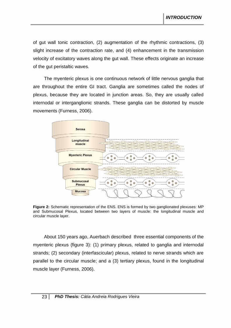
The myenteric plexus is one continuous network of little nervous ganglia that
are throughout the entire GI tract. Ganglia are sometimes called the nodes of
plexus, because they are located in junction areas. So, they are usually called
internodal or interganglionic strands. These ganglia can be distorted by muscle
movements (Furness, 2006).
Figure 2: Schematic representation of the ENS. ENS is formed by two ganglionated plexuses: MP and Submucosal Plexus, located between two layers of muscle: the longitudinal muscle and circular muscle layer.
About 150 years ago, Auerbach described three essential components of the
myenteric plexus (figure 3): (1) primary plexus, related to ganglia and internodal
strands; (2) secondary (interfascicular) plexus, related to nerve strands which are
parallel to the circular muscle; and a (3) tertiary plexus, found in the longitudinal
muscle layer (Furness, 2006).

INTRODUCTION
24 PhD Thesis: Cátia Andreia Rodrigues Vieira
Figure 3: Schematic draw of components of the myenteric plexus.
2.1. Enteric neurons
Enteric neurons are organized in networks of enteric ganglia that are
connected by intraganglionic strands that extend from the upper oesophagus to
the internal anal sphincter (Costa et al., 2000; Furness et al., 1998). There are
different classes of enteric neurons, classified based on their morphology,
electrical properties, neurochemical code and function (Table 1). In that way, it
was identified seventeen different types of neurons in the small intestine, but only
14 are described as functionally important (Furness, 2000; Hansen, 2003). The
majority of the studies done on enteric neurons taxonomy were done in the
guinea-pig small intestine (Costa et al., 1996; Brehmer et al., 1999; Furness, 2000;
Costa, 2000).
2.1.1. Morphology of enteric neurons
Based on their morphology, in 1899, Dogiel proposed a classification to the
enteric neurons. He classified enteric neurons as Dogiel type I, II and III. Later,
were described four more types of enteric neurons (type IV, V, VI, and VII)
(Brehmer et al., 1999). Most of enteric neurons are from types I, II and III
(Furness, 2000). How can these neurons be differentiated? Type I, is
characterized by having a flattened cell body and numerous dendrites projecting
into the musculature. The type II, possess an oval cell body with long processes
projecting to ganglion cells. The type III, is characterized by a rectangular cell body

INTRODUCTION
25 PhD Thesis: Cátia Andreia Rodrigues Vieira
with irregular dendrites. In this latter neurons, the axons project to the mucosa
(Brehmer et al., 1999).
2.1.2. Electrophysiological properties of enteric neurons
Based on different electrophysiological properties, enteric neurons were
classified in two possible ways, AH neurons and S neurons (Furness et al., 1997).
When an action potential is generated in the cell body of AH neurons, it is followed
by a prolonged after-hyperpolarization (AHP), whereas if the same occurs in S
neurons the action potential is followed by prominent fast excitatory post-synaptic
potential (fEPSP). Morphologically S neurons are Dogiel type I, whereas AH
neurons are Dogiel type II (Furness, 2006; Nurgali, 2009).
2.1.3. Neurochemical code of enteric neurons
Primary transmitters, transmitters synthesizing enzymes and
neuromodulators released from enteric neurons include: acetylcholine (ACh),
norepinephrine (NE), adenosine triphosphate (ATP), serotonin (5-HT), dopamine
(DA), cholecystokinin (CCK), substance P (SP), vasoactive intestinal polypeptide
(VIP), somatostatin (SOM), leu-enkephalin (leu-Enk), met-eukephalin (met-Enk)
and bombesin. These compounds provide to the enteric neurons a neurochemical
code. Based on that neurochemical code, most enteric neurons are classified
essentially as nitrergic neurons, cholinergic neurons or peptidergic neurons
(Furness, 2006). To make this distinction, immunolocalization studies using
specifically markers, like nitric oxide synthase (nNOS), choline acetyltransferase
(ChAT), SP, calcitonin-gene related peptide (CGRP) and VIP may be undertaken.
Co-localization studies indicate the subclasses of neurons that co-express
neurotransmitters and neuromodulators (Chiocchetti et al., 2009). The
neurochemical code and density of specific neurons differ between gut regions
and between animal species (Furness, 2006).
2.1.4. Function of enteric neurons
Functionally, enteric neurons were classified as sensory, inhibitory and
excitatory motor neurons, interneurons, secretomotor/ vasomotor neurons and
INTRODUCTION
26 PhD Thesis: Cátia Andreia Rodrigues Vieira
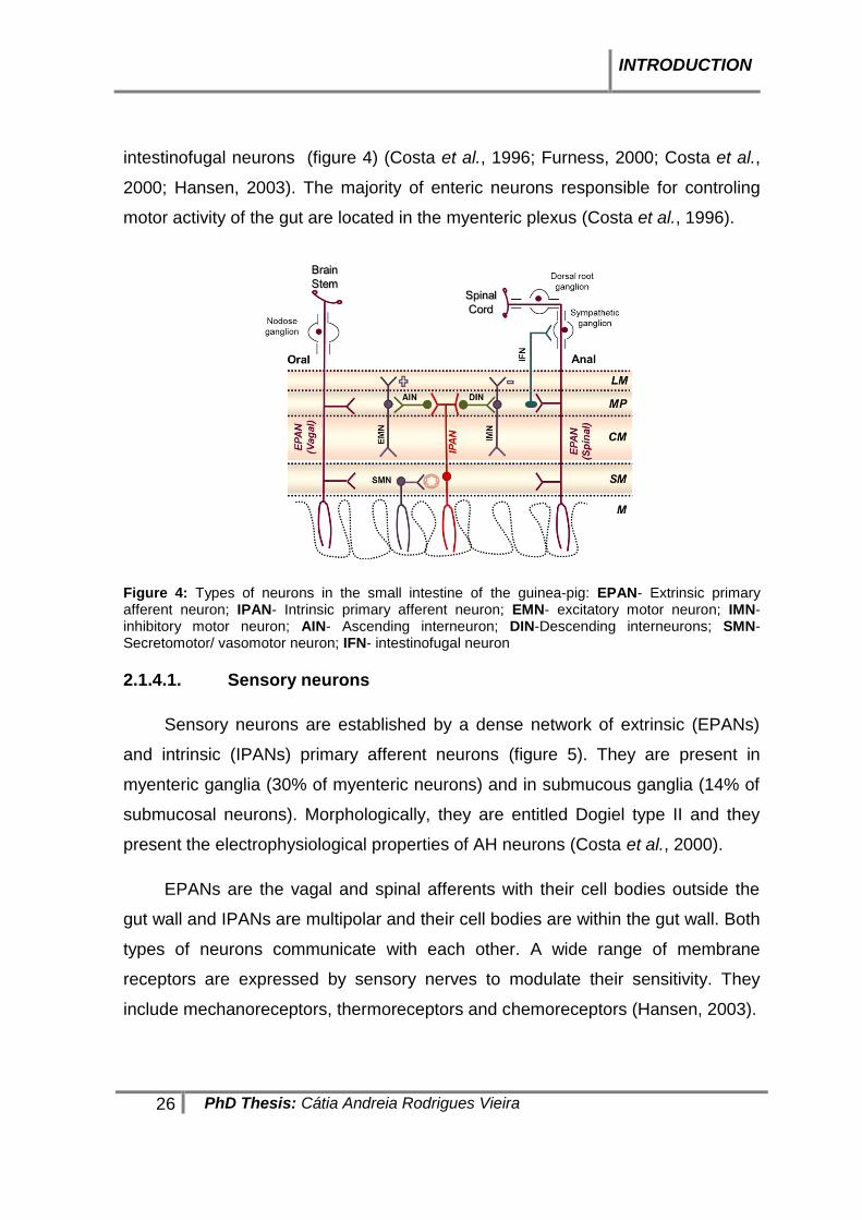
intestinofugal neurons (figure 4) (Costa et al., 1996; Furness, 2000; Costa et al.,
2000; Hansen, 2003). The majority of enteric neurons responsible for controling
motor activity of the gut are located in the myenteric plexus (Costa et al., 1996).
Figure 4: Types of neurons in the small intestine of the guinea-pig: EPAN- Extrinsic primary afferent neuron; IPAN- Intrinsic primary afferent neuron; EMN- excitatory motor neuron; IMN- inhibitory motor neuron; AIN- Ascending interneuron; DIN-Descending interneurons; SMN-Secretomotor/ vasomotor neuron; IFN- intestinofugal neuron
2.1.4.1. Sensory neurons
Sensory neurons are established by a dense network of extrinsic (EPANs)
and intrinsic (IPANs) primary afferent neurons (figure 5). They are present in
myenteric ganglia (30% of myenteric neurons) and in submucous ganglia (14% of
submucosal neurons). Morphologically, they are entitled Dogiel type II and they
present the electrophysiological properties of AH neurons (Costa et al., 2000).
EPANs are the vagal and spinal afferents with their cell bodies outside the
gut wall and IPANs are multipolar and their cell bodies are within the gut wall. Both
types of neurons communicate with each other. A wide range of membrane
receptors are expressed by sensory nerves to modulate their sensitivity. They
include mechanoreceptors, thermoreceptors and chemoreceptors (Hansen, 2003).

INTRODUCTION
27 PhD Thesis: Cátia Andreia Rodrigues Vieira
Figure 5: Types of sensory neurons, extrinsic (EPANs) and intrinsic (IPANs) primary afferent neurons in the ENS. LM, longitudinal muscle; MP, myenteric plexus; CM, circular muscle; SM, submucosal plexus; M, mucosa.
2.1.4.2. Motor neurons
Are described five types of motor neurons: excitatory muscle motor neurons,
inhibitory muscle motor neurons, secretomotor/vasodilator neurons, secretomotor
neurons that are not vasodilator and neurons that are innervating entero-endocrine
cells (Furness, 2000). Motor neurons belongs to the Dogiel type I morphology and
presents the electrophysiological properties of S neurons (Costa et al., 2000)
Longitudinal and circular muscles and the muscularis mucosae are innervated by
muscle motor neurons. They can be excitatory or inhibitory. In that sense, they
release neurotransmitters that promote muscle contractions or relaxations
(Hansen, 2003). Motor neurons are immunoreactive to ChAT (enzyme
responsible for the synthesis of ACh) and to SP (Costa et al., 2000; Furness
2000). Thus, the excitatory muscle motor neurons release transmitters like ACh
and tachykinins (SP and neurokinin A). On the other hand, inhibitory muscle motor
neurons release substances like nitric oxide (NO), VIP, ATP, GABA, neuropeptide
Y (NPY) and carbon monoxide (CO) (Costa et al., 2000; Hansen, 2003).

INTRODUCTION
28 PhD Thesis: Cátia Andreia Rodrigues Vieira
2.1.4.3. Interneurons
The interneurons have both electrophysiological properties described above,
may be AH neurons (morphologically, Dogiel type II) or S neurons
(morphologically, Dogiel type I) (Costa et al., 2000; Hansen, 2003). Ascending
interneurons (one type of interneurons oriented in the oral direction) and
descending interneurons (three types of interneurons oriented in the anal
direction) are the types characterized in the small intestine of the guinea-pig
(Furness, 2000). Ascending interneurons are essentially cholinergic, while the
descending interneurons use as a neurotransmitters VIP, NO and some opioid
peptides (Costa et al., 2000). The three types of descending neurons express
different chemical codings: ChAT/NOS/VIP, ChAT/SOM and ChAT/5-HT (Furness,
2000).
2.1.4.4. Secretomotor and vasomotor neurons
Secretomotor neurons are responsible for the control of secretions, while
vasomotor neurons are responsible for the control of blood flow (Hansen, 2003).
The majority of secretomotor neurons are projected to the mucosa (with their cell
bodies in the mucosal plexus). Some few neurons are projected from the
submucosa to the myenteric plexus (Furness, 2000). There are only 1% of these
neurons in the myenteric ganglia, whereas most of them are located in the
submucosous ganglia (32-42%) (Costa et al., 2000). There are two classes of
secretomotor neurons: cholinergic and non-cholinergic. The cholinergic neurons
release ACh as a transmitter. ACh will act on muscarinic receptors located on
mucosal epithelium. The non-cholinergic neurons release VIP and mediate most of
the local reflex responses (Hansen, 2003; Costa et al., 2000).
2.1.4.5. Intestinofugal neurons
Intestinofugal neurons are the single neurons that are projected from the
myenteric ganglia to the sympathetic prevertebral ganglia (PVG), and that relay
mechanosensory information to the sympathetic prevertebral ganglia neurons.
They release ACh at the sympathetic prevertebral ganglia and evoke fast

INTRODUCTION
29 PhD Thesis: Cátia Andreia Rodrigues Vieira
excitatory postsynaptic potentials (fEPSPs) (Costa et al., 2000; Szurszewski et al.,
2002).
Table 1: Morphologic, electrophysiological and functional characterization of enteric neurons (Furness, 2000; Hansen, 2003, Furness et al., 2003, Sharkey et al., 1998, Wong et al., 2008)
FUNCTION MORPHOLOGY ELECTROPHYSIOLOGY NEUROCHEMICAL
CODING
Sensory (IPANs/ EPANs)
Dogiel type II AH ChAT, Calb, CGRP, SP
Excitatory motor neurons
Dogiel type I S
ChAT, SP, Calret
Inhibitory motor neurons
DYN, ENK, NOS, and VIP
Ascending interneurons
Dogiel type I/ II AH/ S ChAT, Calret, ENK, SP
Descending interneurons
Dogiel type I/ II AH/ S 5-HT, DYN, GRP, NOS, somatostatin, and VIP
Secretomotor neurons (Cholinergic)
Type IV (Stach) S
S
ChAT, CCK, CGRP, DYN, NYP, somastostatin, and VIP.
Secretomotor neurons (Non-Cholinergic)
Dogiel type I
Intestinofugal neurons
Dogiel type I S ChAT, VIP, CCK, ENK, DYN, GRP
ChAT, coline acetyltransferase; Calb, calbindin; CGRP, calcitonin generelated peptide; SP, substance P; DYN, dynorphin; GRP, gastrin releasing peptide, ENK, enkephalins, NOS, nitric oxide synthetase; VIP, vasoactive intestinal polypeptide; 5HT, 5-hydroxytryptamine; NYP, neuropeptide Y.
2.1.5. Enteric neurons in the longitudinal muscle-myenteric plexus (LM-MP)
The experimental work presented in this thesis is based on the use of the
longitudinal muscle-myenteric plexus (LM-MP) preparation of rat ileum described
by Paton and Vizi (1969). This preparation is highly enriched in cholinergic
neurons, especially excitatory motor neurons that are projected to the LM (25%).
They receive inputs from the IPANs (26%) and from the ascending and

INTRODUCTION
30 PhD Thesis: Cátia Andreia Rodrigues Vieira
descending interneurons (17%) (Costa et al., 1996). Although ACh can be
originated in the pre-ganglionic cholinergic nerve endings, this only represents a
small proportion of the total content of ACh measured experimentally. This is due
to the existence of an excessive number of fibers in the ganglion LM-MP
compared to extrinsic pre-ganglionic fibers in the same preparation (Paton & Vizi,
1969).
2.2. Enteric Glial Cells
Enteric glial cells have huge morphological and molecular similarities to
astrocytes found in the central nervous system (CNS). Enteric glial cells and
astrocytes share electrophysiological properties and express two common
proteins: the GFAP-intermediate filament glial fibrillary acidic protein and a
calcium-binding protein S100 (Gulbransen & Sharkey, 2012).
Until very recently, it was thought that enteric glial cells had only a supportive
function to neuronal cells. More recently, countless papers demonstrate that
enteric glial cells are able to modulate enteric neural circuits, but their full role on
this matter is yet to be defined experimentally. Mounting evidences also point
towards enteric glial cells being neuroprotective and able to communicate with
neighbouring neurons (Neunlist et al., 2014; Boesmans et al., 2013). They supply
neurons with neurotransmitter precursors, release neuroactive substances (called
gliotransmitters), express neurotransmitter receptors and respond to
neurotransmitters/ neuromodulators, and have the machinery for sequestering and
degrade neuroactive substances, namely ATP, glutamate, GABA (Rühl, 2005). In
addition, these cells may exert and housekeeping role by expressing enzymes and
transporters in order to control the availability of neuroactive substances that are
toxic to the enteric neurons.
Around 1899, enteric glial cells in the gut were first described by Dogiel. They
display an irregular, stellate-shaped, with an extensive array of highly branched
processes. These cells present a small cell body, with a big nucleus occupying the
all cell body leaving a little cytoplasmic volume (Gulbransen, 1994).

INTRODUCTION
31 PhD Thesis: Cátia Andreia Rodrigues Vieira
Enteric glial cells are present in all layers of the gut wall and are closely
associated with neuronal cells within the ganglia, interganglionic fiber strands and
intramuscular nerve fibres (Neunlist et al., 2014). There are several types of
enteric glial cells throughout the GI tract (figure 6).
Figure 6: Distribution of enteric glial cells along the GI tract. They form a large and extensive network throughout all gut layers.
Hanani was the first to propose a classification for these cells, based only on
morphological differences (Hanani and Reichenbach, 1994). He classified enteric
glial cells in four types that are described in more detail in table 2 below.
To study enteric glial cells there are different markers, such as GFAP, S100β
and Sox-10 (transcription factor SRY box-containing gene 10) (Neunlist et al.,
2014). To the quantification of glial cells versus neurons, the preferential markers
are Sox-10 (which labels the nucleus of glial cells) and HuC/D (which labels the
cell bodies of neurons) (Boesmans et al., 2013). However, nuclear stainings are
unable to define cell boundaries where neurotransmitter receptors and ion
channels are located and, therefore, GFAP and S100β were preferred in this study
(see Table 2). It is also possible to discriminate glial cells from enteric neurons by
their functional characteristics. For instance, enteric neurons display a strong and
fast Ca2+ response to high K+ depolarization, electrical field stimulation (EFS) and
to the activation of nicotinic receptors with 1,1-dimethyl-4-phenylpiperazinium
(DMPP), while enteric glial cells do not respond to these stimuli directly. They
show delayed responses compared to neurons, which can be modulated by

INTRODUCTION
32 PhD Thesis: Cátia Andreia Rodrigues Vieira
several components of the purinergic signalling cascade (Boesmans et al., 2013),
namely NTPDase2 catalysing extracellular ATP hydrolysis and ADP formation
favouring P2Y1 receptor activation, and the danger signal represented by
activation of the ionotropic P2X7 receptor secondary to high levels of ATP
(reviewed in Gulbransen & Sharkey, 2012).
Despite these functional differences, our knowledge about the
pathophysiological significance of enteric glial cells communication to neurons and
vice versa is extremely limited although these cells are ideally positioned to
participate in diverse aspects of GI functions. Regional particularities, intrincate
distribution and intimate relationship between enteric glial cells, neurons and other
non-neuronal cell types like insterstitial cells of Cajal and fibroblast-like cells (see
below) do not facilitate the study of these complexities, but emphasize the
importance of any single report concerning this issue.
Table 2: Types and localization of enteric glial cells (according to Boesmans et al., 2015; Gulbransen & Sharkey, 2012).
Type of enteric glial cells Localization Image
Type I (protoplasmic enteric gliocytes)
Ganglia (between the neurons)
Type II (fibrous enteric gliocytes) Interganglionar
Type III (mucosal enteric gliocytes) Extraganglionar region along
the nerve fibers; can also be present along small blood vessels
Type IV (intramuscular enteric gliocytes)
Intramuscular (circular and longitudinal muscle), along the nerve fibers.
Original immunofluorescence confocal microscopy images of enteric glia from LM-MP of the rat ileum using an antibody against calcium-binding protein S100β (green) (obtained in collaboration with Doctor Fátima Ferreirinha).

INTRODUCTION
33 PhD Thesis: Cátia Andreia Rodrigues Vieira
2.3. Interstitial cells of Cajal
Interstitial cells of Cajal (ICC’s) were discovered by Cajal around 1889. In
that year, he thought that these cells were primitive neurons. But after more than
100 years of studies by several authors, it was shown that they were not neurons
and do not originate from the neural crest (Garcia-Lopez et al., 2009). These cells
have been target of scientific discussion for years, because of their similarities with
neurons and fibroblasts (Sanders et al., 2014). The most important discover in this
field was the expression of the tyrosine kinase receptor (c-Kit) at the surface of
these cells; this receptor plays a crucial role in their development (Maeda et al.,
1992). Nowadays it is recognized that ICC’s are of mesenchymal origin (Wards &
Sanders, 2001). We also know that ICC’s can be differentiated from other cells
expressing high amounts of anoctamin 1, Ano-1 (Tmem16A), which is a calcium-
activated chloride channel (Gomez-Pinilla et al., 2009).
ICC’s are dispersed around nerve plexuses and between smooth muscle
fibres of the gut. They function as pacemaker cells, because of their important role
in the control of spontaneous motility of the gut. ICC’s are responsible for the
propagation of electrical slow waves (Burnstock & Lavin, 2002). These cells have
no basal lamina and form gap junctions with smooth muscle cells (SMC) (Komuro
et al., 1996).
Ganglion level Neuromuscular level
Figure 7: Original immunofluorescence confocal microscopy images of ganglionic (multipolar) and intramuscular (bipolar) ICC from LM-MP of the rat ileum using an antibody against Ano-1 (obtained in collaboration with Doctor Fátima Ferreirinha).

INTRODUCTION
34 PhD Thesis: Cátia Andreia Rodrigues Vieira
Depending of their location within the various layers of the GI tract, ICC’s
have a specific arrangement and cell shape (Komuro, 2006) (see table 3).
Table 3: Nomenclature, localization and morphology of ICC (Komuro, 2006)
Type of ICC Localization Morfology
ICC’s of the subserosa (ICC-SS)
In the subserosal layer.
Stellate cells
ICC’s of longitudinal muscle (ICC-LM) Barely distributed in association
with rather thicker nerve bundles. Bipolar cells
ICC’s of the myenteric plexus (ICC-MP)
Around the myenteric plexus in the space between the circular and longitudinal muscle layers.
Multipolar cells
ICC’s of the circular muscle (ICC-CM) Along the long axis of surrounding
smooth muscle cells. Bipolar cells
ICC’s of the deep muscular plexus (ICC-DMP)
Closely associated with the nerve bundles and circular muscle fibers.
Multipolar cells
ICC’s of the submucosa and submucosal plexus (ICC-SM and ICC-SMP)
At the interface between the submucosal connective tissue and the innermost circular muscle layer.
Multipolar cells
2.4. Fibroblast like cells (FLC)
FLC are another mesenchymal cell type, which is described as a kind of
interstitial cells located on the smooth muscle layer of the GI tract (Kim, 2011).
FLC are located along processes of enteric neurons, between the longitudinal and
circular muscle layers, in juxtaposition with ICC’s, and in the subserosal layer.
These cells are in the vicinity of nerve varicosities and can also form gap junctions
with SMCs (Kurahashi et al., 2011; Iino & Nojyo, 2009). The gap junctions
between FLC and SMCs are smaller than those found between ICC’s and SMCs
(Fujita et al., 2003). This proximity suggests that FLC can be regulated by enteric
nerves and transmit electrical and molecular signals to the smooth muscle fibres
and, ultimately, influence muscle tone (Fujita et al., 2003; Iino & Nojyo, 2009). The
phenotypic characterization of FLC implies the presence of SK3, a small
conductance Ca2+-activated K+ channel (SK channel) (Fujita et al., 2003), of the
INTRODUCTION
35 PhD Thesis: Cátia Andreia Rodrigues Vieira
platelet-derived growth factor receptor α (PDGFR-α) (Kurahashi et al., 2011), and
the cell-cell adhesion factor, CD34 (only found in humans) (Iino & Nojyo, 2009).
According to their morphology, FLC found in the myenteric layer are
multipolar with thin processes, while FLC found in the muscle layers (circular and
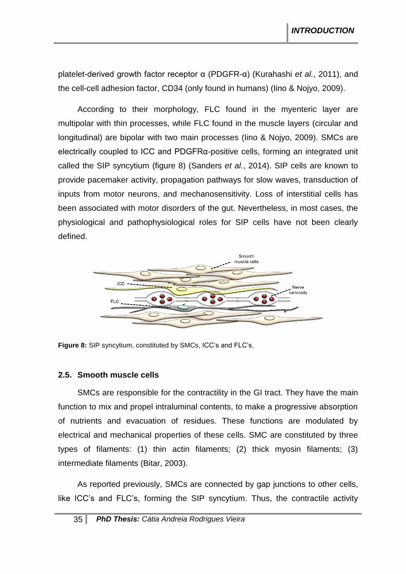
longitudinal) are bipolar with two main processes (Iino & Nojyo, 2009). SMCs are
electrically coupled to ICC and PDGFRα-positive cells, forming an integrated unit
called the SIP syncytium (figure 8) (Sanders et al., 2014). SIP cells are known to
provide pacemaker activity, propagation pathways for slow waves, transduction of
inputs from motor neurons, and mechanosensitivity. Loss of interstitial cells has
been associated with motor disorders of the gut. Nevertheless, in most cases, the
physiological and pathophysiological roles for SIP cells have not been clearly
defined.
Figure 8: SIP syncytium, constituted by SMCs, ICC’s and FLC’s.
2.5. Smooth muscle cells
SMCs are responsible for the contractility in the GI tract. They have the main
function to mix and propel intraluminal contents, to make a progressive absorption
of nutrients and evacuation of residues. These functions are modulated by
electrical and mechanical properties of these cells. SMC are constituted by three
types of filaments: (1) thin actin filaments; (2) thick myosin filaments; (3)
intermediate filaments (Bitar, 2003).
As reported previously, SMCs are connected by gap junctions to other cells,
like ICC’s and FLC’s, forming the SIP syncytium. Thus, the contractile activity
INTRODUCTION
36 PhD Thesis: Cátia Andreia Rodrigues Vieira
produced by SMCs may occur via a mechanism called electromechanical coupling
or via a pharmacomechanical coupling.
The first one, the electromechanical coupling, occurs through several steps
represented in the following sequence:
(1) Depolarization of membrane electrical potentials;
(2) Opening of voltage-gated calcium channels;
(3) Elevation of cytosolic calcium;
(4) Activation of contractile proteins.
The second one, the pharmacomechanical coupling, occurs when chemical
substances released as signals from nerves or from non-neuronal cells located
near to the muscle act as a ligand to its receptor on the muscle membrane. As a
consequence of that occurs the following sequence:
(1) Opening of calcium channels;
(2) Elevation of cytosolic calcium, without any change in the membrane electrical
potential.
3. Purinergic Transmission
The first description of the extracellular action of adenine nucleotides and
nucleosides was published in 1929 by Drury and Szent-Györgyi. These authors
studied the effects of purines in the cardiovascular system of mammals. Despite of
the clearness of the evidences, the recognition of the physiological role of purines
as regulators of intercellular communication has generated some resistance for
many years. In 1972, Burnstock and his collaborators were able to definitively
establish the importance played by adenosine triphosphate (ATP). He coined the
terms "purinergic" (relative to the extracellular mechanisms mediated by purines)
and "purinergic transmission" (denoting to the role of ATP as a neurotransmitter)
(Abbracchio & Burnstock, 1998). Dale’s principle described that the neurons can
synthetize, store and release substances. According to Dale’s principle it was
thought that each neuron released only one neurotransmitter (reviewed in Otsuka,

INTRODUCTION
37 PhD Thesis: Cátia Andreia Rodrigues Vieira
1988). Around 1976, Burnstock introduced the cotransmitter hypothesis; he
demonstrated that ATP is a cotransmitter in the most nerves with classical
neurotransmitters. Nowadays, it is recognized that ATP is released with ACh, NE,
glutamate, GABA, 5-HT and DA in different types of neurons in the CNS
(Burnstock, 2008). ATP can be accumulated into vesicles mediated by a Cl-
dependent vesicular nucleotide transporter (VNUT) (Abbracchio et al., 2008).
Although, the concept that ATP is only stored within synaptic vesicles and
released by exocytosis from nerve terminals and some non-neuronal cells upon
stimulation, has been changing over the past few years. ATP can also be released
by other mechanisms, as follows:
(1) ATP can be released through the activation of ABC proteins (ATP-binding
cassette transporters) (Linton, 2007). This mechanism remains a subject of
controversy. Even though some studies reported that there are systems that
express these ABC proteins capable of transporting ATP, others showed the
inability of these proteins to carry this purine. ABC proteins are the cystic fibrosis
transmembrane conductance regular (CFTR) (Bodin & Burnstock, 2001).
(2) ATP can be released via connexin- and pannexin-containing hemichannels
(Abbracchio et al., 2008). The majority of hemichannels display permeability to
ATP and small dyes, like propidium and ethidium iodide, YoPro and Lucifer yellow.
Membrane depolarization can promote the opening of connexins and pannexins,
but unlike pannexin-1, connexin hemichannels close at normal millimolar Ca2+
concentrations (e.g. 1.8 mM CaCl2 in physiological solutions) and open under Ca2+
depletion conditions (Fasciani et al., 2013). This particularity leaves room for
pannexin-1 hemichannels playing a dominant role in ATP translocation under
normal physiological conditions. Pannexins and connexins have a similar protein
structure: four transmembranar domains, with N and C-terminal located in the
cytosol (Lazarowski et al., 2011).
(3) Huge amounts of ATP can be released from damaged or dying cells upon
membrane cell breaking events.

INTRODUCTION
38 PhD Thesis: Cátia Andreia Rodrigues Vieira
ATP released from cells is quickly hydrolysed by ectonucleotidases (ecto-
enzymes) present in the synaptic cleft, giving rise to biologically active
metabolites, such as ADP, AMP and adenosine (ADO) (Zimmermann, 2000).
Burnstock, in 1978, reported that the physiological effects of adenosine (ADO),
ATP and ADP were mediated by ADO-sensitive P1 receptors and nucleotides-
activated P2 receptors (Abbrancchio & Burnstock, 1998).
Figure 9: Purinergic signalling pathways. ATP is sequentially metabolised into ADP and AMP, by
NTPDases. AMP is then hydrolysed into ADO via ecto-5’-nucleotidase. ADO is deaminated into
inosine (INO) by adenosine desaminase (ADA). The nucleosides can activate P1 receptors and the
nucleotides can activate P2 receptors.
3.1. Ectonucleotidases
Extracellular nucleotides (ATP, ADP and AMP) are hydrolysed into
nucleosides (ADO) through the action of ectoenzymes. The product final of this
metabolic chain, ADO, can be further inactivated by deaminase adenosine (ADA)
into INO. Ectonucleotidases include several members of ENTPDase (ecto-
nucleoside triphosphate diphosphohydrolases), E-NPP (ecto-phosphodiesterase/
nucleotide phosphohydrolases), ecto-alkaline phosphatase and ecto-5’-
nucleotidase (CD73) (Robson et al., 2006). The distribution of these enzymes
throughout tissues is very diverse. They are usually anchored to the membrane,
but some of them can detach from their membrane link and keep activity in the
soluble form, being called exonucleotidases (Zimmermann, 2000). NTPDases are

INTRODUCTION
39 PhD Thesis: Cátia Andreia Rodrigues Vieira
generally cell surface-located enzymes with an extracellular catalytic site, but
some of them can exhibit an intracellular localization (Robson et al., 2006).
Nucleoside triphosphates (ATP and UDP) and diphosphates (ADP and UDP) are
desphosphorylated by NTPDases in the presence of divalent cations (Ca2+ and
Mg2+) (Kukulski et al., 2005).
Eight NTPDases (NTPDase 1 to 8) have been discovered, cloned and
characterized functionally. NTPDases 1, 2, 3 and 8 are expressed on cell surface.
NTPDases 5 and 6 are also expressed intracellularly. NTPDases 4 and 7 are
located exclusively intracellularly (Yegutkin, 2008). NTPDases 4, 5, 6 and 7 are
associated with intracellular organelles, such as the Golgi (Cardoso et al., 2015).
In this work, we will focus in four the ENTPDases that are located on the cell
surface. ENTPDases are extremely important to control signalling through P2
receptors and they can influence ADO formation rate and, subsequently, P1
receptors activation (Kukulski et al., 2005).
Ecto-5’-nucleotidase is an ecto-enzyme responsible for the hydrolysis of
AMP into ADO and phosphate. This enzyme is considered the limiting step for
ADO formation in the majority of the tissues (Cardoso et al., 2015). Ecto-5’-
nucleotidase is attached to the plasma membrane via a glycosyl
phosphatidylinositol (GPI) anchor, which may be cleaved from cell membranes
through hydrolysis of the GPI anchor by phosphatidylinositol-specific
phospholipases or by proteolysis, retaining its catalytic activity in the soluble form
(Sträter, 2006). Ecto-5’-nucleotidase, in both membrane and soluble forms, can be
inhibited by micromolar concentrations of ATP and ADP - feed-forward inhibition
(Vieira et al., 2014). These nucleotides act as competitive inhibitors of this enzyme
because they have the ability to bind to the catalytic site of ecto-5’-nucleotidase
without being hydrolysed.
Adenosine deaminase (ADA) is another important enzyme that is located
predominantly in the cytosol, but can also appear bound to the plasma membrane
in inumerous cells. The irreversible deamination of ADO and 2’-deoxyadenosine
into INO and 2’-deoxyinosine, respectively, is catalysed by ADA (Yegutkin, 2008).
INTRODUCTION
40 PhD Thesis: Cátia Andreia Rodrigues Vieira
Like ecto-5’-nucleotidase, active soluble forms of ADA (exo-ADA) may be released
from activated cells (Correia-de-Sá et al., 2006) or result from membrane cell
damage (Vieira et al., 2014). ADO acts in all P1 receptor subtypes, while INO can
activate A1 and A3 adenosine receptors under certain conditions (Cardoso et al.,
2015).
Alkaline phosphatases (AP) represent a family of enzymes with low affinity
for nucleotides. They are usually attached to the plasma membrane via a GPI
anchor (Zimmermann, 2000). These enzymes promote the release of inorganic
phosphates, pyrophosphates and adenine nucleosides, with a pH optimum for this
catalytic reaction from 8 to 11 (Yegutkin, 2008).
The ecto-nucleotide pyrophosphatase/phosphodiesterase (E-NPP) family
has seven structurally-related ecto-enzymes (NPP1-NPP7). They hydrolyse 5’-
phosphodiester bonds in nucleotides and their derivatives, with the subsequent
release of 5’-nucleoside monophosphates (Goding et al., 2003). This family has a
high substrate specificity to hydrolyse pyrophosphate and phosphodiester bounds
in nucleotides, nucleic acids and nucleotide sugars. However, only the first three
enzymes of this family (NPP1-NPP3) are relevant in the hydrolysis of extracellular
nucleotides, being important to the purinergic signalling cascade. NPP enzymes
are considered really important for the metabolism of extracellular diadenosine
polyphosphates in vertebrate tissues (Yegutkin, 2008). Structurally, they are type II
transmembrane metalloenzymes, characterized by a short intracellular domain N-
terminus, a single transmembrane domain and an extracellular domain containing
a conserved catalytic site (Zimmermann, 2000; Goding et al., 2003). These
enzymes can act as ecto-enzymes, anchored to the cellular membrane, or upon
proteolytic cleavage from cell membranes as exo-enzymes (Goding et al., 2003).

INTRODUCTION
41 PhD Thesis: Cátia Andreia Rodrigues Vieira
Table 4: Role of enzymes of the purinergic cascade and their molecular characteristics (Yegutkin,
2008; Cardoso et al., 2015; Kukulski et al., 2005; Vieira et al., 2014; Robson et al., 2006).
ENZYME STRUCTURE FUNCTION
ENTPDase 1 (ATPDase, CD39 or Apyrase)
Contain two transmembranar domains at the N- and C-terminus
Dephosphorylates ATP directly into AMP, with minimal accumulation of ADP; Hydrolyses ATP, ADP, UTP and UDP with similar efficacy; Converts ATP to ADP and ADP to AMP; Active in pH ranging from 7 to 10.
ENTPDase 2 (ecto-ATPase, CD39L1)
Contain two transmembranar domains at the N- and C-terminus
Preferential triphosphonucleosidase; Hydrolyses ADP 10 to 15 times less efficiently than ATP; Converts ATP and UTP to ADP and UDP; Active at physiological pH as well in acidic conditions. Is inactive at pH > 9
ENTPDase 3 (CD39L3 or HB6)
Contain two transmembranar domains at the N- and C-terminus
Functional intermediate between NTPDase 1 and 2; Active in pH ranging from 5 to 11.
ENTPDase 8 Contain two transmembranar domains at the N- and C-terminus
Functional intermediate between NTPDase 1 and 2.
Ecto-5’-Nucleotidase (CD73)
Contain a glycosyl phosphatidylinositol (GPI) anchor, by which the mature protein is attached to the cell membrane via the C-terminal serine residue linked to a complex oligoglycan and a sphingolipidinositol group.
Dephosphorylates AMP into ADO. Usually the rate limiting step for ADO formation from released adenine nucleotides.
ADA (CD26) More than 90% intracellular It is also located at the cell surface.
Converts ADO to the inactive metabolite, INO.
3.2. Purinergic receptors
Adenine nucleotide and nucleosides modulate cell functions through the
activation of receptors bound to the plama membrane. Purinoceptors are
subdivided in P1 and P2 receptors. P1 receptors are preferentially activated by
ADO, while P2 receptors are activated by both adenine (ATP, ADP, adenosine
polyphosphates) and uracil (UTP, UDP) nucleotides. Receptor classification has
been coined by Burnstock (1978) and further modified to the actual nomenclature

INTRODUCTION
42 PhD Thesis: Cátia Andreia Rodrigues Vieira
adopted by the International Union of Pharmacology (IUPHAR) (Abbrachio &
Burnstock, 1998).
3.2.1. Adenosine Receptors (P1 receptors)
Adenosine receptors are classified in four subtypes, A1, A2A, A2B and A3,
based on molecular cloning, pharmacologic profile and their ability to couple with
different second messenger pathways, including the adenylyl cyclase cyclic AMP
system (table 5). A1 and A2A receptors exhibit high affinity for ADO (activated by
low ADO amounts), while A2B and A3 possess low affinity for the nucleoside and
require higher levels of ADO to become activated, at least in rodents (Fredholm et
al., 2001). It has been proposed that high-affinity P1 receptors are predominantly
activated under physiological processes, while the low-affinity receptors may be
relevant only during pathological conditions, when the extracellular ADO levels
become elevated (Antoniolli et al., 2008; Bozorov et al., 2009). All adenosine
receptors belong to the family of G protein-coupled receptors (GPCR). Structurally,
they show a seven transmembrane α-helical, with an extracellular amino-terminus
and an intracellular carboxy-terminus (Fredholm et al., 2001; Sheth et al., 2014).
Table 5: Classification of adenosine receptors, based on their differential coupling to second
messenger transduction systems (Fredholm et al., 2001; Sheth et al., 2014)
ADENOSINE RECEPTORS
G PROTEIN COUPLING (PRIMARY)
EFFECTS OF G PROTEIN COUPLING
A1 (High affinity) Gi/0 proteins Adenylyl cyclase inhibition; ↓cAMP Inhibition of Ca
2+ currents
Increase in GIRK currents
A2A (High affinity) Gs proteins Adenylyl cyclase activation; ↑cAMP
A2B (Low affinity) Gs proteins/ Gq11 proteins Adenylyl cyclase activation; ↑cAMP¸ Stimulation of phospholipase C /IP3/diacylglycerol; ↑ IP3/DAG (PLC)
A3 (Low affinity in rodents, high affinity in humans)
Gi/0 proteins/ Gq/11 proteins Adenylyl cyclase inhibition; ↓cAMP/ Stimulation of phospholipase C /IP3/diacylglycerol; ↑ IP3/DAG (PLC)

INTRODUCTION
43 PhD Thesis: Cátia Andreia Rodrigues Vieira
As all other metabotropic receptors, activation of adenosine receptors can
lead to desensitization by several mechanisms. The A1 receptor may be
phosphorylated and internalized gradually, with a half-life of several hours; A2A and
A2B receptors desensitization lasts about one hour, and that of the A3 receptor
recovers within a few minutes (Sheth et al., 2014).
3.2.1.1. Adenosine
Production, metabolism and transport are the three main pathways for
generating ADO in living cells. ADO can be produced intracellularly and
extracellularly. ADO produced inside nerve cells is originated from its immediate
precursor, the 5’-adenosine monophosphate (5’-AMP), through the action of
intracellular 5’-nucleotidase. Then, ADO can either be inactivated into INO by ADA
(Sheth et al., 2014) or transported to the extracellular milieu, via equilibrative
nucleoside transporters (ENT) (figure 10).
Figure 10: Intracellular production of ADO.
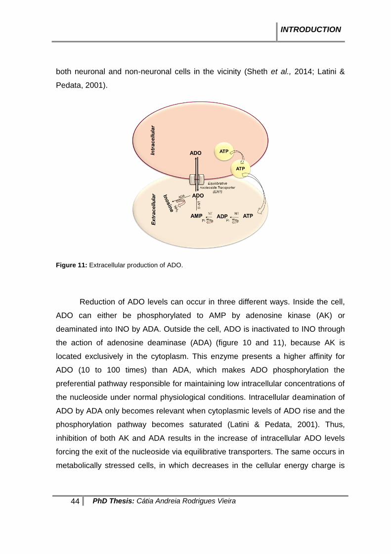
Extracellularly, ADO can be generated from the catabolism of ATP co-
released with classical neurotransmitters. Released ATP is sequential hydrolyzed
by ecto-nucleotidases into ADP, AMP and, finally to ADO (figure 11). All these
metabolites can act on membrane-bound purinoceptors to modulate the activity of
INTRODUCTION
44 PhD Thesis: Cátia Andreia Rodrigues Vieira
both neuronal and non-neuronal cells in the vicinity (Sheth et al., 2014; Latini &
Pedata, 2001).
Figure 11: Extracellular production of ADO.
Reduction of ADO levels can occur in three different ways. Inside the cell,
ADO can either be phosphorylated to AMP by adenosine kinase (AK) or
deaminated into INO by ADA. Outside the cell, ADO is inactivated to INO through
the action of adenosine deaminase (ADA) (figure 10 and 11), because AK is
located exclusively in the cytoplasm. This enzyme presents a higher affinity for
ADO (10 to 100 times) than ADA, which makes ADO phosphorylation the
preferential pathway responsible for maintaining low intracellular concentrations of
the nucleoside under normal physiological conditions. Intracellular deamination of
ADO by ADA only becomes relevant when cytoplasmic levels of ADO rise and the
phosphorylation pathway becomes saturated (Latini & Pedata, 2001). Thus,
inhibition of both AK and ADA results in the increase of intracellular ADO levels
forcing the exit of the nucleoside via equilibrative transporters. The same occurs in
metabolically stressed cells, in which decreases in the cellular energy charge is

INTRODUCTION
45 PhD Thesis: Cátia Andreia Rodrigues Vieira
accompanied by extensive catabolism of adenine nucleotides leading to increased
formation of ADO.
The third pathway used for intracellular ADO inactivation is the reversible
reaction catalysed by S-adenosyl-L-homocysteine hydrolase (SAH hydrolase)
requiring NAD+ as a cofactor. This reaction results in the formation of S-adenosyl-
L-homocysteine from ADO and L-homocysteine (Latini & Pedata, 2001).
3.2.1.2. Adenosine Transport
ADO transport across the plasma membrane is really important to regulate
the extracellular ADO levels. Nucleoside transporters (NT) are members of the
superfamily of solute carrier (SLC) transporters (Köse & Schiedel, 2009). In the
literature, are described two broad families of adenosine transporters: (1) SLC29
family of equilibrative nucleoside transporter (ENT), which are facilitated-diffusion
nucleoside transporters (sodium independent manner), and (2) SLC28 family of
concentrative nucleoside transporters (CNT), which transport nucleosides actively
against their gradient (sodium dependent manner) (Thorn & Jarvis, 1996; Köse &
Schiedel, 2009).
Four types of ENTs exist, which are classified as ENT1, ENT2, ENT3 and
ENT4. All these members of the SLC29 family share the ability of transport ADO,
but they differ in the ability of transport nucleosides and nucleobases (Thorn &
Jarvis, 1996; Yao et al., 1997; Köse & Schiedel, 2009).
Classically, ENTs were classified based in their sensitivity to the inhibition
by the synthetic nucleoside analogue, nitrobenzylthioinosine (NBTI). ENT1
(equilibrative-sensitive transporters, es) are inhibited at low nanomolar
concentrations of NBTI and dipyridamole, while ENT2, ENT3 and ENT4
(equilibrative-insensitive transporters, ei) are inhibited only by micromolar
concentrations of NBTI (Latini & Pedata, 2001). ENT1, ENT2 and ENT4 can be
found at the plasma membrane, while ENT3 is mostly intracellularly (Köse &
Schiedel, 2009).
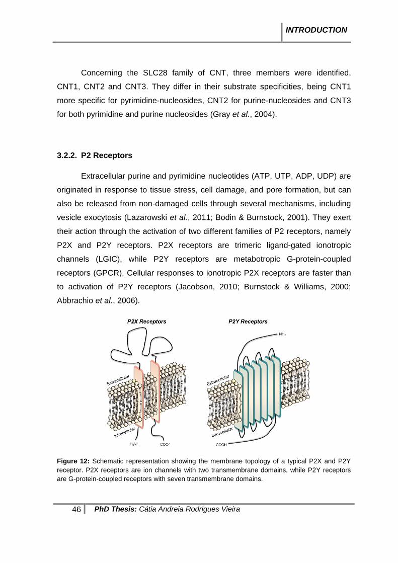
INTRODUCTION
46 PhD Thesis: Cátia Andreia Rodrigues Vieira
Concerning the SLC28 family of CNT, three members were identified,
CNT1, CNT2 and CNT3. They differ in their substrate specificities, being CNT1
more specific for pyrimidine-nucleosides, CNT2 for purine-nucleosides and CNT3
for both pyrimidine and purine nucleosides (Gray et al., 2004).
3.2.2. P2 Receptors
Extracellular purine and pyrimidine nucleotides (ATP, UTP, ADP, UDP) are
originated in response to tissue stress, cell damage, and pore formation, but can
also be released from non-damaged cells through several mechanisms, including
vesicle exocytosis (Lazarowski et al., 2011; Bodin & Burnstock, 2001). They exert
their action through the activation of two different families of P2 receptors, namely
P2X and P2Y receptors. P2X receptors are trimeric ligand-gated ionotropic
channels (LGIC), while P2Y receptors are metabotropic G-protein-coupled
receptors (GPCR). Cellular responses to ionotropic P2X receptors are faster than
to activation of P2Y receptors (Jacobson, 2010; Burnstock & Williams, 2000;
Abbrachio et al., 2006).
Figure 12: Schematic representation showing the membrane topology of a typical P2X and P2Y
receptor. P2X receptors are ion channels with two transmembrane domains, while P2Y receptors
are G-protein-coupled receptors with seven transmembrane domains.

INTRODUCTION
47 PhD Thesis: Cátia Andreia Rodrigues Vieira
3.2.2.1. P2X Receptors
There are 7 subtypes of P2X receptors: P2X1, P2X2, P2X3, P2X4, P2X5,
P2X6 and P2X7. Activation of P2X receptors by binding to extracellular ATP
generates an influx of cations, such as sodium and calcium, leading to
depolarization of excitable cells and activation of cytosolic enzymes (Jacobson,
2010). In neurons, they modulate neurotransmitter release acting presynaptically
or trigger fast excitatory signals in the postsynaptic membrane (Kaczmarek-HájeK
et al., 2012).
The subcellular distribution of the seven P2X receptor subtypes may differ
from cell to cell. Most of them are expressed at the plasma membrane, but some
of them may be in the endoplasmic reticulum (ER) or within endosomes and
lysosomes. Localization of these receptors depends on their trafficking properties
(Robinson & Murrell-Lagnado, 2013). P2X receptors are able to form heteromers
and homomers (Jacobson, 2010). All P2X receptor subunits can form homomers,
with the exception of the P2X6; the same occurs regarding heteromer formation,
the exception in this case is the P2X7 receptor subtype (Ren & Bertrand, 2008).
All these receptors have a similar topology consisting of two transmembrane
domains, a large extracellular ligand binding loop, and intracellular N and C
terminals (Kaczmarek-HájeK et al., 2012).
All subtypes of P2X receptors are activated by ATP, but they can be
distinguished by the required concentrations of the nucleotide. Based on that, P2X
receptors can be grouped in three main classes (Jacobson, 2010; Burnstock &
Williams, 2000):
(1) Receptors with high affinity for ATP (EC50=1 µM), which are rapidly activated
and desensitized: P2X1 and P2X3 receptors.
(2) Receptors with lower affinity for ATP (EC50=10 µM), showing slow
desensitization and sustained depolarizing currents: P2X2, P2X4, P2X5 and
P2X6 receptors.
(3) Receptors with very low affinity for ATP (EC50=300-400 µM), exhibiting little or
no desensitization and capable of forming large pores: P2X7 receptors.

INTRODUCTION
48 PhD Thesis: Cátia Andreia Rodrigues Vieira
3.2.2.2. P2Y Receptors
Purine nucleotides (ATP and ADP) and pyrimidine nucleotides (UTP and
UDP and UDP-glucose) activate metabotropic P2Y receptors. Abbracchio and
collaborators (2006) described eight distinct mammalian subtypes of P2Y
receptors, namely P2Y1, P2Y2, P2Y4, P2Y6, P2Y11, P2Y12, P2Y13 and P2Y14.
According to the pharmacological profile, the P2Y receptors can be
subdivided into four main groups:
(1) Adenine nucleotide-preferring receptors, responding essentially to ATP and
ADP: P2Y1, P2Y12, P2Y13 and human P2Y11;
(2) Uracil nucleotide-preferring receptors, responding respectively to UTP and
UDP: P2Y4 and P2Y6;
(3) Receptors with mixed selectivity: P2Y2, P2Y4 and P2Y11;
(4) Receptors responding to sugar nucleotides, UDP-glucose and UDP-galactose:
P2Y14.
Table 6: Main characteristics of P2Y receptor subtypes: agonist preference and G protein-coupling
(Abbracchio et al., 2006; White et al., 2003).
Receptor Agonist (preferential) Primary G protein coupling/ Second Messenger
P2Y1 ADP (ATP partial agonist) Gq/11; PLC activation; Gi/o; Potassium channel
P2Y2 UTP=ATP Gq/11; PLC activation
P2Y4 UTP>ATP Gq/11; PLC activation
P2Y6 UDP>>UTP (partial agonist) Gq/11; PLC activation
P2Y11 ATP>UTP Gs; AC stimulation; Gq/11; PLC activation
P2Y12 ADP (selective) Gi/0; AC inhibition
P2Y13 ADP>>ATP Gi/0; AC inhibition
P2Y14 UDP>UDP-glucose/UDP galactose
Gi/0; AC inhibition; Gq/11; PLC activation; can activate PLCβ via beta/gamma subunits.
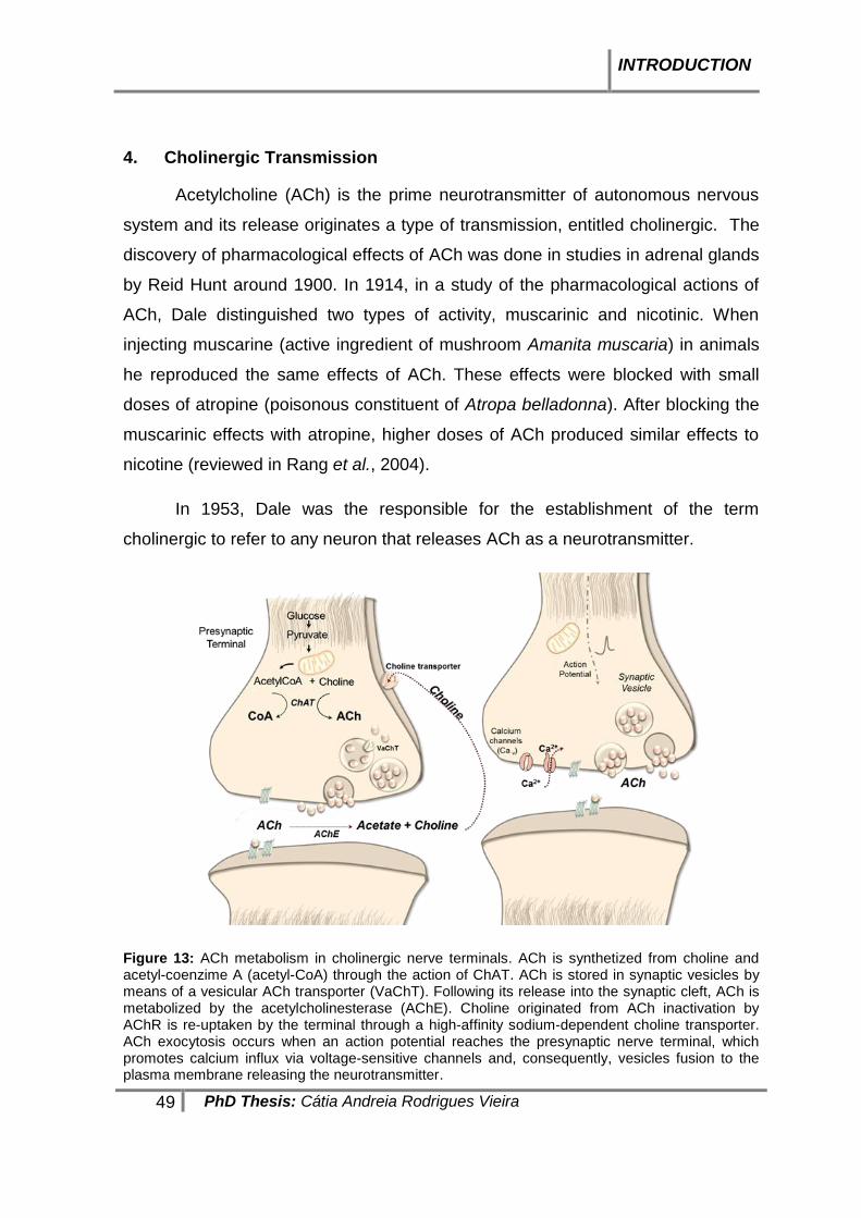
INTRODUCTION
49 PhD Thesis: Cátia Andreia Rodrigues Vieira
4. Cholinergic Transmission
Acetylcholine (ACh) is the prime neurotransmitter of autonomous nervous
system and its release originates a type of transmission, entitled cholinergic. The
discovery of pharmacological effects of ACh was done in studies in adrenal glands
by Reid Hunt around 1900. In 1914, in a study of the pharmacological actions of
ACh, Dale distinguished two types of activity, muscarinic and nicotinic. When
injecting muscarine (active ingredient of mushroom Amanita muscaria) in animals
he reproduced the same effects of ACh. These effects were blocked with small
doses of atropine (poisonous constituent of Atropa belladonna). After blocking the
muscarinic effects with atropine, higher doses of ACh produced similar effects to
nicotine (reviewed in Rang et al., 2004).
In 1953, Dale was the responsible for the establishment of the term
cholinergic to refer to any neuron that releases ACh as a neurotransmitter.
Figure 13: ACh metabolism in cholinergic nerve terminals. ACh is synthetized from choline and acetyl-coenzime A (acetyl-CoA) through the action of ChAT. ACh is stored in synaptic vesicles by means of a vesicular ACh transporter (VaChT). Following its release into the synaptic cleft, ACh is metabolized by the acetylcholinesterase (AChE). Choline originated from ACh inactivation by AChR is re-uptaken by the terminal through a high-affinity sodium-dependent choline transporter. ACh exocytosis occurs when an action potential reaches the presynaptic nerve terminal, which promotes calcium influx via voltage-sensitive channels and, consequently, vesicles fusion to the plasma membrane releasing the neurotransmitter.

INTRODUCTION
50 PhD Thesis: Cátia Andreia Rodrigues Vieira
ACh is synthetized in axonal terminals of cholinergic neurons by the action
of ChAT. ChAT catalyses the reaction between acetyl coenzyme-A (acetyl-CoA)
and choline. ACh and coenzyme-A (CoA) result as reaction products. Acetyl-CoA
is originated in the mitochondria from the complete oxidation of pyruvate. The
formation of acetyl-CoA is catalyzed by an enzyme complex called pyruvate
dehydrogenase complex. Choline that is need for the synthesis of ACh comes
from various sources, including the circulating choline in the plasma, the hydrolysis
of phospholipids of cell membranes and from the recycling of choline resulting
from the action of acetylcholinesterase (AChE) on ACh released into the synaptic
cleft. Reuptake of choline is mediated by a concentrative transporter existing in
cholinergic nerve terminals, which exhibits high-affinity for choline (Km between 1
and 5 µM) and is dependent on temperature and transmembrane Na+ gradient.
Synthesis of ACh is limited by the availability of choline. Synthesized ACh is
transported into synaptic vesicles by an antiport mechanism. When an action
potential reaches nerve terminals, it triggers the influx of Ca2+ from the
extracellular milieu via voltage-sensitive channels, which promotes the fusion
synaptic vesicles to the plasma membrane and, consequently, the exocytotic
release of ACh to the synaptic cleft (Rang et al., 2004). Once in the extracellular
space, released ACh may activate two subtypes of cholinergic receptors,
muscarinic (mAChR) and nicotinic (nAChR).
5. Purinergic and cholinergic transmission in the Myenteric Plexus
ACh is the prime regulator of intestinal motility and the dominant excitatory
neurotransmitter of the myenteric plexus (Costa et al., 1996). In this tissue, ATP
may be co-released with ACh, but also with other neurotransmitters (White, 1982).
Both excitatory and inhibitory effects may result from activation of purinoceptors by
ATP and its metabolites, ADP and ADO, depending on the receptor subtype being
activated, their location, and target cells.
ACh release from cholinergic enteric nerve terminals is modulated by
purines via the activation of both P1 and P2 purinoceptors, which can promote or
inhibit ACh release (De Man et al., 2003).

INTRODUCTION
51 PhD Thesis: Cátia Andreia Rodrigues Vieira
5.1. P1 Receptors
All subtypes of P1 receptors (adenosine A1, A2A, A2B and A3 receptors) were
found in whole-mount preparations of the LM-MP of rat ileum (Vieira et al., 2011).
Results from this study demonstrate substantial heterogeneity in the distribution of
P1 receptors in this tissue. Concerning high-affinity adenosine receptor subtypes,
the adenonsine A1 receptor is predominantly located in neuronal cell bodies of
myenteric ganglia, while the A2A receptor is expressed mainly on VAChT-positive
cholinergic nerve fibres. Regarding to the low affinity adenosine receptor subtypes,
our group showed that the A2B receptor is dominantly expressed on myenteric glial
cells, while the A3 receptor is co-localized with the A1 receptors on neuronal cell
bodies of myenteric ganglia.
From the four adenosine receptor subtypes found in the LM-MP of the rat
ileum, only the A2B receptor is not directly involved in cholinergic
neurotransmission. Besides the well-characterized inhibitory effect of ADO in the
GI tract mediated by A1 receptors, our group demonstrated that endogenously
generated ADO facilitates [3H]ACh release from myenteric neurons through
preferential activation of prejunctional A2A receptors (Duarte-Araújo et al., 2004a).
Upregulation of ACh release was also obtained by stimulating low affinity A3
receptors localized at myenteric cell bodies way from the synaptic region, which
activation may facilitate action potential generation; synergism between A3
receptors activation and the A2A facilitatory receptors localized on axon terminals,
has been observed (Vieira et al., 2011).
The co-existence of high-affinity inhibitory A1 and excitatory A2A receptor
subtypes on cholinergic neurons prompted the question of how endogenous ADO
discriminates between these two receptors to regulate synaptic transmission in the
myenteric plexus of the rat ileum. At the rat myenteric plexus, ADO may be
originated from the extracellular catabolism of released adenine nucleotides
through the ectonucleotidase pathway (Duarte-Araújo et al., 2009). However,
hydrolysis of released ATP contributes only partly to the total extracellular
concentration of the nucleoside; the other component is probably carried out by

INTRODUCTION
52 PhD Thesis: Cátia Andreia Rodrigues Vieira
the outflow of ADO as such via dipyridamole-sensitive ENTs (Correia-de-Sá et al.,
2006). Results from these studies also demonstrate that extracellular ADO
deamination by ADA secreted into the extracellular mileu (Correia-de-Sá et al.,
2006) is the most efficient mechanism to regulate the endogenous levels of the
nucleoside at the myenteric synapse (Duarte-Araújo et al., 2004a). Thus,
extremely high extracellular ADA activity, along with a less-efficient ADO uptake
mechanism, may restrict endogenous ADO actions to the synaptic region near the
release/production sites channelling to the activation of facilitatory A2A receptors
(Duarte-Araújo et al., 2004a; Correia-de-Sá et al., 2006).
5.2. P2X Receptors
P2X receptors are expressed on myenteric and submucosal motor,
sensory, and secretomotor neurones; they are also found on enteric glial cells,
ICCs and smooth muscle fibres (Burnstock, 2012) (see table 7).
In the myenteric plexus, ATP that acting on P2X receptors contributes to
facilitate electrically-evoked fast excitatory postsynaptic potentials (fEPSPs)
(Galligan, 2002). fEPSPs in the myenteric plexus of guinea-pigs have a dominant
purinergic component (67%) mediated by P2X2 and P2X3 receptor subtypes.
Purinergic fEPSPs are more evident in the ileum when compared with other
regions of the intestine. P2X2 receptors (20%) and P2X3 receptors (80%) are also
located in intrinsic sensory neurons (Ren & Bertrand, 2008).
Functional interactions between P2X and nicotinic ACh receptors have
been described. Both ionotropic receptors are co-expressed in myenteric neurons,
indicating that their interaction might be physiologically relevant (Ren & Bertrand,
2008). In this context, our group showed that ATP transiently activates facilitatory
P2X receptors mediating non-stimulated ACh release from myenteric motor
neurons (Duarte-Araújo et al., 2009).
Burnstock & Lavin (2002) identified P2X2 and P2X5 receptors in ICCs of
the guinea-pig myenteric plexus, but no immunostaining was detected for P2X1,
P2X3, P2X4, P2X6 or P2X7. These authors proposed that ATP released from

INTRODUCTION
53 PhD Thesis: Cátia Andreia Rodrigues Vieira
enteric nerves, enteric glial cells or during smooth muscle contractions can
activate P2X2 or P2X5 receptors in ICCs, leading to increases in intracellular
[Ca2+], impacting the activity of slow waves.
Table 7: P2X receptor subtypes and respective locations in the gut (based on data from the following papers: Burnstock, 2012; Galligan, 2002; Liña-Rico et al., 2015; Van Crombruggen et al., 2007; Silva et al., 2015; Burnstock & Lavin, 2002).
RECEPTORS LOCATION
P2X1 Smooth muscle of rat submucosal arterioles. Smooth muscle of the rat distal colon. Smooth muscle of enteric blood vessel.
Enteric glial cells (rat distal colon)
Resident macrophages
Highly expressed in human submucous plexus. Co-localized with P2X2 in the majority of submucous neurons.
P2X2 Myenteric neurons: descending interneurons, NOS-containing motor neurons; Nerve fibres in the myenteric plexus (rat distal colon) Nerve fibres in the submucosal ganglia: inhibitory motor neurons, VIP-containing secretomotor; intrinsic sensory neurons.
Longitudinal and circular muscle of the colon.
Epithelial cells (rat distal colon).
ICC’s.
P2X3 Small subpopulation of myenteric ganglion neurons, but in a larger number of ganglion neurons in the submucous plexus; Cholinergic neurons of submucous plexus of the guinea-pig ileum. Intrinsic sensory neurons; AH-type neurons. Nerve fibres of submucous and myenteric plexus (rat distal colon). Neurons of the human myenteric plexus, but not in the submucous plexus.
Longitudinal smooth muscle fibres.
Epithelial cells (rat distal colon).
P2X4 Only present in resident macrophages (rat distal colon).
P2X5 Longitudinal smooth muscle fibres.
Myenteric plexus: nerve fibers surrounding ganglion cell bodies and possibly on glial cell processes. Submucous plexus: intracellularly and on the plasma membranes of neurons.
ICC’s.
P2X6 Intrinsic sensory neurons.
Enteric Glial cells of myenteric and submucous plexus (rat distal colon).
P2X7 Myenteric and submucosal ganglia in stomach, small intestine, and colon. Surrounding ganglion cell bodies in both myenteric and submucous plexuses.
Enteric glial cells of myenteric and submucous plexus (rat distal colon).
Not present in smooth muscle cells, ICCs, and fibroblast-like cells.

INTRODUCTION
54 PhD Thesis: Cátia Andreia Rodrigues Vieira
5.3. P2Y Receptors
ATP acting on metabotropic P2Y receptors is responsible for slow EPSPs in
the ENS (Ren & Bertrand, 2008). Slow EPSPs are modulated by ACh acting on
muscarinic receptors and by tachykinins acting on neurokinin receptors. Slow
EPSPs is also mediated by ATP acting at P2Y receptors.
It has been proposed that P2Y1 and P2Y2 receptors have a dominant
neuronal localization, while the P2Y4 receptor is located in another cell types, like
enteric glial cells and ICCs. Our group demonstrated that activation of P2Y1
receptors by ADP generated from released ATP via NTPDase2 restrains ACh
release during prolonged stimulation periods of myenteric motor nerves (Duarte-
Araújo et al., 2009). Please refer to table 8 for the distribution of P2Y receptors
along the GI tract.
Mounting evidences are now available about the physiological impact of
neuron-to-glia communication (Boesmans et al., 2013; Gulbransen & Sharkey,
2009; Gulbransen et al., 2010). Enteric glial cells respond to ATP and UTP
released by neurons, via P2 receptors, by increasing intracellular [Ca2+]i,. Previous
studies implicated P2Y4, and possibly the P2Y11, receptors in ATP-mediated
effects in enteric glial cells (Gulbransen & Sharkey, 2009; Gulbransen et al., 2010),
but the P2Y1 receptor activated by ADP generated from the extracellular hydrolysis
of ATP may also be involved (Gulbransen & Sharkey, 2012). Neuron-to-glia
neurotransmission has been proposed in the myenteric plexus of the guinea pig
distal colon. In this model, the stimulation of primary afferent fibres releases
neurotransmitters, such as ATP, CGRP and SP, resulting in the activation of
presynaptic enteric nerve terminals leading to ACh and ATP co-release. These
compounds act respectively on ionotropic nicotinic ACh and P2X receptors on
postsynaptic enteric neurons. Evidence suggest, however, that enteric glia
respond mainly to ATP released from discrete populations of enteric sympathetic
nerves under specified circumstances, which by activating P2Y4 receptors
stimulated Ca2+ responses in the surrounding enteric glia (Gulbransen et al.,
2010).

INTRODUCTION
55 PhD Thesis: Cátia Andreia Rodrigues Vieira
Table 8: P2Y receptor subtypes and respective locations in the gut (based on data from the following papers: Ren & Bertrand, 2008; Burnstock, 2014; Van Crombruggen et al., 2007; Van Nassauw et al., 2006).
RECEPTORS LOCATION
P2Y1 Myenteric and submucous plexus (mouse); NOS immunoreactive neurons (descending interneurons or inhibitory motor neurons); Secretomotor neurons in submucous plexus.
ICC’s, in human and murine small intestine.
FLC.
Enteric glial cells in rat distal colon.
Smooth muscle cells in rat distal colon.
P2Y2 Myenteric plexus of the rat colon (Dogiel type I neurons); Almost a quarter of the neurons co-expressed nNOS.
Co-localize with a majority of calretinin immunoreactive neurons in the myenteric plexus (ascending interneurons and excitatory motor neurons).
All NPY-immunorecative neurons in the submucous plexus (secretomotor neurons)
Enteric glial cells.
P2Y4 ICC’s, in human and murine small intestine.
Enteric glial cells (in rat distal colon and in guinea-pig colon).
P2Y6
Myenteric plexus (all regions of the guinea pig GI tract); Not in the submucous plexus. Co-localize with NOS immunoreactive neurons (32%), descending interneurons and inhibitory motor neurons. Co-localize to calretinin immunoreactivity neurons (45%), ascending interneurons and excitatory motor neurons.
Enteric glial cells and in enteric smooth muscle cells (in rat distal colon).
P2Y11 Enteric glial cells (in rat distal colon).
Smooth muscle cells of the guinea-pig colon.
P2Y12 Localized to calbindin immunoreactive neurons (AH-type sensory neurons), in the guinea pig small intestine.
In the myenteric plexus, in the rat colon.
In neurons exhibiting a Dogiel type II morphology and in uniaxonal neurons (in rat distal colon).
P2Y13 Enterocytes (in rat distal colon).
P2Y14 Some neurons of submucous plexus (in rat distal colon).

INTRODUCTION
56 PhD Thesis: Cátia Andreia Rodrigues Vieira
ICCs and FLCs are electrically coupled to smooth muscle fibres via gap
junctions from a multicellular functional syncytium termed the SIP Syncytium. Cells
that make up the SIP syncytium are highly specialized containing unique
receptors, ion channels and intracellular signalling pathways, which may be
affected by neurotransmitters to regulate the excitability of GI muscles (reviewed in
Blair et al., 2014). For instance, purines activating P2Y1 receptors in FLCs
stimulate the PLC transduction pathway, which culminates in IP3 production and
Ca2+ recruitment from intracellular stores leading to increases in SK3 channels
permeability and cells hyperpolarization (Don Koh & Rhee, 2013).
5.4. Nicotinic and Muscarinic Receptors
ACh activates directly two subclasses of receptors, muscarinic and nicotinic
receptors. Because nicotinic ACh receptors are ligand-gated ion channels, they
mediate fast synaptic transmission in the ENS. It was shown that nicotinic
receptors-mediated neurotransmission prevails in ascending pathways, whereas it
has only a minor contribution in descending pathways (Galligan, 2002). Schneider
and Galligan (2000) showed distinct populations of presynaptic nicotinic receptors
in the myenteric plexus. They demonstrated that nicotinc receptors are located in
the nerve terminals and in the somatodendritic region of AH and S neurons. Our
group provided evidences suggesting that ACh mediates a positive feedback
mechanism via the activation of prejunctinal nicotinic receptors, presumably by
mediating the influx of Ca2+ directly through the receptor pore and, thus,
independently of voltage-sensitive calcium channels (Duarte-Araújo et al., 2004b).
Interestingly, this mechanism seems to be tonically regulated by endogenous ADO
through the activation of A2A receptors coupled to the adenylate cyclase/cyclic
AMP transducing system.
Immuncytochemistry studies showed that nicotinic receptors containig α3-,
α5-, and β2- subunits are most abundant primary cultured myenteric neurons,
whereas the α7- subunit stained only a small number of these neurons (Zhou et
al., 2002). Glushakov and colleagues (2004) showed that submucosal neurons of
the guinea-pig ileum express nicotinic receptors containing α3-, α5- and α7-
INTRODUCTION
57 PhD Thesis: Cátia Andreia Rodrigues Vieira
subunits, but not α4 subunits. Moreover, Obaid and colleagues (2005) using
radioimmunoassays and immunocytochemistry reached the conclusion that guinea
pig enteric ganglia express α3-, α5-, β2-, β4- and α7-subunits.
In addition to nicotinic receptors, ACh can also activate muscarinic
receptors (North et al., 1985). The GI tract of healthy dairy cows contains M1
receptors in the submucosal and myenteric plexus, M2 receptors in SMCs, M3
receptors in the lamina propria, nerve terminals and intermuscular nerve fibers,
and in microvessel myocyte, and M5 in microvessel myocyte and SMCs. M4
receptors were not found in these tissues (Stoffel et al., 2006). At the LM-MP of
the mouse ileum, M2 and M4 receptors may mediate muscarinic autoinhibition in
ACh release (Takeuchi et al., 2005). Using the rat ileum, we demonstrated that
ACh exerts a dual neuromodulatory effect on its own release through the
activation of inhibitory M2 receptors and excitatory M3 receptors, depending on the
stimulation train length (Vieira et al., 2009). The participation of M1 receptors in the
control of ACh release was discarded.
Goyal (2013) revised the role of ICCs in cholinergic neurotransmission in
the gut. It was proposed that ICCs may be intermediates in cholinergic
neuromuscular transmission at the SIP syncytium. ICC’s are endowed with
muscarinic receptors (mainly M3 receptors), which have a prime role in this
process. Activation of muscarinic receptors in ICCs generates a depolarizing
potential and activates different signalling pathways that are driven to smooth
muscle fibres leading to muscle contraction. M3 receptors are coupled to Gq/11
leading to activation of phospholipase C (PLC) resulting in IP3 and DAG formation,
with subsequent recruitment of intracellular Ca2+ and protein kinase C activation,
respectively (Koh & Rhee, 2013). Increases in intracellular Ca2+ can strengthen
activation of the protein kinase C, which may subsequently stimulate ADO outflow
by equilibrative transporter ENT1. Increase in protein kinase C activity may also
stimulate the activity of 5’-nucleotidase inside the cell and/or inhibit AK, enhancing
intracellular accumulation and efflux of ADO from the cell. Thus, the muscarinic M3
positive feedback mechanism facilitating ACh release from stimulated myenteric

INTRODUCTION
58 PhD Thesis: Cátia Andreia Rodrigues Vieira
neurons may be indirectly mediated by the release of ADO from ICCs leading to
activation of prejunctional facilitatory A2A receptors in a novel neuron-to-glia and
back glia-to-neuron communication pathway operated by muscarinic M3 and
adenosine A2A receptors.
Increasing evidences have been produced showing that ICCs are crucial in
the transduction of cholinergic signals to smooth muscle fibres in the GI tract.
However, in the absence of ICCs ACh can couple to Gq12/13 protein and trigger
activation of the Rho-kinase (RhoK) pathway to cause contraction in SMCs (Koh &
Rhee, 2013).
6. Inflammatory bowel diseases
In Portugal, inflammatory bowel diseases (IBD) have a prevalence of 146
patients per 100 000 inhabitants (Azevedo et al., 2010). IBDs include Crohn’s
disease, ulcerative colitis and indeterminate colitis. Ulcerative colitis occurs only in
the colon and within the gut wall tissue damage is restricted to the mucosa and
lamina propria. On the other hand, Crohn’s disease can occur in any region of the
gut and tissue damage may penetrate the entire thickness of the organ (Geboes &
Collins, 1998). Peristaltic changes are common clinical symptoms in IBS. Since in
these chronic GI disorders etiopathogenesis remains unclear, therapeutic
management is usually empiric, extrapolated from ulcerative colitis and Crohn’s
disease. The most typically used drugs are intestinal anti-inflammatory drugs,
systemic corticosteroids, immunosupressants, biologic therapy and antibiotics
(Azevedo et al., 2010). The determination of inflammatory activity is a really
important step for the assessment of disease activity, for choosing an adequate
therapy, and for contributing to ameliorate the quality of patients’ life (Vilela et al.,
2012).
Structural abnormalities of the ENS are observed in IBD. In Crohn’s
Disease are often observed nerve fibre modifications, such as damage,
hypertrophy, and reactive hyperplasia, and enteric glial cells hyperplasia. Neuronal

INTRODUCTION
59 PhD Thesis: Cátia Andreia Rodrigues Vieira
hypertrophy of the Meissner’s plexus is a common pathological feature of
ulcerative colitis (Geboes & Collins, 1998). In both diseases, neutrophils and
lymphocytes (positive for CD4) are the predominant cells in the inflamed mucosa.
Increases in the number of B cells, macrophages, dendritic cells, plasma cells,
eosinophils and mast cells are also very common (Roberts-Thomson et al., 2011).
During an inflammatory insult in the gut, activated leukocytes infiltrate the
gut wall. Various cells of the mucosal immune system produce a number of pro-
and anti-inflammatory cytokines, which induce chemotaxis and leukocytes
proliferation (Neurath, 2014; Schepper et al., 2008). Cytokine levels and cytokine
expression are similar in Ulcerative colitis and Crohn’s disease. Increases in TNF-
α and IFN-γ are consistent with a Th1 response. The proinflammatory cytokines
described in these disorders are: TNFα, IL-12, IL-23, and perhaps IL-17 and IFN-γ
(Roberts-Thomson et al., 2011). The released cytokines also induce morphological
and functional alterations in the neural apparatus, such as sprouting, neuronal
necrosis, and neuronal sensitization (Schepper et al., 2008).
Enteric plasticity comprises a wide range of structural and/or functional
changes in enteric neurons, glial cells and pacemaker cells of Cajal. As a result of
adaptive responses to different types of pathophysiological conditions, enteric
neurons are able to rapidly change their structure, function and chemical
phenotype in order to maintain homeostasis of gut functions. These plasticity
changes occur in distinct situations as a consequence of the wide array of
pathological conditions, ranging from enteric neuropathies to extra-intestinal (e.g.
Parkinson’s disease) or intestinal diseases (e.g. IBD) (Vassina et al., 2006).
Even though the period of inflammation may be brief, and the region of
overt inflammatory damage is circumscribed, its effects on enteric neurons are
long-lasting and significant changes in organ function are observed in regions that
are remote from the inflammatory site (Pontell et al., 2009).

INTRODUCTION
60 PhD Thesis: Cátia Andreia Rodrigues Vieira
6.1. Ileitis
Inflammation of the ileum is called ileitis. It is often caused by Crohn’s
disease, although it may be caused by other diseases, such as ischemia,
neoplasms, medication-induced, eosinophilic enteritis, and others. Inflammation of
the ileum may be acute or in more severe conditions, can be chronic (DiLauro et
al., 2010).
6.2. Animal models of inflammatory bowel diseases
A wide variety of animal models of IBD appeared in last years (table 9). IBD
animal models have proven to be important tools for detecting potential
therapeutic agents and for investigating the mechanisms involved in its
pathogenesis (Jurjus et al., 2004; Wirtz and Neurath, 2007a,b). It is considered an
appropriate animal model, the one that display certain key characteristics:
(1) Morphologic alterations;
(2) Inflammation;
(3) Pathophysiology and course similar to human IBD.
The most widely accepted models are those resulting from the instillation of
chemical irritants using trinitrobenzene sulfonic acid (TNBS) or dextran sodium
sulphate (DSS) (Jurjus et al., 2004; Wirtz and Neurath, 2007a,b). In the present
work, it will be used the chemically-induced model by the instillation of TNBS into
the lumen of the ileum. So, now I will focus mostly in this model.

INTRODUCTION
61 PhD Thesis: Cátia Andreia Rodrigues Vieira
Table 9: Animal models categories and subcategories of IBD.
Models (categories)
Subcategories
Gene Knockout (KO) models
(1) Interkeukin-2 KO/IL-2 receptor (R)α KO mice (2) IL-10 KO mice models (3) T cell receptor (TCR) mutante mice (4) TNF-3’ untranslated region (UTR) KO mice (5) Trefoil factor-deficient mice
Transgenic mouse and rat models
(1) IL-7 transgenic mice (2) Signal transducer and activating transcription (STAT)-4 transgenic mice (3) HLA B27 transgenic rats
Spontaneous Colitis models (1) C3H/HejBir mice (2) SAMP1/Yit mice
Inducible models (Chemically induced)
(1) Acetic acid (2) Iodoacetamide (3) Indomethacin (4) TNBS (5) Oxazolone (6) DSS (7) Peptidoglycan –polysaccharide (PG-PS) colitis
Adoptive transfer models
(1) Colitis induced by transfer of heat shock protein (hsp) 60-specific CD8 T cells. (2) CD45RB transfer model
6.2.1. TNBS-Induced ileitis
Ileitis can be induced by a intraluminal instillation of 2,4,6-
trinitrobenzenesulfonic acid (TNBS) in ethanol, which is required to break the
mucosal barrier (Martinolle et al., 1997; Wirtz et al., 2007).
This model is also frequently used to induce colitis. The model in the colon
seems to be more aggressive than in the ileum. In colon, the chronic phase is
more easily achieved than in the ileum. This difference may be due to the absence
of specific ileum resident bacteria found in the colon. The resident flora is needed
to keep the inflammatory response to TNBS (Moreels et al., 2001). These authors
sustain that TNBS-induced ileitis in the rat lacks the chronic phase and it is

INTRODUCTION
62 PhD Thesis: Cátia Andreia Rodrigues Vieira
accompanied by functional hypocontractility of longitudinal SMCs during the acute
inflammatory phase. They claim that the contractility of the longitudinal muscle is
increased in this phase due to structural changes. Moreels and colleagues (2001)
concluded that the rat model of TNBS-induced ileitis is characterized by an acute
transmural inflammation.
In 1992, Selve & Wöhrmann published a study where they described a
model of chronic IBD with TNBS, in the rat ileum. In this work, chronic
inflammation was only achieved 3 to 4 weeks after starting TNBS application. The
protocol implicates a previous immunization and a daily application of TNBS (via a
catheter). This chronic phase in the ileum is characterized by the following
pathological alterations:
(1) Intense hyperemia and oedema of the mucosal surface;
(2) Inflammatory cell infiltration (by lymphocytes and histiocytes in all layers of the
gut wall;
(3) Without neutrophil infiltration;
(4) Transmural granulomatous inflammation of the gut wall.
In the model of TNBS-induced ileitis used more often, pathological
alterations are the following:
(1) Substantial increase in the thickness of the intestinal wall (Tsujikawa et al.,
1999);
(2) Transmural lesions, that involves Th-1 mediated macrophage expression of
cytokines, such as TNF-α, IFN-γ and IL-12 (Merritt et al., 2002).
(3) Mucosal infiltration by mononuclear and polynuclear cells (Martinolle et al.,
1997);
(4) Increased production of inflammatory mediators such as leukotrienes,
prostaglandins, platelet-activating factor, SP and NO (Martinolle et al., 1997).
(5) T lymphocytes and eosinophils are found more close to myenteric ganglia
(Pontell et al., 2009).

INTRODUCTION
63 PhD Thesis: Cátia Andreia Rodrigues Vieira
(6) Increased neuronal excitability persists for at least 8 weeks (Pontell et al.,
2009).
(7) Critical changes in spontaneous contractile activity of circular and longitudinal
muscle (Martinolle et al., 1997).
TNBS-induced ileitis results in different effects on the electrophysiological
properties of distinct types of neurons of the small intestine. Dogiel type I neurons
from inflamed ileum of the guinea-pig had prominent late afterhyperpolarizing
potentials (AHPs) after a single action potential triggered by an intracellular current
pulse. Dogiel type II neurons were hyperexcitable compared to the controls
(Nurgali et al., 2007). It was described also by Stewart and colleagues, in 2003,
that there is an increase of excitability of intestinal sensory neurons by modulation
of ion channels (Na+ and K+) in guinea-pig ileitis. The appearance of tetrodotoxin-
resistant sodium currents is increased, while potassium currents are supressed.
One group has provided insights on the effects of inflammation on cell
proliferation in the myenteric plexus of the guinea-pig ileum (Bradley et al., 1997).
These authors concluded that inflammation of the ileum does not stimulate the
appearance of new myenteric neurons, but stimulates significantly enteric glia cells
proliferation.They also observed an increase in the number of macrophages and
T-cells in the tissue. It appears that the increased number of macrophages is not a
result of cells proliferation, but may be a consequence of the infiltration of resident
cells. Apparently inflammatory cell infiltrates spare the myenteric ganglia and, for
this reason, they designate myenteric ganglia as an immune-privileged tissue
where immunocytes do not penetrate.
Reports from the literature refer to impairments of the purinergic modulation
of enteric neurotransmission during chronic inflammation of the mouse ileum (De
Man et al., 2003). During an inflammatory insult, the P2Y11 receptor is activated in
immune cells by low concentrations of ATP, while the P2X7 receptor requires high
concentrations of ATP (>100 µM) to become activated. The release of
inflammatory mediators may be accomplished by the activation of P2X3 and P2X7
receptors (Sanger, 2012). Increases of the P2X3 receptor expression has been

INTRODUCTION
64 PhD Thesis: Cátia Andreia Rodrigues Vieira
observed in human patients with IBD, which implicates a potential role for this
receptor in enteric dismotility and pain (Yiangou et al., 2001). Increase expression
of the P2Y6 receptor has also been observed in epithelial cells, thus promoting the
epithelial release of cytokines and chemokines that indirectly modulate the severity
of GI inflammation (Grbic et al., 2008; Roberts et al., 2012)
Nowadays, there is an increasing interest on the involvement of ADO in the
pathophysiology of intestinal disorders (Antonioli et al., 2008). In addition, an
active role of extracellular ATP and its receptors in the alterations motivated by
intestinal inflammation has been suggested (Burnstock, 2008). In IBD models, not
only morphological but also changes in the excitability and synaptic properties of
enteric neurons might justify the GI dysfunction.

GOALS
65 PhD Thesis: Cátia Andreia Rodrigues Vieira
V. GOALS
The main goals of this study were to investigate the following aspects in a model
of postinflammatory ileitis caused by intraluminal instillation of TNBS in the rat:
(1) The kinetics of the extracellular catabolism of ATP and ADO formation/
inactivation cycle in order to comprehend the overall homeostatic role of the
purinergic cascade on enteric excitability.
(2) The action of endogenous adenosine on electrically evoked [3H]ACh
release from myenteric motor neurons in postinflammatory conditions, using
selective antagonists to adenosine receptors.
(3) Changes in the electrically-evoked release of purines (ATP and ADO) and
ACh in the myenteric plexus of control and TNBS-treated rats.
(4) Changes in purine releasing sites, their relation to cell type and density
changes, in order to propose novel therapeutic targets for inflammatory
intestinal diseases.
Understanding how ubiquitous neuromodulators, such as adenine
nucleotides and ADO, affect plastic adaptations of enteric neurotransmission
during inflammatory insults may prompt for novel therapeutic targets to manage
inflammatory intestinal diseases.

ORIGINAL RESEARCH ARTICLES
66 PhD Thesis: Cátia Andreia Rodrigues Vieira
VI. ORIGINAL RESEARCH PAPERS
The results presented in this thesis were published/ submitted for publication as
original research papers, as follows:
Paper 1: Vieira C, Magalhães-Cardoso MT, Ferreirinha F, Silva I, Dias AS, Pelletier J, Sévigny J & Correia-de-Sá P (2014). Feed-Forward Inhibition of CD73 and Upregulation of Adenosine Deaminase Contribute to the Loss of Adenosine Neuromodulation in Postinflammatory Ileitis. Mediators of Inflammation. Volume 2014 (2014), Article ID 254640, 19 pages. (DOI: 10.1155/2014/254640)
Paper 2: Vieira C, Magalhães-Cardoso MT, Ferreirinha F, Silva I & Correia-de-Sá P (2015). Postinflammatory ileitis induces neurochemical adjustments of purine cellular resources in the myenteric plexus. (Manuscript submitted for publication)
Data from the following papers published by the author were also used and/or
discussed in this thesis:
Vieira C, Ferreirinha F, Silva I, Duarte-Araújo M, Correia-de-Sá P. Localization and function of adenosine receptor subtypes at the longitudinal muscle-myenteric plexus of the rat ileum. Neurochem Int. 2011. 59(7):1043-1055.
Vieira C, Duarte-Araújo M, Adães S, Magalhães-Cardoso T, Correia-de-Sá P. Muscarinic M3 facilitation of acetylcholine release from rat myenteric neurons depends on adenosine outflow leading to activation of excitatory A2A receptors. Neurogastroenterol Motil. 2009. 21(10):1118-e95.
Correia-de-Sá P, Adães S, Timóteo MA, Vieira C, Magalhães-Cardoso T, Nascimento C, Duarte-Araújo M. Fine-tuning modulation of myenteric motoneurons by endogenous adenosine: On the role of secreted adenosine deaminase. Auton Neurosci. 2006. 126-
127:211-224.
Author contributions to the papers:
Author participated in all aspects of the work and performed autonomously the surgical procedures,
in vivo and in vitro experiments, analyzed the data, discussed the results and drafted the papers
until final approval. Paulo Correia-de-Sá (MD, PhD) supervised the project, designed the
experiments, analyzed the data, discussed the results and drafted/approved the papers. The
author received collaborations from Isabel Silva (MSc, PhD Student) – quantification of ATP by the
luciferin-luciferase bioluminescence assay; Fátima Ferrreirinha (PhD) - image acquisition and
analysis of immunofluorescence confocal micrographs; and Teresa Magalhães-Cardoso (PharmD)
– quantification of adenine nucleotides and nucleosides by HPLC.
ORIGINAL RESEARCH ARTICLES
67 PhD Thesis: Cátia Andreia Rodrigues Vieira
PAPER 1
Mediators of Inflammation. 2014. 2014, 19 pages.
DOI: 10.1155/2041/254640

ORIGINAL RESEARCH ARTICLES
68 PhD Thesis: Cátia Andreia Rodrigues Vieira
Feed-forward inhibition of CD73 and upregulation of adenosine deaminase
contribute to the loss of adenosine neuromodulation in postinflammatory
ileitis
Cátia Vieira1, Maria Teresa Magalhães-Cardoso1, Fátima Ferreirinha1, Isabel
Silva1, Ana Sofia Dias1, J. Pelletier2, J. Sévigny2,3 & Paulo Correia-de-Sá1,
1Laboratório de Farmacologia e Neurobiologia, UMIB and MedInUP, Instituto de Ciências
Biomédicas Abel Salazar (ICBAS), Universidade do Porto (UP), L. Prof. Abel Salazar 2,
4099-033 Porto, Portugal, 2 Centre de Recherche du CHU de Québec, Québec, QC,
Canada. and 3 Département de Microbiologie-Infectiologie et d’Immunologie, Faculté de
Médecine, Université Laval, Québec, QC, Canada.
ABSTRACT
Purinergic signalling is remarkably plastic during GI inflammation. Thus,
selective drugs targeting the “purinome” may be helpful for inflammatory GI
diseases. The myenteric neuromuscular transmission of healthy individuals is fine-
tuned controlled by ADO acting on A1 inhibitory and A2A excitatory receptors. Here,
we investigated the neuromodulatory role of ADO in TNBS-inflamed LM-MP of the
rat ileum. Seven-day postinflammation ileitis lacks ADO neuromodulation, which
may contribute to accelerate GI transit. The loss of ADO neuromodulation results
from deficient accumulation of the nucleoside at the myenteric synapse despite
that increases in ATP release were observed. Disparity between ATP outflow and
ADO deficit in postinflammatory ileitis is ascribed to feed-forward inhibition of ecto-
5’-nucleotidase/CD73 by high extracellular ATP and/or ADP. Redistribution of
NTPDase2, but not of NTPDase3, from ganglion cell bodies to myenteric nerve
terminals leads to preferential ADP accumulation from released ATP, thus
contributing to prolong inhibition of muscle-bound ecto-5’-nucleotidase/CD73 and
to delay ADO formation at the inflamed neuromuscular synapse. On the other
hand, depression of endogenous ADO accumulation may also occur due to
enhancement of ADA activity. Both membrane-bound and soluble forms of ecto-5’-
nucleotidase/CD73 and ADA were detected in the inflamed myenteric plexus.
These findings provide novel therapeutic targets for inflammatory gut motility
disorders.

ORIGINAL RESEARCH ARTICLES
69 PhD Thesis: Cátia Andreia Rodrigues Vieira
Keywords: Ectonucleotidases; enteric nervous system; intestinal inflammation; myenteric
plexus; purinergic receptors; soluble enzymes.
INTRODUCTION
The ENS undergoes a series of adaptive responses to different pathological
conditions (e.g. inflammatory and/or ischemic insults) (Sharkey et al., 2001;
Vassina et al., 2006). For instance, enteric neurons rapidly change their structure,
function or chemical phenotype in order to maintain gut homeostasis. Even if the
inflammatory insult is brief and the damage circumscribed, its repercussion on
enteric neurons may be long-lasting leading to significant changes in intestinal
function, which can be observed in remote regions from the inflammation site. The
postinflammatory status is frequently accompanied by substantial increases in
enteric motility (Pontell et al., 2009).
Inflammation of the GI tract causes marked changes in the release of
purines leading to subsequent adaptive modifications of purinoceptors expression
and/or function (reviewed in Roberts et al., 2012). The underlying mechanisms of
disturbed purinergic modulation are not completely understood, in part because
the study of purinoceptors may be hampered by the presence of distinct nucleotide
release sites and cell surface enzymes that rapidly break down extracellular
nucleotides into nucleosides (Kukulski et al., 2011). In healthy individuals, ATP is
released predominantly from stimulated enteric neurons (White & Leslie, 1982),
but its release from non-neuronal cells (e.g. smooth muscle fibres, ICC) might also
occur (Nitahara et al., 1995). Four members of the ecto-nucleoside triphosphate
diphosphohydrolase (E-NTPDase) family, namely NTPDase1, NTPDase2,
NTPDase3 and NTPDase8, and two members of the ecto-nucleotide
pyrophosphatases/phosphodiesterases (E-NPP) family, NPP1 and NPP3, are
located at the plasma membrane and hydrolyse extracellular nucleotides (Kukulski
et al., 2011; Kukulski et al., 2005; Stefan et al., 2005). The relative contribution of
distinct ecto-enzymes to the modulation of purinergic signalling depends on
ORIGINAL RESEARCH ARTICLES
70 PhD Thesis: Cátia Andreia Rodrigues Vieira
differential tissue and cell distribution, regulation of expression, targeting to
specific membrane domains, but also on substrate preference and availability.
Regarding the substrate preference, NTPDase1 (CD39 or apyrase)
dephosphorylates ATP directly into AMP, with minimal accumulation of ADP.
NTPDase2 (ATPase) is a preferential nucleoside triphosphatase hydrolysing ADP
10 to 15 times less efficiently than ATP, leading to minimal AMP accumulation
(Kukulski et al., 2005). NTPDase3 and NTPDase8 are functional intermediates
between NTPDase1 and NTPDase2 (Kukulski et al., 2005). Because of their
involvement in physiological processes, namely blood clotting, vascular
inflammation, immune reactions and certain types of cancer, NTPDases are now
considered as potential drug targets (Gendron et al., 2002). As for NPP1 and
NPP3, they release nucleoside 5’-monophosphate from a variety of nucleotides,
but intriguingly, their phosphorylated product (e.g. AMP) bind to NPPs with a
higher affinity than substrates do, and thus inhibit catalysis (Stefan et al., 2009).
Finally, AMP is hydrolysed to ADO and inorganic phosphate by ecto-5’-
nucleotidase (CD73), which is concentrated in the myenteric smooth muscle cell
layer (Nitahara et al., 1995). Interestingly, ecto-5’-nucleotidase (CD73) may be
cleaved from cell membranes through hydrolysis of the GPI anchor by
phosphatidylinositol-specific phospholipases or by proteolysis while retaining its
catalytic activity in the soluble form (Sträter et al., 2006).
At the myenteric neuromuscular synapse, ATP is primarily metabolized into
AMP, which is then dephosphorylated into ADO; alternative conversion of ATP into
ADP is more relevant at high ATP concentrations (Duarte-Araújo et al., 2009).
Thus, gradients of ATP and its breakdown products (ADP and ADO) may provide
fine tuning control of peristaltic motor performance in the gut during stressful
situations, such as sustained neuronal activity, ischemia and inflammation, when
extracellular ATP reaches high levels (see Milusheva et al., 1990; De Man et al.,
2003). ATP transiently facilitates ACh release from non-stimulated nerve terminals
via prejunctional P2X (probably P2X2) receptors. Hydrolysis of ATP directly into
AMP and subsequent formation of ADO activates inhibitory A1 receptors in smooth

ORIGINAL RESEARCH ARTICLES
71 PhD Thesis: Cátia Andreia Rodrigues Vieira
muscle fibres (Nicholls et al., 1996) and in ganglionic cell bodies of myenteric
neurons (Vieira et al., 2011), where it downmodulates the release of excitatory
neurotransmitters, such as SP (Moneta et al., 1997) and ACh (Duarte-Araújo et
al., 2004). Besides the well characterized inhibitory A1 receptors, myenteric nerve
terminals are endowed with prejunctional A2A receptors mediating facilitation of
ACh release (Duarte-Araújo et al., 2004; Correia-de-Sá et al., 2006; Vieira et al.,
2011).
Previous findings from our group demonstrated that the ecto-nucleotidase
pathway contributes only partially to the total interstitial ADO concentration in the
myenteric plexus (Correia-de-Sá et al., 2006). ADO released as such from either
neuronal or non-neuronal cells seems to be the main source of endogenous ADO
in the ENS (Duarte-Araújo et al., 2004). This release is sought to be mediated by
facilitated diffusion via nucleoside transporters, which can be regulated by
endogenous signaling molecules, such as ACh via muscarinic M3 receptors
(Correia-de-Sá et al., 2006; Vieira et al., 2011). On the other hand, we
demonstrated the extracellular deamination by ADA represents the most effective
mechanism regulating synaptic ADO levels in the LM-MP of the ileum from healthy
animals (Duarte-Araújo et al., 2004; Correia-de-Sá et al., 2006). ADA is a
ubiquitous purine metabolic enzyme localized on the cell surface (ecto-ADA)
(Pettengil et al., 2013). Upon proteolytic cleavage from cell membranes, soluble
forms of ADA (exo-ADA) retain their catalytic activity and may be found in
interstitial fluids (Franco et al., 1997). We detected a relatively high ADA activity
(~0.6 U/ml) in superfusates collected after stimulating LM-MP preparations, which
is in keeping with the hypothesis that “ADA secretion” may restrict endogenous
ADO actions to the synaptic region near the release / production sites (Correia-de-
Sá et al., 2006). ADA secretion increases in various diseases and may be
originated from monocyte/macrophage lineages, thus reflecting the involvement of
the cellular immune system. De Man and colleagues (2003) reported that chronic
intestinal inflammation enhanced the enteric contractile activity in part due to an
impairment of purinergic modulation of cholinergic nerve activity (De Man et al.,
2003).

ORIGINAL RESEARCH ARTICLES
72 PhD Thesis: Cátia Andreia Rodrigues Vieira
Because, adenine nucleotides and nucleosides (and ADA itself) may be
released from activated inflammatory cells (Marquard et al., 1984), as well as from
neighbouring neuronal and non-neuronal enteric cells (Stead et al., 1989; Bogers
et al., 2000), there is an increasing interest in the neuromodulatory effects exert by
ADO during inflammatory insults. The therapeutic potential of ADO-related
compounds for controlling intestinal motility and inflammation (De Man et al., 2003;
Antonioli et al., 2008), prompted us to investigate the kinetics of ADO formation/
inactivation cycle in order to understand the overall homeostatic role of the
nucleoside on enteric excitability in postinflammatory ileitis caused by intraluminal
instillation of TNBS in the rat.
MATERIAL AND METHODS
Animals
Rats of either sex (Wistar, ~200 g) (Charles River, Barcelona, Spain) were
kept at a constant temperature (21) and a regular light (06.30-19.30 h)-dark
(19.30-06.30 h) cycle, with food and water) and a regular light (06.30-19.30 h)-
dark (19.30-06.30 h) cycle, with food and water ad libitum. All studies involving
animal experiments are reported in agreement with ARRIVE guidelines. Animal
handling and experiments were in accordance with the guidelines prepared by
Committee on Care and Use of Laboratory Animal Resources (National Research
Council, USA) and followed the European Communities Council Directive
(86/609/EEC).
TNBS-induced intestinal inflammation model
Intestinal inflammation was produced by the instillation of 2,4,6-
trinitrobenzenesulfonic acid (TNBS) into the lumen of the rat ileum. After a fasting
period of 4-8 hours with free access to drinking water, rats undergo median
laparotomy under anaesthesia with an association of medetomidine (10 mg/Kg)
and ketamine (75 mg/Kg) given subcutaneously. At the end of the procedure
animals were retrieved with atipamezole (10 mg/Kg). The terminal ileal loop was

ORIGINAL RESEARCH ARTICLES
73 PhD Thesis: Cátia Andreia Rodrigues Vieira
gently exteriorized, and TNBS (40 mM) was injected through the enteric wall into
the lumen of the ileum, 10 centimeters proximal to the ileocolonic junction.
Controls received 1 ml of 0.9% saline. Sixty minutes after surgery, the rats were
allowed to eat and drink ad libitum. After surgery, pain was controlled with
tramadol hydrochloride (10 mg/Kg). To have a control of time course of body
weight loss and recovery after injection of TNBS, rats were weighed prior to TNBS
administration and daily following surgery. Animals with intestinal inflammation
(TNBS) transiently lose weight for three to four days after surgery, and regain
weight thereafter. Seven days after surgery, animals were sacrificed following an
overnight fasting period.
Histological findings and gastrointestinal motility
The postinflammatory phase of TNBS-induced ileitis was characterized in
haematoxylin-eosin stained sections using Pontell and Jergens criteria, which is
based in the loss of normal tissue architecture, epithelial damage and infiltration of
inflammatory cells (Pontell et al., 2009; Jergens, 1999; Engel et al., 2009; Wirtz &
Neurath, 2007). Evaluation of GI motility was performed in vivo by oral
administration of a gavage containing the methylene blue dye. Dye progression in
the GI tract was evaluated after 30 minutes, both in control and TNBS-injected
animals: the total length of the small intestine was used to normalize data.
3H-Acetylcholine release experiments
Sections of the rat ileum not including the terminal five centimetres were
used. The longitudinal muscle strip with the myenteric plexus attached was
separated from the underlying circular muscle according to the method of Paton
and Vizi (1969). This preparation is highly enriched in cholinergic neurons, mainly
excitatory neurons projecting to the longitudinal muscle (25%) that receive inputs
from intrinsic primary afferents (26%) and from ascending and descending
pathways (17%) (Costa et al., 1996).
The procedures used for labelling the preparations and measuring evoked
[3H]-Acetylcholine ([3H]-ACh) release were previously described (Duarte-Araújo et

ORIGINAL RESEARCH ARTICLES
74 PhD Thesis: Cátia Andreia Rodrigues Vieira
al., 2004; Duarte-Araújo et al., 2009; Correia-de-Sá et al., 2006; Vieira et al., 2011)
and used with minor modifications. LM-MP strips were mounted in 3 ml capacity
vertical perfusion chambers heated at 37 C. After a 30 min equilibration period,
myenteric neurons were labelled for 40 min with 1 µM [3H]-choline (specific activity
2.5 µCi nmol-1) under EFS (1 Hz-frequency, 1 ms pulse width). Following loading,
the washout Tyrode’s solution contained hemicholinium-3 (10 µM) to prevent
choline uptake. After a 60 min period of washout, bath samples (3 ml) were
automatically collected every 3 min using a fraction collector (Gilson, FC203B,
France). Tritium content of the samples was measured by liquid scintillation
spectrometry (% counting efficiency: 40±2%).
Test drugs were added 15 min before S2. The change in the ratio between
the evoked [3H]-ACh release during the two stimulation periods (S2/S1) relative to
that observed in control situations (in the absence of test drugs) was taken as a
measure of the effect of the tested drugs. None of the drugs changed significantly
(P>0.05) basal tritium outflow.
Kinetic experiments of the extracellular catabolism of purines and HPLC
analysis
For the kinetic experiments of the extracellular catabolism of adenine
nucleotides and ADO, strips from the longitudinal muscle with the myenteric
plexus attached (LM-MP) were mounted in a 2 ml organ bath. All experiments
were performed at 37C. Preparations were superfused with gassed (95% O2 and
5% CO2) Tyrode’s solution containing (mM): NaCl 137, KCl 2.7, CaCl2 1.8, MgCl2
1, NaH2PO4 0.4, NaHCO3 11.9 and glucose 11.2. After a 30 min equilibration
period, the preparations we incubated for 45 min with Tyrode’s solution to
eliminate endogenous interfering compounds. Following a washout period of 10
min, the preparations were then incubated with 30 µM of ATP, ADP, AMP or ADO
(zero time). Samples of 75 µl were collected from the organ bath at different times
up to 45 min for HPLC (with UV detection) analysis of the variation of substrate
disappearance and product formation (Duarte-Araújo et al., 2004; Duarte-Araújo et
al., 2009; Correia-de-Sá et al., 2006; see also Pinheiro et al., 2013). The

ORIGINAL RESEARCH ARTICLES
75 PhD Thesis: Cátia Andreia Rodrigues Vieira
concentrations of the substrate and products were plotted as a function of time
(progress curves). In some experiments, we measured unbound 5’-nucleotidase
and ADA activities in the fluid retained in the bath after removing the LM-MP
preparation. We followed a similar experimental protocol to that used for studying
the kinetics of the extracellular catabolism of adenine nucleotides and ADO in the
presence of the preparations.
Release of ATP and adenine nucleosides (adenosine plus inosine)
Experiments were performed using an automated perfusion system for
sample collecting for given time periods, therefore improving the efficacy of HPLC
(with diode array detection) and bioluminescence analysis. After the 30 min
equilibration period, the preparations were incubated with 1.8 ml gassed Tyrode’s
solution, which was automatically changed every 15 min by emptying and refilling
the organ bath with the solution in use. The preparations were electrically
stimulated once, 15 min after starting sample collection (zero time), using 3000
square wave pulses of 1 ms duration delivered at a 5 Hz frequency. In these
experiments, only the sample collected before stimulus application and the two
samples collected after stimulation, were retained for analysis. Bath aliquots (50-
250 µL) were frozen in liquid nitrogen immediately after collection, stored at -20C
(the enzymes are stable for at least 4 weeks) and analysed within 1 week of
collection by HPLC with diode array detection (Finigan Thermo Fisher Scientific
System LC/DAD, equipped with an Accela Pump coupled to an Accela
Autosample, a diode array detector and an Accela PDA running the X-Calibur
software chromatography manager). Chromatographic separation was carried out
through a Hypersil GOLD C18 column (5 µM, 2.1 mm x 150 mm) equipped with a
guard column (5 µm, 2.1 mm x 1 mm) using an elution gradient composed of
ammonium acetate (5 mM, with a pH of 6 adjusted with acetic acid) and methanol.
During the procedure the flow rate was set at 200 µl/min and the column
temperature was maintained at 20°C. The autosampler was set at 4°C and 50 µl of
standard or sample solution was injected, in duplicate, for each HPLC analysis. In
order to obtain chromatograms and quantitative analysis with maximal sensibility,

ORIGINAL RESEARCH ARTICLES
76 PhD Thesis: Cátia Andreia Rodrigues Vieira
the diode array detection wavelength was set at 259 nm for ADO and 248 nm for
INO. In parallel, the ATP content of the same samples was evaluated with the
luciferin-luciferase ATP bioluminescence assay kit HS II (Roche Applied Science,
Indianapolis, Indiana) (see e.g. Pinheiro et al., 2013; Timóteo et al., 2014).
Luminescence was determined using a multi detection microplate reader (Synergy
HT, BioTek Instruments). Stimulation-evoked release of adenine nucleotides and
nucleosides was calculated by subtracting the basal release, measured in the
sample collected before stimulation, from the total release of adenine nucleotides
and nucleosides determined after stimulus application.
Immunofluorescence staining and confocal microscopy observation
LM-MP fragments were isolated from the rat ileum as previously described.
The LM-MP fragments were stretched to all directions and pinned onto Petri
dishes coated with Sylgard®. The tissues, then, were fixed in PLP solution
(paraformaldehyde 2%, lysine 0.075 M, sodium phosphate 0.037 M, sodium
periodate 0.01 M) for 16 h at 4 °C. Following fixation, the preparations were
washed three times for 10 min each using 0.1 M phosphate buffer. At the end of
the washout period, tissues were cryoprotected during 16 h with a solution
containing anhydrous glycerol 20% and phosphate buffer 0.1 M at 4 °C and, then,
stored at -20 °C for further processing. Once defrosted, tissue fragments were
washed with tamponated phosphate saline buffer (PBS) and incubated with a
blocking buffer, consisting in foetal bovine serum 10%, bovine serum albumin 1%,
Triton X-100 0.3% in PBS, for 2 h; washout was facilitated by constant stirring of
the samples. After blocking and permeabilization, samples were incubated with
selected primary antibodies (see Table 10) diluted in the incubation buffer (foetal
bovine serum 5%, serum albumin 1%, Triton X-100 0.3% in PBS), at 4 °C, for 48
h. For double immunostaining, antibodies were combined before application to
tissue samples. Following the washout of primary antibodies with PBS
supplemented with Triton-X 0.3% (3 cycles of 10 min), tissue samples were
incubated with species-specific secondary antibodies in the dark for two hours, at
room temperature. Finally, tissue samples were mounted on optical-quality glass
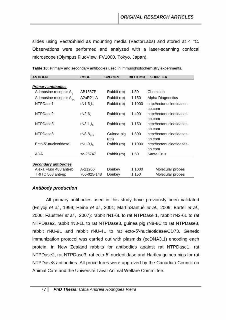
ORIGINAL RESEARCH ARTICLES
77 PhD Thesis: Cátia Andreia Rodrigues Vieira
slides using VectaShield as mounting media (VectorLabs) and stored at 4 °C.
Observations were performed and analyzed with a laser-scanning confocal
microscope (Olympus FluoView, FV1000, Tokyo, Japan).
Table 10: Primary and secondary antibodies used in immunohistochemistry experiments.
ANTIGEN CODE SPECIES DILUTION SUPPLIER
Primary antibodies
Adenosine receptor A1 AB1587P Rabbit (rb) 1:50 Chemicon
Adenosine receptor A2A
A2aR21-A Rabbit (rb) 1:150 Alpha Diagnostics
NTPDase1 rN1-6LI4 Rabbit (rb) 1:1000 http://ectonucleotidases-
ab.com
NTPDase2 rN2-6L Rabbit (rb) 1:400 http://ectonucleotidases-
ab.com
NTPDase3 rN3-1LI5 Rabbit (rb) 1:150 http://ectonucleotidases-
ab.com
NTPDase8 rN8-8CI5 Guinea-pig
(gp)
1:600 http://ectonucleotidases-
ab.com
Ecto-5'-nucleotidase rNu-9LI5 Rabbit (rb) 1:1000 http://ectonucleotidases-
ab.com
ADA sc-25747 Rabbit (rb) 1:50 Santa Cruz
Secondary antibodies
Alexa Fluor 488 anti-rb A-21206 Donkey 1:1000 Molecular probes
TRITC 568 anti-gp 706-025-148 Donkey 1:150 Molecular probes
Antibody production
All primary antibodies used in this study have previously been validated
(Enjyoji et al., 1999; Heine et al., 2001; MartínSantué et al., 2009; Bartel et al.,
2006; Fausther et al., 2007): rabbit rN1-6L to rat NTPDase 1, rabbit rN2-6L to rat
NTPDase2, rabbit rN3-1L to rat NTPDase3, guinea pig rN8-8C to rat NTPDase8,
rabbit rNU-9L and rabbit rNU-4L to rat ecto-5′-nucleotidase/CD73. Genetic
immunization protocol was carried out with plasmids (pcDNA3.1) encoding each
protein, in New Zealand rabbits for antibodies against rat NTPDase1, rat
NTPDase2, rat NTPDase3, rat ecto-5’-nucleotidase and Hartley guinea pigs for rat
NTPDase8 antibodies. All procedures were approved by the Canadian Council on
Animal Care and the Université Laval Animal Welfare Committee.

ORIGINAL RESEARCH ARTICLES
78 PhD Thesis: Cátia Andreia Rodrigues Vieira
Materials and solutions
Adenosine 5’-triphosphate (ATP), adenosine 5′-diphosphate (ADP),
adenosine 5′-monophosphate (AMP), ADO, INO, hypoxanthine (HX); 2,4,6-
trinitrobenzenesulphonic acid (TNBS); choline chloride, paraformaldehyde (prills),
lysine, sodium periodate, anhydrous glycerol, fetal bovine serum (Sigma, St Louis,
MO, USA); serum albumin, Triton X-100, metanol, potassium dihydrogen
phosphate (KH2PO4) (Merck, Darmstadt, Germany); dipyridamole (Boehringer
Ingelheim, Germany); 1,3-dipropyl-8-cyclopentylxanthine (DPCPX) (Research
Biochemicals, Natick, MA, USA); mibefradil dihydrochloride, tetrodotoxin (TTX), (4-
(2-[7-amino-2-(2-furyl{1,2,4}-triazolo{2,3-a{1,3,5}triazin-5-yl-aminoethyl) phenol
(ZM 241385) (Tocris Cookson Inc., UK); [methyl-3H]-choline chloride (ethanol
solution, 80 Ci mmol-1) (Amersham, UK); Pic®A reagent (Waters corporation,
Milford, USA); ATP bioluminescence assay kit HS II (Roche Applied Science,
Indianapolis, Indiana); medetomidine hydrochloride (Domitor®, Pfizer Animal
Health); atipamezole hydrochloride (Antisedan®, Orion, Espoo, Finland); ketamine
hydrochloride (Imalgene®, Merial, Lyon, France); Sodium chloride 0,9%, tramadol
hydrochloride (Labesfal, Santiago de Besteiros, Portugal).
All drugs were prepared in distilled water. All stock solutions were stored as
frozen aliquots at -20°C. Dilutions of these stock solutions were made daily and
appropriate solvent controls were done. No statistically significant differences
between control experiments, made in the absence or in the presence of the
solvents at the maximal concentrations used (0.5 % v/ v), were observed. The pH
of the perfusion solution did not change by the addition of the drugs in the
maximum concentrations applied to the preparations.
Presentation of data and statistical analysis
The values are expressed as mean ± SEM, with n indicating the number of
animals used for a particular set of experiments. Statistical analysis of data was
carried out using paired or unpaired Student’s t-test or one-way analysis of
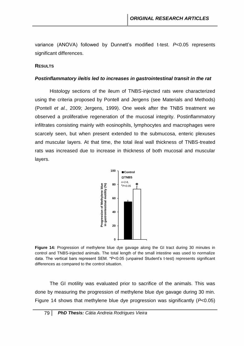
ORIGINAL RESEARCH ARTICLES
79 PhD Thesis: Cátia Andreia Rodrigues Vieira
variance (ANOVA) followed by Dunnett’s modified t-test. P<0.05 represents
significant differences.
RESULTS
Postinflammatory ileitis led to increases in gastrointestinal transit in the rat
Histology sections of the ileum of TNBS-injected rats were characterized
using the criteria proposed by Pontell and Jergens (see Materials and Methods)
(Pontell et al., 2009; Jergens, 1999). One week after the TNBS treatment we
observed a proliferative regeneration of the mucosal integrity. Postinflammatory
infiltrates consisting mainly with eosinophils, lymphocytes and macrophages were
scarcely seen, but when present extended to the submucosa, enteric plexuses
and muscular layers. At that time, the total ileal wall thickness of TNBS-treated
rats was increased due to increase in thickness of both mucosal and muscular
layers.
0
20
40
60
80
100
Pro
gre
ss
ion
of
Me
thyle
ne
blu
ein
gas
tro
inte
sti
na
l m
oti
lity
(%
)
Control
TNBS
n=2-8,
*P<0.05 *
Figure 14: Progression of methylene blue dye gavage along the GI tract during 30 minutes in
control and TNBS-injected animals. The total length of the small intestine was used to normalize
data. The vertical bars represent SEM. *P<0.05 (unpaired Student’s t-test) represents significant
differences as compared to the control situation.
The GI motility was evaluated prior to sacrifice of the animals. This was
done by measuring the progression of methylene blue dye gavage during 30 min.
Figure 14 shows that methylene blue dye progression was significantly (P<0.05)

ORIGINAL RESEARCH ARTICLES
80 PhD Thesis: Cátia Andreia Rodrigues Vieira
faster in TNBS-injected animals than in control animals, indicating that GI
propulsion is increased in postinflammatory ileitis. Although TNBS-ileitis in the rat
lacks a typical chronic phase, the postinflammatory phase is characterized by
increases in motility (see e.g. Moreels et al., 2001).
Adenosine neuromodulation is impaired in postinflammation ileitis
Previous reports suggested that the purinergic control of cholinergic nerve
activity may be significantly impaired during chronic intestinal inflammation, both in
the colon of rabbits and guinea-pigs (Deporotere et al., 2002; Strong et al., 2010),
and in mice ileum (De Man et al., 2003). Despite the differences in the
mechanisms used to produce intestinal inflammation among these groups, all are
unanimous on the indication for the need of more studies in order to investigate
the underlying pathways responsible for disturbed purinergic neuromodulation.
Hence, we focused our attention on the net tonic action of endogenous ADO on
electrically-evoked [3H]-ACh release from myenteric motoneurons seven days after
an inflammatory insult to the rat ileum. In preparations from healthy rats, the A1
receptor antagonist, DPCPX (10 nM) increased the release of [3H]-ACh by 27±4%
(n=4) (Figure 15A). Conversely, selective blockade of adenosine A2A receptors
with ZM 241385 (50 nM) significantly decreased the evoked tritium outflow by
37±10% (n=6). The results indicate that endogenous adenosine exerts a dual role
on evoked [3H]-ACh release via the activation of inhibitory A1 and excitatory A2A
receptors in the rat ileum (cf. Duarte-Araújo et al., 2004; Correia-de-Sá et al.,
2006). However, when similar experiments were conducted in the LM-MP seven
days after ileal inflammation none of the adenosine receptor antagonists caused
any measurable change on evoked transmitter release (Figure 15A). These results
indicate that ADO modulation of cholinergic nerve activity is severely impaired in
postinflammation ileitis, as previously suggested using both myographic
recordings and electrophysiology methods (see above).
Several authors hypothesized that purinoceptors downregulation in the ENS
can occur during prolonged contact with purines. This was predicted since purines,
such as ATP and ADO, can be released from activated inflammatory cells
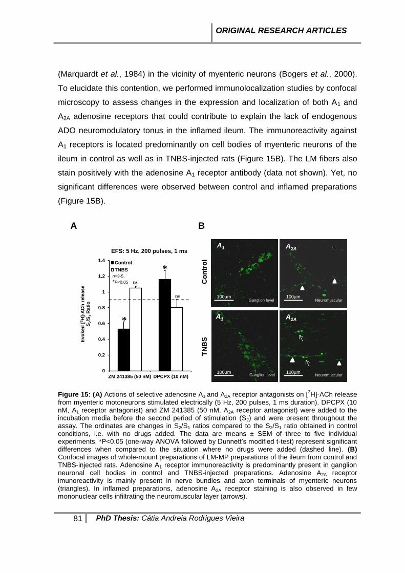
ORIGINAL RESEARCH ARTICLES
81 PhD Thesis: Cátia Andreia Rodrigues Vieira
(Marquardt et al., 1984) in the vicinity of myenteric neurons (Bogers et al., 2000).
To elucidate this contention, we performed immunolocalization studies by confocal
microscopy to assess changes in the expression and localization of both A1 and
A2A adenosine receptors that could contribute to explain the lack of endogenous
ADO neuromodulatory tonus in the inflamed ileum. The immunoreactivity against
A1 receptors is located predominantly on cell bodies of myenteric neurons of the
ileum in control as well as in TNBS-injected rats (Figure 15B). The LM fibers also
stain positively with the adenosine A1 receptor antibody (data not shown). Yet, no
significant differences were observed between control and inflamed preparations
(Figure 15B).
100µm
100µm
Co
ntr
ol
Ganglion level
A1
TN
BS
A1
Ganglion level100µm
A2A
100µm
A2A
Neuromuscular
A B
Neuromuscular0
0.2
0.4
0.6
0.8
1
1.2
1.4
ZM 241385 (50 nM) DPCPX (10 nM)
Evo
ked
[3H
]-A
Ch
rele
ase
S2/S
1R
ati
o
EFS: 5 Hz, 200 pulses, 1 ms
Control
TNBS
*
n=3-5,
*P<0.05
*
ns
ns
Figure 15: (A) Actions of selective adenosine A1 and A2A receptor antagonists on [
3H]-ACh release
from myenteric motoneurons stimulated electrically (5 Hz, 200 pulses, 1 ms duration). DPCPX (10 nM, A1 receptor antagonist) and ZM 241385 (50 nM, A2A receptor antagonist) were added to the incubation media before the second period of stimulation (S2) and were present throughout the assay. The ordinates are changes in S2/S1 ratios compared to the S2/S1 ratio obtained in control conditions, i.e. with no drugs added. The data are means ± SEM of three to five individual experiments. *P<0.05 (one-way ANOVA followed by Dunnett’s modified t-test) represent significant differences when compared to the situation where no drugs were added (dashed line). (B) Confocal images of whole-mount preparations of LM-MP preparations of the ileum from control and TNBS-injected rats. Adenosine A1 receptor immunoreactivity is predominantly present in ganglion neuronal cell bodies in control and TNBS-injected preparations. Adenosine A2A receptor imunoreactivity is mainly present in nerve bundles and axon terminals of myenteric neurons (triangles). In inflamed preparations, adenosine A2A receptor staining is also observed in few mononuclear cells infiltrating the neuromuscular layer (arrows).
ORIGINAL RESEARCH ARTICLES
82 PhD Thesis: Cátia Andreia Rodrigues Vieira
Likewise, the A2A receptor immunoreactivity, which is more evident on
myenteric nerve terminals, also did not significantly differ among control and
inflamed tissues (Figure 15B). Interestingly, we observed staining against the A2A
receptor in few mononuclear cells (probably lymphocytes) infiltrating the
neuromuscular level, which might be responsible for the immunosuppressant
effect of A2A receptors activation in experimental ileitis (Odashima et al., 2005;
Cavalcante et al., 2006). Mononuclear infiltrates containing A2A-positive immune
cells may contribute to the increased mRNA expression of this receptor found in
chronic inflamed intestinal tissues (Antonioli et al., 2011). Although we cannot
exclude at this stage differences in the intracellular signaling pathway triggered by
activation of the two high affinity adenosine receptor subtypes, A1 and A2A, in the
LM-MP of the rat ileum, our data suggest that receptors expression is fairly
conserved in the ileum seven days following an inflammatory insult and,
apparently, does not underscore the lack of ADO neuromodulation tonus in this
condition (see e.g., De Man et al., 2003; Antonioli et al., 2011; Antonioli et al.,
2013).
Postinflammatory ileum releases more ATP, which is not followed by
adenosine formation
It has been hypothesized, but not yet proven, that disruption in ATP release
and/or breakdown as most likely explanations for the suppression of purinergic
neuromuscular transmission in inflamed regions of the distal colon (Strong et al.,
2010). It is possible that inflammatory mediators affect mitochondrial function, and
therefore ATP synthesis, in the ulcerated region of the chronic inflamed intestine.
Another possibility is that there may be increased expression of ectonucleotidases,
which are responsible for the breakdown of ATP (Duarte-Araújo et al., 2009;
Neshat et al., 2009), as has been demonstrated in the purinergic sympathetic
regulation of submucosal blood vessels (Neshat et al., 2009). Here, we measured
the extracellular accumulation of ATP (by bioluminescence) in parallel with ADO
plus INO content (by HPLC with diode array detection) in samples collected
immediately before and after electrical stimulation of LM-MP preparations of the

ORIGINAL RESEARCH ARTICLES
83 PhD Thesis: Cátia Andreia Rodrigues Vieira
ileum of both control and TNBS-injected rats. Figure 16 shows that, in healthy
animals, stimulation of the LM-MP at a frequency of 5 Hz (3000 pulses of 1 ms
duration) yield increased amounts of ATP in the incubation fluid, which was
followed by even higher extracellular accumulation of adenine nucleosides,
consisting mostly of ADO and INO. This pattern was totally reversed seven days
after inflammation of the rat ileum. Basal levels of ATP significantly (P<0.05)
increase (Figure 16A), whereas the baseline of endogenous ADO plus INO
decreased (Figure 16B), in the fluid collected from TNBS-treated LM-MP of the rat
ileum as compared to control tissues. Although the amount of ATP and adenine
nucleosides increased from baseline following electric stimulation of LM-MP
preparations in both animal groups, released ATP levels reached higher values in
inflamed preparations than in controls (Figure 16A), but the opposite was
observed regarding the content of ADO plus INO (Figure 16B).
0
0.002
0.004
0.006
0.008
0.01
0.012
pre-EFS post-EFS
AT
P (
pm
ol/ m
g)
Control
TNBS
*
*n=4*P<0.05
0
5
10
15
20
25
30
35
40
45
pre-EFS post-EFS
AD
O +
IN
O (
pm
ol/ m
g)
Control
TNBS
*
*
n=12-29*P<0.05
A B
Figure 16: Effect of EFS on extracellular accumulation of purines in the LM-MP of control and inflamed rat ileum. The preparations were incubated with Tyrode’s solution for 15 min and then subject to EFS (5 Hz frequency, 3000 pulses of 1ms duration). Samples collected from the incubation media were analysed by HPLC to separate and quantify purine nucleosides; ATP outflow was assessed using the luciferin-luciferase bioluminescence assay. Illustrated are ATP (A) and purine nucleosides (B) content of bath samples collected immediately before (Pre-EFS) and after (Post-EFS) LM-MP stimulation expressed in pmol/mg of tissue. Data are means ± SEM of an n number of individual experiments. *P <0.05 (one-way ANOVA followed by Dunnett’s modified t test) represent significant differences as compared to control animals.

ORIGINAL RESEARCH ARTICLES
84 PhD Thesis: Cátia Andreia Rodrigues Vieira
These results indicate that, in contrast to previous hypothesis, the amount
of ATP released in basal conditions and following electric stimulation of TNBS-
treated ileal preparations was significantly higher than in control tissues, although
one could not discard at this stage that ATP accumulation could also result from a
decrease in the extracellular breakdown of the nucleotide (see below). Despite this
increase in ATP accumulation, the content of ADO plus INO in the superfusates
from TNBS-treated animals was severely decreased, which might contribute to
explain the lack of endogenous ADO neuromodulatory tonus in postinflammatory
ileitis (Figure 15; see also Antonioli et al., 2011).
In healthy tissue, but not in the ileum following the inflammatory insult, stimulus-
evoked ADO release was partially dependent on neuronal activity. This was
evidenced because pretreatment of the preparations with tetrodotoxin (TTX),
applied in a concentration (1 µM) that essentially abolished evoked [3H]-ACh
release, reduced by about 50% adenine nucleosides outflow from control tissues
with almost no effect in TNBS-treated preparations (Figure 17). These findings
indicate that barely half of the extracellular ADO released from stimulated LM-MP
preparations from healthy rats comes from TTX-sensitive neuronal cells, but this
source may be severely affected in the postinflammation phase of TNBS-ileitis due
to neuronal dysfunction, as demonstrated by several authors. Under conditions
leading to myenteric neuronal dysfunction such as chronic inflammation, ATP
release may be shifted from a neuronal origin towards other cell sources (e.g.
smooth muscle fibers, glial cells, ICC, infiltrating immune cells) (see e.g., Nitahara
et al., 1995). As a matter of fact, evoked ATP release from TTX-resistant non-
neuronal sources increased (P<0.05) above baseline from 7.4±0.6 fmol/mg (n=4)
in control animals to 13.2±0.2 fmol/mg (n=4) in TNBS-treated preparations.
Smaller amounts of adenosine released from inflamed ileum originates from
activated pacemaker interstitial cells of Cajal via equilibrative nucleoside
transporters
Considering that ADO may also be released as such from non-neuronal
cells at the tripartite myenteric synapse, we tested whether stimulus-evoked
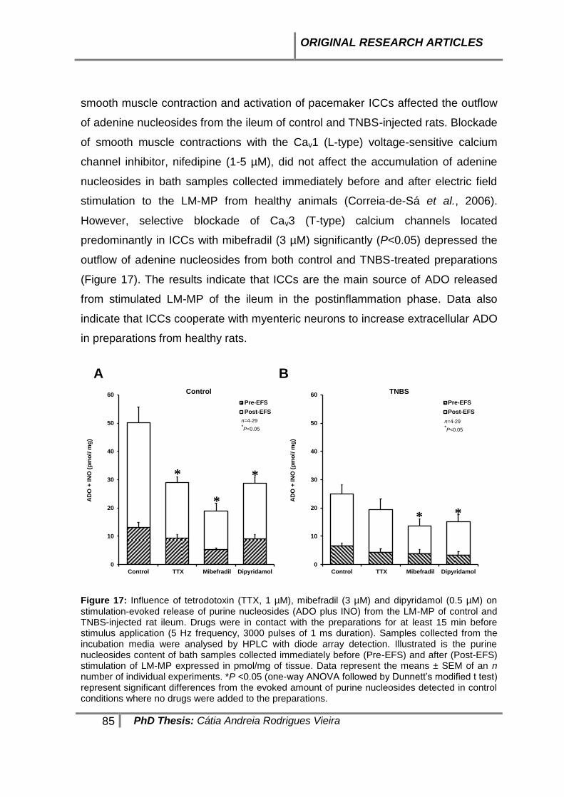
ORIGINAL RESEARCH ARTICLES
85 PhD Thesis: Cátia Andreia Rodrigues Vieira
smooth muscle contraction and activation of pacemaker ICCs affected the outflow
of adenine nucleosides from the ileum of control and TNBS-injected rats. Blockade
of smooth muscle contractions with the Cav1 (L-type) voltage-sensitive calcium
channel inhibitor, nifedipine (1-5 µM), did not affect the accumulation of adenine
nucleosides in bath samples collected immediately before and after electric field
stimulation to the LM-MP from healthy animals (Correia-de-Sá et al., 2006).
However, selective blockade of Cav3 (T-type) calcium channels located
predominantly in ICCs with mibefradil (3 µM) significantly (P<0.05) depressed the
outflow of adenine nucleosides from both control and TNBS-treated preparations
(Figure 17). The results indicate that ICCs are the main source of ADO released
from stimulated LM-MP of the ileum in the postinflammation phase. Data also
indicate that ICCs cooperate with myenteric neurons to increase extracellular ADO
in preparations from healthy rats.
0
10
20
30
40
50
60
Control TTX Mibefradil Dipyridamol
AD
O +
IN
O (
pm
ol/
mg
)
Control
Pre-EFS
Post-EFS
*
*
*
n=4-29*P<0.05
0
10
20
30
40
50
60
Control TTX Mibefradil Dipyridamol
AD
O +
IN
O (
pm
ol/
mg
)
TNBS
Pre-EFS
Post-EFS
* *
n=4-29*P<0.05
A B
Figure 17: Influence of tetrodotoxin (TTX, 1 µM), mibefradil (3 µM) and dipyridamol (0.5 µM) on stimulation-evoked release of purine nucleosides (ADO plus INO) from the LM-MP of control and TNBS-injected rat ileum. Drugs were in contact with the preparations for at least 15 min before stimulus application (5 Hz frequency, 3000 pulses of 1 ms duration). Samples collected from the incubation media were analysed by HPLC with diode array detection. Illustrated is the purine nucleosides content of bath samples collected immediately before (Pre-EFS) and after (Post-EFS) stimulation of LM-MP expressed in pmol/mg of tissue. Data represent the means ± SEM of an n number of individual experiments. *P <0.05 (one-way ANOVA followed by Dunnett’s modified t test) represent significant differences from the evoked amount of purine nucleosides detected in control conditions where no drugs were added to the preparations.

ORIGINAL RESEARCH ARTICLES
86 PhD Thesis: Cátia Andreia Rodrigues Vieira
Given that ADO is neither stored nor released as a classical
neurotransmitter and previous findings from our laboratory demonstrated that the
ecto-nucleotidase pathway contributes only partially to the total interstitial ADO
concentration in the myenteric plexus (Correia-de-Sá et al., 2006), we tested the
involvement of ENTs to extracellular ADO released from tissues isolated from
control and TNBS-injected rats. The nucleoside transport inhibitor, dipyridamole
(0.5 µM), decreased proportionally and by a similar magnitude to that obtained
with mibefradil (3 µM) the outflow of adenine nucleosides from both control and
inflamed tissues (Figure 17). Thus, it is likely that extracellular ADO detected in
ileum following an inflammatory insult originates predominantly from activated
ICCs via ENTs.
Another potential source of endogenous ADO in the rat ileum may be
adenosine 3',5'-cyclic monophosphate (cAMP) extruded from activated cells,
which may be converted to AMP and then to ADO by ecto-phosphodiesterase and
ecto-5'nucleotidase/CD73, respectively (Giron et al., 2008). Despite the existence
of a functional extracellular cAMP-adenosine pathway in the rat ileum, we were
unable to detect any measurable amounts of cAMP by HPLC analysis (with diode
array detection) of the samples used to quantify adenine nucleosides, even though
we could identify a chromatographic peak corresponding to cAMP spectrum but
with a higher retention time than ADO using a 30 µM external standard (data not
shown). These results suggest that, under the present experimental conditions,
the cAMP-adenosine pathway does not account significantly for endogenous ADO
formation in the LM-MP of the rat ileum.

ORIGINAL RESEARCH ARTICLES
87 PhD Thesis: Cátia Andreia Rodrigues Vieira
The kinetics of the extracellular catabolism of adenine nucleotides (ATP and
ADP) was not different in control and postinflammation preparations of the
rat ileum
Despite the observation that the ileum in the postinflammation phase
releases more ATP than control preparations, we observed a decrease in the
extracellular ADO content in samples collected both under basal and stimulated
conditions. It is, therefore, possible that disruption of ATP catabolism into ADO via
ecto-NTPDases may occur in the inflamed LM-MP of the ileum. Figure 18
illustrates the time course of the extracellular catabolism of ATP and ADP in the
LM-MP of the rat ileum from control and TNBS-treated rats. No significant
differences (P>0.05) were observed among the half-degradation times of ATP (30
µM) and ADP (30 µM) in control and TNBS-treated preparations, i.e. the half
degradation time of the two adenine nucleotides ranged from 6 to 7 min
independently of the experimental condition.
Extracellular ATP (30 µM) was metabolized into ADP, AMP, ADO, INO and
HX, whose concentrations increased with time. Higher amounts of AMP as
compared to ADP were detected in the bath at all time points following ATP (30
µM) application (Figure 18A). ADP (30 µM) catabolism led to AMP, ADO, INO and
HX (Figure 18B). Interestingly, we detected differences in the accumulation of
AMP and its degradation product, ADO, between control and postinflamed
preparations incubated either with ATP (30 µM) or ADP (30 µM). The extracellular
AMP content increased (orange bars), whereas ADO decreased (green bars),
proportionally in the incubation fluid of TNBS-treated preparations as compared to
those isolated from control rats following incubation with either ATP (30 µM) or
ADP (30 µM) (Figure 18). Increases in AMP content of bath samples from
postinflammation ileum reached the control levels 45 min after starting incubation
with either ATP (30 µM) or ADP (30 µM), i.e. when the concentration of adenine
nucleotides reached minimum (Figure 18).

ORIGINAL RESEARCH ARTICLES
88 PhD Thesis: Cátia Andreia Rodrigues Vieira
A B
Figure 18: Kinetics of the extracellular catabolism of ATP (A) and ADP (B) in the LM-MP of the ileum of control and TNBS-injected rats. ATP and ADP (30 µM) were incubated at zero time (see Methods). Collected samples were analysed by HPLC with UV detection to separate and quantify ATP (black), ADP (red), AMP (orange), ADO (green), INO (dark grey) and HX (light grey). Average results obtained in four experiments.
Feed-forward inhibition of ecto-5’-nucleotidase/CD73 by adenine nucleotides
controls adenosine formation in postinflammation ileitis
Data from Figure 18 clearly indicate that differences in ADO formation from
the extracellular catabolism of adenine nucleotides in postinflammation ileitis
implicate AMP dephosphorylation by ecto-5’-nucleotidase, which is the rate limiting
enzyme for ADO formation in the rat myenteric plexus (Duarte-Araújo et al., 2009).
Surprisingly, we did not observe significant changes (P>0.05) in the half
degradation time of AMP (30 µM) between control and TNBS-treated preparations
(Figure 19A). If any difference has to be mentioned, is the fact that the inflamed
ileum accumulated proportionally less ADO in the extracellular milieu as compared
to control tissues when AMP (30 µM) was used as substrate (Figure 19A). Overall,
the results indicate that ADO is being rapidly converted into INO by ADA in the
extracellular space of both control and TNBS-treated preparations (see below),
given that stoichiometry of AMP conversion into ADO, INO and HX is kept

ORIGINAL RESEARCH ARTICLES
89 PhD Thesis: Cátia Andreia Rodrigues Vieira
unaltered ruling out a significant contribution of the nucleoside uptake system (cf.
Duarte-Araújo et al., 2004).
t½(AMP)=14�2 min. t½(AMP)=12�1 min. t½(AMP)=66�8 min. t½(AMP)=21�6 min.
A B
C
Figure 19: Kinetics of the extracellular catabolism AMP (30 µM) in the LM-MP of the ileum of
control and TNBS-injected rats (A). The residual ecto-5’-nucleotidase/CD73 activity (soluble form)
in the incubation fluid after removing the preparations from the organ bath, is also shown (B). AMP
(30 µM) was incubated at zero time (see Methods). Collected samples were analysed by HPLC
with UV detection to separate and quantify AMP (orange), ADO (green), INO (dark grey) and HX
(light grey). Average results obtained in four experiments. Panel (C) shows that immunoreactivity
against ecto-5’-nucleotidase/CD73 in longitudinal smooth muscle fibres of the ileum from TNBS-
treated rats is much more evident than in control animals.

ORIGINAL RESEARCH ARTICLES
90 PhD Thesis: Cátia Andreia Rodrigues Vieira
Since ecto-5’-nucleotidase/CD73 may be cleaved from its membrane GPI
anchor by proteolysis retaining its catalytic activity in the incubation fluid, we
decided to test whether binding of this enzyme to the plasma membrane was
somehow affected during chronic inflammation in order to explain the differences
detected in ADO formation from the hydrolysis of adenine nucleotides. To this end,
we evaluated the 5’-nucleotidase activity in the fluid remaining in the bath after
removing the preparation following a 45-min incubation period (Figure 19B). Under
these experimental conditions, the half-life of AMP (30 µM) was significantly
(P<0.05) decreased in TNBS-treated preparations (t½=21±6 min, two-times slower
than with the tissue present) as compared to controls (t½=66±8 min, almost five-
times slower than with the tissue present). Data suggest that postinflammation
ileitis causes an increase in unbound soluble forms of 5’-nucleotidase, which
contribute to dephosphorylation of extracellular AMP into ADO without changing
the global enzyme activity in the tissue. Notwithstanding, functional repercussions
may be expected from this fact taking into consideration that ecto-5’-
nucleotidase/CD73 is concentrated in the myenteric smooth muscle cell layer of
healthy ileum (Nitahara et al., 1995), whereas unbound soluble forms of the
enzyme may generate ADO from extracellular AMP way from the most common
site at the myenteric neuromuscular synapse in the inflamed ileum.
Given that we did not observe any significant differences in the AMP
catabolism between control and TNBS-treated preparations (see Figure 19A) and
we, still, detected increases in ecto-5’-nucleotidase/CD73 immunoreactivity in the
longitudinal neuromuscular layer of the inflamed rat ileum (Figure 19C; see also
(Antonioli et al., 2011), we hypothesized that ecto-5’-nucleotidase/CD73 could be
feed-forwardly inhibited by high extracellular levels of ATP and/or ADP which bind
to the catalytic site of the enzyme blunting ADO formation from AMP in inflamed
preparations (Figure 16; see also Magalhães-Cardoso et al., 2003). The ecto-5’-
nucleotidase activity was evaluated by quantifying the ratio [Nucleosides]:[Total
nucleotides] per min, which is a direct measure of the activity of ecto-5’-
nucleotidase, using either ATP (30 µM) or ADP (30 µM) as substrates. Figure 20A
shows that this ratio increased progressively with time upon consumption of ATP

ORIGINAL RESEARCH ARTICLES
91 PhD Thesis: Cátia Andreia Rodrigues Vieira
or ADP in the incubation media. The activity of ecto-5’nucleotidase/CD73 was
significantly (P<0.05) impaired in the LM-MP of the ileum following an
inflammatory insult, particularly when ADP (30 µM) was used as substrate. These
findings agree with data from Figure 18, showing that ADO formation is delayed
with a compensatory increase in AMP accumulation in the inflamed ileum as
compared to control preparations which is compatible with our hypothesis that
ATP and/or ADP feed-forwardly inhibit ecto-5’-nucleotidase/CD73 in
postinflammation ileitis.
0.00
0.05
0.10
0.15
0.20
0 5 10 15 20 25 30
(Nu
cle
os/
Nu
cle
ot)
.min
-1
Time (min)
ATP (30 µM) as substrate
Control
TNBS
n=4
*P<0.05
*0.00
0.05
0.10
0.15
0.20
0 5 10 15 20 25 30
(Nu
cle
os/
Nu
cle
ot)
.min
-1
Time (min)
ADP (30 µM) as substrate
Control
TNBS
* * **
n=4,
*P<0.05
A B
Figure 7
Figure 20: Activity of ecto-5’-nucleotidase/CD73 when ATP (A) and ADP (B) were used as
substrates of the ectonucleotidase cascade in the LM-MP of the ileum of control and TNBS-injected
rats. ATP and ADP (30 µM) were incubated at zero time. Ecto-5’-nucleotidase/CD73 activity was
evaluated by quantifying the ratio [Nucleosides]:[Nucleotides]/min. Average results obtained in four
experiments; the vertical bars represent the SEM and are shown when they exceed the symbols in
size. *P <0.05 (one-way ANOVA followed by Dunnett’s modified t test) represent significant
differences as compared to control animals.
Tissue distribution changes of NTPDase2, NTPDase3 and NTPDase8 in
postinflammation ileitis
The relative amounts of ATP and/or ADP near the ecto-5’-
nucleotidase/CD73, which is concentrated in the myenteric smooth muscle layer
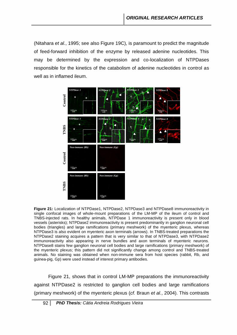
ORIGINAL RESEARCH ARTICLES
92 PhD Thesis: Cátia Andreia Rodrigues Vieira
(Nitahara et al., 1995; see also Figure 19C), is paramount to predict the magnitude
of feed-forward inhibition of the enzyme by released adenine nucleotides. This
may be determined by the expression and co-localization of NTPDases
responsible for the kinetics of the catabolism of adenine nucleotides in control as
well as in inflamed ileum.
Co
ntr
ol
Figure 8
TN
BS
NTPDase 3
NTPDase 2
100µm
NTPDase 3
100µm
NTPDase 2
100µm 100µm
NTPDase 8
100µm
NTPDase 8
100µm
NTPDase 1
NTPDase 1
* *
**
100µm
100µm
Co
ntr
ol
TN
BS
Non-immune (Rb)
100µm
100µm
Non-immune (Rb)
Non-immune (Gp)
Non-immune (Gp)
100µm
100µm
Figure 21: Localization of NTPDase1, NTPDase2, NTPDase3 and NTPDase8 immunoreactivity in single confocal images of whole-mount preparations of the LM-MP of the ileum of control and TNBS-injected rats. In healthy animals, NTPDase 1 immunoreactivity is present only in blood vessels (asterisks); NTPDase2 immunoreactivity is present predominantly in ganglion neuronal cell bodies (triangles) and large ramifications (primary meshwork) of the myenteric plexus, whereas NTPDase3 is also evident on myenteric axon terminals (arrows). In TNBS-treated preparations the NTPDase2 staining acquires a pattern that is very similar to that of NTPDase3, with NTPDase2 immunoreactivity also appearing in nerve bundles and axon terminals of myenteric neurons. NTPDase8 stains few ganglion neuronal cell bodies and large ramifications (primary meshwork) of the myenteric plexus; this pattern did not significantly change among control and TNBS-treated animals. No staining was obtained when non-immune sera from host species (rabbit, Rb, and guinea-pig, Gp) were used instead of interest primary antibodies.
Figure 21, shows that in control LM-MP preparations the immunoreactivity
against NTPDase2 is restricted to ganglion cell bodies and large ramifications
(primary meshwork) of the myenteric plexus (cf. Braun et al., 2004). This contrasts

ORIGINAL RESEARCH ARTICLES
93 PhD Thesis: Cátia Andreia Rodrigues Vieira
with the localization of NTPDase3 immunoreactivity, which is also evident on
myenteric nerve trunks and terminals corresponding to secondary and tertiary
neuronal meshworks, respectively (Lavoie et al., 2011). At this time, we cannot
exclude the presence of this enzyme on intramuscular ICCs. This pattern changes
in the postinflammation ileum. In TNBS-treated preparations, the NTPDase2
immunoreactivity is also observed in the secondary and tertiary neuronal
meshwork, meaning that ADP formation from released ATP by stimulated enteric
neurons (White & Leslie, 1982) and smooth muscle fibres (Nitahara et al., 1995)
may contribute to prolong feed-forward inhibition of muscle-bound ecto-5’-
nucleotidase/CD73 at the myenteric neuromuscular synapse following an
inflammatory insult.
Apparently no significant differences were found in the expression and
localization of NTPDase3 in the inflamed LM-MP of the rat ileum. Regarding
NTPDase8, we were able to demonstrate its presence in few neuronal cell bodies
of myenteric ganglia, with much lesser expression at the neuromuscular layer
(Figure 21). Phenotypic characterization of NTPDase8 positive neuronal cell
bodies deserves future investigations. Like NTPDase3, we found no gross
changes in NTPDase8 immunoreactivity between control and TNBS-treated
animals. We focused our attention on these three ectoenzymes, because we
detected no immunoreactivity against NTPDase1 in the LM-MP of the rat ileum
besides few blood vessels (Figure 21).
Postinflammation ileitis is accompanied by higher adenosine deaminase
(ADA) activity: contribution of membrane-bound (ecto-ADA) and soluble
(exo-ADA) forms of the enzyme
The bath concentrations of ADO (30 µM) decrease progressively with time
yielding to the formation of INO and HX in the LM-MP of the rat ileum (see
Correia-de-Sá et al., 2006). The extracellular catabolism of ADO (30 µM) was
faster (t½=24±3 min, n=4) in TNBS-treated than in control preparations (t½=52±10
min, n=4). The rate of extracellular ADO inactivation can be best appreciated by

ORIGINAL RESEARCH ARTICLES
94 PhD Thesis: Cátia Andreia Rodrigues Vieira
calculating ADA activity in the present experimental conditions, which is
represented in Figure 22A.
100µm 100µm
100µm 100µm
Con
trol
TN
BS
ADA ADA
ADA ADA
Neuromuscular level Ganglion level
B
0.0000
0.0002
0.0004
0.0006
0.0008
0.0010
0 5 10 15 20 25 30
AD
A a
ctiv
ity
(U
)
Time (min)
ADO (30 µM) as a substrate
Control
TNBS
n=4,
*P<0.05
* * **
0.000
0.002
0.004
0.006
0.008
0.010
0.012
0 5 10 15 20 25 30
AD
A S
olu
ble
form
act
ivit
y (
U)
Time (min)
ADO (30 µM) as a substrate
Control
TNBS
n=4,
*P<0.05
*
*
*
A
Figure 9
Figure 22: (A) Activity of ADA during the extracellular catabolism of ADO in the LM-MP of the ileum of control and TNBS-injected rats. ADO (30 µM) was incubated at zero time. Average results obtained in four experiments; the vertical bars represent the SEM and are shown when they exceed the symbols in size. *P <0.05 (one-way ANOVA followed by Dunnett’s modified t test) represent significant differences as compared to control animals. (B) Localization of ADA immunoreactivity in single confocal images of whole-mount preparations of the LM-MP of the ileum of control and TNBS-injected rats, taken both at ganglia and neuromuscular layers. Please note that ADA immunoreactivity is much more exuberant in preparations from TNBS-treated rats than in control animals. The figure insert details ADA staining in mononuclear inflammatory cells infiltrating myenteric ganglia.

ORIGINAL RESEARCH ARTICLES
95 PhD Thesis: Cátia Andreia Rodrigues Vieira
Data indicate that the LM-MP of the ileum following an inflammatory insult
has increasing amounts of ADA that we confirmed by immunofluorescence
confocal microscopy (Figure 22B). ADA immunoreactivity was detected both at the
ganglion level, as well as at the neuromuscular layer probably attached to smooth
muscle fibers. In TNBS-treated preparations, ADA staining was also observed in
mononuclear cells, most probably T lymphocytes and macrophages, infiltrating the
myenteric plexus at the ganglion level (Martin et al., 1995; Conlon & Law, 2004).
Likewise, Antonioli and col. (2010) demonstrated by Western blot analysis
increases in the expression of ADA at the level of inflamed colonic tissues. Like
the ecto-5’-nucleotidase/CD73, extracellular ADA can be found attached to the
plasma membrane (ecto-ADA) (Pettengill et al., 2013), as well as in soluble forms
after proteolytic cleavage from cell membranes (exo-ADA) (Franco et al., 1997).
Both forms retain catalytic activity. Figure 22A, also shows that the activity of the
soluble form of ADA retained in the incubation fluid is highest during the first 5 min
after removing the LM-MP preparation from the bath. Soluble ADA activity was not
accompanied by any modification of lactate dehydrogenase activity, thus indicating
that its activity is not due to damaged cells (see e.g. Correia-de-Sá et al., 2006).
The soluble ADA activity was further increased in the incubation fluid which
had been in contact for 45 min with preparations treated with TNBS. This implies
that ADA is secreted in high amounts from the inflamed ileum, thus contributing to
explain the lack of ADO neuromodulatory tonus. Even though INO may
accumulate in postinflammation ileitis as a result of increased extracellular ADO
deamination (see Figures 18 and 19), we failed to detect any variation on evoked
[3H]-ACh release when INO was applied to the incubation fluid of control and
TNBS-treated preparations in concentrations as high as 1 mM (data not shown).
Catabolic degradation of ADO by soluble ADA may also result in the impairment of
immune modulation by endogenous ADO (via A2A receptors on mononuclear cells,
see Figure 15B) and the consequent worsening of inflammation and tissue injury
(Antonioli et al., 2010).

ORIGINAL RESEARCH ARTICLES
96 PhD Thesis: Cátia Andreia Rodrigues Vieira
DISCUSSION AND CONCLUSIONS
Enteric plasticity comprises a wide range of structural and/or functional
changes in enteric neurons, glial cells and pacemaker ICCs, which are located
between the muscle layers of GI tract. Different types of pathophysiological
conditions drive to distinct adaptive responses. Enteric neurons are known to
control all GI functions. They are able to rapidly change their structure, function or
chemical coding to maintain the gut homeostasis (e.g. during inflammatory
disorders) (Lakhan et al., 2010). Despite heterogeneity of enteric adaptive
responses, involvement of ubiquitous purine nucleotides and nucleosides provide
fine tuning control of peristaltic motor performance in the gut during stressful
situations, such as sustained neuronal activity, ischemia and inflammation
(reviewed in Pettengill et al., 2013). Numerous studies have described the
potential role of purinergic function regulators in inflammation and immunity
(Robson et al., 2006; Antonioli et al., 2013; Louis et al., 2008; Sotnikov & Louis,
2010). There is recent evidence that blockade of adenosine kinase, which results
in increased endogenous levels of ADO, downregulates the inflammatory
response in experimental colitis (Siegmund et al., 2001). In addition, Mabley et al.
(2003) showed that INO resulting from the breakdown of ADO effectively reduced
the inflammatory response in a murine model of colitis, despite we showed in this
study that INO accumulation might not be sufficient to explain the purinergic
neuromodulatory changes observed in postinflammation ileitis. These, and many
other findings, suggest that ADO can mediate both pro-inflammatory (via A1 or A2B
receptors) and/or anti-inflammatory/ immunosuppressant activities (mainly through
A2A and A3 receptors) in inflammatory gut diseases, and they have potential for the
treatment of these diseases (reviewed in Antonioli et al., 2013). However, much is
unknown to what extent the immune-modulatory role of purines during
inflammation interferes with neuromodulation of the ENS (De Man et al., 2003).
Control of cellular activity by purines depends on the activation of two
families of membrane-bound purinoceptors, P1 and P2, which are sensitive to
ADO and to adenine and/or uracil nucleotides, respectively. Interestingly, P1 and

ORIGINAL RESEARCH ARTICLES
97 PhD Thesis: Cátia Andreia Rodrigues Vieira
P2 purinoceptors are also present on immune cells and there is evidence that
ADO and ATP are generated at sites of inflammation (see, for a review (Cronstein,
1994). Purinoceptors activation is fine-tuned regulated by the extracellular
conversion of released adenine and uracil nucleotides leading to the formation of
other biologically active products, namely ADP/UDP and ADO, via a cascade of
membrane-bound ectonucleotidases (Kukulski et al., 2005; Yegutkin, 2008). Thus,
cellular expression and topology of ectonucleotidase enzymes, as well as factors
that extrinsically affects their catalytic activity (e.g. ionic concentration, nucleotide
binding), create gradients of adenine nucleotides and their breakdown products,
which may discriminate which nearby purinoceptor is more likely to be activated.
For instance, under basal conditions ATP transiently facilitates ACh from non-
stimulated myenteric neurons via prejuctional P2X (probably P2X2) receptors;
however, increases in the amount of ATP released from stimulated neurons create
conditions favourable to ADP accumulation at the myenteric synapse, leading to
downregulation of transmitter release via the P2Y1 receptor (Duarte-Araújo et al.,
2009). Strategic manipulation of the activity of ectonucleotidases has been
proposed as a novel therapeutic approach to modify pathogenic purinergic
signalling cascades (reviewed in Burnstock & William’s, 2000).
At the myenteric plexus, the rate limiting enzyme responsible for
extracellular ADO formation from released adenine nucleotides is ecto-5’-
nucleotidase/CD73 (Duarte-Araújo et al., 2004, Duarte-Araújo et al., 2009). This
enzyme acts in a concerted manner with ADO inactivation pathways, both cellular
uptake and ADA, to control extracellular levels of the nucleoside and, thereby, the
activation of co-localized P1 receptors (Correia-de-Sá et al., 2006). Interestingly,
although much less has been explored in functional terms, the enzymes most
relevant to control ADO levels in the extracellular milieu, both ecto-5’-
nucleotidase/CD73 and ADA, may be cleaved from their anchor to the plasma
membrane while retaining their catalytic activity in the soluble form (Pettengill et
al., 2013). Coincidently or not, significant increases in ecto-5’-nucleotidase/CD73
and ADA mRNA expression were observed in inflamed colonic tissues (Antonioli
et al., 2014). Fragile docking of these enzymes to the plasma membrane is more
ORIGINAL RESEARCH ARTICLES
98 PhD Thesis: Cátia Andreia Rodrigues Vieira
likely to occur within the context of inflammatory reactions, given that recruitment
of inflammatory cells release a huge number of cytokines, chemokines and
enzymes, some of these with proteolytic activity. One of these enzymes is
glycosylphosphatidylinositol-specific phospholipase, which plays a role in
inflammation since it can hydrolyse the GPI anchor of several membrane proteins
and its hydrolytic products up-regulate cytokine expression in macrophages
(O’Brien et al., 1999). The widespread distribution of soluble forms of ecto-5’-
nucleotidase/CD73 and ADA create conditions to unbalanced bulk production
and/or inactivation of ADO way from its original location, thus affecting mass organ
function. Likewise, inflammatory infiltrates including T lymphocytes, which are
endowed with NTPDase1/CD39 and ecto-5’-nucleotidase/CD73 enzymes, may
additionally contribute to ADO accumulation disturbance and to unpredictable P1-
receptor-mediated responses in inflamed tissues. In light of these compelling
hypotheses, this work was designed to investigate the kinetics of the catabolism of
adenine nucleotides and ADO accumulation in the myenteric plexus of the ileum
following an inflammatory insult.
Data from this study evidenced a disparity between higher amounts of ATP
detected in the extracellular fluid released from TNBS-treated ileal preparations
and deficits in the ADO levels measured in the same samples, both under baseline
conditions and after electrical stimulation. This puzzling contradiction has not been
detected before. On the contrary, indirect evidences made by other authors led to
the hypothesis that the disruption of ATP release could be due to mitochondrial
dysfunction induced by inflammatory mediators (Neshat et al., 2009; Strong et al.,
2010), which was not confirmed in the present study. In postinflammation ileitis,
higher ATP amounts can be originated from infiltrating immune and non-neuronal
cells, such as glial cells, ICCs and smooth muscle contractions (see e.g., (Nitahara
et al., 1995); reviewed in Burnstock & Lavin, 2002, considering the proposed
inflammatory neuronal dysfunction. Hemichannels containing pannexins may act
as conduits for ATP release from non-neuronal cells in response to physiological
and pathological stimuli. These channels are able to form signalling complexes
with the purinergic P2X7 receptor, which activation leads to large pore formation

ORIGINAL RESEARCH ARTICLES
99 PhD Thesis: Cátia Andreia Rodrigues Vieira
and consequently to further release of ATP (Diezmos et al., 2013; Gulbransen et
al., 2012). These authors proposed a model where high extracellular levels of ATP
chronically activate neuronal P2X7, pannexins and caspases. This relationship
between P2X7 and pannexins has been related to the loss of enteric neurons
during inflammatory conditions (Lakhan & Kirchgessner, 2010; Gulbransen et al.,
2012, Sanger, 2012). Moreover, our data add some information to explain the
impairment of purinergic signalling during intestinal inflammation detected in other
reports (De Man et al., 2003, Depoortere et al., 2002, Strong et al., 2010) which
has been expanded in this study by showing a lack of ADO neuromodulatory
control of ACh release from postinflammation myenteric plexus. We provided
compelling evidences that most of the ADO originated in the inflamed myenteric
plexus of the rat ileum was derived from activated ICC’s by the release of the
nucleoside as such via dipyridamole-sensitive equilibrative transporters. This also
agrees with previous findings from our group showing that ADO formation from
released ATP, via the ecto-nucleotidase cascade, contributes only partially to the
total interstitial ADO concentration in the myenteric plexus of healthy rats (Correia-
de-Sá et al., 2006).
The apparent disturbance of ADO generated from the extracellular
catabolism of released ATP may be due to feed-forward inhibition of ecto-5’-
nucleotidase/CD73 as a consequence of high extracellular levels of ATP, and
particularly ADP, accumulated in the inflamed myenteric synapse [cf. Magalhães-
Cardoso et al., 2003). ATP and ADP are competitive inhibitors of ecto-5’-
nucleotidase/CD73 with inhibition constants in the low micromolar range. This
indicates that they can bind to the active site of this enzyme, like AMP, but they
cannot be hydrolysed (Sträter, 2006; Fausther et al., 2012); recovery of ecto-5’-
nucleotidase/CD73 activity occurs once extracellular ATP and ADP levels
decrease below the micromolar range. Redistribution of NTPDase2 from ganglion
cell bodies, which is the preferential localization of this enzyme in healthy animals,
towards myenteric nerve terminals affords the most probable explanation for the
unpredicted ADP accumulation in postinflammation neuromuscular synapse. This
is possible because NTPDase2 is a preferential nucleoside triphosphatase

ORIGINAL RESEARCH ARTICLES
100 PhD Thesis: Cátia Andreia Rodrigues Vieira
hydrolysing ADP 10 to 15 times less efficiently than ATP (Kukulski et al., 2005),
and we detected no changes in the distribution of NTPDase3 and NTPDase8
present in the myenteric plexus. Notably, no evidence of NTPDase1 expression
was detected in the rat myenteric plexus, besides a few blood vessels. Thus,
during postinflammation ileitis the hydrolysis of ATP into ADO at the myenteric
synapse may be transiently interrupted by feed-forward inhibition of ecto-5’-
nucleotidase/CD73, which makes a shift from P1 receptors activation (by ADO) to
a preferential P2 receptors activation (by ATP or ADP). These data are in
agreement with the hydrolysis of ATP by NTPDase2 and ecto-5’-
nucleotidase/CD73 in the rat liver (Cunha, 2001). Indeed, depending with which
NTPDase subtype ecto-5’-nucleotidase/CD73 co-localizes different amounts of
ADO will be generated (Cunha, 2001). The functional interpretation of these
findings in order to justify the increase in GI motility may be complicated by
disturbances in compartmentalization originated by the release of significant
amounts of soluble forms of the enzyme (Pettengil et al., 2013; Sträter, 2006).
Nevertheless, our results fully agree with other authors suggesting that ecto-5’-
nucleotidase/CD73 might play a significant role in the modulation of purinergic
signalling during enteric inflammation (Louis et al., 2008; Antonioli et al., 2013).
Previous findings from our group demonstrated that extracellular
deamination by ADA is the most efficient mechanism regulating synaptic ADO
levels and, consequently, tonic activation of facilitatory A2A receptors in myenteric
nerve terminals of healthy rats (Duarte-Araújo et al., 2004). Besides the very high
level of activity of ecto-ADA, a less-efficient nucleoside transport system sensitive
to dipyridamole may also contribute to inactivation of extracellular ADO. Owing to
the effectiveness of both inactivation mechanisms, endogenous ADO actions may
be restricted to the release/production region at the myenteric cholinergic synapse,
while exogenously added ADO seems to activate preferentially extrajunctional
inhibitory A1 receptors (Duarte-Araújo et al., 2004; Vieira et al., 2011). A question
remains unanswered regarding the tonic effect of endogenous ADO on low affinity
excitatory A3 receptors, although selective agonists to this receptor have been
shown to beneficially influence inflammation in experimental models (Guzma et al.,

ORIGINAL RESEARCH ARTICLES
101 PhD Thesis: Cátia Andreia Rodrigues Vieira
2006, Antonioli et al., 2010). Tandem localization of adenosine A3 (on cell bodies)
and A2A (on nerve varicosities) receptors along myenteric neurons explains why
the A3 receptor activation may be prevented by ZM 241385 (50 nM), a selective
A2A receptor antagonist with low affinity for A3 receptors (Ki>10 µM). This implies
that endogenous ADO acts preferentially on prejunctional A2A under normal
conditions making the activation of low affinity A3 receptors by ADO high unlikely
during the postinflammatory phase of TNBS-ileitis due to suppression/inactivation
of the nucleoside (this study). The putative activation of A3 receptors by
endogenously formed INO via ADA also cannot explain the loss of the
neuromodulatory influence of nucleosides in postinflammation ileitis. While we are
uncertain regarding the extracellular levels of ADO at both particular locations,
both in vivo and in vitro models suggest that the balance between inhibitory A1 and
excitatory A2A may be important in regulating intestinal motility. For instance, De
Man et al. (2003) reported that chronic intestinal inflammation enhanced enteric
contractile activity in part due to a loss of the cholinergic neuromodulation
mediated by A1 receptors (De Man et al., 2003). It is worth noting that human post-
ganglionic myenteric neurons co-express adenosine A1 and A2A receptors, which
exhibit a heterogeneous distribution (Christofi, 2001). Therefore, there is a
considerable interest in the neuroprotective effects exerted by ADO during
inflammatory (and ischemic) insults, and it is conceivable that adenosine
accumulation disturbances may contribute to enteric excitability during
pathological conditions.
Besides the proposed interruption of the ectonucleotidase cascade at the
level of ecto-5’-nucleotidase/CD73 leading to AMP accumulation and low ADO
formation in the inflamed myenteric plexus, our data also show that ADA activity is
significantly enhanced in postinflammation ileitis thus contributing to decrease the
extracellular ADO levels and, thereby, tonic activation of P1 receptors. ADA is the
enzyme responsible for the deamination of ADO into INO. This enzyme has a wide
phylogenetic distribution and its amino acid sequence is highly conserved,
suggesting that ADA is a key enzyme in purine metabolism. Besides the classical
intracellular form, ADA is reported to bind to the cell surface of T lymphocytes, via

ORIGINAL RESEARCH ARTICLES
102 PhD Thesis: Cátia Andreia Rodrigues Vieira
the activation cell marker CD26 (Cristalli et al., 2001). The presence of ADA in
nerve terminals has also been demonstrated in many brain regions and it was
hypothesize that ADA-containing terminals may release ADO or the enzyme itself,
which could serve to regulate purinergic neurotransmission (Nagy et al., 1990;
Nagy et al., 1996). In the rat myenteric plexus, we first described that ADA activity
increased in bath samples collected after tissue stimulation in parallel with the
interstitial accumulation of ADO and its metabolites (Correia-de-Sá et al., 2006).
This implies that the amount of ADO detected following stimulation of the
preparation was largely underestimated as compared to the levels of the
nucleoside in the biophase. Secretion of ADA may occur in several diseases,
namely those affecting hematopoietic and immune (Ungerer et al., 1992). To our
knowledge, this is the first study reporting an increase in the activity of soluble
ADA in the chronic inflamed myenteric plexus of the rat ileum. Our findings unravel
the mechanism by which inhibition of ADA might attenuate inflammation in
experimental colitis (Antonioli et al., 2007).
In conclusion, data indicate that postinflammation ileitis suppresses ADO
neuromodulation, which may contribute to increase GI motility. Impairment of ADO
neuromodulation is most probably due to deficient accumulation of the nucleoside
at the myenteric synapse despite the paradoxical increase in ATP release.
Discrepancy between ATP outflow and ADO deficiency in the chronic inflamed
ileum can be ascribed to feed-forward inhibition of ecto-5’-nucleotidase/CD73 by
high extracellular levels of ATP and/or ADP. Interestingly, redistribution of
NTPDase2, but not of NTPDase3, from ganglion cell bodies to myenteric nerve
terminals leads to preferential ADP accumulation from released ATP, thus
contributing to prolong inhibition of muscle-bound ecto-5’-nucleotidase/CD73 and
to delay ADO formation at the inflamed neuromuscular synapse. On the other
hand, depression of endogenous ADO accumulation may also occur due to
enhancement of ADA activity. We observed a remarkably increase in the activity of
soluble forms of ecto-5’-nucleotidase/CD73 and ADA in the postinflammatory
phase of TNBS-ileitis, which may contribute to unbalanced bulk production and/or
inactivation of ADO way from its location in the native tissue thus affecting organ

ORIGINAL RESEARCH ARTICLES
103 PhD Thesis: Cátia Andreia Rodrigues Vieira
function. Thus, our findings suggest that pharmacological inhibition of ADA, and/or
the use of AMP-derived prodrugs able to selectively activate P1 receptor co-
localized with ecto-5’-nucleotidase/CD73, represent promising therapeutic
strategies to ameliorate motility disturbances and immune reactivity inherent to
inflammatory enteric disorders.
This research was partially supported by FCT (FCT project Pest-
OE/SAU/UI215/2011) with the participation of FEDER funding. CV and IS were in receipt
of PhD studentships by FCT (SFRH/BD/79091/2011 and SFRH/BD/88855/2012,
respectively). J.S. received support from the Canadian Institutes of Health Research
(CIHR; MOP-102472) and he was also a recipient of a senior Scholarship from the Fonds
de recherche du Québec-Santé (FRQS).
The authors acknowledge the valuable collaboration of Professor Margarida Duarte-
Araújo in the implementation of TNBS-induced intestinal inflammation rat model in our lab.
Blind histological characterization of inflammatory infiltrates in the ileum of control versus
TNBS-treated rats was initially performed by Professor Justina Oliveira (Departamento de
Ciências Veterinárias, UTAD, Vila Real, Portugal) (data not shown). The authors also wish
to thank Mrs. Maria Helena Costa e Silva and Belmira Silva for technical assistance.

ORIGINAL RESEARCH ARTICLES
104 PhD Thesis: Cátia Andreia Rodrigues Vieira
PAPER 2
(Manuscript submitted for publication)

ORIGINAL RESEARCH ARTICLES
105 PhD Thesis: Cátia Andreia Rodrigues Vieira
Postinflammatory ileitis induces neurochemical adjustments of purine
cellular resources in the myenteric plexus
Cátia Vieira, Fátima Ferreirinha, Maria Teresa Magalhães-Cardoso, Isabel Silva &
Paulo Correia-de-Sá*
Laboratório de Farmacologia e Neurobiologia, Center for Drug Discovery and Innovative
Medicines (MedInUP), Instituto de Ciências Biomédicas de Abel Salazar (ICBAS),
Universidade do Porto, Porto, Portugal.
ABSTRACT
Uncoupling between ATP overflow and extracellular adenosine formation
changes purinergic signalling in postinflammatory ileitis. Loss of adenosine
neuromodulation was ascribed to feed-forward inhibition of ecto-5’-
nucleotidase/CD73 by high extracellular adenine nucleotides in the inflamed ileum.
Here, we hypothesized that inflammation-induced changes in cellular density may
also account to unbalance the release of purines and their influence on [3H]ACh
release from TNBS-treated longitudinal muscle-myenteric plexus preparations of
the rat ileum. The population of S100β-positive glial cells increase and a loss of
ANO-1-positive interstitial cells of Cajal/fibroblast-like cells (ICC/FLC) were noted
in the ileum 7-days after the inflammatory insult. In the absence of changes on
VAChT-positive cholinergic nerves by confocal microscopy, the inflamed myenteric
plexus released smaller amounts of [3H]ACh, which also became less sensitive to
neuronal block with TTX (1µM). Instead, [3H]ACh release was attenuated by
sodium fluoroacetate (5mM), carbenoxolone (10µM) and A438079 (3µM), which
prevent activation of glial cells, pannexin-1 hemichannels and P2X7 receptors,
respectively. Sodium fluoroacetate also decreased ATP release without
significantly affecting adenosine levels indicating that surplus ATP overflow
parallels reactive gliosis in postinflammatory ileitis. Conversely, loss of ICC/FLC
may explain the lower adenosine levels in TNBS-treated preparations, since
blockade of Cav3 (T-type) channels existing in ICC/FLC with mibefradil (3µM) or
inhibition of equilibrative nucleoside transporters with dipyridamole (0.5µM), both
decreased extracellular adenosine. Data suggest that postinflammatory ileitis

ORIGINAL RESEARCH ARTICLES
106 PhD Thesis: Cátia Andreia Rodrigues Vieira
operates a shift on purinergic neuromodulation reflecting the upregulation of ATP-
releasing enteric glial cells and the depletion of ICC/FLC that account for
decreased adenosine overflow via nucleoside transporters.
Keywords: Postinflammatory ileitis; Acetylcholine release; Adenosine release; ATP
release; Myenteric plexus; Enteric glia; Interstitial cells.
INTRODUCTION
Inflammation of the gastrointestinal (GI) tract triggers a series of adaptive
morphological, chemical and functional changes in the cellular components
responsible for maintaining gut homeostasis (Sharkey & Kroese, 2001). These
involve the number and chemical coding of enteric neurons, but also the relative
abundance and activity of adjacent non-neuronal cells such as enteric glia,
interstitial cells of Cajal, fibroblast-like cells and smooth muscle fibers, directly or
indirectly influenced by inflammatory infiltrates. Adaptive cellular responses may
impact on the coordination of motor function, local blood flow, GI secretions and
also on the endocrine and immune reactions (Costa et al., 2000). As a matter of
fact, the postinflammatory status is frequently accompanied by significant changes
in enteric motility (Pontell et al., 2009; Vieira et al., 2014).
Changes in the release of purines together with adaptive modifications of
purinoceptors expression and/or function are hallmarks of inflammatory reactions
in most tissues, with the GI tract being no exception (Roberts et al., 2012).
Although purinergic signaling modifications underlying inflammatory responses of
the GI tract are not fully understood, the extreme plasticity of the purinergic system
and its pathophysiological impact on immune reactions, enteric neuronal
networking and cellular communication make drugs targeting the purinergic
cascade ideal candidates for treating inflammatory GI diseases. Purines, such as
ATP and adenosine, are released from activated infiltrating inflammatory cells
(Marquardt et al., 1984), as well from resident neuronal and non-neuronal enteric
cells (Stead et al., 1989; Bogers et al., 2000). ATP released in response to

ORIGINAL RESEARCH ARTICLES
107 PhD Thesis: Cátia Andreia Rodrigues Vieira
inflammatory mediators is crucial for neutrophil activation and immune defense
(Lazarowski et al., 2011), but can also function as a danger signal preventing cells
invasion of immune-privileged tissues, like myenteric ganglia (Bradley et al.,
1997).
In healthy individuals, ATP is co-released by vesicular exocytosis from
enteric neurons with other neurotransmitters, like acetylcholine (ACh) (Burnstock,
1976). Mounting evidences indicate that ATP release from non-neuronal cells is
also critical to control organ functions, in both normal and stressful conditions
(Bodin & Burnstock, 2001; Lazarowski et al., 2011; Mutafova-Yambolieva, 2012;
Silva et al., 2015). Non-neuronal release of ATP may be carried out by vesicular
ATP transporters (VNUT) (Sawada et al., 2008; Lazarowski et al., 2011), as well
as via other mechanisms involving activation of ABC proteins and hemichannels
containing connexins and/or pannexins (Bodin & Burnstock, 2001; Lazarowski et
al., 2011; Timóteo et al., 2014; Carneiro et al., 2014; Silva et al., 2015).
Once released from either neuronal or non-neuronal cells, ATP modifies
organ functions by activating directly ionotropic P2X and metabotropic P2Y
purinoceptors or indirectly, via P1 receptors, after being metabolized into
adenosine through ecto-nucleotidases. At the myenteric neuromuscular synapse,
ATP transiently facilitates [3H]ACh release from non-stimulated nerve terminals via
the activation of P2X (most probably P2X2 or P2X2/3) receptors (Duarte-Araújo et
al., 2009). Fast conversion of ATP directly into AMP catalyzed by NTPDases 2
and 3 (Vieira et al., 2015) and, subsequent, formation of adenosine by ecto-5’-
nucleotidase/CD73, controls evoked [3H]ACh release through stimulation of high-
affinity excitatory A2A and/or inhibitory A1 receptors located on nerve terminals and
ganglion cells bodies of myenteric nerves, respectively (Duarte-Araújo et al.,
2004; 2009; Vieira et al., 2011). However, inflammation may lead to
overexpression of NTPDase2 at the myenteric synapse (Vieira et al., 2014), which
is a preferential nucleoside triphosphatase hydrolysing ADP 10 to 15 times less
efficiently than ATP (Kukulski et al., 2011). This promotes ADP accumulation and

ORIGINAL RESEARCH ARTICLES
108 PhD Thesis: Cátia Andreia Rodrigues Vieira
subsequent downregulation of transmitter release via prejunctional inhibitory P2Y1
receptors (Duarte-Araújo et al., 2009).
In contrast to ATP, adenosine is not stored nor released from synaptic
vesicles. Our group demonstrated that the ecto-nucleotidase pathway contributes
only partially to the total interstitial adenosine concentration in the myenteric
plexus (Correia-de-Sá et al., 2006). Adenosine released as such from either
neuronal or non-neuronal cells seems to be the main source of endogenous
adenosine in the enteric nervous system (Duarte-Araújo et al., 2004). This release
is sought to be mediated by facilitated diffusion via equilibrative nucleoside
transporters (Gu et al., 1995; Duarte-Araújo et al., 2004; Correia-de-Sá et al.,
2006) existing predominantly in interstitial cells of Cajal and/or fibroblast-like cells
(ICC/FLC) (Vieira et al., 2014). Another potential source of endogenous adenosine
could be adenosine 3',5'-cyclic monophosphate (cAMP) extruded from activated
cells, which can be converted to AMP and then to adenosine by ecto-
phosphodiesterase and ecto-5'nucleotidase/CD73, respectively (Giron et al.,
2008), but this pathway does not account significantly for endogenous adenosine
formation in the myenteric plexus of the rat ileum (Vieira et al., 2014).
Uncoupling between ATP overflow and extracellular adenosine formation
has been observed in postinflammatory ileitis (Vieira et al., 2014). This situation,
which disrupts the purinergic control of gut motility, has been ascribed to feed-
forward inhibition of ecto-5’-nucleotidase/CD73 by high extracellular levels of
adenine nucleotides together with an enhancement of adenosine deaminase
(ADA) activity in the inflamed ileum. While these findings explain, at least partially,
the loss of adenosine neuromodulation in the inflamed ileum, they miss the point
regarding enhancement of the ATP-mediated tone. In this study, we hypothesized
that inflammation-induced changes in the density of specific enteric resident cells
could also account to unbalance the release of purines and, thus, their influence
on evoked [3H]ACh release from longitudinal muscle-myenteric plexus
preparations of rats with TNBS-induced postinflammatory ileitis.
ORIGINAL RESEARCH ARTICLES
109 PhD Thesis: Cátia Andreia Rodrigues Vieira
MATERIAL AND METHODS
TNBS-induced intestinal inflammation rat model
Rats of either sex (Wistar,∼200 g) (CharlesRiver, Barcelona, Spain) were
kept at a constant temperature (21°) and a regular light- (06.30–19.30 h) dark
(19.30–06.30 h) cycle, with food and water) and a regular light- (06.30–19.30 h)
dark (19.30–06.30 h) cycle, with food and water ad libitum. Intestinal inflammation
was produced by the instillation of 2,4,6-trinitrobenzenesulfonic acid (TNBS) into
the lumen of the rat ileum. After a fasting period of 4–8 hours with free access to
drinking water, rats undergo median laparotomy under anaesthesia with an
association of medetomidine (10 mg/Kg) and ketamine (75 mg/Kg) given
subcutaneously. At the end of the procedure animals were retrieved with
atipamezole (10 mg/Kg). The terminal ileal loop was gently exteriorized, and TNBS
(40 mM) was injected through the enteric wall into the lumen of the ileum, 10
centimeters proximal to the ileocolonic junction. Controls received 1 mL of 0.9 %
saline. Sixty minutes after surgery, the rats were allowed to eat and drink ad
libitum. After surgery, pain was controlled with tramadol hydrochloride (10 mg/Kg).
To have a control of time course of body weight loss and recovery after injection of
TNBS, rats were weighed prior to TNBS administration and daily following surgery.
Animals with intestinal inflammation (TNBS) transiently lose weight for three to
four days after surgery and regain weight thereafter. Seven days after surgery,
animals were sacrificed following an overnight fasting period.
[3H]-acetylcholine release experiments
Sections of the rat ileum not including the terminal five centimetres were
used. The longitudinal muscle strip with the myenteric plexus attached was
separated from the underlying circular muscle according to the method of Paton
and Vizi (1969). This preparation is highly enriched in cholinergic neurons, mainly
excitatory neurons projecting to the longitudinal muscle (25%) that receive inputs
from intrinsic primary afferents (26%) and from ascending and descending
pathways (17%) (Costa et al., 1996).

ORIGINAL RESEARCH ARTICLES
110 PhD Thesis: Cátia Andreia Rodrigues Vieira
The procedures used for labelling the preparations and measuring evoked
[3H] acetylcholine ([3H]ACh) release were previously described (Duarte-Araújo et
al., 2004, 2009; Correia-de-Sá et al., 2006; Vieira et al., 2011) and used with minor
modifications. Longitudinal muscle-myenteric plexus (LM-MP) strips were mounted
in 365 µL chambers of a semi-automated 12-sample superfusion system (SF-12
Suprafusion 1000, Brandel, Gaithersburg, MD, USA) heated at 37°C. After a 30-
min equilibration period, nerve terminals were labelled during 40 min with 1 µM of
[3H]choline (specific activity 5 µCi/nmol) under electrical field stimulation (1 Hz
frequency, 1 ms pulse width) using two platinum-made grid electrodes placed
above and below the muscle strip (transmural EFS stimulation). Following loading,
the washout superfusion (1 ml/min) of the preparations was performed during 80
min with Tyrode’s solution supplemented with the choline uptake inhibitor,
hemicholinium-3 (10 µM). Tritium outflow was evaluated by liquid scintillation
spectrometry (TriCarb2900TR, Perkin Elmer, and Boston, USA; % counting
efficiency: 56±2%) in 0.6 ml bath samples automatically collected every 1 min
using the SF-12 suprafusion system. [3H]ACh release was evoked by two periods
of EFS (S1 and S2), each consisting of 200 square wave pulses of 1 ms duration
delivered at a 5-Hz frequency.
Test drugs were added 8 min before S2 and were present up to the end of
the experiments. The change in the ratio between the evoked [3H]ACh release
during the two stimulation periods (S2/ S1) relative to that observed in control
situations (in the absence of test drugs) was taken as a measure of the effect of
the tested drugs. Positive and negative values represent facilitation and inhibition
of evoked [3H]ACh release, respectively. None of the drugs significantly (P>0.05)
changed the basal tritium outflow.
Release of ATP and adenine nucleosides (adenosine plus inosine)
The procedures used to measure ATP and adenine nucleosides were
previously described (Vieira et al., 2014). Experiments were performed using an
automated perfusion system for sample collecting for given time periods, therefore
improving the efficacy of HPLC (with diode array detection) and bioluminescence

ORIGINAL RESEARCH ARTICLES
111 PhD Thesis: Cátia Andreia Rodrigues Vieira
analysis. After a 30-min equilibration period, the preparations were incubated with
1.8 mL gassed Tyrode’s solution, which was automatically changed every 15 min
by emptying and refilling the organ bath with the solution in use. The preparations
were electrically stimulated once, 15 min after starting sample collection (zero
time), using 3000 square wave pulses of 1ms duration delivered at a 5-Hz
frequency. In these experiments, only the sample collected before stimulus
application and the two samples collected after stimulation were retained for
analysis. Bath aliquots (50–250 𝜇L) were frozen in liquid nitrogen immediately after
collection, stored at −20C (the enzymes are stable for at least 4 weeks) and
analysed within 1 week of collection by HPLC with diode array detection (Finigan
Thermo Fisher Scientific System LC/DAD, equipped with an Accela Pump coupled
to an Accela Autosample, a diode array detector and an Accela PDA running the
X-Calibur software chromatography manager). Chromatographic separation was
carried out through a Hypersil GOLD C18 column (5 𝜇M, 2.1mm × 150 mm)
equipped with a guard column (5 𝜇m, 2.1mm × 1 mm) using an elution gradient
composed of ammonium acetate (5mM, with a pH of 6 adjusted with acetic acid)
and methanol. During the procedure the flow rate was set at 200 𝜇L/min and the
column temperature was maintained at 20C. The autosampler was set at 4C and
50 𝜇L of standard or sample solution was injected, in duplicate, for each HPLC
analysis. In order to obtain chromatograms and quantitative analysis with maximal
sensibility, the diode array detection wavelength was set at 259 nm for adenosine
and 248 nm for inosine.
In parallel, the ATP content of the same samples was evaluated with the
luciferin-luciferase ATP bioluminescence assay kit HS II (Roche Applied Science,
Indianapolis, Indiana). Luminescence was determined using a multi detection
microplate reader (SynergyHT, BioTek Instruments).
Stimulation-evoked release of adenine nucleotides and nucleosides was
calculated by subtracting the basal release, measured in the sample collected
before stimulation, from the total release of adenine nucleotides and nucleosides
determined after stimulus application.

ORIGINAL RESEARCH ARTICLES
112 PhD Thesis: Cátia Andreia Rodrigues Vieira
Myographic recordings of ileal contractile activity
The contractile activity of the LM-MP was recorded as previously described
(Vieira et al. 2009, 2011; Mendes et al., 2015). Ileum strips from control and
TNBS-treated rats were mounted along the longitudinal axis in 14 ml capacity
perfusion chambers connected to isometric force transducers. The changes in
tension were recorded continuously with a PowerLab data acquisition system
(Chart 5, v.4.2; AD Instruments, USA). Tissues were preloaded with 0.5 g of
tension and allowed to equilibrate for 90 min under continuous superfusion with
gassed (95% O2 and 5% CO2) Tyrode’s solution at 37°C.
Immunofluorescence staining and confocal microscopy observation
LM-MP fragments were isolated from the rat ileum as previously described.
The LM-MP fragments were stretched to all directions and pinned onto Petri
dishes coated with Sylgard®. The tissues, then, were fixed in PLP solution
(paraformaldehyde 2%, lysine 0.075M, sodium phosphate 0.037M, sodium
periodate 0.01 M) for 16 h at 4C. Following fixation, the preparations were
washed three times for 10 min each using 0.1M phosphate buffer. At the end of
the washout period, tissues were cryoprotected during 16 h with a solution
containing anhydrous glycerol 20% and phosphate buffer 0.1M at 4C and, then,
stored at −20C for further processing. Once defrosted, tissue fragments were
washed with tamponated phosphate saline buffer (PBS) and incubated with a
blocking buffer, consisting in foetal bovine serum 10%, bovine serum albumin 1%,
triton X-100 1% in PBS, for 2 hours; washout was facilitated by constant stirring of
the samples. After blocking and permeabilization, samples were incubated with
selected primary antibodies (see Table 1) diluted in the incubation buffer (foetal
bovine serum 5%, serum albumin 1%, Triton X-100 1% in PBS), at 4C, for 48 h.
For double immunostaining, antibodies were combined before application to tissue
samples. Following the washout of primary antibodies with PBS supplemented
with Triton X 1% (3 cycles of 10 min), tissue samples were incubated with species-
specific secondary antibodies in the dark for two hours, at room temperature.
Finally, tissue samples were mounted on optical-quality glass slides using

ORIGINAL RESEARCH ARTICLES
113 PhD Thesis: Cátia Andreia Rodrigues Vieira
VectaShield as mounting media (VectorLabs) and stored at 4C. Observations
were performed and analyzed with a laser scanning confocal microscope
(Olympus FluoView, FV1000, Tokyo, Japan).
Table 11: Primary and secondary antibodies used in immunohistochemistry experiments
ANTIGEN CODE SPECIES DILUTION SUPPLIER
Primary antibodies
PGP 9.5 7863-1004 Mouse (ms) 1:500 Serotec
NF200 ab8135 Rabbit (rb) 1:1000 ABCAM
VaChT AB1588 Guinea-pig (gp) 1:500 Chemicon
nNOS ab1376 Goat (gt) 1:300 ABCAM
GFAP MAB360 Mouse (ms) 1:600 Chemicon
S100 Ab868 Rabbit (rb) 1:400 ABCAM
Vimentin M0725 Mouse (ms) 1/150 Dako
Ano-1 Ab53212 Rabbit (rb) 1:100 ABCAM
P2X7 APR-004 Rabbit (rb) 1/50 Alomone
CD11B (OX42) Sc-53086 Mouse (ms) 1/50 Santa Cruz
Secondary antibodies
Alexa Fluor 488 anti-rb A-21206 Donkey 1:1000 Molecular probes
Alexa Fluor 488 anti-ms A21202 Donkey 1:1000 Molecular probes
Alexa Fluor 568 anti-gt A11057 Donkey 1:1000 Molecular probes
Alexa Fluor 568 anti-ms A-10037 Donkey 1:1000 Molecular probes
Alexa Fluor 633 anti-ms A21052 Goat 1:1000 Molecular probes
TRITC 568 anti-gp 706-025-148 Donkey 1:150 Jackson Immuno
Research
Dylight 649 anti-gp 706-025-148 Donkey 1:100 Jackson Immuno
Research
Materials and solutions
2,4,6-trinitrobenzenesulphonic acid (TNBS); carbenoxolone, choline
chloride, paraformaldehyde (prills), lysine, sodium periodate, anhydrous glycerol,
fetal bovine serum (Sigma, St Louis, MO, USA); serum albumin, triton X-100,
metanol, potassium dihydrogen phosphate (KH2PO4) (Merck, Darmstadt,
Germany); A438079, mibefradil dihydrochloride, L-NAME, tetrodotoxin (TTX)
ORIGINAL RESEARCH ARTICLES
114 PhD Thesis: Cátia Andreia Rodrigues Vieira
(Tocris Cookson Inc., UK); Sodium Fluoroacetate (Supelco); [methyl-3H]-choline
chloride (ethanol solution, 80 Ci mmol−1) (Amersham, UK); ATP bioluminescence
assay kit HS II (Roche Applied Science, Indianapolis, Indiana); medetomidine
hydrochloride (Domitor, Pfizer Animal Health); atipamezole hydrochloride
(Antisedan, Orion, Espoo, Finland); ketamine hydrochloride (Imalgene, Merial,
Lyon, France); Sodium chloride 0.9%, tramadol hydrochloride (Labesfal, Santiago
de Besteiros, Portugal).
All drugs were prepared in distilled water. All stock solutions were stored as
frozen aliquots at −20C. Dilutions of these stock solutions were made daily and
appropriate solvent controls were done. No statistically significant differences
between control experiments, made in the absence or in the presence of the
solvents at the maximal concentrations used (0.5% v/v), were observed. The pH of
the perfusion solution did not change by the addition of the drugs in the maximum
concentrations applied to the preparations.
Presentation of data and statistical analysis
The values are expressed as mean ± SEM, with 𝑛 indicating the number of
animals used for a particular set of experiments. Statistical analysis of data was
carried out using paired or unpaired Student’s t-test with Welch correction. P<0.05
represents significant differences.

ORIGINAL RESEARCH ARTICLES
115 PhD Thesis: Cátia Andreia Rodrigues Vieira
RESULTS
Postinflammatory ileitis causes an increase in enteric glial cells (types III
and IV) and a partial loss of pacemaker interstitial cells of Cajal / fibroblast-
like cells
Structural changes accompanied by neuronal cell death have been
observed in chronic intestinal inflammation (Sanovic et al., 1999; Linden et al.,
2005; Venkataramana et al., 2015). However, we found no obvious changes in the
amount of neurons stained positively against (1) neurofilament NF200 expressed
predominantly in Dogiel type I and II neurons (Hu et al., 2002), and (2) the pan-
neuronal marker, protein gene product 9.5 (PGP 9.5), in the myenteric plexus 7
days after instillation of TNBS into the lumen of ileum compared to control rats
treated with saline (Figure 23c-f). This is compatible with Moreels et al. (2001)
findings showing that TNBS-induced ileitis in the rat lacks the chronic inflammatory
phase and is characterized by an acute transmural inflammation that is
accompanied by functional abnormalities of neuronal activity which persists for at
least 8 weeks without obvious neuronal loss (Stewart et al., 2003; Nurgali et al.,
2007). Conversely, these authors found alterations in longitudinal muscle
contractility which was attributed to structural thickness of the ileal wall.
Confocal micrographs show that neuronal cells are grouped in small
clusters (enteric ganglia), which are interconnected by nerve fibre bundles from
whom emerge small diameter nerve terminals (see e.g. Furness, 2006).
Inflammatory infiltrates consisting of monocytes/ macrophages exhibiting
immunoreactivity against the integrin CD11B/OX42 were found surrounding the
myenteric plexus of TNBS-treated rats (Figure 23b), but not in preparations from
control animals (Figure 23a). The absence of inflammatory cells inside myenteric
ganglia has been observed before (see e.g. Figures 24B and 27B) and this is why
myenteric ganglia are considered an immune-privileged tissue (Bradley et al.,
1997).

ORIGINAL RESEARCH ARTICLES
116 PhD Thesis: Cátia Andreia Rodrigues Vieira
100 µm100 µm100 µm
100 µm100 µm100 µm
GFAP S100
Co
ntr
ol
TN
BS
NF200 PGP 9.5CD11B/αM
a
i
j
b
Vimentin
Co
ntr
ol
TN
BS
c
d
e
f
100 µm100 µm 100 µm
100 µm100 µm 100 µm
k
l
g
h
Figure 23: Confocal micrographs of whole-mount preparations of the longitudinal muscle-myenteric plexus of the ileum of control and TNBS-treated rats. Z-stacks illustrate the immunoreactivity against CD11B/αM (OX42) (marker of inflammatory cells) (a,b), NF200 (neurofilament expressed in neurons) (c,d), PGP9.5 (pan-neuronal cell marker) (e,f), vimentin (intermediate filament of mesenchymal cells, like ICCs and FLCs) (g,h), GFAP (i,j) and S100β (k,l) (enteric glial cells markers). Images are representative of at least four individuals per group. Scale bars = 100 µm.
Enteric glial cells express astrocytic cell markers, including the intermediate
filament glial fibrillary acidic protein (GFAP) and the calcium-binding protein S100β
(Gulbrasen & Sharkey, 2012). Subtypes of enteric glia are classified in: type I,
intraganglionic cells; type II, within interganglionic fibers; type III, form a matrix in
the extraganglionic region remaining in close association with neuronal bundles;
and type IV, elongated glia running with nerve fibres within the musculature
(Boesmans et al., 2015). Figure 23i-l shows that the myenteric plexus of rats
treated with TNBS exhibits increased amounts of GFAP- and S100β-positive glial
cells, which are particularly evident in extraganglionic (glial type III) and

ORIGINAL RESEARCH ARTICLES
117 PhD Thesis: Cátia Andreia Rodrigues Vieira
intramuscular regions (glial type IV). Thus, in contrast to the absence of significant
structural changes in the neuronal cell population, TNBS-induced ileitis stimulates
enteric glial cells proliferation (Bradley et al., 1997) or, at least, the increase in
expression and/or synthesis of glial protein cell markers (Cabarrocas et al., 2003),
in a similar manner to that observed in astrocytes of the CNS (Lhermitte et al.
1980; Rühl et al., 2004).
Interstitial cells of Cajal and fibroblast-like cells (ICC/FLC) are known to act
as pacemaker cells and integrators of nerve activity and smooth muscle
contraction, respectively (Sanders, 1996). The number of these cells can
significantly change in pathological conditions (Ekblad et al., 1998). Using
vimentin, a characteristic intermediate filament of mesenchymal cells like
ICC/FLC, we show here that the density of vimentin-positive cells decrease
significantly in the myenteric plexus of TNBS-induced ileitis (Figure 23h) compared
to control preparations (Figure 23g). Likewise, staining with anoctamine-1 (Ano-1),
a calcium-activated chloride channel involved in the pacemaker activity of ICCs
(Gomez-Pinilla et al., 2009), also decreased in density 7-days after instillation of
TNBS into the lumen of the rat ileum (Figure 24A). The partial loss of Ano-1
immunoreactivity was detected by confocal microscopy focusing in the ganglion
layer (myenteric stellate cells; Figure 24Aa and 24Ab) and in the neuromuscular
region (intramuscular spindle-shape cells, Figure 24Ac and 24Ad) of inflamed LM-
MP preparations. The presence of auto-fluorescent inflammatory cells (coloured in
red) was more visible surrounding the myenteric ganglia than at the intramuscular
layer of TNBS-treated rats (see inset of Figure 24A).
The loss of ICC/FLC may affect the frequency of spontaneous enteric
contractions (Kinoshita et al., 2007). In fact, myographic recordings demonstrate
that the frequency of spontaneous contractions of LM-MP preparations of the
ileum of TNBS-treated animals were less frequent (P<0.05) than those observed in
control animals (Figure 24A).

ORIGINAL RESEARCH ARTICLES
118 PhD Thesis: Cátia Andreia Rodrigues Vieira
Control TNBS
An
o-1
100 µm 100 µm
An
o-1
Ga
ng
lion
leve
lN
euro
mu
scu
lar
leve
l
100 µm100 µm
A B
a b
c d
0.00
0.10
0.20
0.30
0.40
0.50
0.60
0.70
0.80
Control TNBSF
req
uen
cy o
f sp
on
tan
eou
s co
ntr
act
ion
s
n=10-20
*P<0.05
*
Figure 24: The partial loss of anoctamine-1 (Ano-1)-positive interstitial cells in the myenteric plexus of TNBS-treated rats correlates with the reduction in the frequency of spontaneous contractions of the rat ileum. (A) Confocal micrographs of whole-mount preparations of the ileum of control and TNBS-injected rats taken at the myenteric ganglion level (a,b) and at the longitudinal smooth muscle layer (c,d). Shown is the immunoreactivity against Ano-1, a calcium-activated chloride channel involved in the pacemaker activity of ICCs. At the ganglion level, but not at the neuromuscular junction layer, it is possible to visualize autofluorescent inflammatory cells (stained in red, inset) infiltrating the myenteric plexus of TNBS-treated rats. Images are representative of at least five individuals per group. Scale bars = 100 µm. (B) Histograms representing the frequency of spontaneous myographic contractions of the ileum from control rats and TNBS-treated animals. The data are means ± SEM of a n number of animals. *P<0.05 (unpaired Student’s t-test with
Welch correction) represents a significant difference from control animals.
Myenteric neurons from the ileum of TNBS-treated rats release smaller
amounts of [3H]ACh, but no relationship exists between cholinergic
hypoactivity and nitrergic inhibitory signals
ACh is the prime regulator of intestinal motility and is the most important
excitatory neurotransmitter in the myenteric plexus (Costa et al., 1996). Figure 25A
shows that cholinergic neurons from the myenteric plexus of TNBS-treated rats
release significantly smaller amounts of [3H]ACh (see also Collins et al., 1992a;
Davis et al., 1998), despite no obvious changes were detected by confocal
microscopy in the density of cholinergic nerve fibres stained specifically against

ORIGINAL RESEARCH ARTICLES
119 PhD Thesis: Cátia Andreia Rodrigues Vieira
the vesicular acetylcholine transporter (VAChT) (Arvidsson et al., 1997) (Figure
25B).
A B
Co
ntr
ol
TN
BS
VAChT
100 µm 100 µm
100 µm 100 µm
0
10000
20000
30000
40000
50000
60000
Control TNBS
Ev
ok
ed
[3H
]-A
Ch
rele
ase
S1
(DP
M/g
)
n=32-35
*P<0.05
*
Figure 25: Electrically-stimulated myenteric neurons of TNBS-treated rats release smaller amounts of [
3H]ACh. (A) Shown is the amount of [
3H]ACh released from electrically-stimulated (5 Hz, 1 ms,
200 pulses) myenteric neurons of the ileum of control and TNBS-treated rats during S1 in desintegrations per min (DPM)/g of wet tissue. (B) Confocal micrographs of whole-mount preparations of the longitudinal muscle-myenteric plexus of the ileum from control and TNBS-treated rats stained against the vesicular ACh transporter (VAChT). The z-stacks shown are representative of at least five individuals per group. Scale bar = 100 µm.
These results prompted us to investigate whether cholinergic nerve
hypoactivity had any relationship with increased inhibitory neurotransmission
operated by NO, which can be released from neighbouring nitrergic nerves, enteric
glia cells and/or infiltrating immunocytes (MacEachern et al., 2015; see also Figure
26A). This was hypothesized because up-regulation of nitric oxide synthase (NOS)
activity and enhancement of NO-mediated inhibitory neurotransmission were
demonstrated in chronic inflammatory bowel diseases (Miller et al., 1993). Figure
26B shows that inhibition of NOS activity with L-NAME (100 µM) had no effect on
evoked [3H]ACh release from the LM-MP of the ileum of control and TNBS-treated
rats, indicating that NO does not mediate inhibition of [3H]ACh release in TNBS-
induced ileitis. This was concluded despite the relative increase in neuronal NOS-
immunoreactivity in enteric ganglia cells of TNBS-treated animals (Figure 26Aii).

ORIGINAL RESEARCH ARTICLES
120 PhD Thesis: Cátia Andreia Rodrigues Vieira
A BControl TNBS
nN
OS 100 µm 100 µm
i
iii
100 µm 100 µm
Ga
ng
lion
leve
lN
euro
mu
scu
lar
leve
l
ii
iv
0.00
0.20
0.40
0.60
0.80
1.00
1.20
Control L-Name (100 µM)
Ev
ok
ed
[3H
]AC
h r
ele
ase
S2/S
1ra
tio
Drug in S2
Control
TNBS
n=4-6
*P<0.05
VA
Ch
T/
nN
OS
100 µm
Z-s
tack
v
Figure 26: (A) Confocal micrographs of whole-mount preparations of the longitudinal muscle-myenteric plexus of the ileum from control and TNBS-treated rats taken at the myenteric ganglion level (i,ii) and at the longitudinal smooth muscle layer (iii,iv). Shown is the immunoreactivity against neuronal NOS (nNOS); myenteric ganglia from TNBS-treated rats exhibit a higher number of nNOS-positive neuronal cell bodies than control tissues. The z-stack presented in panel (v) shows that nNOS-positive nitrergic nerve fibres (red) are adjacent, but do not co-localize, with VAChT immunoreactivity (green). Images are representative of at least four individuals per group. Scale bar = 100 µM. (B) Effect of the nitric oxide synthase (NOS) inhibitor, L-Name (100 µM), on [
3H]ACh
released from stimulated myenteric neurons of the ileum of control and TNBS-treated rats. After loading and washout periods, [
3H]ACh release was elicited by two trains (S1 and S2) of electrical
field stimulation, each consisting of 200 pulses delivered at a 5 Hz frequency. L-Name (100 μM) was applied 8 min before S2. The ordinates are changes in S2/S1 ratios compared to the S2/S1 ratio obtained without addition of any drug. The data are means ± SEM of four to six individuals. *P<0.05 (unpaired Student’s t-test with Welch correction) represent significant differences from
control animals.
Postinflammatory shift from neuronal to non-neuronal control of evoked
[3H]ACh release from stimulated myenteric neurons
Figure 27A shows that [3H]ACh release from electrical-stimulated ileal
myenteric neurons of healthy rats was almost prevented (P<0.05) by the blockage
of nerve action potentials with tetrodotoxin (TTX, 1 µM). This confirms our previous
observations, where we also demonstrated that [3H]ACh exocytosis from

ORIGINAL RESEARCH ARTICLES
121 PhD Thesis: Cátia Andreia Rodrigues Vieira
depolarized myenteric nerve terminals depend on Ca2+ influx through voltage-
sensitive channels (Duarte-Araújo et al., 2004a; 2004b; Correia-de-Sá et al.,
2006). Interestingly, inhibition of the electrically-driven [3H]ACh outflow from
TNBS-treated preparations was much less sensitive to TTX (1 µM) than controls
(Figure 27A), suggesting that [3H]ACh outflow in TNBS-induced ileitis is likely due
to direct axonal activation by a non-neuronal released activator (Broadhead et al.,
2012; Gulbransen et al., 2012).
Under the present experimental conditions, the influence of smooth muscle
contractions was ruled out since the Cav1 (L-type) channel blocker, nifedipine (1
µM), depressed ACh-induced contractions of the longitudinal muscle without
affecting the release of [3H]ACh from stimulated LM-MP of both control (Correia-
de-Sá et al., 2006) and TNBS-treated rats (unpublished observations). Likewise,
we discarded the participation ICC/FLC in the regulation of evoked [3H]ACh
release in both animal groups, because the transmitter release was not (P>0.05)
affected when the experiments were performed in the presence of selective
inhibitors of ICC/FLC activity, namely mibefradil (3 µM) (Figure 27A) and apamin
(3 µM) (data not shown), which block specifically voltage-sensitive Cav3 (T-type)
and small-conductance KCa2 (SK3) channels characteristic of these cells,
respectively (Fujita et al., 2003).
Therefore, we tested whether inhibition of enteric glial metabolism with
sodium fluoroacetate (5 mM) (MacEachern et al., 2015) could affect [3H]ACh
release from stimulated myenteric neurons. Figure 27A, shows that while sodium
fluoroacetate (5 mM) was unable to change transmitter release in preparations
from healthy rats, it significantly (P<0.05) decreased evoked [3H]ACh release from
myenteric neurons of TNBS-treated animals. These results led us to the
hypothesis that maintenance of minimal cholinergic tone in postinflammatory ileitis
depends on the release of an excitatory gliotransmitter from proliferating enteric
glial cells.

ORIGINAL RESEARCH ARTICLES
122 PhD Thesis: Cátia Andreia Rodrigues Vieira
0.00 0.20 0.40 0.60 0.80 1.00 1.20
Control
TTX (1 µM)
Sodium Fluoroacetate (5 mM)
Mibefradil (3 µM)
Carbenoxolone (10 µM)
A438079 (3 µM)
Evoked [3H]ACh release (S2/S1ratio)
Drugs in S2:
TNBS
Control
n=4-7,
*P<0.05
*
*
*
*
A B
100 µm 100 µm
Control TNBS
P2X
7S
100β
100 µm 100 µm
Figure 27: (A) Effects of TTX (1 μM, a nerve action potential blocker), sodium fluoroacetate (5 mM, a glial cell metabolism uncoupler), mibefradil (3 μM, an inhibitor of Cav3 (T-type) channels existing
in ICC/FLC), carbenoxolone (10 µM, a pannexin-1 inhibitor) and A438079 (3 µM, a P2X7 receptor antagonists) on electrically-evoked [
3H]-ACh release from the LM-MP of the rat ileum of control and
TNBS-treated rats. After loading and washout periods, [3H]ACh release was elicited by two trains
(S1 and S2) of electrical field stimulation, each consisting of 200 pulses delivered at a 5 Hz frequency. Drugs were applied 8 min before S2. Abscissa is changes in S2/S1 ratios compared to the S2/S1 ratio obtained without addition of any drug. The data are means ± SEM of four to seven individuals. *P<0.05 (unpaired Student’s t-test with Welch correction) represent significant differences from the situation where no drugs were added (dashed vertical line). (B) Confocal micrographs of whole-mount preparations of the longitudinal muscle-myenteric plexus of the ileum from control and TNBS-treated rats. Shown is the immunoreactivity against the P2X7 receptor and
the enteric glial cell marker, S100β. Please note the presence of autofluorescent inflammatory cells
(red dots) outside myenteric ganglia in TNBS-treated preparations. Z-stacks shown in the pictures are representative of at least four individuals per group. Scale bar=100 µm.
ATP may be the putative excitatory gliotransmitter responsible for
maintaining the cholinergic tone in postinflammatory ileitis
The nature of the putative excitatory gliotransmitter regulating ACh release
from inflamed myenteric neurons is uncertain. Considering our previous
observations that (1) ATP can be released from both neuronal and non-neuronal
cells, and that (2) it can increase the release of [3H]ACh from resting myenteric
nerve terminals through the activation of ionotropic P2X receptors in the presence
of TTX (Duarte-Araújo et al., 2009), we designed experiments to test if

ORIGINAL RESEARCH ARTICLES
123 PhD Thesis: Cátia Andreia Rodrigues Vieira
endogenous ATP could play any role in the regulation of ACh release in TNBS-
induced ileitis.
Recently, our group demonstrated that ATP can be released from non-
neuronal cells through hemichannels containing pannexin-1 and this mechanism
can lead to a “vicious cycle” where ATP can induce the release of ATP via the
activation of ionotropic P2X and metabotropic P2Y receptors under normal and
pathological conditions (Pinheiro et al., 2013; Timóteo et al., 2014; Noronha-Matos
et al., 2014; Silva et al., 2015; Certal et al., 2015). High extracellular ATP levels,
such as those detected after an inflammatory insult, can stimulate low-affinity
P2X7 receptors, which often but not exclusively couple to pannexin-1 to promote
ATP outflow, via the pannexin-1 hemichannel or the P2X7 receptor pore (Diezmos
et al., 2013; Gulbransen et al., 2012). This prompted us to investigate the role of
pannexin-1 hemichannels and P2X7 receptors on [3H]ACh release from stimulated
LM-MP preparations of control and TNBS-treated rats using specific inhibitors.
Figure 27A, shows that selective blockage of pannexin-1 hemichannels and
P2X7 receptors respectively with carbenoxolone (10 µM) and A438079 (3 µM)
significantly (P<0.05) decreased [3H]ACh release from myenteric neurons of
TNBS-treated rats, but not of their control littermates. The magnitude of the
inhibitory effects on transmitter release produced by carbenoxolone (10 µM) and
A438079 (3 µM) was about the same that observed upon blocking glial cells
metabolism with sodium fluoroacetate (5 mM) (Figure 27A). Moreover, the
distribution of the P2X7 immunoreactivity in the myenteric plexus of both groups of
animals is remarkably similar to that of the glial cell marker, S100β (Figure 27B).
Co-localization of the P2X7 receptor and the S100β glial marker was
demonstrated in the rat myenteric plexus before (Vanderwinden et al., 2003;
reviewed in Gulbransen & Sharkey, 2012).
Postinflammatory myenteric glial cells release higher amounts of ATP
In a previous study from our group, we demonstrated that ATP is released
in higher amounts from electrically-stimulated TNBS-treated preparations than in
control tissues (Vieira et al., 2014). High postinflammatory ATP levels are

ORIGINAL RESEARCH ARTICLES
124 PhD Thesis: Cátia Andreia Rodrigues Vieira
comparable to those obtained in control LM-MP preparations submitted to
blockage of action potentials generation, of glial cells metabolism and of ICC/FLC
activation respectively with TTX (1 µM), sodium fluoroacetate (5 mM) and
mibefradil (3 µM) (Figure 28A). These findings indicate that all these cells
contribute to keep low extracellular ATP levels in normal physiological conditions.
However, this scenario changes considerably in TNBS-induced ileitis. From the
inhibitors used to target specifically the three resident cell types in the myenteric
plexus, only sodium fluoroacetate (5 mM) was able to decrease significantly
(P<0.05) the release of ATP in postinflammatory ileitis, while TTX (1 µM) and
mibefradil (3 µM) were both ineffective (Figure 28A).
0.000
0.002
0.004
0.006
0.008
0.010
0.012
0.014
0.016
0.018
Ev
ok
ed A
TP
rel
ease
(pm
ol/
mg)
CTR
TNBS
*
*
n=4-6
*,#P<0.05 *
#
0.0
5.0
10.0
15.0
20.0
25.0
30.0
35.0
40.0
45.0
Ev
ok
ed A
DO
rel
ease
(pm
ol/
mg)
CTR
TNBS
*
*#
*n=4-6
*,#P<0.05
A B
Figure 28: Differential effects of TTX (1 μM), sodium fluoroacetate (5 mM) and mibefradil (3 μM) on ATP and adenosine (plus inosine) ouflow from stimulated longitudinal muscle-myenteric plexus preparations of the ileum from control and TNBS-treated rats. Drugs were in contact with the preparations for at least 15 min before stimulus application (5 Hz frequency, 3000 pulses of 1 ms duration). Samples from the incubation media were collected and analysed in parallel using the luciferin-luciferase bioluminescence assay and HPLC with diode array detection to evaluate their content in ATP (A) and adenine nucleosides (B), respectively. Ordinates represent stimulation-induced increases in ATP (A) and adenosine (plus inosine) (B) above baseline levels determined immediately before the stimulus application; results are expressed in pmol/mg of tissue. The data are means ± SEM of four to six individuals. *P<0.05 (unpaired Student’s t-test with Welch correction) represent significant differences from the amount of purines released in control conditions where no drugs were added to the preparations (dashed horizontal lines).
ORIGINAL RESEARCH ARTICLES
125 PhD Thesis: Cátia Andreia Rodrigues Vieira
Thus, increased overflow of ATP after an inflammatory insult may possibly
result from activation of proliferating glial cells. Interestingly, the pharmacology
affecting ATP overflow from stimulated ileal preparations of TNBS-treated rats
(Figure 28A) looks like that verified by measuring the release of [3H]ACh (Figure
27A), further strengthening our hypothesis that ATP may be the putative excitatory
gliotransmitter responsible for maintaining the cholinergic tone in TNBS-induced
ileitis.
Deficient extracellular adenosine accumulation in postinflammatory ileitis
parallels the loss of ICC/FLC
Although adenosine may be released from activated inflammatory cells
(Marquardt et al., 1984) in the vicinity of myenteric neurons (Bogers et al., 2000),
the loss of adenosine neuromodulatory control of evoked [3H]ACh release in
TNBS-induced ileitis detected in a previous report results mainly from deficient
accumulation of the nucleoside at the myenteric synapse (Vieira et al., 2014); this
was verified despite the increase in ATP content of the same samples. Uncoupling
between ATP overflow and adenosine levels in postinflammatory ileitis was
ascribed to feed-forward inhibition of ecto-5’-nucleotidase/CD73 and upregulation
of adenosine deaminase. Here, we decided to evaluate the putative contribution of
inflammation-induced type-specific cell density changes in the myenteric plexus to
adenosine deficiency. To this end, we measured the extracellular concentration of
adenosine (plus inosine) in parallel with the ATP levels before and after electrical
stimulation of LM-MP preparations of both control and TNBS-treated rats in the
presence of cell-specific activity inhibitors. Figure 28B shows that the amounts of
adenosine released into the extracellular fluid following stimulation of the LM-MP
are much higher in healthy controls than in TNBS-treated rats.
Stimulus-evoked adenosine release was partially dependent on neuronal
activity in preparations from healthy rats, but not in the ileum of TNBS-treated
animals. This was assumed because pre-treatment of the preparations with TTX
(1 µM) reduced by about 50% the outflow of adenine nucleosides in control tissues
with no effect on TNBS-treated preparations. This means that the neuronal source

ORIGINAL RESEARCH ARTICLES
126 PhD Thesis: Cátia Andreia Rodrigues Vieira
of adenosine may be severely affected in TNBS-induced ileitis due to neuronal
cells dysfunction (see above). Even though we considered unlikely that
proliferating glial cells could contribute to adenosine deficits in the inflamed
myenteric plexus of the rat ileum, we tested the effect of the glial cell metabolic
uncoupler, sodium fluoroacetate (5 mM). Inhibition of glial cells metabolism had
only a limited effect on adenosine outflow from stimulated LM-MP preparations of
both control and TNBS-treated rats (Figure 28B). The inhibitory action of sodium
fluoroacetate (5 mM) was slightly more evident in healthy tissues than in TNBS-
treated preparations, which is in agreement with the proposed uncoupling between
ATP overflow and adenosine formation secondary to the inflammatory insult.
Prevention of smooth muscle contractions with the voltage-sensitive Cav1
(L-type) channel inhibitor, nifedipine (5 µM), did not affect the release of adenosine
(plus inosine) in both animal groups (Correia-de-Sá et al., 2006; Vieira et al., 2009;
2014). However, blockade of Cav3 (T-type) channels expressed in ICC/FLC with
mibefradil (3 µM) significantly (P<0.05) attenuated the release of adenosine (plus
inosine) from both control and TNBS-treated animals. Taking into consideration
previous findings from our laboratory demonstrating that the nucleoside transport
inhibitor, dipyridamole (0.5 µM), decreased proportionally and by a similar amount
to that obtained with mibefradil (3 µM) the outflow of adenosine (plus inosine) from
both control and inflamed tissues (Vieira et al., 2014), it is very likely that
adenosine accumulated in the ileum following an inflammatory insult originates
predominantly from activated ICC/FLC via equilibrative nucleoside transport.
DISCUSSION AND CONCLUSIONS
A number of key cellular players may be involved in the reaction to
inflammatory insults by the gastrointestinal system. These include inflammatory
cells, such as granulocytes, lymphocytes, mast cells, monocytes and
macrophages, which release numerous cytokines, pro-inflammatory peptides,
neuroactive and neurotrophic factors, noxious compounds, inflammatory enzymes

ORIGINAL RESEARCH ARTICLES
127 PhD Thesis: Cátia Andreia Rodrigues Vieira
and other danger signalling molecules. Enteric plasticity comprises a wide range of
structural and/or functional changes in enteric neurons, glial cells and interstitial
cells (ICC/FLC). As a result of adaptive responses to different types of
pathophysiological conditions, enteric neurons are able to rapidly change their
structure, function and chemical phenotype in order to maintain homeostasis of gut
functions. In contrast to long-term functional abnormalities in the activity of
neuronal cells described by several authors (Stewart et al., 2003; Nurgali et al.,
2007) and confirmed in the present study by denoting hypoactivity of cholinergic
neurotransmission, we found no obvious modifications in cellular density and
distribution of specific neuronal cell markers (e.g. NF200, PGP 9.5) in TNBS-
induced ileitis. In this sense, ileitis caused by TNBS differs from other chronic
inflammatory disease models because it lacks the chronic inflammatory phase
(Moreels et al., 2001) that is usually accompanied by neuronal cell loss (Sanovic
et al., 1999; Linden et al., 2005; Venkataramana et al., 2015). Notwithstanding,
this constrain may be turned into an advantage characteristic of this animal model
if one wants to investigate the adaptive changes of neuronal to non-neuronal cells
communication after a transient inflammatory insult, as we aimed in this work.
Results show that stimulated myenteric neurons release smaller amounts of
[3H]ACh in the ileum of TNBS-treated rats compared to their control littermates,
without evidence of any alteration of VAChT-positive cholinergic nerve fibres in the
myenteric plexus. Likewise, significant decreases in [3H]choline uptake,
acetylcholine release and contractile responses to stimulation of enteric nerves
have been observed in rats with colitis induced by TNBS (Poli et al., 2001). These
results imply a functional loss, not corresponding to cellular depletion, of
cholinergic neurotransmission in inflamed tissues (Collins et al.,1992a). The
mechanism underlying attenuation of ACh release in the inflamed rat intestine has
been a matter of debate in the literature, but so far there is no unifying theory
given the contradictory findings when looking at different immune cell players and
inflammatory mediators (e.g. Davis et al., 1998). This is so, even though
evidences have been produced showing that cytokines may directly change
neurotransmitters content and release (Collins et al., 1992b). This strengthens our

ORIGINAL RESEARCH ARTICLES
128 PhD Thesis: Cátia Andreia Rodrigues Vieira
hypothesis that functional and structural adaptations of resident myenteric cell
populations of the rat ileum may also play a relevant role in the mechanisms
underlying downregulation of cholinergic neurotransmission in TNBS-induced
ileitis.
Using immunofluorescence confocal microscopy, we show here that the
LM-MP of the rat ileum becomes deficient in Ano-1 positive ICCs, both in the
ganglion layer (myenteric stellate cells) and at the neuromuscular region
(intramuscular spindle-shape cells). The same occurred regarding immunostaining
against vimentin, which is an intermediate filament protein also present in FLC.
Notwithstanding the fact that vimentin can stain more strongly ICC compared to
FLC, since the former is enriched in intermediate filament fibres (Fujita et al.,
2003; Komuro et al., 1996), we were unable to conclusively distinguish the two cell
types in this work and, for the sake of transparency, we prefer referring to these
cells as interstitial cells or ICC/FLC. Investigations about many aspects of the role
played by each of these cell types are deeply needed. In the more severe colitis
rat model, muscularis resident macrophages have been implicated in the loss of
ICC/FLC detected 7-days after instillation of TNBS (Kinoshita et al., 2007). An
association between low numbers of CD206-positive anti-inflammatory
macrophages with a loss of interstitial cells have been observed in patients with
diabetic gastroparesis (Bernard et al., 2014). Porcher et al. (2002) demonstrated
an almost complete abolition of interstitial cells in biopsy samples from patients
with Crohn’s disease. The loss of ICCs may account for the decrease in the
frequency of spontaneous contractions of LM-MP preparations of the ileum of
TNBS-treated rats (this study), since these cells have been implicated in the
generation of electrical slow waves regulating the phasic contractile activity of the
gastrointestinal smooth muscle (Kinoshita et al., 2007).
To our knowledge, this is the first time that a loss of ICC/FLC in the
myenteric plexus of the rat ileum following an inflammatory insult is associated
with the depletion of the extracellular adenosine levels compared to normal
physiological condition. This was suggested because the lowest extracellular
ORIGINAL RESEARCH ARTICLES
129 PhD Thesis: Cátia Andreia Rodrigues Vieira
adenosine levels in the ileum were detected upon blocking ICC/FLC activity with
the Cav3 (T-type) channel inhibitor, mibefradil, both in control conditions and after
the inflammatory insult with TNBS. Therefore, it is highly likely that this
mechanism, together with the feed-forward inhibition of ecto-5’-nucleotidase/CD73
and upregulation of adenosine deaminase shown in a previous study (see Vieira et
al., 2014), may concur to prevent adenosine-mediated actions in TNBS-induced
ileitis. Considering that, under normal physiological conditions, endogenous
adenosine facilitates [3H]ACh release from stimulated myenteric neurons through
the preferential activation of prejunctional A2A receptors (Duarte-Araújo et al.,
2004a), one may speculate that cholinergic hypoactivity in TNBS-induced ileitis is
dependent on the loss of adenosine A2A receptor-mediated neurofacilitation.
Moreover, one must not forget that neurons are also an important source of
extracellular adenosine in the ileum of healthy animals (Correia-de-Sá et al., 2006)
and this resource may turn to be deficient accompanying inflammation-induced
functional abnormalities of myenteric neurons, which cannot be compensated by
the loss of ICC/FLC.
Purinergic re-enforcement to maintain cholinergic neurotransmission during
high enteric nerve activity or whenever endogenous levels of adenosine become
elevated has been observed in hypoxia, inflammation and postoperative ileum
(Milusheva et al., 1990; De Man et al., 2003; Kadowaki et al., 2003). In a previous
study, we provided evidences suggesting that muscarinic M3 receptors activation
by neuronally-released ACh triggers a positive feedback loop leading to facilitation
of transmitter release that is indirectly mediated by adenosine outflow and
activation of prejunctional A2A receptors (Vieira et al., 2009), but at that time we
had no information regarding the cellular players of the SIP syncytium involved in
this pathway. Nowadays, we learned that ICC/FLC are endowed with muscarinic
M3 receptors coupled to Gq/11 (reviewed in Goyal, 2013) and these receptors can
be activated by inhibiting endogenous ACh breakdown with physostigmine
resulting in adenosine overflow from the myenteric plexus of healthy rats (Vieira et
al., 2009). Activation of phospholipase C (PLC) by muscarinic M3 receptors causes
the formation of IP3 and DAG with subsequent recruitment of intracellular Ca2+ and
ORIGINAL RESEARCH ARTICLES
130 PhD Thesis: Cátia Andreia Rodrigues Vieira
protein kinase C activation, respectively (Koh & Rhee, 2013). Increases in
intracellular Ca2+ can strengthen activation of protein kinase C, which
subsequently stimulates adenosine outflow via the equilibrative transporter ENT1
(Coe et al., 2002). Increase in protein kinase C activity might also stimulate 5’-
nucleotidase inside cells (Obata et al., 2001) and/or inhibit adenosine kinase
(Sinclair et al., 2000), enhancing intracellular adenosine accumulation and the
efflux of the nucleoside from the cells. These hypotheses were confirmed in the
myenteric plexus of healthy rats incubated with the protein kinase C activator,
phorbol 12-myristate 13-acetate, whereas extracellular adenosine accumulation
was prevented by inhibiting the equilibrative nucleoside transport system with
dipyridamole (Vieira et al., 2009).
In this context, our current vision about these processes is that the loss of
ICC/FLC expressing muscarinic M3 receptors in TNBS-induced ileitis contributes to
reduce extracellular adenosine accumulation, breaking down the amplification loop
initiated by ACh release from myenteric neurons, mediated by muscarinic M3
receptors-induced adenosine overflow from ICC/FLC, and concluded through the
activation of facilitatory A2A receptors on cholinergic nerve terminals. To our
knowledge, this is the first time adenosine release from myenteric ICC/FLC is
implicated in functional cholinergic nerve abnormalities in postinflammatory ileitis.
While the findings discussed so far may justify the downregulation of
cholinergic neurotransmission in TNBS-induced ileitis, they fail to explain why ACh
release from inflamed myenteric neurons becomes resistant to blockade of nerve
action potentials with TTX and the mechanism(s) underlying the TTX-resistant
transmitter release that is still significant in inflamed preparations. The appearance
of TTX-resistant sodium currents has been observed before in TNBS-induced
ileitis (Stewart et al., 2003). Interestingly, the pharmacology concerning the
modulation of [3H]ACh release from myenteric motor neurons of TNBS-treated rats
is remarkably similar to that obtained when attempting to control ATP release in
the same preparations. Notwithstanding the fact that inflammation-induced
variations in the myenteric concentrations of ACh and ATP diverge (i.e. ACh

ORIGINAL RESEARCH ARTICLES
131 PhD Thesis: Cátia Andreia Rodrigues Vieira
decreases and ATP increases), the release of the two transmitters were
significantly reduced in the presence of the gliotoxin, sodium fluoroacetate, but it
was not affected by the neuronal activation blocker, TTX. These results led us to
suggest that enteric glia cells are crucial to maintain ACh release from inflamed
myenteric neurons and that ATP released from these cells may be the excitatory
gliotransmitter responsible for keeping a minimal cholinergic tone in TNBS-induced
ileitis.
As a matter of fact, we observed increases in GFAP- and S100β-
immunoreactivities in TNBS-induced ileitis suggesting that the inflammatory insult
may stimulate enteric glial cells proliferation (Bradley et al., 1997) and/or activate
the expression/synthesis of glial protein cell markers (Cabarrocas et al., 2003).
The release of cytokines and growth factors from inflammatory and immune cells
may promote glial cells proliferation (Fields & Burnstock, 2006). On the other,
enteric glial cells may secrete high amounts of neurotrophic factors, like NGF and
GDNF, which may change the neurochemical code and content of
neurotransmitters in myenteric neurons, even if they do not vary in terms of
density (Von Boyen & Steinkamp, 2010). Enteric glia cells may also be a source
of pro-inflammatory cytokines, such as IL-6 and IL-1β (Stoffels et al., 2014), with
IL-1β being associated with the suppression of ACh release from the myenteric
plexus (Van Assche & Collins, 1996). However, if this were the case in TNBS-
induced ileitis, blockade of enteric glia cells metabolism would enhance, rather
than decrease, evoked [3H]ACh release from myenteric motor neurons. Moreover,
enteric glia contain neurotransmitter precursors, uptake and degrade neuroactive
substances, express neurotransmitter receptors and provide neurosupportive
actions (Vasina et al., 2006). Regarding recovery from inflammatory adaptive cell
changes, it appears that enteric glial dysfunction does not persist after the
resolution of intestinal inflammation in TNBS-treated animals, indicating that
attempts to correct glial cells dysfunction may be restricted to the course of
inflammatory adaptations (MacEachern et al., 2015).

ORIGINAL RESEARCH ARTICLES
132 PhD Thesis: Cátia Andreia Rodrigues Vieira
The function of enteric glial cells is intimately tied to the levels of purines
and membrane-bound ectonucleotidases, which regulate the availability of P2
receptor ligands (Braun et al., 2004; Lavoie et al., 2011). Data from this study
suggest that surplus ATP released by the myenteric plexus after an inflammatory
insult may originate from proliferating glial cells, because ATP accumulation was
reduced to control levels in the presence of the glial metabolic uncoupler, sodium
fluoroacetate. ATP released from glial cells, acting most likely via prejunctional
homodimer P2X2 or heterodimer P2X2/3 receptors, may trigger the release of
ACh directly from cholinergic nerve terminals (Galligan, 2002; Castelluci et al.,
2002; Ren et al., 2003; Ohta et al., 2005; Duarte-Araújo et al., 2009). Ionotropic
P2X receptor subtypes are Ca2+ permeable non-selective cation channels (also
permeable to Na+), which can also inhibit membrane potassium conductance in
enteric neurons (Barajas-López et al., 1994). Via these mechanisms, ATP can
cause membrane depolarization leading to a secondary opening of voltage-gated
Ca2+ channels and to transmitter release in the absence of action potential
generation (Duarte-Araújo et al., 2009; see also Barthó et al., 2006). Redistribution
of NTPDase2, but not NTPDase3 from ganglia to the neuromuscular region leads
to preferential ADP accumulation from released ATP, particularly when
extracellular ATP levels reach the Vmax for NTPDase3 (Duarte-Araújo et al., 2009)
like that occurring in inflamed neuromuscular synapses (Vieira et al., 2014).
Inflammation-induced increase in the expression of NTPDase2 in intramuscular
glia may also contribute to restrain nerve-evoked ACh release in TNBS-induced
ileitis via the activation of ADP-sensitive inhibitory P2Y1 receptors in myenteric
nerve terminals (Duarte-Araújo et al., 2009), yet this hypothesis needs
experimental confirmation.
Next we tried to investigate the mechanism underlying the control shift from
neuronal to non-neuronal of ACh release in TNBS-induced ileitis. Results show
that evoked ACh release from inflamed myenteric neurons was attenuated by
blocking the P2X7 receptor-pannexin-1 pathway respectively with A438079 and
carbenoxolone in a similar manner to that verified with sodium fluoroacetate.
Interestingly, the P2X7 receptor immunoreactivity in the myenteric plexus of the rat

ORIGINAL RESEARCH ARTICLES
133 PhD Thesis: Cátia Andreia Rodrigues Vieira
ileum is remarkably similar to that obtained for the glial cell marker, S100β,
indicating that they may co-localize (Vanderwinden et al., 2003; reviewed in
Gulbransen & Sharkey, 2012). Despite this, the P2X7 receptor is also expressed
in neuronal soma and nerve terminals, but it is absent from smooth muscle fibres,
ICC and FLC.
The ionotropic P2X7 receptor is a non-selective cation channel allowing Na+
and Ca2+ influx and K+ efflux in the presence of high-micromolar ATP
concentrations (Morandini et al., 2014). This receptor has been largely associated
with inflammatory diseases, including inflammatory bowel diseases (Diezmos et
al., 2013), and it can signal through caspase-1 to cause the production of IL-1β
and IL-18 (Kurashima et al., 2012), as well as TNF-α and nitric oxide (Alves et al.,
2013), mostly from inflammatory cells. Likewise, pannexin-1 is expressed in a
variety of immune cells and is required for caspase-1 cleavage in response to
P2X7 receptor activation (Pelegrin & Surprenant, 2006). Interestingly,
downregulation of pannexin-1 is associated with pro-inflammatory responses,
whereas the opposite promotes anti-inflammatory reactions (Sáez et al., 2014).
Pannexin-1, along with pannexin-2, is expressed in all layers of the human colon,
including the mucosa, muscularis mucosa, submucosa and muscularis externa,
where it is found preferentially in enteric ganglia, but also in the endothelium of
blood vessels, epithelial cells and goblet cells (Diezmos et al., 2013; Maes et al.,
2015). The P2X7 receptor and pannexin-1 hemichannels form signalling
complexes in several cell types and both structures may concur to release ATP
from non-neuronal cells under physiological and pathological conditions.
Interestingly, the selective P2X7 receptor antagonist, A438079, and the specific
pannexin-1 inhibitor, carbenoxolone, did not modify evoked ACh release in the
ileum of healthy rats where myenteric ATP levels are low, but they significantly
decreased [3H]ACh overflow from preparations of TNBS-treated rats in conditions
where extracellular ATP release from proliferating glial cells is assumed to be high.
It has also been shown that activation of the P2X7 receptor in enteric neurons may
elicit ATP release through pannexin-1 hemichannels and this may act as a danger
signal for neurons (Gulbransen et al., 2012). However, this mechanism is unlikely
ORIGINAL RESEARCH ARTICLES
134 PhD Thesis: Cátia Andreia Rodrigues Vieira
to modulate [3H]ACh outflow in postinflammatory ileitis because no substantial
neuronal loss was observed in our experimental conditions and the transmitter
release in TNBS-induced ileitis was TTX-resistant, yet fluoroacetate-sensitive,
indicating a dominant participation of enteric glial cells.
It has been proposed that enteric glial cells may play a critical role in
maintaining the integrity of the bowel and that their loss or dysfunction might
contribute to cellular mechanisms of inflammatory bowel diseases. In this respect,
a fulminating and fatal jejuno-ileitis is produced 7-14 days after ablation of enteric
glia in adult transgenic mice, with almost none effects in other parts of the GI tract
including the colon (Bush et al., 1998). The most striking finding from that study is
that this pathology can be verified with only moderate degeneration of myenteric
neurons, resulting in moderate distension of the bowel and minor changes in stool
pellet propulsion, suggesting that neuronal dysfunction is more relevant than
structural abnormalities in order to disrupt gut motility, as we detected in the
TNBS-induced ileitis rat model. Pathological lesions are patchy and start always at
the mucosal surface progressing to the muscularis externa, including pronounced
dilation of capillaries with inflammatory cell infiltrates and erythrostasis, but are
relatively independent of bacterial overgrowth resulting from disturbed gut motility
due to neuronal degeneration. The functional defects observed in the transgenic
animal model are comparable to those detected when glial ablation was produced
with fluorocitrate, where alterations in the mucosal function were not seen
(reviewed in Gulbransen & Sharkey, 2012). These findings suggest that it is the
inflammatory insult, and not altered glial function, that underlies mucosal
alterations. However, it remains to be explored whether deficits in ATP outflow
from deficient glial cells play a role in triggering this disease process. This would
be clinically relevant, since we predicted here that the nucleotide may be an
important gliotransmitter responsible for sustaining cholinergic tone after an
inflammatory insult and this might contribute to avoid degeneration of myenteric
neurons as already proved for glutamate in the CNS (Bush et al., 1999). In this
context, the vasoactive properties of released adenine nucleotides and

ORIGINAL RESEARCH ARTICLES
135 PhD Thesis: Cátia Andreia Rodrigues Vieira
nucleosides may also contribute to microvascular disturbances and certain
changes resembling ischemic bowel (see e.g. Mendes et al., 2015).
Thus, similarities in the appearance of end-stage pathology in various
bowel conditions (e.g. inflammation, hypoxia/ischemia, diabetes) suggest that the
gut may have limited ways of responding when it is defective (Bush et al., 1998).
For this reason it is important to identify single events that are able to trigger
complex pathological cascades. Our working hypothesis in this study was that
inflammation-induced cell density changes in the myenteric plexus may unbalance
the release of purines and, thus, their influence on ACh release from stimulated
enteric nerves. We clearly demonstrated that inflammation-induced cholinergic
hypoactivity is deeply dependent on the purinergic shift from a preferential
adenosinergic tone under physiological conditions to a more prevalent ATP-
mediated control of ACh release in TNBS-induced ileitis. The adenosine
neuromodulation deficit parallels the partial loss of ICC/FLC population in the
inflamed myenteric plexus and contributes to explain the brake of the cholinergic
amplification loop operated by facilitatory muscarinic M3 and adenosine A2A
receptors, which is responsible for keeping the safety margin of myenteric
neuromuscular transmission under stressful conditions. On the other hand,
proliferating and/or reactive enteric glial cells markedly influence the amount of
ATP at the inflamed myenteric synapse by creating a vicious cycle that promote
the release of the nucleotide through a mechanism involving the P2X7
receptor/pannexin-1 pathway. Data suggest that ATP may be the excitatory
gliotransmitter responsible for keeping the cholinergic tone above a critical
minimum in postinflammatory ileitis. Inflammation may, thus, recapitulate an
ancestral mechanism by which ATP released by glial cells directly (via ionotropic
P2X receptors) stimulates ACh release from cholinergic nerve terminals even
when these cells are disturbed and unable to generate normal action potentials. In
conclusion, this study provides a deeper understanding of the pathophysiological
impact of purines in the communication between non-neuronal cells and neurons
underlying the dysregulation of cholinergic neurotransmission in postinflammatory
ileitis. Data suggest that the purinergic cascade (e.g. ATP release mechanisms,

ORIGINAL RESEARCH ARTICLES
136 PhD Thesis: Cátia Andreia Rodrigues Vieira
ecto-nucleotidase enzymes and purinoceptors activation) may offer novel
therapeutic targets to overcome long-term cholinergic neurotransmission deficits
affecting motility in inflammatory bowel diseases.
This research was partially supported by FCT (FCT project Pest-OE/SAU/UI215/2014)
with the participation of FEDER funding. CV and IS were in receipt of PhD studentships by
FCT (SFRH/BD/79091/2011 and SFRH/BD/88855/2012, respectively).
The authors acknowledge the valuable collaboration of Professor Margarida Duarte-
Araújo in the implementation of TNBS-induced intestinal inflammation rat model in our lab.
The authors also wish to thank Mrs. Maria Helena Costa e Silva and Belmira Silva for
technical assistance.

CONCLUDING REMARKS
137 PhD Thesis: Cátia Andreia Rodrigues Vieira
VII. CONCLUDING REMARKS
Inflammation of the intestine leads to long-lasting structural and functional
changes in the ENS, which may be observed in remote regions from the
inflammation site. These modifications are usually accompanied by notorious
changes in the purinergic signalling cascade affecting both immune responses and
synaptic neurotransmission, although the underlying mechanisms and
consequences of this diversity are not completely understood. This study aimed at
investigating the nature and activity of resident purine-releasing cells in the
myenteric plexus, the mechanisms responsible for the release of these mediators
from cells, the kinetics of their extracellular metabolism/ inactivation cycle and the
purinoceptor subtypes most implicated in postinflammatory ileitis. Because
adenine nucleotides and nucleosides can be released from infiltrating
inflammatory cells, as well as from resident neuronal and non-neuronal enteric
cells, there is an increasing interest in studying the mechanism by which purines
can control inflammatory reactions, synaptic neurotransmission and
motility/secretion in the gut following inflammatory insults. Fulfiling these
objectives, one may be able to suggest novel therapeutic targets to control enteric
inflammatory diseases.
Most studies pursuiting this goal, were done in experimental colitis animal
models induced by TNBS or DSS. These are severe models of inflammatory
colitis, where lesions span the whole thickness of the gut wall, exhibit
characteristics of chronic inflammation and causes irreversible losses of enteric
neurons. However, in this study we wanted to investigate neuronal to non-
neuronal cell communication adaptations carried out reversibly by purines and,
therefore, we focused on the postinflammatory ileitis rat model induced by
intraluminal instillation of TNBS in the rat 7 days before experimental protocols.
Under these circunstances, the inflammatory reaction affects mainly the
submucosa, with the presence of inflammatory infiltrates such as eosinophils,
lymphocytes, macrophages, mononuclear and giant cells reaching myenteric
CONCLUDING REMARKS
138 PhD Thesis: Cátia Andreia Rodrigues Vieira
ganglia, without penetrating the boundaries of this structure. In this sense, ileitis
caused by TNBS differs from other chronic inflammatory disease models because
it lacks the chronic inflammatory phase (Moreels et al., 2001b) that is usually
accompanied by neuronal cell loss (Sanovic et al., 1999; Linden et al., 2005;
Venkataramana et al., 2015).
Postinflammatory ileitis increased the gastrointestinal transit measured by
methylene blue gavage progression, while, paradoxically, we detected
hypocontractility of the longitudinal muscle and a decrease in the frequency of
spontaneous contractions that is compatible with the observed loss of pacemaker
interstitial cells of Cajal. Several authors attributed muscle contraction changes to
structural abnormalities of the smooth muscle layer due to oedema and smooth
muscle hypertrophy. Structural modifications of the enteric wall may also reduce
the lumen size of the gut resulting in faster progression of liquids (e.g. methylene
blue), although one cannot exclude the existence of plastic changes in the
neuronal network as a cause for acceleration in the gastrointestinal transit.
Data from this and other studies show that cholinergic neurotransmission is
impaired in postinflammatory ileitis (see e.g. Poli et al., 2001; Collins et al., 1992a),
without evidence of any modifications in the density and cellular distribution of
VAChT-positive cholinergic neurons. The mechanisms underlying downregulation
of ACh release in the inflamed ileum have been a matter of debate in the literature,
but no unifying theory is available. Contradictory findings were produced when
looking at the pathophysiological influence of immune cells and inflammatory
mediators. Here, we put forward several hypotheses to explain inflammation-
induced cholinergic hypoactivity based on the purinergic shift from a preferential
adenosinergic tone under healthy conditions to a dominant ATP-mediated control
of ACh release in TNBS-induced ileitis.
Regarding the adenosine neuromodulation deficit observed in
postinflammatory ileitis, it may result, at least in part, from the partial loss of
interstitial cells (ICC/FLC) population in the inflamed myenteric plexus. These cells
are endowed with muscarinic M3 receptors coupled to PLC/ IP3 / PKC, whose
CONCLUDING REMARKS
139 PhD Thesis: Cátia Andreia Rodrigues Vieira
activation facilitates adenosine release from ENTs. This mechanism is significantly
impaired after an inflammatory insult because the number of ICC/FLC decreases
and ACh released from dysfunctional cholinergic nerve terminals might not reach
high enough concentrations to stimulate M3 receptors in these cells. In addition,
we demonstrated that adenosine deficits may also result from deficient conversion
of adenine nucleotides (AMP) into adenosine due to feed-forward inhibition of
ecto-5’-nucleotidase/CD73 by high amounts of ATP and/or ADP generated in the
inflamed myenteric synapse. The lack of adenosine release per se via equilibrative
nucleotide transporters and/or its formation from released adenine nucleotides is
affected further by the upregulation of ADA activity, which inactivates adenosine
into inosine in the extracellular fluid (Vieira et al., 2014). Interestingly, both
membrane-bound and soluble forms of ecto-5’-nucleotidase/CD73 and ADA were
detected in the inflamed myenteric plexus. Secretion of these enzymes is
increased in TNBS-induced ileitis, thus contributing to unbalance the bulk
production and/or inactivation of adenosine way from its location in the native
tissue – the myenteric neuromuscular synapse. The result from these adaptations
is more widespread organ affection. All these mechanisms ultimately contribute to
brake of the cholinergic amplification loop operated by facilitatory muscarinic M3
and adenosine A2A receptors that is responsible for keeping the safety margin of
myenteric neuromuscular transmission under stressful conditions.
Inflammation-induced glial cells proliferation significantly increases the
amount of ATP in the myenteric synapse. This may be due to a vicious cycle that
promotes the release of the nucleotide from these cells through a mechanism
involving P2X7 receptors and pannexin-1 hemichannels. While ATP is critical for
keeping the cholinergic tone above a minimum in postinflammatory ileitis, it upon
hydrolysis into ADP by NTPDase2 may contribute to restrain nerve-evoked ACh
release from inflamed cholinergic motor neurons. The major difference between
these apparent contradictory actions of ATP is that the nucleotide can stimulate
(via ionotropic P2X receptors) ACh release directly from cholinergic nerve
terminals even when these cells are pathologically affected and unable to

CONCLUDING REMARKS
140 PhD Thesis: Cátia Andreia Rodrigues Vieira
generate normal action potentials, whereas activation of metabotropic P2Y1
receptors requires nerve stimulation in order to downregulate ACh release.
Redistribution of NTPDase2, but not NTPDase3, from the ganglion region
to the neuromuscular myenteric synapse leads to preferential ADP accumulation
from released ATP, thus contributing to prolong inhibition of muscle-bound ecto-5’-
nucleotidase/CD73, thereby delaying adenosine formation, and to promote
activation of prejunctional inhibitory P2Y1 receptors at the inflamed myenteric
synapse.
Thus, this study provides a deeper understanding of the pathophysiological
impact of purines in the communication between non-neuronal cells and neurons
underlying dysregulation of cholinergic neurotransmission in postinflammatory
ileitis. Data suggest that the purinergic cascade (e.g. ATP release mechanisms,
ecto-nucleotidase enzymes and purinoceptors activation) may offer novel
therapeutic targets to overcome long-term cholinergic neurotransmission deficits
affecting motility in inflammatory enteric diseases.
REFERENCES
141 PhD Thesis: Cátia Andreia Rodrigues Vieira
VIII. REFERENCES
Abbracchio MP, Burnstock G, Verkhratsky A, Zimmermann H. Purinergic signalling
in the nervous system: an overview. Trends Neurosci. 2009. 32(1):19-29.
Abbracchio MP, Burnstock G. Purinergic signalling: pathophysiological roles. Jpn J
Pharmacol. 1998. 78:113-145.
Alves LA, Bezerra RJ, Faria RX, Ferreira LG, da Silva Frutuoso V. Physiological
Roles and Potential Therapeutic Applications of the P2X7 Receptor in
Inflammation and Pain. Molecules. 2013. 18(9):10953-10972.
Antonioli L, Colucci R, Pellegrini C, Giustarini G, Tuccori M, Blandizzi C, Fornai M.
The role of purinergic pathways in the pathophysiology of gut diseases:
Pharmacological modulation and potential therapeutic applications.
Pharmacol Ther. 2013. 139(2):157-188.
Antonioli L, Fornai M, Awwad O, Giustarini G, Pellegrini C, Tuccori M, Caputi V,
Qesari M, Castagliuolo I, Brun P, Giron MC, Scarpignato C, Blandizzi C,
Colucci R. Role of the A2B receptor-adenosine deaminase complex in colonic
dysmotility associated with bowel inflammation in rats. Br J Pharmacol. 2014.
171:1314-1329.
Antonioli L, Fornai M, Colucci R, Awwad O, Ghisu N, Tuccori M, Da Settimo F, La
Motta C, Natale G, Duranti E, Virdis A, Blandizzi C. The blockade of
adenosine deaminase ameliorates chronic experimental colitis through the
recruitment of adenosine A2A and A3 receptors. J Pharmacol Exp Ther. 2010.
335:434-42.
Antonioli L, Fornai M, Colucci R, Awwad O, Ghisu N, Tuccori M, Del Tacca M,
Blandizzi C. Differential recruitment of high affinity A1 and A2A adenosine
receptors in the control of colonic neuromuscular function in experimental
colitis. Eur J Pharmacol. 2011. 650(2-3):639-649.
REFERENCES
142 PhD Thesis: Cátia Andreia Rodrigues Vieira
Antonioli L, Fornai M, Colucci R, Ghisu N, Da Settimo F, Natale G, Kastsiuchenka
O, Duranti E, Virdis A, Vassalle C, La Motta C, Mugnaini L, Breschi MC,
Blandizzi C, Del Taca M. Inhibition of adenosine deaminase attenuates
inflammation in experimental colitis. J Pharmacol Exp Ther. 2007.
322(2):435-42.
Antonioli L, Fornai M, Colucci R, Ghisu N, Tuccori, M, Del Tacca M, Blandizzi C.
Regulation of enteric functions by adenosine: Pathophysiological and
pharmacological implications. Pharmacol Ther. 2008. 120(3): 233–253.
Antonioli L, Pacher P, Vizi ES, Haskó G. CD39 and CD73 in immunity and
inflammation. Trends Mol Med. 2013. 19:6.
Arvidsson U, Riedl M, Elde R, Meister B. Vesicular acetylcholine transporter
(VAChT) protein: a novel and unique marker for cholinergic neurons in the
central and peripheral nervous systems. J Comp Neurol. 1997. 378(4):454-
67.
Azevedo LF, Magro F, Portela F, Lago P, Deus J, Cotter J, Cremers I, Vieira A,
Peixe P, Caldeira P, Lopes H, Gonçalves R, Reis J, Cravo M, Barros L,
Ministro P, Lurdes M, Duarte A, Campos M, Carvalho L, Costa-Pereira A.
Estimating the prevalence of inflammatory bowel disease in Portugal using a
pharmaco-epidemiological approach. Pharmacoepidemiol Drug Saf. 2010.
19(5):499-510.
Barajas-López C, Huizinga JD, Collins SM, Gerzanich V, Espinosa-Luna R, Peres
AL P2X-purinoceptors of myenteric neurones from the guinea-pig ileum and
their unusual pharmacological properties. Br J Pharmacol. 1996. 119: 1541–
1548.
Bartel DL, Sullivan SL, Lavoie EG, Sévigny J, Finger TE. Nucleoside triphosphate
diphosphohydrolase-2 is the ecto-ATPase of type I cells in taste buds. J
Comp Neurol. 2006. 497(1):1–12.

REFERENCES
143 PhD Thesis: Cátia Andreia Rodrigues Vieira
Barthó L, Undi S, Benkó R, Wolf M, Lázár Z, Lénárd L Jr, Maggi CA. Multiple
motor effects of ATP and their inhibition by P2 purinoceptor antagonist,
pyridoxalphosphate-6-azophenyl-2′,4′-disulphonic acid in the small intestine
of the guine-pig. Basic Clin Pharmacol Toxicol. 2006. 98: 488–495.
Bernard CE, Gibbons SJ, Mann IS, Froschauer L, Parkman HP, Harbison S, Abell
TL, Snape WJ, Hasler WL, McCallum RW, Sarosiek I, Nguyen LA, Koch KL,
Tonascia J, Hamilton FA, Kendrick ML, Shen KR, Pasricha PJ, Farrugia G;
NIDDK Gastroparesis Clinical Research Consortium (GpCRC). Association
of low numbers of CD206-positive cells with loss of ICC in the gastric body of
patients with diabetic gastroparesis. Neurogastroenterol Motil. 2014.
26(9):1275-84.
Bitar KN. Function of gastrointestinal smooth muscle: from signaling to contractile
proteins. Am J Med. 2003;115(Suppl 3A):15S-23S.
Blair PJ, Rhee PL, Sanders KM, Ward SM. The significance of interstitial cells in
neurogastroenterology. J Neurogastroenterol Motil. 2014.31;20(3):294-317.
Bodin P, Burnstock G. Purinergic signalling: ATP release. Neurochem Res. 2001.
26(8-9):959-969.
Boesmans W, Lasrado R, Vanden Berghe P, Pachnis V. Heterogeneity and
phenotypic plasticity of glial cells in the mammalian enteric nervous system.
Glia. 2015. 63(2):229-241.
Boesmans W, Martens MA, Weltens N, Hao MM,Tack J, Cirillo C, Vanden Berghe
P. Imaging neuron-glia interactions in the enteric nervous system. Front Cell
Neurosci. 2013. 7:183.
Bogers J, Moreels T, De Man J, Vrolix G, Jacobs W, Pelckmans P, Van Marck E.
Schistosoma mansoni infection causing diffuse enteric inflammation and
damage of the enteric nervous system in the mouse small intestine.
Neurogastroenterol. Motil. 2000. 12(5): 431–440.
REFERENCES
144 PhD Thesis: Cátia Andreia Rodrigues Vieira
Bozarov A, Wang YZ, Yu JG, Wunderlich J, Hassanain HH, Alhaj M, Cooke HJ,
Grants I, Ren T, Christofi FL. Activation of adenosine low-affinity A3 receptors
inhibits the enteric short interplexus neural circuit triggered by histamine. Am
J Physiol Gastrointest Liver Physiol. 2009. 297(6):G1147-G1162.
Bradley JS Jr, Parr EJ, Sharkey KA. Effects of inflammation on cell proliferation in
the MP of the guinea-pig ileum. Cell Tissue Res. 1997. 289(3):455-461.
Braun N, Sévigny J, Robson SC, Hammer K, Hanani M, Zimmermann H.
Association of the ecto-ATPase NTPDase2 with glial cells of the peripheral
nervous system. Glia. 2004. 45(2):124-32.
Brehmer A, Schrodl F, Neuhuber W. Morphological classifications of enteric
neurons-100 years after Dogiel. Anat Embryol (Berl). 1999. 200(2):125-135.
Broadhead MJ, Bayguinov PO, Okamoto T, Heredia DJ, Smith TK. Ca2+ transients
in myenteric glial cells during the colonic migrating motor complex in the
isolated murine large intestine. J. Physiol. 2012. 590, 335–350.
Burnstock G, Lavin S. Interstitial cells of Cajal and purinergic signalling. Auton
Neurosci. 2002. 97(1):68-72.
Burnstock G, Williams M. P2 purinergic receptors: modulation of cell function and
therapeutic potential. J Pharmacol Exp Ther. 2000. 295(3):862-869.
Burnstock G. Do some nerve cells release more than one transmitter?
Neuroscience. 1976. (4):239-48.
Burnstock G. P2X receptors in the gut. Wiley Interdiscip Rev Membr Transp
Signal. 2012. 1:269-279.
Burnstock G. Purinergic signalling in the gastrointestinal tract and related organs
in health and disease. Purinergic Signal. 2014. 10(1):3-50.
Burnstock G. The journey to establish purinergic signalling in the gut.
Neurogastroenterol Motil. 2008. 20 (Suppl 1):8-19.
REFERENCES
145 PhD Thesis: Cátia Andreia Rodrigues Vieira
Bush TG, Puvanachandra N, Horner CH, Polito A, Ostenfeld T, Svendsen CN,
Mucke L, Johnson MH, Sofroniew MV. Leukocyte infiltration, neuronal
degeneration, and neurite outgrowth after ablation of scar-forming, reactive
astrocytes in adult transgenic mice. Neuron. 1999. 23(2):297-308.
Bush TG, Savidge TC, Freeman TC, Cox HJ, Campbell EA, Mucke L, Johnson
MH, Sofroniew MV. Fulminant jejuno-ileitis following ablation of enteric glia in
adult transgenic mice. Cell. 1998. 93(2):189-201.
Cabarrocas J, Savidge TC, Liblau RS. Role of enteric glial cells in inflammatory
bowel disease. Glia. 2003. 41(1):81-93.
Cardoso AM, Schetinger MR, Correia-de-Sá P, Sévigny J. Impact of
ectonucleotidases in autonomic nervous functions. Auton Neurosci. 2015.
191:25-38.
Carneiro I, Timóteo MA, Silva I, Vieira C, Baldaia C, Ferreirinha F, Silva-Ramos M,
Correia-de-Sá P. Activation of P2Y6 receptors increases the voiding
frequency in anaesthetized rats by releasing ATP from the bladder
urothelium. Br J Pharmacol. 2014. 171(14):3404-19.
Castelucci P, Robbins HL, Poole DP, Furness JB. The distribution of purine
P2X(2) receptors in the guinea-pig enteric nervous system. Histochem Cell
Biol. 2002.117(5):415-22. Epub 2002 Apr 26.
Cavalcante IC, Castro MV, Barreto AR, Sullivan GW, Vale M, Almeida PR, Linden
J, Rieger JM, Cunha FQ, Guerrant RL, Ribeiro RA, Brito GAC. Effect of novel
A2A adenosine receptor agonist ATL 313 on Clostridium difficile toxin A-
induced murine ileal enteritis. Infect Immun. 2006. 74 (5):2606-2612.
Certal M, Vinhas A, Pinheiro AR, Ferreirinha F, Barros-Barbosa AR, Silva I, Costa
MA, Correia-de-Sá P. Calcium signaling and the novel anti-proliferative effect
of the UTP-sensitive P2Y11 receptor in rat cardiac myofibroblasts. Cell
Calcium. 2015 Nov;58(5):518-33.

REFERENCES
146 PhD Thesis: Cátia Andreia Rodrigues Vieira
Chiocchetti R, Bombardi C, Mongardi-Fantaguzzi C, Venturelli E, Russo D,
Spadari A, Montoneri C, Romagnoli N, Grandis A. Intrinsic innervation of the
horse ileum. Res Vet Sci. 2009. 87(2):177-185.
Christofi FL. Unlocking mysteries of gut sensory transmission: is adenosine the
key? News Physiol Sci. 2001. 16:201-207.
Coe I, Zhang Y, McKenzie T, Naydenova Z. PKC regulation of the human
equilibrative nucleoside transporter, hENT1. FEBS Lett. 2002- 517: 201–5.
Collins SM, Blennerhassett P, Vermillion DL, Davis K, Langer J, Ernst PB.
Impaired acetylcholine release in the inflamed rat intestine is T cell
independent. Am J Physiol. 1992(a). 263(2 Pt 1):G198-201
Collins SM, Hurst SM, Main C, Stanley E, Khan I, Blennerhassett P, Swain M.
Effect of inflammation of enteric nerves. Cytokine-induced changes in
neurotransmitter content and release. Ann N Y Acad Sci. 1992(b). 664:415-
24.
Conlon BA, Law WR. Macrophages are a source of extracellular adenosine
deaminase-2 during inflammatory responses. Clin Exp Immunol. 2004.
138(1):14-20.
Correia-de-Sá P, Adães S, Timóteo MA, Vieira C, Magalhães-Cardoso T,
Nascimento C, Duarte-Araújo M. Fine-tuning modulation of myenteric
motoneurons by endogenous adenosine: On the role of secreted adenosine
deaminase. Auton Neurosci. 2006. 126-127:211-224.
Costa M, Brookes S, Hennig G. Anatomy and physiology of the enteric nervous
system. Gut. 2000. 47(Suppl 4):iv15-iv19.
Costa M, Brookes SJ, Steele PA, Gibbins I, Burcher E, Kandiah CJ.
Neurochemical classification of myenteric neurons in the guinea-pig ileum.
Neuroscience. 1996. 75(3):949-967.
REFERENCES
147 PhD Thesis: Cátia Andreia Rodrigues Vieira
Cristalli G, Costanzi S, Lambertucci C, Lupidi G, Vittori S, Volpini R, Camaioni E.
Adenosine deaminase: functional implications and different classes of
inhibitors. Med Res Rev. 2001. 21(2):105-128.
Cronstein BN. Adenosine, an endogenous anti-inflammatory agent. J Appl Physiol,
1994. 76(1):5-13.
Cunha RA. Regulation of the ecto-nucleotidase pathway in rat hippocampal nerve
terminals. Neurochem. Res. 2001. 26:979-991.
Davis KA, Masella J, Blennerhassett MG. Acetylcholine metabolism in the
inflamed rat intestine. Exp Neurol. 1998. 152(2):251-258.
De Man JG, Seerden TC, De Winter BY, Van Marck EA, Herman AG, Pelckmans
PA. Alteration of the purinergic modulation of enteric neurotransmission in
the mouse ileum during chronic intestinal inflammation. Br. J. Pharmacol.
2003. 139(1):172–184
De Schepper HU, De Man JG, Moreels TG, Pelckmans PA, De Winter BY. Review
article: gastrointestinal sensory and motor disturbances in inflammatory
bowel disease-clinical relevance and pathophysiological mechanisms.
Aliment Pharmacol Ther. 2008. 27(8):621-637.
Depoortere I, Thijs T, Peeters TL. Generalized loss of inhibitory innervation
reverses serotonergic inhibition into excitation in a rabbit model of TNBS-
colitis. Br J Pharmacol. 2002. 135(8):2011-2019.
Diezmos EF, Sandow SL, Markus I, Shevy Perera D, Lubowski DZ, King DW,
Bertrand PP, Liu L. Expression and localization of pannexin-1 hemichannels
in human colon in health and disease. Neurogastroenterol Motil. 2013.
25(6):e395-e405.
DiLauro S, Crum-Cianflone NF. Ileitis: When It Is Not Crohn’s Disease. Curr
Gastroenterol Rep. 2010. 12(4):249-258.
REFERENCES
148 PhD Thesis: Cátia Andreia Rodrigues Vieira
Duarte-Araújo M, Nascimento C, Timóteo MA, Magalhães-Cardoso MT, Correia-
de-Sá P. Relative contribution of ecto-ATPase and ecto-ATPDase pathways
to the biphasic effect of ATP on acetylcholine release from myenteric
motoneurons. Br J Pharmacol. 2009. 156(3):519-533.
Duarte-Araújo M, Nascimento C, Timóteo MA, Magalhães-Cardoso MT, Correia-
de-Sá P. Dual effects of adenosine on acetylcholine release from myenteric
motoneurons are mediated by junctional facilitatory A2A and extrajunctional
inhibitory A1 receptors. Br J Pharmacol. 2004(a). 141(6):925-934.
Duarte-Araújo M, Timóteo MA, Correia-de-Sá P. Adenosine activating A2A-
receptors coupled to adenylate cyclase/cyclic AMP pathway downregulates
nicotinic autoreceptor function at the rat myenteric nerve terminals.
Neurochem Int. 2004(b). 45(5):641-651
Ekblad E, Sjuve R, Arner A, Sundler F. Enteric neuronal plasticity and a reduced
number of interstitial cells of Cajal in hypertrophic rat ileum. Gut. 1998.
42(6):836-844.
Engel MA, Kellermann CA, Rau T, Burnat G, Hahn EG, Konturek PC. Ulcerative
colitis in AKR mice is attenuated by intraperitoneally administered
anandamide. J. Physiol Pharmacol. 2008. 59 (4):673-689.
Enjyoji K, Sévigny J, Lin Y, Frenette PS, Christie PD, Esch JS 2nd., Imai M,
Edelberg JM, Rayburn H, Lech M, Beeler DL, Csizmadia E, Wagner DD,
Robson SC, Rosenberg RD. Targeted disruption of cd39/ATP
diphosphohydrolase results in disordered hemostasis and thromboregulation.
Nat Med.1999. 5(9):1010–1017.
Fasciani I, Temperan A, Perez-Atencio LF et al: Regulation of connexin
hemichannel activity by membrane potential and the extracellular calcium in
health and disease. Neuropharmacology. 2013. 75: 479.
Fausther M, Lecka J, Kukulski F, Lévesque SA, Pelletier J, Zimmermann H,
Dranoff JA, Sévigny J. Cloning, purification, and identification of the liver

REFERENCES
149 PhD Thesis: Cátia Andreia Rodrigues Vieira
canalicular ecto-ATPase as NTPDase8. Am J Physiol Gastrointest Liver
Physiol. 2007. 292(3):G785–G795.
Fausther M, Lecka J, Soliman E, Kauffenstein G, Pelletier J, Sheung N, Dranoff JA
& Sévigny J. Co-expression of ecto-5'-nucleotidase/CD73 with specific
NTPDases differentially regulates adenosine formation in the rat liver. Am. J.
Physiol. Gastrointest. Liver Physiol. 2012. 302(4):G447-59.
Fields RD, Burnstock G. Purinergic signalling in neuron-glia interactions. Nat Rev
Neurosci. 2006. 7(6):423-36.
Franco R, Casadó V, Ciruela F, Saura C, Mallol J, Canela EI, Lluis C. Cell surface
adenosine deaminase: Much more than a ectoenzyme. Prog Neurobiol.
1997. 52(4):283-294.
Fredholm BB, IJzerman a P, Jacobson K a, Klotz KN, Linden J. International
Union of Pharmacology. XXV. Nomenclature and classification of adenosine
receptors. Pharmacol Rev. 2001. 53(4):527-552.
Fujita A, Takeuchi T, Jun H, Hata F. Localization of Ca2+-Activated K+ Channel,
SK3, in Fibroblast-Like Cells Forming Gap Junctions With Smooth Muscle
Cells in the Mouse Small Intestine. J Pharmacol Sci. 2003. 92(1):35-42.
Furness JB & Costa M. Types of nerves in the enteric nervous system.
Neuroscience. 1980. 5(1):1-20
Furness JB, Alex G, Clark MJ, Lal VV. Morphologies and projections of defined
classes of neurons in the submucosa of the guinea-pig small intestine. Anat
Rec A Discov Mol Cell Evol Biol. 2003. 272(2):475-483.
Furness JB, Kunze WA, Bertrand PP, Clerc N, Bornstein JC. Intrinsic primary
afferent neurons of the intestine. Prog Neurobiol. 1998. 54(1):1-18.
Furness JB. The Enteric Nervous System. Blackwell publishing. 2006.

REFERENCES
150 PhD Thesis: Cátia Andreia Rodrigues Vieira
Furness JB. Types of neurons in the enteric nervous system. J Auton Nerv Syst.
2000;81(1-3):87-96.
Galligan JJ. Pharmacology of synaptic transmission in the enteric nervous system.
Curr Opin Pharmacol. 2002. 2(6):623-629.
Garcia-Lopez P, Garcia-Marin V, Martínez-Murillo R, Freire M. Updating old ideas
and recent advances regarding the Interstitial Cells of Cajal. Brain Res Rev.
2009. 61(2):154-169.
Geboes K, Collins S. Structural abnormalities of the nervous system in Crohn's
disease and ulcerative colitis. Neurogastroenterol Motil. 1998. 10(3):189-202.
Gendron FP, Benrezzak O, Krugh BW, Kong Q, Weisman GA, Beaudoin AR.
Purine signaling and potential new therapeutic approach: possible
Giron MC, Bin A, Brun P, Etteri S, Bolego C, Florio C, Gaion RM. Cyclic AMP in
rat ileum: evidence for the presence of an extracellular cyclic AMP-adenosine
pathway. Gastroenterology. 2008 Apr;134(4):1116-26.
Glushakov AV, Voytenko LP, Skok MV, Skok V. Distribution of neuronal nicotinic
acetylcholine receptors containing different alpha-subunits in the submucosal
plexus of the guinea-pig. Auton Neurosci. 2004. 110(1):19-26.
Goding JW, Grobben B, Slegers H. Physiological and pathophysiological functions
of the ecto-nucleotide pyrophosphatase/phosphodiesterase family. Biochim
Biophys Acta. 2003. 1638(1):1-19.
Gomez-Pinilla PJ, Gibbons SJ, Bardsley MR, Lorincz A, Pozo MJ, Pasricha PJ,
Van de Rijn M, West RB, Sarr MG, Kendrick ML, Cima RR, Dozois EJ,
Larson DW, Ordog T, Farrugia G. Ano1 is a selective marker of interstitial
cells of Cajal in the human and mouse gastrointestinal tract. Am J Physiol
Gastrointest Liver Physiol. 2009. 296(6):G1370-G1381.
Goyal RK. Revised role of interstitial cells of Cajal in cholinergic neurotransmission
in the gut. J Physiol. 2013. 591(Pt 21):5413-5414.
REFERENCES
151 PhD Thesis: Cátia Andreia Rodrigues Vieira
Gray JH, Owen RP, Giacomini KM. The concentrative nucleoside transporter
family, SLC28. Pflugers Arch. 2004. 447(5):728-734.
Grbic DM, Degagné E, Langlois C, Dupuis AA, Gendron FP. Intestinal
inflammation increases the expression of the P2Y6 receptor on epithelial cells
and the release of CXC chemokine ligand 8 by UDP. J Immunol.
2008;180(4):2659-2668.
Gu JG, Foga IO, Parkinson FE, Geiger JD. Involvement of bidirectional adenosine
transporters in the release of L-[3H]adenosine from rat brain synaptosomal
preparations. J Neurochem. 1995. May;64(5):2105-10.
Gulbransen BD, Bains JS, Sharkey KA. Enteric Glia Are Targets of the
Sympathetic Innervation of the MP in the Guinea Pig Distal Colon. J
Neurosci. 2010. 30(19):6801-6809.
Gulbransen BD, Bashashati M, Hirota SA, Gui X, Roberts JA, MacDonald AJ,
Muruve DA, McKay DM, Beck PL, Mawe GM, Thompson RJ, Sharkey KA.
Activation of neuronal P2X7 receptor-Pannexin-1 mediates death of enteric
neurons during colitis. Nat Med. 2012. 18(4):600-604.
Gulbransen BD, Sharkey KA. Novel functional roles for enteric glia in the
gastrointestinal tract. Nat Rev Gastroenterol Hepatol. 2012. 9(11):625-632.
Gulbransen BD, Sharkey KA. Purinergic neuron-to-glia signaling in the enteric
nervous system. Gastroenterology. 2009. 136(4):1349-1358.
Guzman J, Yu JG, Suntres Z, Bozarov A, Cooke H, Javed N, Auer H, Palatini J,
Hassanain HH, Cardounel AJ, Javed A, Grants I, Wunderlich JE, Christofi
FL. ADOA3R as a therapeutic target in experimental colitis: proof by
validated high-density oligonucleotide microarray analysis. Inflamm Bowel
Dis. 2006. 12(8):766–789.

REFERENCES
152 PhD Thesis: Cátia Andreia Rodrigues Vieira
Hanani M, Reichenbach A. Morphology of horseradish peroxidase (HRP)-injected
glial cells in the myenteric plexus of the guinea-pig. Cell Tissue Res. 1994.
278(1):153-60.
Hansen MB. The Enteric Nervous System I: Organisation and Classification. C
Pharmacol Toxicol. 2003. 92(3):105-113.
Heine P, Braun N, Sévigny J, Robson SC, Servos J, Zimmermann H. The C-
terminal cysteine-rich region dictates specific catalytic properties in chimeras
of the ectonucleotidases NTPDase1 and NTPDase2. Eur J Biochem. 2001.
268(2):364–373.
Hu HZ, Gao N, Lin Z, Gao C, Liu S, Ren J, Xia Y, Wood JD. Chemical coding and
electrophysiology of enteric neurons expressing neurofilament 145 in guinea
pig gastrointestinal tract. J Comp Neurol. 2002. 442(3):189-203.
Iino S, Nojyo Y. Immunohistochemical demonstration of c-Kit-negative fibroblast-
like cells in murine gastrointestinal musculature. Arch Histol Cytol. 2009.
72(2):107-115.
Jacobson KA. P2X and P2Y Receptors. Tocris Biosci Sci Rev Ser. 2010. 33:1-15.
Jergens AE. Inflammatory bowel disease. Current perspectives. Vet Clin North Am
Small Anim Pract. 1999. 29(2):501-521.
Jurjus AR, Khoury NN, Reimund JM. Animal models of inflammatory bowel
disease. Journal of Pharmacological and Toxicological Methods. 2003.
50(2):81-92.
Kaczmarek-Hájek K, Lörinczi E, Hausmann R, Nicke A. Molecular and functional
properties of P2X receptors-recent progress and persisting challenges.
Purinergic Signal. 2012. 8(3):375-417.
Kadowaki M, Nagakura Y, Tokita K, Hanaoka K, Tomoi M. Adenosine A1 receptor
blockade reverses experimental postoperative ileus in rat colon. Eur. J.
Pharmacol. 2003. 458, 197– 200.

REFERENCES
153 PhD Thesis: Cátia Andreia Rodrigues Vieira
Kim HJ. A functional role for the “fibroblast-like cells” in gastrointestinal smooth
muscles. J Neurogastroenterol Motil. 2011. 17(4):425-426.
Kinoshita K, Horiguchi K, Fujisawa M, Kobirumaki F, Yamato S, Hori M, Ozaki H.
Possible involvement of muscularis resident macrophages in impairment of
interstitial cells of Cajal and myenteric nerve systems in rat models of TNBS-
induced colitis. Histochem Cell Biol. 2007. 127(1):41-53.
Koh SD, Rhee PL. Ionic Conductance(s) in Response to Post-junctional
Potentials. J Neurogastroenterol Motil. 2013. 19(4):426-432.
Komuro T, Tokui K, Zhou DS. Identification of the interstitial cells of Cajal. Histol
Histopathol. 1996. 11(3):769-786.
Komuro T. Structure and organization of interstitial cells of Cajal in the
gastrointestinal tract. J Physiol. 2006. 576(Pt 3):653-658.
Köse M, Schiedel AC. Nucleoside/nucleobase transporters: drug targets of the
future? Future Med Chem. 2009. 1(2):303-326.
Kukulski F, Lévesque SA, Lavoie ÉG, Lecka J, Bigonnesse F, Knowles AF,
Robson SC, Kirley T L, Sévigny J. Comparative Hydrolysis of P2 receptor
agonists by NTPDases 1, 2, 3 and 8. Purinergic Signal, 2005. 1(2):193-204.
Kukulski F, Lévesque SA, Sévigny J. Impact of ectoenzymes on P2 and P1
receptor signaling. Adv. Pharmacol. 2011. 61: 263-299.
Kurahashi M, Zheng H, Dwyer L, Ward SM, Koh SD, Sanders KM. A functional
role for the “fibroblast-like cells” in gastrointestinal smooth muscles. JPhysiol.
2011. 589(PT3):697-710.
Kurashima Y, Amiya T, Nochi T, Fujisawa K, Haraguchi T, Iba H, Tsutsui H, Sato
S, Nakajima S, Iijima H, Kubo M, Kunisawa J, Kiyono H. Extracellular ATP
mediates mast cell-dependent intestinal inflammation through P2X7
purinoceptors. Nat Commun. 2012. 3:1034.

REFERENCES
154 PhD Thesis: Cátia Andreia Rodrigues Vieira
Lakhan S, Kirchgessner A. Neuroinflammation in inflammatory bowel disease. J
Neuroinflammation. 2010. 7: 37.
Latini S, Pedata F. Adenosine in the central nervous system: Release
mechanisms and extracellular concentrations. J Neurochem. 2001.
79(3):463-484.
Lavoie EG, Gulbransen BD, Martín-Satué M, Aliagas E, Sharkey KA & Sévigny J.
Ectonucleotidases in the digestive system: focus on NTPDase3 localization.
2011. Am. J. Physiol. Gastrointest. Liver Physiol. 2011. 300(4): G608-20.
Lazarowski ER, Sesma JI, Seminario-Vidal L, Kreda SM. Molecular Mechanisms
of Purine and Pyrimidine Nucleotide Release. Adv Pharmacol. 2011. 61:221-
61.
Lhermitte F, Gray F, Lyon-Caen O, Pertuiset BF, Bernard P. Paralysis of digestive
tract with lesions of myenteric plexuses. A new paraneoplastic syndrome.
Rev Neurol (Paris). 1980;136(12):825-36.
Liñán-Rico A, Wunderlich JE, Enneking JT, Tso DR, Grants I, Williams KC, Otey
A, Michel K, Schemann M, Needleman B, Harzman A, Christofi FL.
Neuropharmacology of purinergic receptors in human submucous plexus:
Involvement of P2X1, P2X2, P2X3 channels, P2Y and A3 metabotropic
receptors in neurotransmission. Neuropharmacology. 2015. 95:83-99.
Linden DR, Couvrette JM, Ciolino A, McQuoid C, Blaszyk H, Sharkey KA. Mawe
GM. Indiscriminate loss of myenteric neurones in the TNBS-inflamed guinea-
pig distal colon. Neurogastroenterol Motil. 2005. 17(5):751-760.
Linton KJ. Structure and function of ABC transporters. Physiology (Bethesda).
2007. 22:122-130.
Louis NA, Robinson AM, MacManus CF, Karhausen J, Scully M, Colgan SP.
Control of IFN-αA by CD73: Implications for Mucosal inflammation. J
Immunol. 2008. 180(6):4246-4255.

REFERENCES
155 PhD Thesis: Cátia Andreia Rodrigues Vieira
Lundgren O, Svanvik J, Jivegård L. Enteric nervous system.I.Physiology and
pathophysiology of the intestinal tract. Dig Dis Sci. 1989. 34(2):264-83.
Mabley JG, Pacher P, Liaudet L, Soriano FG, Haskó G, Marton A, Szabo C,
Salzman AL. Inosine reduces inflammation and improves survival in a murine
model of colitis. Am J Physiol Gastrointest Liver Physiol. 2003. 284(1):G138-
G144.
MacEachern SJ, Patel BA, Keenan CM, Dicay M, Chapman K, McCafferty DM,
Savidge TC, Beck PL, MacNaughton WK, Sharkey KA. Inhibiting Inducible
Nitric Oxide Synthase in Enteric Glia Restores Electrogenic Ion Transport in
Mice with Colitis. Gastroenterology. 2015. 149(2):445-455.e3.
Maeda H, Yamagata A, Nishikawa S, Yoshinaga K, Kobayashi S, Nishi K,
Nishikawa S. Requirement of c-kit for development of intestinal pacemaker
system. Development. 1992. 116(2):369-375.
Maes M, Cogliati B, Crespo Yanguas S, Willebrords J, Vinken M. Roles of
connexins and pannexins in digestive homeostasis. Cell Mol Life Sci. 2015.
72(15):2809-21.
Magalhães-Cardoso MT, Pereira MF, Oliveira L, Ribeiro JA, Cunha RA, Correia-
de-Sá P. Ecto-AMP deaminase blunts the ATP-derived adenosine A2A
receptor facilitation of acetylcholine release at rat motor nerve endings. J
Physiol. 2003. 549(Pt 2): 399-408.
Marquardt DL, Gruber HE, Wasserman SI. Adenosine release from stimulated
mast cells. Proc Natl Acad Sci U S A. 1984. 81(19):6192-6196.
Martín M, Huguet J, Centelles JJ, Franco R. Expression of ecto-adenosine
deaminase and CD26 in human T cells triggered by the TCR-CD3 complex.
Possible role of adenosine deaminase as costimulatory molecule. J Immunol.
1995. 155(10):4630-4643.

REFERENCES
156 PhD Thesis: Cátia Andreia Rodrigues Vieira
Martinolle JP, Garcia-Villar R, Fioramonti J, Bueno L. Altered contractility of
circular and longitudinal muscle in TNBS-inflamed guinea pig ileum. Am J
Physiol. 1997. 272(5 Pt 1):G1258-G1267.
Martín-Satué M, Lavoie EG, Pelletier J, Fausther M, Csizmadia E, Guckelberger
O, Robson SC, Sévigny J. Localization of plasma membrane bound
NTPDases in the murine reproductive tract. Histochem Cell Biol. 2009.
131(5):615–628.
Mendes CE, Palombit K, Vieira C, Silva I, Correia-de-Sá P, Castelucci P. The
Effect of Ischemia and Reperfusion on Enteric Glial Cells and Contractile
Activity in the Ileum. Dig Dis Sci. 2015. 60(9):2677-2689.
Merritt AM, Buergelt CD, Sanchez LC. Porcine ileitis model induced by TNBS-
ethanol instillation. Dig Dis Sci. 2002. 47(4):879-885.
Miller MJ, Sadowska-Krowicka H, Chotinaruemol S, Kakkis JL, Clark DA.
Amelioration of chronic ileitis by nitric oxide synthase inhibition. J Pharmacol
Exp Ther. 1993. 264(1):11-16.
Milusheva E, Sperlágh B, Kiss B, Szporny L, Pásztor E, Papasova M, Vizi ES.
Inhibitory effect of hypoxic condition on acetylcholine release is partly due to
the effect of adenosine released from the tissue. Brain Res Bull. 1990. 24(3):
369-73.
Moneta NA, McDonald T J, Cook MA. Endogenous adenosine inhibits evoked
substance P release from perifused networks of myenteric ganglia. Am J
Physiol Gastrointest. 1997. 272(1Pt 1): G38–G45.
Morandini AC, Savio LE, Coutinho-Silva R. The role of P2X7 receptor in infectious
inflammatory diseases and the influence of ectonucleotidases. Biomed J.
2014.37(4):169-177.
Moreels TG, De Man JG, Bogers JJ, De Winter BY, Vrolix G, Herman AG, Van
Marck EA, Pelckmans PA. Effect of Schistossoma mansoni-induced
REFERENCES
157 PhD Thesis: Cátia Andreia Rodrigues Vieira
granulomatous inflammation on murine gastrointestinal motility. Am J Physiol
Gastrointest liver Physiol. 2001 (a). 280(5):G1030-1042.
Moreels TG, De Man JG, Dick JM, Nieuwendijk RJ, De Winter BY, Lefebvre RA,
Herman AG, Pelckmans PA. Effect of TNBS-induced morphological changes
on pharmacological contractility of the rat ileum. Eur J Pharmacol. 2001(b).
423(2-3):211-22.
Mutafova-Yambolieva VN. Neuronal and extraneuronal release of ATP and
NAD(+) in smooth muscle. IUBMB Life. 2012. 64(10):817-824.
Nagy JI, Geiger JD, Staines WA. Adenosine deaminase and purinergic
neuroregulation. Neurochem Int. 1990. 16(3):211-21.
Nagy JI, Yamamoto T, Uemura H, Schrader WP. Adenosine deaminase in rodent
median eminence: detection by antibody to the mouse enzyme and co-
localization with adenosine deaminase-complexing protein (CD26).
Neuroscience. 1996. 73: 459-471.
Neshat S, deVries M, Barajas-Espinosa AR, Skeith L, Chisholm SP, Lomax AE.
Loss of purinergic vascular regulation in the colon during colitis is associated
with upregulation of CD39. Am J Physiol Gastrointest Liver Physiol. 2009.
296(2):G399-G405.
Neunlist M, Rolli-Derkinderen M, Latorre R, Van Landeghem L, Coron E,
Derkinderen P, De Giorgio R. Enteric glial cells: recent developments and
future directions. Gastroenterology. 2014. 147(6):1230-1237.
Neurath MF. Cytokines in inflammatory bowel disease. Nature Reviews
Immunology. 2014.14:329–342.
Nicholls J, Brownhill VR, Hourani SM O. Characterization of P1-purinoceptors on
rat isolated duodenum longitudinal muscle and muscularis mucosae. Br. J.
Pharmacol. 1996. 117(1):170–174.

REFERENCES
158 PhD Thesis: Cátia Andreia Rodrigues Vieira
Nitahara K, Kittel A, Liang SD, Vizi ES. A1-receptor-mediated effect of adenosine
on the release of acetylcholine from the myenteric plexus: role and
localization of ecto-ATPase and 5'-nucleotidase. Neuroscience. 1995.
67(1):159-168.
Noronha-Matos JB, Coimbra J, Sá-e-Sousa A, Rocha R, Marinhas J, Freitas R,
Guerra-Gomes S, Ferreirinha F, Costa MA, Correia-de-Sá P. P2X7-induced
zeiosis promotes osteogenic differentiation and mineralization of
postmenopausal bone marrow-derived mesenchymal stem cells. FASEB J.
2014. 28(12):5208-22.
North RA, Slack BE, Surprenant A. Muscarinic M1 and M2 receptors mediate
depolarization and presynaptic inhibition in guinea-pig enteric nervous
system. J Physiol. 1985. 368:435-452.
Nurgali K, Nguyen T V, Matsuyama H, Thacker M, Robbins HL, Furness JB.
Phenotypic changes of morphologically identified guinea-pig myenteric
neurons following intestinal inflammation. J Physiol. 2007. 583(Pt 2):593-609.
Nurgali K. Plasticity and ambiguity of the electrophysiological phenotypes of
enteric neurons. Neurogastroenterol Motil. 2009. 21(9):903-913.
O´Brien KD, Pineda C, Chiu WS, Bowen R, Deeg MA.
Glycosylphosphatidylinositol-specific phospholipase D is expressed by
macrophages in human atherosclerosis and colocalizes with oxidation
epitopes. J Am heart Assoc. 1999. 99(22):2876-2882.
Obaid A L, Nelson ME, Lindstrom J, Salzberg BM. Optical studies of nicotinic
acetylcholine receptor subtypes in the guinea-pig enteric nervous system. J
Exp Biol. 2005. 208(Pt 15):2981-3001.
Obata T, Kubota S, Yamanaka Y. Histamine increases interstitial adenosine
concentration via activation of ecto-5’-nucleotidase in rat hearts in vivo. J
Pharmacol Exp Ther. 2001. 298: 71–6.

REFERENCES
159 PhD Thesis: Cátia Andreia Rodrigues Vieira
Odashima M, Bamias G, Rivera-Nieves J, Linden J, Nast CC, Moskaluk CA,
Marini M, Sugawara K, Kozaiwa K, Otaka M, Watanabe S, Cominelli F.
Activation of A2A adenosine receptor attenuates intestinal inflammation in
animal models of inflammatory bowel disease. Gastroenterology. 2005.
129(1):26-33.
Ohta T, Kubota A, Murakami M, Otsuguro K, Ito S. P2X2 receptors are essential
for [Ca2+]i increases in response to ATP in cultured rat myenteric neurons.
Am J Physiol Gastrointest Liver Physiol. 2005. 289(5):G935-48.
Otsuka M. Dale’s principle and the one neuron‐one transmitter concept. Nihon
Yakurigaku Zasshi. 1988. 91(6):335-340.
Paton WDM, Vizi ES. The inhibitory action of noradrenaline and adrenaline on
acetylcholine output by guinea pig-ileum longitudinal muscle strip. Br. J.
Pharmacol. 1969. 35(1):10–28.
Pelegrin P, Surprenant A. Pannexin-1 mediates large pore formation and
interleukin-1β release by the ATP-gated P2X7 receptor. EMBO J. 2006.
25(21):5071–5082.
Pettengill M, Robson S, Tresenriter M, Millán JL, Usheva A, Bingham T, Belderbos
M, Bergelson I, Burl S, Kampmann LG, Kollmann T, Bont L, Levy O. Soluble
ecto-5´-nucleotidase (5’-NT), alkaline phosphatase, and adenosine
deaminase (ADA1) activities in neonatal blood favor elevated extracellular
adenosine. J. Biol. Chem. 2013. 288(38):27315-27326.
Pinheiro AR, Paramos-de-Carvalho D, Certal M, Costa MA, Costa C, Magalhães-
Cardoso MT, Ferreirinha F, Sévigny J, Correia-de-Sá P. Histamine induces
ATP release from human subcutaneous fibroblasts, via pannexin-1
hemichannels, leading to Ca2+ mobilization and cell proliferation. J Biol
Chem. 2013. 288(38):27571-27583.

REFERENCES
160 PhD Thesis: Cátia Andreia Rodrigues Vieira
Poli E, Lazzaretti M, Grandi D, Pozzoli C, Coruzzi G. Morphological and functional
alterations of the myenteric plexus in rats with TNBS-induced colitis.
Neurochem Res. 2001. 26(8-9):1085-93.
Pontell L, Castelucci P. Bagyánszki M, Jovic T, Thacker M, Nurgali K, Bron R,
Furness JB. Structural changes in the epithelium of the small intestine and
immune cell infiltration of enteric ganglia following acute mucosal damage
and local inflammation. Virchows Arch. 2009. 455(1):55-65.
Porcher C, Baldo M, Henry M, Orsoni P, Julé Y, Ward SM. Deficiency of interstitial
cells of Cajal in the small intestine of patients with Crohn's disease. Am J
Gastroenterol. 2002. 97(1):118-25.
Rang HP, Dale MM, Ritter, JM and Moore PK. Farmacologia. Tradução da 5ª.
edição americana: Elsevier, 2004.
Ren J, Bertrand PP. Purinergic receptors and synaptic transmission in enteric
neurons. Purinergic Signal. 2008. 4(3):255-266.
Ren J, Bian X, DeVries M, Schnegelsberg B, Cockayne DA, Ford AP, Galligan JJ.
P2X2 subunits contribute to fast synaptic excitation in myenteric neurons of
the mouse small intestine. J Physiol. 2003. 552(Pt 3):809-21. Epub 2003 Aug
22.
Roberts JA, Lukewich MK, Sharkey KA, Furness JB, Mawe GM, Lomax AE. The
roles of purinergic signaling during gastrointestinal inflammation. Curr Opin
Pharmacol. 2012. 12(6):659-666.
Roberts-Thomson IC, Fon J, Uylaki W, Cummins AG, Barry S. Cells, cytokines
and inflammatory bowel disease: a clinical perspective. Expert Rev
Gastroenterol Hepatol. 2011. 5(6):703-16
Robinson LE, Murrell-Lagnado RD. The trafficking and targeting of P2X receptors.
Front Cell Neurosci. 2013. 7:233

REFERENCES
161 PhD Thesis: Cátia Andreia Rodrigues Vieira
Robson SC, Sévigny J, Zimmermann H. The E-NTPDase family of
ectonucleotidases: Structure function relationships and pathophysiological
significance. Purinergic Signal. 2006. 2(2):409-430.
Rühl A, Nasser Y, Sharkey KA. Enteric glia. Neurogastroenterol Motil. 2004.
16(suppl 1):44-49.
Rühl A. Glial cells in the gut. Neurogastroenterol Motil. 2005. 17(6):777-790.
Sáez PJ, Shoji KF, Aguirre A, Sáez JC. Regulation of hemichannels and gap
junction channels by cytokines in antigen-presenting cells. Mediators
Inflamm. 2014. 2014:742734.
Sanders KM, Ward SM, Koh SD. Interstitial Cells: Regulators of Smooth Muscle
Function. Physiol Rev. 2014. 94(3):859-907.
Sanders KM. A case for interstitial cells of Cajal as pacemakers and mediators of
neurotransmission in the gastrointestinal tract. Gastroenterology. 1996.
111(2):492-515.
Sanger GJ. New target profiling for gastrointestinal disorders: where are we
going? Curr Opin Pharmacol. 2012. 12(6):629-631.
Sanovic S, Lamb DP, Blennerhassett MG. Damage to the Enteric Nervous System
in Experimental Colitis. Am J Pathol. 1999. 155(4):1051-1057.
Sawada K, Echigo N, Juge N, Miyaji T, Otsuka M, Omote H, Yamamoto A,
Moriyama Y. Identification of a vesicular nucleotide transporter. Proceedings
of the National Academy of Sciences of the United States of America. 2008.
105(15):5683-5686.
Schneider DA, Galligan JJ. Presynaptic nicotinic acetylcholine receptors in the
myenteric plexus of guinea pig intestine. Am J Physiol. 2000. 279(3):G528-
G535.
REFERENCES
162 PhD Thesis: Cátia Andreia Rodrigues Vieira
Selve N, Wöhrmann T. Intestinal inflammation in TNBS sensitized rats as a model
of chronic inflammatory bowel disease. Mediators Inflamm. 1992. 1(2):121-
126.
Sharkey KA, Kroese AB. Consequences of intestinal inflammation on the Enteric
Nervous System: Neuronal Activation Induced by inflammatory mediators.
Anat Rec. 2001. 262(1): 79-90.
Sharkey KA, Lomax AE, Bertrand PP, Furness JB. Electrophysiology, shape, and
chemistry of neurons that project from guinea pig colon to inferior mesenteric
ganglia. Gastroenterology. 1998. 115(4):909-918.
Sheth S, Brito R, Mukherjea D, Rybak LP, Ramkumar V. Adenosine Receptors:
Expression, Function and Regulation. Int J Mol Sci. 2014. 15(2):2024-2052.
Siegmund B, Rieder F, Albrich S, Wolf K, Bidlingmaier C, Firestein GS, Boyle D,
Lehr HA, Loher F, Hartmann G, Endres S, Eigler A. Adenosine kinase
inhibitor GP515 improves experimental colitis in mice. J Pharmacol Exp Ther.
2001. 296(1):99-105.
Silva I, Ferreirinha F, Magalhães-Cardoso MT, Silva-Ramos M, Correia-de-Sá P.
Activation of P2Y6 Receptors Facilitates Nonneuronal Adenosine
Triphosphate and Acetylcholine Release from Urothelium with the Lamina
Propria of Men with Bladder Outlet Obstruction. J Urol. 2015.194(4):1146-54.
Silva MV, Marosti AR, Mendes CE, Palombit K, Castelucci P. Differential effects of
experimental ulcerative colitis on P2X7 receptor expression in enteric
neurons. Histochem Cell Biol. 2015. 143(2):171-184.
Sinclair CJD, Shepel PN, Geiger JD, Parkinson FE. Stimulation of nucleoside
efflux and inhibition of adenosine kinase by A1 adenosine receptor activation.
Biochem Pharmacol. 2000.59: 477–83.
Sotnikov I, Louis NA. CD73-dependent regulation of interferon αA and interleukin-
10 in the inflamed mucosa. Scientific World Journal. 2010. 10:2167-2180.

REFERENCES
163 PhD Thesis: Cátia Andreia Rodrigues Vieira
Stakenborg N, Di Giovangiulio M, Boeckxstaens GE, Matteoli G. The Versatile
Role of the Vagus Nerve in the Gastrointestinal Tract. EMJ Gastroenterol.
2013. 1:106-114.
Stead RH, Dixon MF, Bramwell NH, Riddell RH, Bienenstock J. Mast cells are
closely apposed to nerves in the human gastrointestinal mucosa.
Gastroenterology. 1989. 97(3):575–585.
Stefan C, Jansen S, Bollen M. NPP-type ectophosphodiesterases: unity in
diversity. Trends Biochem Sci. 2005. 30(10): 542-550.
Stewart T, Beyak MJ, Vanner S. Ileitis modulates potassium and sodium currents
in guinea pig dorsal root ganglia sensory neurons. J Physiol. 2003. 552(Pt
3):797-807.
Stoffel MH, Monnard CW, Steiner A, Mevissen M, Meylan M. Distribution of
muscarinic receptor subtypes and interstitial cells of Cajal in the
gastrointestinal tract of healthy dairy cows. Am J Vet Res. 2006.
67(12):1992-1997.
Stoffels B, Hupa KJ, Snoek SA, van Bree S, Stein K, Schwandt T, Vilz TO, Lysson
M, Veer CV, Kummer MP, Hornung V, Kalff JC, de Jonge WJ, Wehner S.
Postoperative ileus involves interleukin-1 receptor signaling in enteric glia.
Gastroenterology. 2014. 146(1):176-87.e1.
Sträter N. Ecto-5′-nucleotidase: Structure function relationships. Purinergic Signal.
2006. 2(2):343-350.
Straub RH, Wiest R, Strauch UG, Härle P, Schölmerich J. The role of the
sympathetic nervous system in intestinal inflammation. Gut. 2006.
55(11):1640-1649.
Strong DS, Cornbrooks CF, Roberts JA, Hoffman JM, Sharkey KA, Mawe GM.
Purinergic neuromuscular transmission is selectively attenuated in ulcerated

REFERENCES
164 PhD Thesis: Cátia Andreia Rodrigues Vieira
regions of inflamed guinea pig distal colon. J Physiol. 2010. 588(Pt 5):847–
859.
Szurszewski JH, Ermilov LG, Miller SM. Prevertebral ganglia and intestinofugal
afferent neurones. Gut. 2002. 51(Suppl 1:i6-i10)
Takeuchi T, Fujinami K, Goto H, Fujita A, Taketo MM, Manabe T, Matsui M, Hata
F. Roles of M2 and M4 Muscarinic Receptors in Regulating Acetylcholine
Release From Myenteric Neurons of Mouse Ileum. J Neurophysiol. 2005.
93(5):2841-2848.
Thorn JA, Jarvis SM. Adenosine transporters. Gen Pharmacol. 1996. 27(4):613-
620.
Timóteo MA, Carneiro I, Silva I, Noronha-Matos JB, Ferreirinha F, Silva-Ramos M,
Correia-de-Sá P. ATP released via pannexin-1 hemichannels mediates
bladder overactivity triggered by urothelial P2Y6 receptors. Biochem
Pharmacol. 2014. 87(2): 371-379.
Tsujikawa T, Ohta N, Nakamura T, Satoh J, Uda K, Ihara T, Okamoto T, Araki Y,
Andoh A, Sasaki M, Fujiyama Y, Bamba T. Medium-chain triglycerides
modulate ileitis induced by trinitrobenzene sulfonic acid. J Gastroenterol
Hepatol. 1999. 14(12):1166-1172.
Ungerer JP, Oosthuizen HM, Bissbort SH, Vermaak WJ. Serum adenosine
deaminase: isoenzymes and diagnostic application. Clin Chem. 1992.
38(7):1322-1326.
Van Assche G, Collins SM. Leukemia inhibitory factor mediates cytokine-induced
suppression of myenteric neurotransmitter release from rat intestine.
Gastroenterology. 1996. 111(3):674-81.
Van Crombruggen K, Van Nassauw L, Timmermans JP, Lefebvre RA. Inhibitory
purinergic P2 receptor characterisation in rat distal colon.
Neuropharmacology. 2007. 53(2):257-271.
REFERENCES
165 PhD Thesis: Cátia Andreia Rodrigues Vieira
Van Nassauw L, Costagliola A, Van Op den Bosch J, Cecio A, Vanderwinden JM,
Burnstock G, Timmermans JP. Region-specific distribution of the P2Y4
receptor in enteric glial cells and interstitial cells of Cajal within the guinea-pig
gastrointestinal tract. Auton Neurosci. 2006. 126-127:299-306.
Vanderwinden JM, Timmermans JP, Schiffmann SN. Glial cells, but not interstitial
cells, express P2X7, an ionotropic purinergic receptor, in rat gastrointestinal
musculature. Cell Tissue Res. 2003. 312:149–154.
Vasina V, Barbara G, Talamonti L, Stanghellini V, Corinaldesi R, Tonini M, De
Ponti F, De Giorgio, R. Enteric neuroplasticity evoked by inflammation. Auton
Neurosci. 2006. 126-127:264-272.
Venkataramana S, Lourenssen S, Miller KG, Blennerhassett MG. Early
inflammatory damage to intestinal neurons occurs via inducible nitric oxide
synthase. Neurobiol Dis. 2015. 75:40-52.
Vieira C, Duarte-Araújo M, Adães S, Magalhães-Cardoso T, Correia-de-Sá P.
Muscarinic M3 facilitation of acetylcholine release from rat myenteric neurons
depends on adenosine outflow leading to activation of excitatory A2A
receptors. Neurogastroenterol Motil. 2009. 21(10):1118-e95.
Vieira C, Ferreirinha F, Silva I, Duarte-Araújo M, Correia-de-Sá P. Localization and
function of adenosine receptor subtypes at the longitudinal muscle-myenteric
plexus of the rat ileum. Neurochem Int. 2011. 59(7):1043-1055.
Vieira C, Magalhães-Cardoso MT, Ferreirinha F, Silva I, Dias AS, Pelletier J,
Sévigny J, Correia-de-Sá P. Feed-forward inhibition of CD73 and
upregulation of adenosine deaminase contribute to the loss of adenosine
neuromodulation in postinflammatory ileitis. Mediators Inflamm. 2014. 2014:
254640.
Vilela EG, Torres HO, Martins FP, Ferrari Mde L, Andrade MM, Cunha AS.
Evaluation of inflammatory activity in Crohn’s disease and ulcerative colitis.
World J Gastroenterol. 2012. 18(9):872-881.

REFERENCES
166 PhD Thesis: Cátia Andreia Rodrigues Vieira
Von Boyen G, Steinkamp M. The role of enteric glia in gut inflammation. Neuron
Glia Biol. 2010. 6(4):231-6.
Ward SM, Sanders KM. Interstitial cells of Cajal: Primary targets of enteric motor
innervation. Anat Rec. 2001. 262(1):125-135.
White PJ, Webb TE, Boarder MR. Characterization of a Ca2+ response to both
UTP and ATP at human P2Y11 receptors: evidence for agonist-specific
signaling. Mol Pharmacol. 2003. 63(6):1356-1363.
White TD, Leslie RA. Depolarization-induced release of adenosine 5'-triphosphate
from isolated varicosities derived from the myenteric plexus of the guinea pig
small intestine. J Neurosci. 1982. 2(2):206-215.
Wirtz S, Neufert C, Weigmann B, Neurath MF. Chemically induced mouse models
of intestinal inflammation. Nat Protoc. 2007. 2(3):541-546.
Wirtz S, Neurath MF. Mouse models of inflammatory bowel disease. Adv Drug
Deliv Rev. 2007. 59(11):1073-1083.
Wong V, Blennerhassett M, Vanner S. Electrophysiological and morphological
properties of submucosal neurons in the mouse distal colon.
Neurogastroenterol Motil. 2008. 20(6):725-734.
Yao SY, Ng AM, Muzyka WR, Griffiths M, Cass CE, Baldwin SA, Young JD.
Molecular cloning and functional characterization of nitrobenzylthioinosine
(NBMPR)-sensitive (es) and NBMPR-insensitive (ei) equilibrative nucleoside
transporter proteins (rENT1 and rENT2) from rat tissues. J Biol Chem. 1997.
272(45):28423-28430.
Yegutkin GG. Nucleotide- and nucleoside-converting ectoenzymes: Important
modulators of purinergic signalling cascade. Biochim Biophys Acta. 2008.
1783(5):673-694.
Yiangou Y, Facer P, Baecker PA, Ford AP, Knowles CH, Chan CL, Williams NS,
Anand P. ATP-gated ion channel P2X3 is increased in human inflammatory

REFERENCES
167 PhD Thesis: Cátia Andreia Rodrigues Vieira
bowel disease Western blotting. Neurogastroenterol Motil. 2001. 13(4):365-
369.
Zhou X, Ren J, Brown E. Schneider D, Caraballo-Lopez Y, Galligan JJ.
Pharmacological properties of nicotinic acetylcholine receptors expressed by
guinea pig small intestinal myenteric neurons. J Pharmacol Exp Ther. 2002.
302(3):889-897.
Zimmermann H. Extracellular metabolism of ATP and other nucleotides. Naunyn
Schmiedebergs Arch Pharmacol. 2000. 362(4-5):299-309.