The effect of bite-blocks with and without repelling … effect of bite-blocks with and without...
10
The effect of bite-blocks with and without repelling magnets studied histomorphometrically in the rhesus monkey (Macaca mulatta) B. Melsen, DDS, PhD, J. A. McNamara, Jr., DDS, PhD, and D. C. Hoenie, DDS, MS Aarhus, Denmark, Ann Arbor, Mich., and Cincinnati, Ohio The effect of bite-blocks with and without repelling magnets as proposed for the treatment of open bite was analyzed. Twelve male juvenile monkeys were divided into three groups of four. Group A was used as control, group B was given bite-blocks containing samarium cobalt disks, and group C received identical bite-blocks without active magnets. The monkeys were observed for 24 weeks before death. Histomorphometric evaluation was then performed on the molar roots, their periodontal tissues, the zygomaticotemporal suture, and the pterygomaxillary suture. The root surfaces of the molars in both the bite-block gorup and the magnetic group were characterized by pronounced resorption that sometimes was active and occasionally undergoing repair with bony tissue. The sutures also clearly reflected the effect of both appliances used, although more markedly in the cases of bite-blocks containing active magnets. The surface density expressing the sutural area, was increased significantly, possibly as an adaptation to the altered functional demand. The cellular activity of the sutural surfaces also was increased markedly in both appliance groups, reflecting an ongoing adapation. A steady state had not been reached. The study demonstrated a widespread effect of the force developed by bite-blocks with and without magnets. The final quantity and the reversibility of the effect is not known, however. More long-.term studies should be undertaken to obtain this information. (AM J ORTHODDENTOFAC ORTHOP 1995;108:500-9.) Although it was once thought that ante- rior open bite was related to a short mandibular ramus, 1 it is now generally recognized that anterior open bite is highly related to excessive development of the molar region, leading to a posterior rotation of the mandible? -6 Both surgical and nonsurgical approaches to treatment have been advocated, de- pending on the severity of the existing malocclu- sion, the degree of skeletal development, and the age of the patient. Differing perceptions of the form-function re- lationship have resulted in varied approaches to the nonsurgical treatment of anterior open bite. One group of authors, 7-1° has advocated restriction of the tongue space, since they feel that hypermobility of the tongue was causing the open bite. With the same basic philosophy, they recommended speech therapy as part of the treatment regimen. Still focusing on the tongue and its volume, surgical aDepartment of Orthodontics, Royal Dental College, Aarhus, Denmark. bDepartment of Orthodontics and Pediatric Dentistry and Center for Human Growth and Development, the University of Michigan, Ann Arbor, Mich. tin private practice, Cincinnati, Ohio. Copyright © 1995 by the American Association of Orthodontists. 0889-5406/95/$5.00 + 0 8/1/56460 reduction of the tongue also has been recom- m e n d e d . 1a-14 Another approach to treatment is based on the assumption that function is secondary to structure. Thus, such investigators as Speidel et al. 4 and Profitt and Mason 15 have recommended orthodon- tic treatment of the existing malocclusion as the primary therapeutic procedure. In addition, Pear- son 16 recommended the use of extraoral traction, by using a high-pull chincup to produce posterior intrusion of the dentition as part of the orthodontic treatment. A recent approach that has been advocated for the nonsurgical treatment of open bite is the use of the active vertical corrector, an appliance charac- terized by repelling magnets placed in bite-blocks that cover the posterior teeth. 17 Dellinger 17 indi- cated that closure of the anterior open bite may be enhanced by one or more of the following modes of action: (1) the constant intrusive force deliv- ered by the active vertical corrector (AVC), (2) the increased cellular activity that occurs when tissues are subjected to time-varying magnetic field and "that the possibility of microcurrent flow should be considered a positive tissue stimulator" with saliva acting as an electrolyte. The last effect, however, is questionable as saliva can also be 500
Transcript of The effect of bite-blocks with and without repelling … effect of bite-blocks with and without...
The effect of bite-blocks with and without repelling magnets studied histomorphometrically in the rhesus monkey (Macaca mulatta)
B. Melsen, DDS, PhD, J. A. McNamara, Jr., DDS, PhD, and D. C. Hoenie, DDS, MS Aarhus, Denmark, Ann Arbor, Mich., and Cincinnati, Ohio
The effect of bite-blocks with and without repelling magnets as proposed for the treatment of open bite was analyzed. Twelve male juvenile monkeys were divided into three groups of four. Group A was used as control, group B was given bite-blocks containing samarium cobalt disks, and group C received identical bite-blocks without active magnets. The monkeys were observed for 24 weeks before death. Histomorphometric evaluation was then performed on the molar roots, their periodontal tissues, the zygomaticotemporal suture, and the pterygomaxillary suture. The root surfaces of the molars in both the bite-block gorup and the magnetic group were characterized by pronounced resorption that sometimes was active and occasionally undergoing repair with bony tissue. The sutures also clearly reflected the effect of both appliances used, although more markedly in the cases of bite-blocks containing active magnets. The surface density expressing the sutural area, was increased significantly, possibly as an adaptation to the altered functional demand. The cellular activity of the sutural surfaces also was increased markedly in both appliance groups, reflecting an ongoing adapation. A steady state had not been reached. The study demonstrated a widespread effect of the force developed by bite-blocks with and without magnets. The final quantity and the reversibility of the effect is not known, however. More long-.term studies should be undertaken to obtain this information. (AM J ORTHOD DENTOFAC ORTHOP 1995;108:500-9.)
A l t h o u g h it was once thought that ante- rior open bite was related to a short mandibular ramus, 1 it is now generally recognized that anterior open bite is highly related to excessive development of the molar region, leading to a posterior rotation of the mandible? -6 Both surgical and nonsurgical approaches to treatment have been advocated, de- pending on the severity of the existing malocclu- sion, the degree of skeletal development, and the age of the patient.
Differing perceptions of the form-function re- lationship have resulted in varied approaches to the nonsurgical treatment of anterior open bite. One group of authors, 7-1° has advocated restriction of the tongue space, since they feel that hypermobility of the tongue was causing the open bite. With the same basic philosophy, they recommended speech therapy as part of the treatment regimen. Still focusing on the tongue and its volume, surgical
aDepartment of Orthodontics, Royal Dental College, Aarhus, Denmark. bDepartment of Orthodontics and Pediatric Dentistry and Center for Human Growth and Development, the University of Michigan, Ann Arbor, Mich. tin private practice, Cincinnati, Ohio. Copyright © 1995 by the American Association of Orthodontists. 0889-5406/95/$5.00 + 0 8/1/56460
reduction of the tongue also has been recom- mended. 1a-14
Another approach to treatment is based on the assumption that function is secondary to structure. Thus, such investigators as Speidel et al. 4 and Profitt and Mason 15 have recommended orthodon- tic treatment of the existing malocclusion as the primary therapeutic procedure. In addition, Pear- son 16 recommended the use of extraoral traction, by using a high-pull chincup to produce posterior intrusion of the dentition as part of the orthodontic treatment.
A recent approach that has been advocated for the nonsurgical treatment of open bite is the use of the active vertical corrector, an appliance charac- terized by repelling magnets placed in bite-blocks that cover the posterior teeth. 17 Dellinger 17 indi- cated that closure of the anterior open bite may be enhanced by one or more of the following modes of action: (1) the constant intrusive force deliv- ered by the active vertical corrector (AVC), (2) the increased cellular activity that occurs when tissues are subjected to time-varying magnetic field and "that the possibility of microcurrent flow should be considered a positive tissue stimulator" with saliva acting as an electrolyte. The last effect, however, is questionable as saliva can also be
500
American Journal of Orthodontics and Dentofacial Orthopedics Melsen, McNamara, and Hoenie 501 Volume 108, No. 5
thought of as a short circuiting environment, dimin- ishing any bioeffect.
Kalra et al. 18 examined the treatment effects produced by repelling magnetic appliances. They reported an increase in mandibular length, intru- sion of teeth, and an upward and forward autoro- tation of the mandible in 20 patients treated with fixed magnetic appliances. Barbre and Sinclair 19 reported maxillary and mandibular molar intrusion and autorotation of the mandible in 25 open bite cases treated with the active vertical corrector ap- pliance. However, they noted only minimal skeletal changes in the sagittal direction.
The effect of opening the bite vertically, with posterior bite-blocks, must be differentiated from the effect of bite-blocks that contain repelling mag- nets. A series of experimental studies in monkeys considered the effect of opening the vertical di- mension with bite-blocks without magnets. 2°-24 Cast onlays were used to open the bite posteriorly. The adaptation to the appliance by the experimental animal involved the temporary lengthening of the masseter and other elevator muscles. In juvenile animals, the most striking and consistent finding was a marked reorientation of the growth of the entire midfacial complex. The growth of the mid- face was directed anteriorly and markedly superi- orly. This superior and anterior displacement of the maxilla was most noticeable in the premaxillary region. 22 Adaptations were also observed in the growth of the mandible. The bite opening splints were found to decrease the downward growth of the mandible, while the anterior growth was in- creased slightly. 22 These observations also were reported by Kalra et al., 18 who perceived this man- dibular growth change as a result of stimulated condylar growth.
Varied results have been reported in the litera- ture regarding the relative intrusion of the poste- rior teeth, after bite opening with nonmagnetized splints in experimental animals. McNamara 2° re- ported no intrusion of the maxillary or mandibular teeth, although the eruption of these teeth was apparently inhibited by the appliance. In contrast, Altuna and Woodside 25 have reported considerable depression of the maxillary molars in a similar experimental model. Recently, Darendeliler and Joho 26 presented a removable appliance with repel- ling magnets posteriorly and attracting magnets anteriorly, but they did not report any results.
Another cephalometric study by Hoenie 27 was designed with the purpose of separating the effect
of the magnets from that of the bite-block in the rhesus monkey. Three groups of four juvenile mon- keys were analyzed, one group serving as controls, one group receiving maxillary and mandibular bite opening splits, containing nonmagnetized sa- marium-cobalt magnets and the last group receiv- ing identical splints containing the same rare earth magnets that were energized to saturation. The findings from the nonmagnetized group were simi- lar to the studies discussed previously. The intro- duction of the energized repelling magnets gave rise to unexpected changes in the transverse dimen- sion, with skeletal asymmetries being produced in three of the four animals studied. This lateral shearing effect of repelling magnets has now been eliminated by Dellinger, who has redesigned the acrylic bases of the vertical corrector with restrict- ing acrylic flanges. True dental intrusion of the buccal segments could not be shown in any of the two bite-block groups. The observed changes were related to skeletal rather than dentoalveolar adap- tations. When Kiliaridis et al. 28 compared the effect of magnets and bite-blocks on open bite in 20 patients, no evidence of clinical significant molar intrusion was presented. Although there was an improvement in the severity of the open bite, asym- metries corresponding to those described by Hoe- nie 27 developed in several of the patients.
Magnetic forces were introduced in orthodon- tics by Blechman and Smiley, 29 who performed a clinical and radiographic study of intramaxillary magnets in cats. Postmortem analysis of the cats revealed no pathologic findings, although the au- thors warned that lengthy treatment might result in periodontal disturbances and root resorption. Blechman 3° later reported on the clinical results of both intramaxillary and intermaxillary samarium- cobalt magnets. However, only a few studies have analyzed the possible biologic effect of magnetic fields. Linder-Aronson and Lindskog 31 reported that the effect of a static field was a significant increase in resorbing areas underneath magnets that had been fixed to the tibia of young rats. They suggested that the resorption activity was related to an inhibition of the developing osteoblast, i.e., an uncoupling. 32 The effect seemed to increase with time over the 4-week observation period and was present both in cases of repelling and attracting magnets. The design of this study was, however, greatly criticized in letters to the editor, in which further research in the area was encouraged. 33'34 A more recent study on the effect of a static magnetic
502 Melsen, McNamara, and Hoenie American Journal of Orthodontics and Dentofacial Orthopedics November 1995
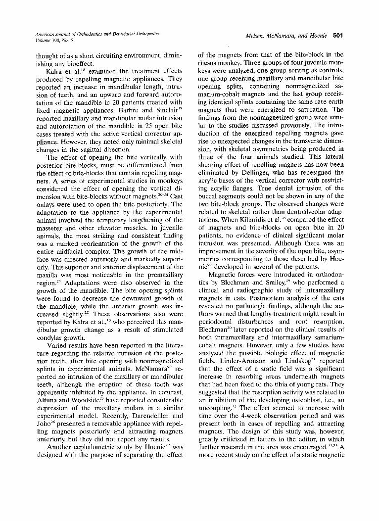
Fig. 1. Microscopic field with superimposed test system. Sur- face density is estimated from intersections between cycloides and bony surface.
field does, indeed, fail to demons t ra te any long- term effect of a magnet placed o+er the sagittal suture. 35 A compar ison of the biologic effect of the magnets is, however, not simple as there is a large variat ion in bo th force magnitude, flux density, and materials used in the articles cited.
Repel l ing magnets that are incorpora ted in ap- pliances used in the t rea tment of open bite are usually worn for an extended per iod of time and the tissue react ion and any potential iatrogenic response to this appliance is therefore of consider- able interest.
The purpose of this repor t was therefore to per form a h i s tomorphomet r ic analysis of the mo- lars, their per iodonta l tissue, and the temporal- zygomatic and pterygomaxil lary sutures in monkeys wearing magnet ized and nonmagnet ized bite- blocks. The results of these studies are compared with a ma tched control popuplat ion. The monkeys had been previously studied cephalometrical ly by Hoenie . 27
MATERIALS AND METHODS Sample
Twelve male juvenile rhesus monkeys (Macaca mu- latta), obtained from the Caribbean Primate Research Center in Puerto Rico, were used in this study. The animals were divided into three groups of four and were
comparable with regard to the stage of dental develop- ment. All animals had an intact deciduous dentition and fully erupted first molars. According to the tooth erup- tion data of Hurme and Van Wagenen, 36 these animals were approximately 18 to 24 months of age at the beginning of the study.
Appliances
Bite opening appliances with magnetized disks in a repelling configuration were inserted in the animals of the first group. Four additional animals were given simi- lar bite-blocks containing samarium-cobalt disks, but these disks were not energized. The last four animals received no appliances and were used as controls.
The splints consisted of bilateral acrylic blocks, con- nected and reinforced by a 0.036-inch stainless steel framework. Samarium-cobalt disks, covered by stainless steel casings as advocated by Dellinger, 17 were imbedded in each block. To produce repelling forces, the polarity of the maxillary and mandibular magnets was the same. The stainless steel casings containing the magnets that were provided by Active Vertical Corrector Inc. had a diameter of 5 mm and a vertical height of 2 ram. Every effort was made to place the metal disks in positions that minimized the vertical opening produced. The magnets were adapted to the size of the monkeys and had a peak repelling force of 358 gm at 0 mm air gap. This force level was, however, never reached even when the monkeys were clenching their teeth because the casings were covered with a thin layer of methacrylate. At 1 mm air gap, the force was 148 gm and at a distance of 2 mm, the magnets were separated by 78 gm of force. When the monkeys occluded, the maxi- mum force was estimated to 250 gin.
Before bonding, the acrylic appliances were equili- brated to allow a balanced occlusion in the posterior region. The appliances were then bonded with Excel bonding resin (Reliance Orthodontic Products, Itasca, Ill.).
Histologic analysis At the end of the treatment interval (24 weeks), the
appliances were removed, and final cephalogram was taken, and then the animals were killed with a perfusion of saline, followed by a 10% solution of neutral buffered formalin. The jaws were excised and replaced in the solution of neutral buffered formalin. Tissue blocks com- prising the maxillary molars, the pterygomaxillary fissure, and the zygomaticotemporal suture were excised and decalcified in EDTA, before embedding in celloidin paraffin. All sections were stained with hematoxylin and eosin.
The alveolar processes were cut parasagitally in 8 Ixm thick sections. Ten sections, all including the full exten- sion of the pulp of at least two teeth, were evaluated.
To avoid bias in the quantitation of the sutures because of the anisotrophy of the structures to be evalu- ated, a stereologic estimation of surface density, which
American Journal of Orthodontics and Dentofacial Orthopedics Melsen, McNamara, and Hoenie 503 Volume 108, No. 5
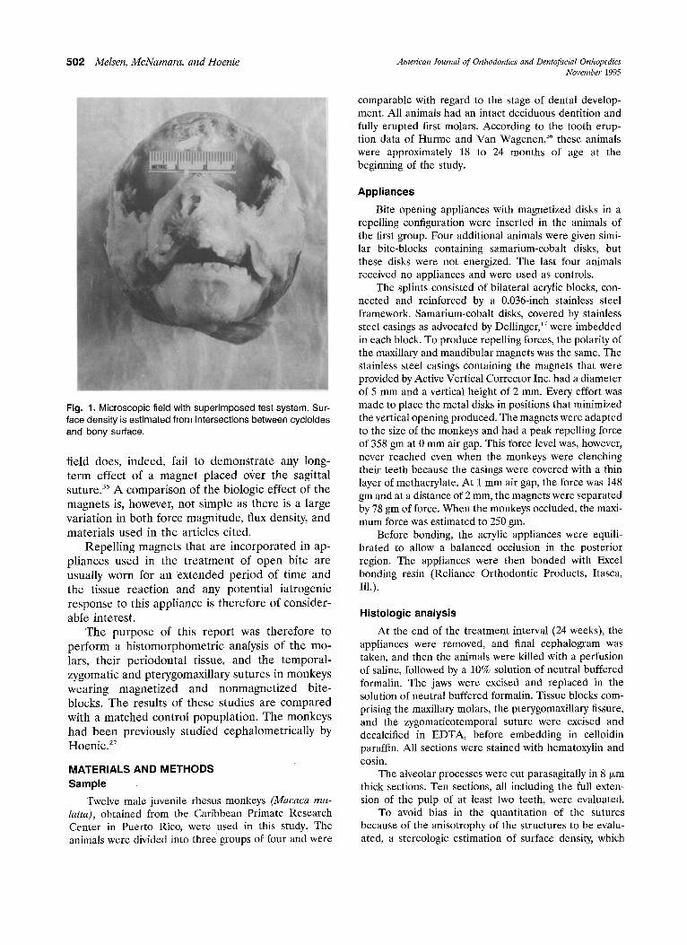
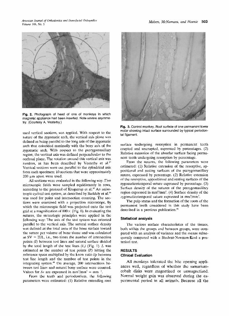
Fig. 2. Photograph of head of one of monkeys in which magnetic appliance had been inserted. Note severe asymme- try. (Courtesy A. Vesterby.)
used vertical sections, was applied. With respect to the suture of the zygomatic arch, the vertical axis plane was defined as being parallel to the long axis of the zygomatic arch that coincided maximally with the bony axis of the zygomatic arch. With respect to the pterygomaxillary region, the vertical axis was defined perpendicular to the occlusal plane. The rotation around this vertical axis was random, as has been described by Vesterby et al. 37 Vertical sections were cut parallel to the cylindrical axis from each specimen; 10 sections that were approximately 200 pom apart were used.
All sections were evaluated in the following way: Five microscopic fields were sampled equidistantly in rows, according to the protocol of Kragstrup et al. 3s An aniso- tropic cycloid test system, as described by Baddely et al? 9 was used for point and intersection counting. The sec- tions were examined with a projection microscope, by which the microscopic field was projected onto the test grid at a magnification of 100 x (Fig. 1). In evaluating the sutures, the stereologic principles were applied in the following way: The axis of the test system was oriented parallel to the vertical axis. The sutural surface density was defined as the total area of the bone surface toward the suture per volume of bone tissue and was calculated as SV = 2I/L, i.e., two times the number of intersection points (I) between test lines and sutural surface divided by the total length of the test lines (L) (Fig. 1). L was estimated as the number of test points (P) hitting the reference space multiplied by the know ratio l/p between test line length and the number of test points in the integrating system, g° On average, 200 intersections be- tween test lines and sutural bony surface were counted. Values for Sv are expressed in mm2/mm 3 = mm 1.
From the teeth and periodontium, the following parameters were estimated: (1) Relative extending root
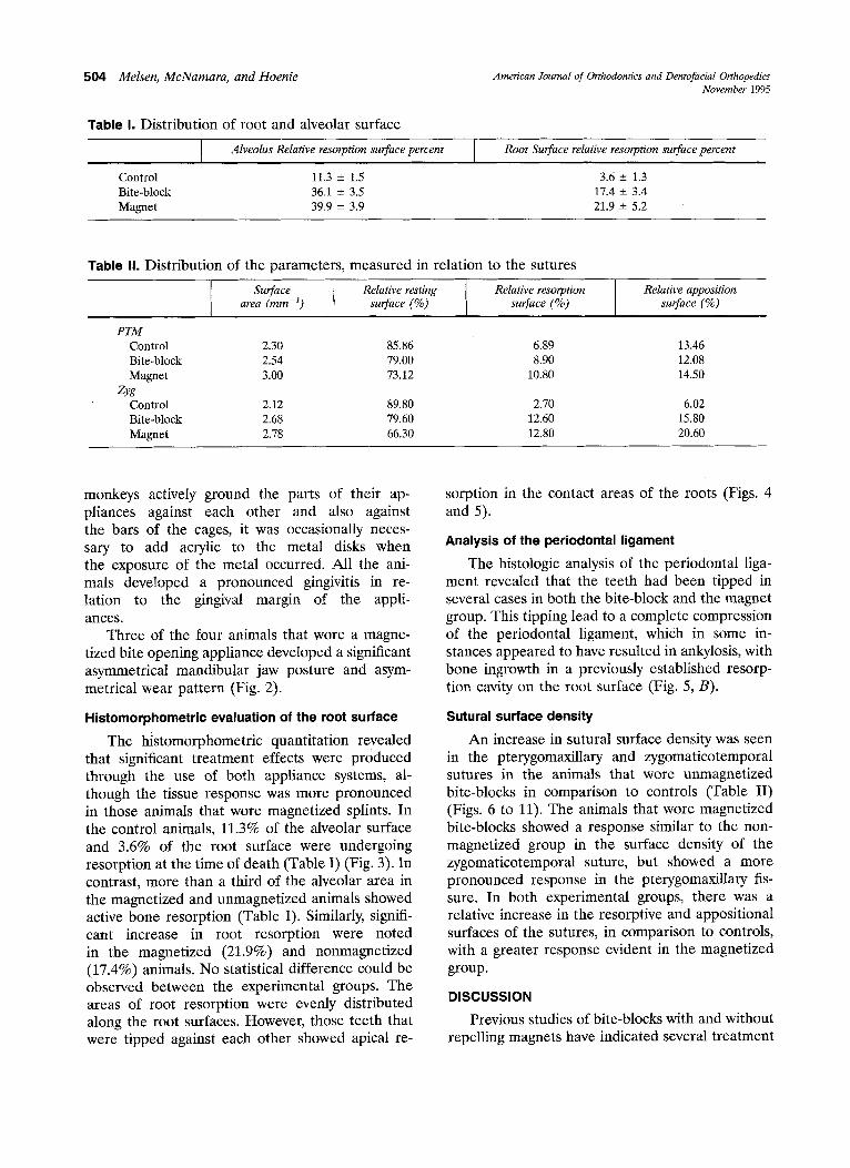
Fig. 3. Control monkey, Root surface of one permanent lower molar showing intact surface surrounded by typical periodon- tal ligament.
surface undergoing resorption in permanent teeth erupted and unerupted, expressed by percentages. (2) Relative extension of the alveolar surface facing perma- nent teeth undergoing resorption by percentage.
From the sutures, the following parameters were estimated: (1) Relative extension of the resorptive, ap- positional and resting surfaces of the pterygomaxillary suture, expressed by percentage. (2) Relative extension of the resorptive, appositional and resting surfaces of the zygomaticotemporal suture expressed by percentage. (3) Surface density of the sutures of the pterygomaxillary region expressed in mm2/mm 3. (4) Surface density of the zygomaticotemporal suture expressed in mm2/mm 3.
The pulp status and the formation of the roots of the permanent teeth considered in this study have been described in a previous publication, gl
Statistical analysis
The various surface characteristics of the tissues, both within the groups and between groups, were com- pared with an analysis of variance and the means subse- quently compared with a Student-Newman-Keul a pos- teriori test.
RESULTS Clinical Evaluation
A l l monkeys t o l e r a t e d the b i te open ing appl i - ances well, r ega rd les s of w h e t h e r the samar ium- coba l t disks were magne t i z ed or unmagne t i zed . N o r m a l weight gain was obse rved dur ing the ex- p e r i m e n t a l p e r i o d in all animals . Because all the
5 0 4 Melsen, McNamara, and Hoenie American Journal of Orthodontics and Dentofacial Orthopedics November 1995
Table h Distribution of root and alveolar surface
Alveolus Relative resorption surface percent Root Surface relative resorption surface percent
Control 11.3 4- 1.5 3.6 _+ 1.3 Bite-block 36.1 - 3.5 17.4 _+ 3.4 Magnet 39.9 -+ 3.9 21.9 _+ 5.2
Table Ih Distribution of the parameters, measured in relation to the sutures
Surface t Relative resting Relative resorption I Relative apposition area (mrn ~) surface (%) surface (%) surface (%)
PTM Control 2.30 85.86 6.89 13.46 Bite-block 2.54 79.00 8.90 12.08 Magnet 3.00 73.12 10.80 14.50
Zyg Control 2.12 89.80 2.70 6.02 Bite-block 2.68 79.60 12.60 15.80 Magnet 2.78 66.30 12.80 20.60
monkeys actively ground the parts of their ap- pliances against each other and also against the bars of the cages, it was occasionally neces- sary to add acrylic to the metal disks when the exposure of the metal occurred. All the ani- mals developed a pronounced gingivitis in re- lation to the gingival margin of the appli- ances.
Three of the four animals that wore a magne- tized bite opening appliance developed a significant asymmetrical mandibular jaw posture and asym- metrical wear pattern (Fig. 2).
Histomorphometric evaluation of the root surface
The histomorphometric quantitation revealed that significant treatment effects were produced through the use of both appliance systems, al- though the tissue response was more pronounced in those animals that wore magnetized splints. In the control animals, 11.3% of the alveolar surface and 3.6% of the root surface were undergoing resorption at the time of death (Table I) (Fig. 3). In contrast, more than a third of the alveolar area in the magnetized and unmagnetized animals showed active bone resorption (Table I). Similarly, signifi- cant increase in root resorption were noted in the magnetized (21.9%) and nonmagnetized (17.4%) animals. No statistical difference could be observed between the experimental groups. The areas of root resorption were evenly distributed along the root surfaces. However, those teeth that were tipped against each other showed apical re-
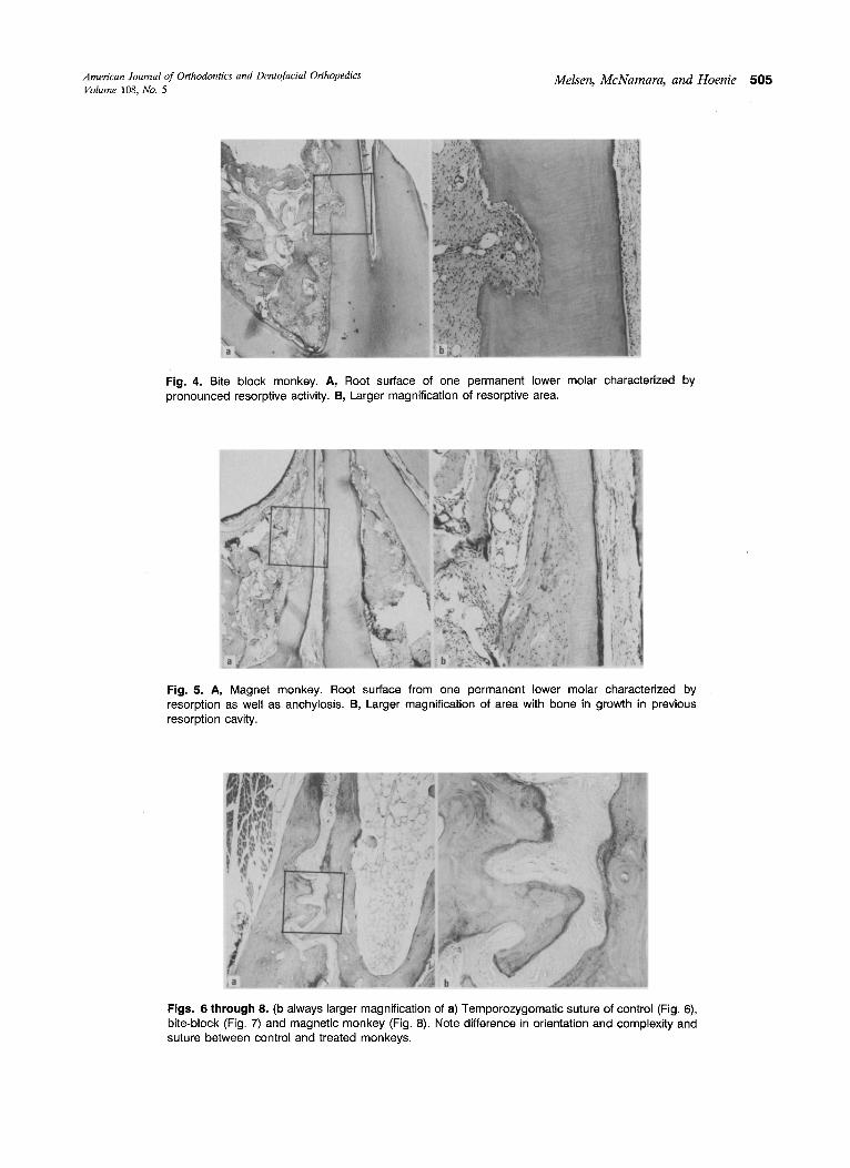
sorption in the contact areas of the roots (Figs. 4 and 5).
Analysis of the periodontal ligament
The histologic analysis of the periodontal liga- ment revealed that the teeth had been tipped in several cases in both the bite-block and the magnet group. This tipping lead to a complete compression of the periodontal ligament, which in some in- stances appeared to have resulted in ankylosis, with bone ingrowth in a previously established resorp- tion cavity on the root surface (Fig. 5, B).
Sutural surface density
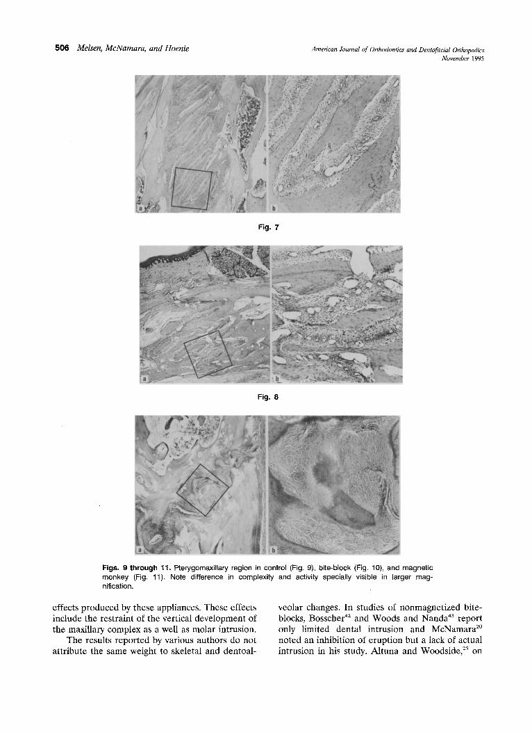
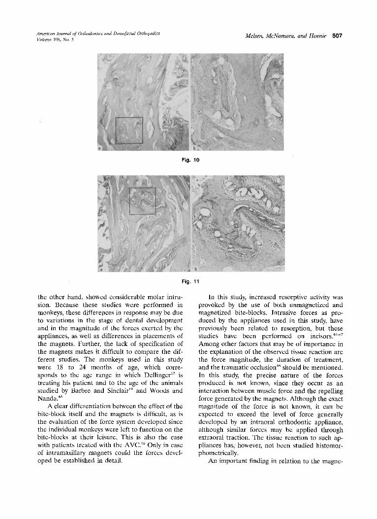
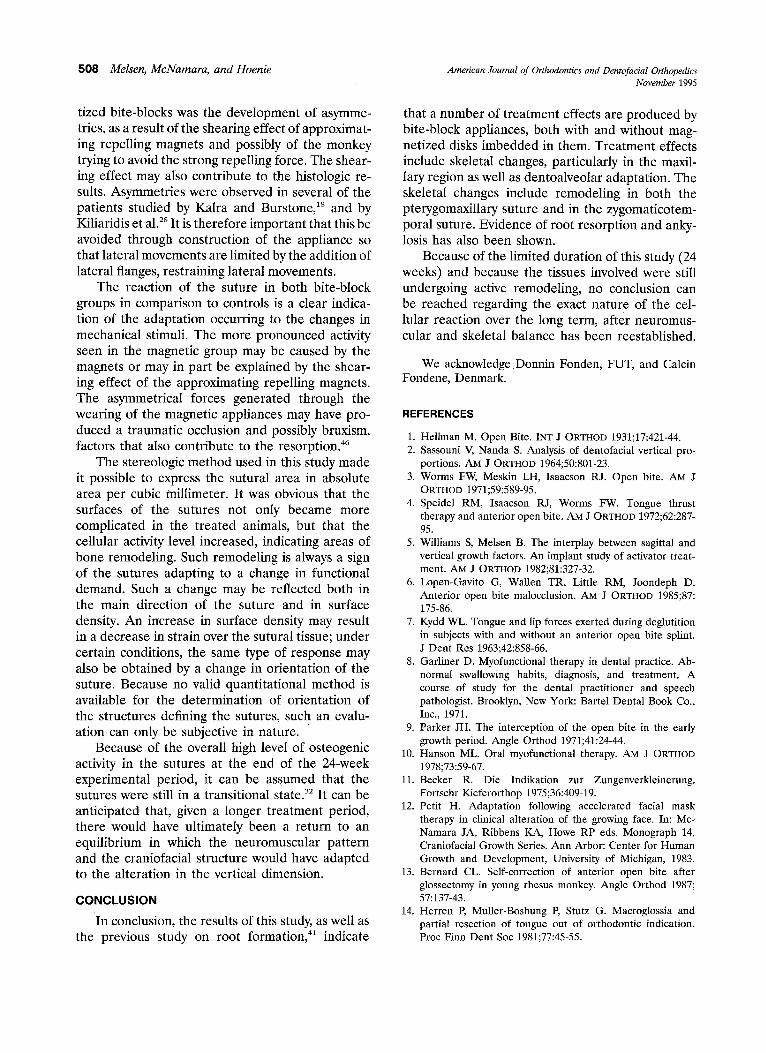
An increase in sutural surface density was seen in the pterygomaxillary and zygomaticotemporal sutures in the animals that wore unmagnetized bite-blocks in comparison to controls (Table II) (Figs. 6 to 11). The animals that wore magnetized bite-blocks showed a response similar to the non- magnetized group in the surface density of the zygomaticotemporal suture, but showed a more pronounced response in the pterygomaxillary fis- sure. In both experimental groups, there was a relative increase in the resorptive and appositional surfaces of the sutures, in comparison to controls, with a greater response evident in the magnetized group.
DISCUSSION
Previous studies of bite-blocks with and without repelling magnets have indicated several treatment
American Journal of Orthodontics and Dentofacial Orthopedics Melsen, McNamara, and Hoenie 505 Volume 108, No. 5
Fig. 4. Bite block monkey. A, Root surface of one permanent lower molar characterized by pronounced resorptive activity. B, Larger magnification of resorptive area.
! i l . . . . . . . . . . . .
Fig. 5. A, Magnet monkey. Root surface from one permanent lower molar characterized by resorption as well as anchylosis. B, Larger magnification of area with bone in growth in previous resorption cavity.
Figs. 6 through 8. (b always larger magnification of a) Temporozygomatic suture of control (Fig. 6), bite-block (Fig. 7) and magnetic monkey (Fig. 8). Note difference in orientation and complexity and suture between control and treated monkeys.
506 Melsen, McNamara, and Hoenie American Journal of Orthodontics and Dentofacial Orthopedics November 1995
Fig. 7
Fig. 8
Figs. 9 through 11. Pterygomaxillary region in control (Fig. 9), bite-block (Fig. 10), and magnetic monkey (Fig. 11). Note difference in complexity and activity specially visible in larger mag- nification.
effects produced by these appliances. These effects include the restraint of the vertical development of the maxillary complex as a well as molar intrusion.
The results reported by various authors do not attribute the same weight to skeletal and dentoal-
veolar changes. In studies of nonmagnetized bite- blocks, Bosscher 42 and Woods and Nanda 43 report only limited dental intrusion and McNamara 2° noted an inhibition of eruption but a lack of actual intrusion in his study. Altuna and Woodside, 25 on
American Journal of Orthodontics and Dentofacial Orthopedics Melsen, McNamara, and Hoenie 507 Volume 108, No. 5
Fig. 10
Fig. 11
the other hand, showed considerable molar intru- sion. Because these studies were performed in monkeys, these differences in response may be due to variations in the stage of dental development and in the magnitude of the forces exerted by the appliances, as well as differences in placements of the magnets. Further, the lack of specification of the magnets makes it difficult to compare the dif- ferent studies. The monkeys used in this study were 18 to 24 months of age, which corre- sponds to the age range in which Dellinger 17 is treating his patient and to the age of the animals studied by Barbre and Sinclair 19 and Woods and Nanda. 43
A clear differentiation between the effect of the bite-block itself and the magnets is difficult, as is the evaluation of the force system developed since the individual monkeys were left to function on the bite-blocks at their leisure. This is also the case with patients treated with the AVCY Only in case of intramaxillary magnets could the forces devel- oped be established in detail.
In this study, increased resorptive activity was provoked by the use of both unmagnetized and magnetized bite-blocks. Intrusive forces as pro- duced by the appliances used in this study, have previously been related to resorption, but these studies have been performed on incisors. 44-47 Among other factors that may be of importance in the explanation of the observed tissue reaction are the force magnitude, the duration of treatment, and the traumatic occlusion 46 should be mentioned. In this study, the precise nature of the forces produced is not known, since they occur as an interaction between muscle force and the repelling force generated by the magnets. Although the exact magnitude of the force is not known, it can be expected to exceed the level of force generally developed by an intraoral orthodontic appliance, although similar forces may be applied through extraoral traction. The tissue reaction to such ap- pliances has, however, not been studied histomor- phometrically.
An important finding in relation to the magne-
508 Melsen, McNamara, and Hoenie Amelican Journal of Orthodontics and Dentofacial Orthopedics November 1995
tized bite-blocks was the development of asymme- tries, as a result of the shearing effect of approximat- ing repelling magnets and possibly of the monkey trying to avoid the strong repelling force. The shear- ing effect may also contribute to the histologic re- suits. Asymmetries were observed in several of the patients studied by Kalra and Burstone, 18 and by Kiliaridis et al. ~8 It is therefore important that this be avoided through construction of the appliance so that lateral movements are limited by the addition of lateral flanges, restraining lateral movements.
The reaction of the suture in both bite-block groups in comparison to controls is a clear indica- tion of the adaptat ion occurring to the changes in mechanical stimuli. The more pronounced activity seen in the magnetic group may be caused by the magnets or may in part be explained by the shear- ing effect of the approximating repelling magnets. The asymmetrical forces generated through the wearing of the magnetic appliances may have pro- duced a traumatic occlusion and possibly bruxism, factors that also contribute to the resorpt ion? 6
The stereologic method used in this study made it possible to express the sutural area in absolute area per cubic millimeter. It was obvious that the surfaces of the sutures not only became more complicated in the t reated animals, but that the cellular activity level increased, indicating areas of bone remodeling. Such remodeling is always a sign of the sutures adapting to a change in functional demand. Such a change may be reflected both in the main direction of the suture and in surface density. An increase in surface density may result in a decrease in strain over the sutural tissue; under certain conditions, the same type of response may also be obtained by a change in orientation of the suture. Because no valid quantitational method is available for the determination of orientation of the structures defining the sutures, such an evalu- ation can only be subjective in nature.
Because of the overall high level of osteogenic activity in the sutures at the end of the 24-week experimental period, it can be assumed that the sutures were still in a transitional s tate? 2 It can be anticipated that, given a longer t rea tment period, there would have ultimately been a return to an equilibrium in which the neuromuscular pat tern and the craniofacial structure would have adapted to the alteration in the vertical dimension.
CONCLUSION
In conclusion, the results of this study, as well as the previous study on root formation, 41 indicate
that a number of t reatment effects are produced by bite-block appliances, both with and without mag- netized disks imbedded in them. Trea tment effects include skeletal changes, particularly in the maxil- lary region as well as dentoalveolar adaptation. The skeletal changes include remodeling in both the pterygomaxillary suture and in the zygomaticotem- poral suture. Evidence of root resorption and anky- losis has also been shown.
Because of the limited duration of this study (24 weeks) and because the tissues involved were still undergoing active remodeling, no conclusion can be reached regarding the exact nature of the cel- lular reaction over the long term, after neuromus- cular and skeletal balance has been reestablished.
We acknowledge Donnin Fonden, FUT, and Calcin Fondene, Denmark.
REFERENCES
1. Hellman M. Open Bite. INT J ORTHOD 1931;17:421-44. 2. Sassouni V, Nanda S. Analysis of dentofacial vertical pro-
portions. AM J ORTHOD 1964;50:801-23. 3. Worms FW, Meskin LH, Isaacson RJ. Open bite. AM J
ORTHOD 1971;59:589-95. 4. Speidel RM, Isaacson RJ, Worms FW. Tongue thrust
therapy and anterior open bite. AM J ORTHOD 1972;62:287- 95.
5. Williams S, Melsen B. The interplay between sagittal and vertical growth factors. An implant study of activator treat- ment. AM J ORTHOD 1982;81:327-32.
6. Lopen-Gavito G, Wallen TR, Little RM, Joondeph D. Anterior open bite malocclusion. AM J ORTHOD 1985;87: 175-86.
7. Kydd WL. Tongue and lip forces exerted during deglutition in subjects with and without an anterior open bite splint. J Dent Res 1963;42:858-66.
8. Garliner D. Myofunctional therapy in dental practice. Ab- normal swallowing habits, diagnosis, and treatment. A course of study for the dental practitioner and speech pathologist. Brooklyn, New York: Bartel Dental Book Co., Inc., 1971.
9. Parker JH. The interception of the open bite in the early growth period. Angle Orthod 1971;41:24-44.
10. Hanson ML. Oral myofunctional therapy. AM J ORTHOD 1978;73:59-67.
11. Becker R. Die Indikation zur Zungenverkleinerung. Fortschr Kieferorthop 1975;36:409-19.
12. Petit H. Adaptation following accelerated facial mask therapy in clinical alteration of the growing face. In: Mc- Namara JA, Ribbens KA, Howe RP eds. Monograph 14, Craniofacial Growth Series. Ann Arbor: Center for Human Growth and Development, University of Michigan, 1983.
13. Bernard CL. Self-correction of anterior open bite after glossectomy in young rhesus monkey. Angle Orthod 1987; 57:137-43.
14. Herren P, Muller-Boshung P, Stutz G. Macroglossia and partial resection of tongue out of orthodontic indication. Proc Finn Dent Soc 1981;77:45-55.
American Journal of Orthodontics and Dentofacial Orthopedics Volume 108, No. 5
15. Proffit WR, Mason RM. Myofunctional therapy for tongue thrusting: background and recommendations. J Am Dent Assoc 1975;90:403-11.
16. Pearson LE. Vertical control in treatment of patients having backward-rotational growth tendencies. Angle Orthod 1978; 45(2):130-40.
17. Dellinger E. A clinical assessment of the Active Vertical Corrector. A nonsurgical alternative for skeletal open bite treatment. AM J ORTHOD 1986;89:428-36.
18. Kalra V, Burstone C, Nanda R. Effects of a fixed magnetic appliance on the dentofacial complex. AM J ORTHOD DENTOFAC ORTHOP 1989;95:467-78.
19. Barbre RE, Sinclair PM. A cephalometric evaluation of anterior open bite correction with the magnetic active ver- tical corrector. Angle Orthod 1991;61:93-109.
20. McNamara JA Jr. An experimental study of increased vertical dimension in the growing face. AM J ORTHOD 1977;71:382-95.
21. Maxwell LC, Carlson DS, McNamara JA Jr, Faulkner JA. Adaptations of the masseter and temporalis muscles follow- ing alteration in length, with or without surgical detach- ment. Anat Rec 1981;200:127-37.
22. Carlson DS, Ellis E, Schneiderman ED, Ungerleider JC. Experimental models of surgical intervention in the growing face: cephalometric analysis of facial growth and relapse. In: McNamara JA Jr, Carlson DS, Ribbens KA, ed. The effects of surgical intervention on craniofacial growth. Monographs 12. Craniofacial growth series. Ann Arbor: Center for Hu- man Growth and Development, University of Michigan, 1982.
23. Schneiderman ED, Carlson DS. Cephalometric analysis of condylar adaptations through altered mandibular position in adult rhesus monkeys (Macaca mulatta). Arch Oral Biol 1985;30:49-54.
24. Rowe TK, Carlson DS. The effect of bite-opening appli- ances on mandibular rotational growth and remodeling in the rhesus monkey (Macaca mulatta). AM J ORTHOD DENTOFAC ORTHOP 1990;98:544-9.
25. Altuna G, Woodside DG. Response of the midface to treatment with increased vertical occlusal forces; treatment and posttreatment effects in monkeys. Angle Orthod 1985; 55:251-63.
26. Darendeliler MA, Joho J-P. Magnetic activator device II (MADII) for correction of Class II, Division 1 malocclu- sions. AM J ORTHOD DENTOFAC ORTHOP 1993;103:223-39.
27. Hoenie DC. The effect of interocclusal repelling magnets in a bite opening splint on the growth of the craniofacial complex on juvenile Macaca mulatta. [Dissertation]. Ann Arbor: University of Michigan, 1986.
28. Kiliarides S, Egermark I, Thilander B. Anterior open bite treatment with magnets. Eur J Orthod 1990;12:447-57.
29. Blechman AM, Smiley H. Magnetic force in orthodontics. AM J ORTHOD 1978;74:435-43.
30. Blechman AM. Magnetic force systems in orthodontics. AM J ORTHOD 1985;87:201-10.
31. Linder-Aronson S, Lindskog S. A morphometric study of
Melsen, McNamara, and Hoenie 509
bone surface and skin reactions after stimulation with static magnetic fields in rats. AM J ORTHOD 1991;99:44-8.
32. Frost HM. Transient-steady state phenomena in microdam- age physiology: a proposed algorithm for lamellar bone. Calcif Tissue Int 1989;44:367-81.
33. Blechman AM. Comments on static magnetic fields. AM J ORTHOD DENTOFAC ORTHOP 1991;90:18A-20A.
34. DeVincenzo J. More on static magnetic fields. AM J ORTHOD DENTOFAC ORTHOP 1991;90:21A.
35. Camilleri S, McDonald F. Static magnetic field effects on the sagittal suture in Rattus Norvegicus. AM J ORTHOD DENTOFAC ORTHOP 1993;103:240-6.
36. Hurme VO, Van Wagenen G. Basic data on the emergence of permanent teeth in the monkey. J Dent Res 1961;40:732.
37. Vesterby A, Kragstrup J, Gundersen HJG, Melsen F. Un- biased stereologic estimation of surface density in bone using vertical sections. Bone 1987;8:13-7.
38. Kragstrup J, Gundersen HJG, Melsen F, Mosekilde L. Estimation of the three dimensional wall thickness of com- pleted remodelling sites in trabecular bone. Metal Bone Dis Rel Res 1982;4:113-9.
39. Baddely AJ, Oundersen HJG, Cruz-Orive LM. Estimation of surface area from vertical sections. J Microsc 1986;142: 259-76.
40. Jensen EB, Gundersen HJG. Stereological ratio estimation based on counts from intergral test system. J Microsc 1982;125:51-65.
41. Melsen B, McNamara J, Hoenie D. Effect of biteblocks and repelling magnets on root formation of unerupted premo- lars in macacca monkeys. Proc Finn Dent Soc 1991;87:no 1.
42. Bosscher P Jr. Adaptations in the maxillary complex in- duced by alterations in muscle length. [Dissertation.] Ann Arbor: University of Michigan, 1985.
43. Woods MG, Nanda RS. Intrusion of posterior teeth and magnets. An experiment in growing baboons. Angle Orthod 1988;58:136-50.
44. De Schields RW. A study of root resorption in treated class II Division I malocclusions. Angle Orthod 1969;39:231-45.
45. Dermaut LR, De Munck A. Apical root resorption of upper incisors caused by intrusive tooth movement. A radiographic study. AM J ORTHOD 1986;90:321-6.
46. Linge L, Linge OB. Patient characteristics and treatment variables associated with apical root resorption during or- thodontic treatment. AM J ORTHOD DENTOFAC ORTHOP 1991;99:35-43.
47. Melsen B, Agerbaek N, Markenstam G. Intrusion of incisors in adult patients with marginal bone loss. AM J ORTHOD DENTOFAC ORTHOP 1989;96:232-41.
Reprint requests: Dr. Birte Melsen Institute of Orthodontics Royal Dental College Vennelyst Blvd. DK-8000 Aarhus C Denmark