The Drosophila pipsqueakgene encodes a nuclear BTB...
13
INTRODUCTION Proper development of the Drosophila early egg chamber is required to generate polarity in the egg and embryo. During the middle stages of oogenesis, RNAs and proteins required for germ cell determination and for the generation of embryonic abdominal segments are localized to the posterior pole of the oocyte (reviewed by St. Johnston, 1993). The local- ization of the germ cell determinant(s) and the mRNA encoding Nanos, the posterior morphogen, is accomplished by a common mechanism, which requires the activity of at least 9 maternal-effect genes. Mothers homozygous for mutations in any of these genes produce embryos that lack abdominal segments and pole cells (the germline precursors). The pipsqueak (psq) gene has been shown to be required maternally for correct abdominal segmentation and pole cell formation in the embryo (Siegel et al., 1993). It has been suggested that the psq embryonic defect in posterior pattern- ing might be due to insufficient expression of the maternal posterior group gene vasa in psq mutants (Siegel et al., 1993). In addition, we have shown that certain psq alleles also exhibit dorsal/ventral defects in eggshell structure (Horowitz and Berg, 1995). Establishment of the dorsal/ventral axis is set into motion with the localization of gurken (grk) mRNA in the developing oocyte. The Grk protein serves as a signal to determine follicle cell fate at two distinct times during oogenesis (reviewed by Anderson, 1995). During mid-oogenesis, Grk signals to the follicle cells at the posterior of the egg chamber, inducing them to adopt a posterior fate (Roth et al., 1995; González-Reyes et al., 1995). Later, in stages 8-10 of oogenesis, Grk is localized as a cap above the oocyte nucleus (Roth et al., 1995) where it signals to adjacent follicle cells to determine their fate as dorsal. This signaling cascade culminates in the proper estab- lishment of dorsal/ventral polarity in the embryo, and in the formation of the two dorsolateral respiratory appendages of the eggshell (reviewed by Schüpbach and Roth, 1994; Chasan and Anderson, 1993). We report here the cloning and molecular characterization of the psq gene. Our analyses indicate that psq is a complex and large gene encoding multiple transcripts and protein isoforms. The PsqA isoform contains a BTB domain; these domains have been shown to mediate protein-protein interac- tions (Bardwell and Treisman, 1994; Chen et al., 1995). psq mutants that produce a truncated protein harboring this domain exhibit defects in the dorsal/ventral patterning of the eggshell and embryo. MATERIALS AND METHODS Fly stocks P[lacZ; ry + ] (PZ) insertional mutations at the psq locus (alleles psq 2403 , psq 0115 , psq 8109 and psq 0482 ) were generated in a screen described in Karpen and Spradling (1992); psq is referred to as zeppelin in Spradling (1993). Excision lines psq RF9 and psq RF13 were created from allele psq 2403 as described previously (Horowitz and Berg, 1995). The deficiency, Df(2R)psq-lola∆18 (∆18) was generated 1859 Development 122, 1859-1871 (1996) Printed in Great Britain © The Company of Biologists Limited 1996 DEV5061 Mutations at the pipsqueak locus affect early patterning in the Drosophila egg and embryo. We have cloned pipsqueak and found that it is a large and complex gene, encoding multiple transcripts and protein isoforms. One protein, PsqA, is absent in all of the mutants that we have examined. We show that PsqA is a nuclear protein present in the germ cells and somatically derived follicle cells throughout oogenesis and that it is required prior to stage one of oogenesis. PsqA contains a BTB (POZ) domain at its amino terminus; additionally, we have identified an evolutionar- ily conserved motif of unknown function present four times in tandem at the C terminus of the protein. PZ pipsqueak mutants produce a putative fusion protein containing the pipsqueak BTB domain fused to sequences resident on the PZ element (H. Horowitz and C. Berg, 1995 Genetics 139, 327-335). We demonstrate here that expression of this fusion protein in wild-type flies has a dominant effect, resulting in infertility and eggshell defects. These dominant phenotypes are discussed in light of current theories on the role of the BTB domain in protein-protein interactions. Key words: pipsqueak, psq, BTB, dominant-negative, Drosophila, oogenesis SUMMARY The Drosophila pipsqueak gene encodes a nuclear BTB-domain-containing protein required early in oogenesis Heidi Horowitz and Celeste A. Berg Department of Genetics, P.O. Box 357360, University of Washington, Seattle, Washington 98195-7360, USA Author for correspondence (e-mail: [email protected])
Transcript of The Drosophila pipsqueakgene encodes a nuclear BTB...
1859Development 122, 1859-1871 (1996)Printed in Great Britain © The Company of Biologists Limited 1996DEV5061
The Drosophila pipsqueak gene encodes a nuclear BTB-domain-containing
protein required early in oogenesis
Heidi Horowitz and Celeste A. Berg
Department of Genetics, P.O. Box 357360, University of Washington, Seattle, Washington 98195-7360, USA
Author for correspondence (e-mail: [email protected])
Mutations at the pipsqueak locus affect early patterning inthe Drosophila egg and embryo. We have cloned pipsqueakand found that it is a large and complex gene, encodingmultiple transcripts and protein isoforms. One protein,PsqA, is absent in all of the mutants that we have examined.We show that PsqA is a nuclear protein present in the germcells and somatically derived follicle cells throughoutoogenesis and that it is required prior to stage one ofoogenesis. PsqA contains a BTB (POZ) domain at its aminoterminus; additionally, we have identified an evolutionar-ily conserved motif of unknown function present four timesin tandem at the C terminus of the protein. PZ pipsqueak
mutants produce a putative fusion protein containing thepipsqueak BTB domain fused to sequences resident on thePZ element (H. Horowitz and C. Berg, 1995 Genetics 139,327-335). We demonstrate here that expression of thisfusion protein in wild-type flies has a dominant effect,resulting in infertility and eggshell defects. These dominantphenotypes are discussed in light of current theories on therole of the BTB domain in protein-protein interactions.
Key words: pipsqueak, psq, BTB, dominant-negative, Drosophila,oogenesis
SUMMARY
INTRODUCTION
Proper development of the Drosophila early egg chamber isrequired to generate polarity in the egg and embryo. Duringthe middle stages of oogenesis, RNAs and proteins requiredfor germ cell determination and for the generation ofembryonic abdominal segments are localized to the posteriorpole of the oocyte (reviewed by St. Johnston, 1993). The local-ization of the germ cell determinant(s) and the mRNAencoding Nanos, the posterior morphogen, is accomplished bya common mechanism, which requires the activity of at least9 maternal-effect genes. Mothers homozygous for mutations inany of these genes produce embryos that lack abdominalsegments and pole cells (the germline precursors).
The pipsqueak (psq) gene has been shown to be requiredmaternally for correct abdominal segmentation and pole cellformation in the embryo (Siegel et al., 1993). It has beensuggested that the psq embryonic defect in posterior pattern-ing might be due to insufficient expression of the maternalposterior group gene vasa in psq mutants (Siegel et al., 1993).In addition, we have shown that certain psq alleles also exhibitdorsal/ventral defects in eggshell structure (Horowitz andBerg, 1995).
Establishment of the dorsal/ventral axis is set into motionwith the localization of gurken (grk) mRNA in the developingoocyte. The Grk protein serves as a signal to determine folliclecell fate at two distinct times during oogenesis (reviewed byAnderson, 1995). During mid-oogenesis, Grk signals to thefollicle cells at the posterior of the egg chamber, inducing them
to adopt a posterior fate (Roth et al., 1995; González-Reyes etal., 1995). Later, in stages 8-10 of oogenesis, Grk is localizedas a cap above the oocyte nucleus (Roth et al., 1995) where itsignals to adjacent follicle cells to determine their fate asdorsal. This signaling cascade culminates in the proper estab-lishment of dorsal/ventral polarity in the embryo, and in theformation of the two dorsolateral respiratory appendages of theeggshell (reviewed by Schüpbach and Roth, 1994; Chasan andAnderson, 1993).
We report here the cloning and molecular characterizationof the psq gene. Our analyses indicate that psq is a complexand large gene encoding multiple transcripts and proteinisoforms. The PsqA isoform contains a BTB domain; thesedomains have been shown to mediate protein-protein interac-tions (Bardwell and Treisman, 1994; Chen et al., 1995). psqmutants that produce a truncated protein harboring this domainexhibit defects in the dorsal/ventral patterning of the eggshelland embryo.
MATERIALS AND METHODS
Fly stocksP[lacZ; ry+] (PZ) insertional mutations at the psq locus (allelespsq2403, psq0115, psq8109 and psq0482) were generated in a screendescribed in Karpen and Spradling (1992); psq is referred to aszeppelin in Spradling (1993). Excision lines psqRF9 and psqRF13 werecreated from allele psq2403 as described previously (Horowitz andBerg, 1995). The deficiency, Df(2R)psq-lola∆18 (∆18) was generated

1860 H. Horowitz and C. A. Berg
by excising the two PZ elements resident in allele psq0482; such anexcision is likely to delete all of the DNA between the two P elements(Cooley et al., 1990). The ∆18 deficiency fails to complement bothpsq and a nearby lethal (lola, E. Giniger, personal communication);Southern blot analysis (data not shown) demonstrates that the ∆18deficiency has lost DNA beginning at the PZ element within psq andextending through the length of our cloned DNA 5′ to the psq gene(see Fig. 6).
The EMS allele psqHK38 (Schüpbach and Wieschaus, 1991) wasoriginally recovered as a second mutation on a cappucino mutantchromosome. Our studies employed a recombinant chromosomelacking the cappucino mutation.
The P[lacZ, w+] (PW) line psqfs1 and the excision allele psq1-30 aredescribed by Siegel et al. (1993). These authors refer to psqfs1 as psqP1
and psq1-30 as psqX1-30.
Transformant lines were produced by germline transformationcarried out according to Spradling (1986).
Embryo and ovary preparationsCuticles were prepared according to Wieschaus and Nüsslein-Volhard(1986). Ovaries and individual egg chambers were prepared accordingto Verheyen and Cooley (1994). In situ hybridizations were carriedout as described previously (Gillespie and Berg, 1995). Whole-mountanalyses using antibodies to the Psq protein were carried out asfollows: ovaries from 2- to 3-day-old females were dissected inmodified EBR and fixed in devitellinizing buffer for 10 minutes(Verheyen and Cooley, 1994). All washes were carried out for 5minutes unless otherwise specified. Ovaries were rinsed three timesin 1× PBS and subsequently extracted in 1% saponin in 1× PBS forone hour at room temperature. The ovaries were rinsed twice in 0.1%saponin in PBS and once in PBTB (1× PBS, 0.1% Triton-X-100, 0.2%BSA), then incubated for 30 minutes in 5% normal goat serum (NGS)in PBTB. Rabbit serum was diluted (1:500) into PBTB with 5% NGSand overnight incubation was carried out at 4°C. The tissue was rinsedfour times for 15 minutes each in PBTB and incubated with a 1:100dilution of BODIPY-conjugated goat anti-rabbit secondary antibody(Molecular Probes) in PBTB overnight at 4°C. The samples were sub-sequently rinsed four times for 15 minutes each in PBTB and mountedas described in Theurkauf et al. (1992). Fluorescence was observedon a Bio-Rad MRC-600 confocal microscope, utilizing the Bio-RadCOMOS program. Images were processed using the NIH Imageprogram.
Cloning and characterization of psq DNAPreparation of DNA and RNA, and northern and Southern blotanalyses were described previously (Horowitz and Berg, 1995).
Genomic DNA flanking the PZ psq2403 insertion and the PW psqfs1
insertion were kindly provided by Haifan Lin and Vivian Siegel,respectively. Genomic DNA spanning the exons downsteam of the PZinsertions was isolated by probing a cosmid library (gift of MarcChampe, Genentech, San Francisco) with the 1.0 kb partial psqcDNA, 13-13, described previously (Horowitz and Berg, 1995). The5.1 kb psq-1 cDNA, pHPT7-9, was isolated from an ovarian cDNAlibrary (Stroumbakis et al., 1994) using a 300 bp 5′ UTR probe (seeFig. 6). Alternative cDNAs pHH62 and pHH63 (psq-2) were isolatedby rescreening the library with the complete pHPT7-9 cDNA.
DNA sequencing was performed using the dideoxynucleotide chaintermination method with the Sequenase kit (US Biochemical). Bases615-624 (TATACTGCAG, Fig. 4A) were not present in pHPT7-9 butwere found in both cDNA 13-13 and genomic DNA (TATACTG-GAG). The most likely cause of this deletion is an error by the reversetranscriptase during the cDNA library construction: this regioncontains direct repeats and significant dyad symmetry.
Determination of genomic structure was performed by a combina-tion of restriction analysis of cloned DNA, Southern blot analysis ofwhole fly DNA and DNA sequencing. For all introns, sequence deter-mination of both the 5′ and 3′ junctions in the genomic DNA
permitted unambiguous identification of the position of each intronrelative to the cDNA (see Fig. 4A). We identified introns between thefollowing pairs of nucleotides in the psq-1 cDNA (numbering as inFig. 4A): (614, 615), (883, 884), (1048,1049), (2014, 2015), (2153,2154), (2693, 2694), (3332, 3333), (3435, 3436), and (3619, 3620).Nucleotide pairs presented above in bold indicate positions at whichthe introns were sequenced in their entirety. The presence of the intronat (883, 884) was inferred based on Southern analysis of genomicDNA and homology to consensus sequences; the precise position ofthis intron was determined by Zollman et al. (1994). The genomicstructure that we have determined differs somewhat from thatpresented by Weber et al. (1995).
Sequence was compiled using the IntelliGenetics program.Database searches were carried out using the BLAST program fromthe NIH (Altschul et al. 1990).
The sequences of the three classes of embryonic psq transcriptsidentified by Weber et al. (1995) are distinct from the ovarian tran-scripts that we present here. The primary differences are as follows:The 5′ ends of all the embryonic transcripts differ from the ovariantranscripts psq-1 and psq-2. The first 210 nucleotides of the E1A exonof Weber et al. (1995) are unique, but are followed by sequencestarting at position 402 of the psq-1 transcript (numbering as in Fig.4A). Our genomic sequencing indicates that nucleotides 1-614 of psq-1 are present in a single exon, suggesting that the 5′ end of E1A isderived from sequence 5′ to the psq-1 transcript. The sequences ofexons E1B and E1C of Weber et al. (1995) are not present in any ofthe ovarian transcripts that we have identified. The 3′ ends of theembryonic transcripts are extended by approximately 1 to 1.2 kbcompared to the ovarian transcripts psq-2 and psq-1, respectively.
The predicted proteins from all embryonic transcripts differ fromthose of the ovarian transcripts: the predicted embryonic proteins have24 unique amino acids C-terminal to amino acid 1062 of the proteinencoded by psq-1 (see Fig. 4A). Comparison with our sequence showsthat the embryonic transcripts lack the sequence from 3920 to 4096of psq-1, suggesting that an additional splicing event occurs in theembryonic tissues, permitting translation to proceed somewhat furtherthan predicted for the ovarian proteins.
PlasmidsDrosophila transformation constructs
P[hs-psq]An XbaI fragment containing sequences from position 1 to 4619 ofcDNA pHPT7-9 was cloned into the unique XbaI site of the trans-formation vector pCaSpeRhs (Thummel and Pirrotta, 1991), whichuses the w+ gene as a marker.
P[hs-psq-l(3)S12]A 570 bp EcoRI-SalI fragment isolated from a RT-PCR product ofthe psq-l(3)S12 truncation mRNA (see Fig. 4A, Horowitz and Berg,1995) was cloned into pCaSpeRhs. This fragment contains the entirecoding region of the predicted fusion protein, as well as 47 bp of psq5′ UTR and 29 bp of sequence 3′ to the predicted l(3)S12 stop codon(Dutton and Chovnick, 1991).
Protein expression constructspHH14
A BamHI fragment of psq extending from nucleotide 864 to 5162 ofthe pHPT7-9 cDNA (amino acids 45-1065) was cloned into theBamHI and BglII sites of expression vector pQE-13 (Qiagen).
pHH70A fragment extending from the NcoI site (position 2038 in Fig. 4A)to the 3′ end (NotI site) of the psq-2 cDNA pHH63 (see Results) wascloned into the NcoI and NotI sites of the expression vector, pET-28C(Novagen). Expression of pHH70 produces a protein of 69×103 Mr,derived entirely of psq sequence (amino acids 436-1065 in Fig. 4).
1861Drosophila psq gene
pHH69This is identical to pHH70, except that the fragment was derived fromcDNA pHH62 (see Results), and thus harbors a deletion of aminoacids 719-736 (Fig. 4), producing a protein of 67×103 Mr.
Polyclonal antibody productionA 6xHis-Psq fusion protein generated from pHH14 was isolated fromE. coli inclusion bodies. Protein purified by preparative SDS-poly-acrylamide gel electrophoresis was used to immunize rabbits (R&RRabbitry, Stanwood, WA). Antisera from two different rabbits (AS1and AS2) showed slightly different immunoreactivity on westerns(see Results).
Western blot analysisDrosophila ovary lysates were prepared by homogenizing ovaries inbuffer (50 mM Tris, pH 7.5, 3 mM EDTA, 1% NP-40, 0.1% SDS, 1mM N-ethylmaleimide, 2 µg/ml leupeptin, 200 KIU/ml Traysol, 100µg/ml PMSF), and protein concentration was determined using theBio-Rad Protein Assay reagent. Approximately 5 µg of protein wereloaded per lane, subjected to SDS-polyacrylamide gel electrophor-esis, and transfered to nitrocellulose. Membranes were probed withpolyclonal antiserum at a 1:5000 dilution using the ECL Westernblotting analysis system (Amersham).
Heat-shock experimentsRescue of psq2403 mutant phenotypeThe strain referred to as X-3; psq2403 has the genotype w1118 P[hs-psq]; cn psq2403 ; +/− ry506. w1118 flies were used as a control. 3- to5-day-old females (n=40-50) were aged with males for 2 days inembryo collection bottles at 25°C. Flies were subjected to a 1 hourheat pulse at 37°C in prewarmed vials, after which new males wereprovided. Timed collections on apple juice/agar plates wereperformed and hatch rates determined 48 hours postcollection. Fig. 7shows combined data from two experiments; the curves were plottedby splining the data. For examination of heat-shock-induced Psqproteins, shocked flies were permitted to recover with new males at25°C for 2 hours, after which ovaries were isolated and proteinextracts prepared as described above.
Expression of the Psq-l(3)S12 fusion protein in wild-type flies Six strains containing insertions of P[hs-psq-l(3)S12] in a w1118 back-
Table 1. pipsqAllele Phenotype
fs1 posterior group, grandchildless 1-30 posterior group, grandchildless
HK38 posterior group, weakly dorsalizes eggshell and embryo
2403 posterior group, moderately dorsalizes eggshell and embryo
RF9 viable, fertile
RF13 viable, fertile
8109 decreased viability, posterior group, strongly dorsalizeseggshell and embryo, early oogenesis defects
0115 decreased viability, posterior group, strongly dorsalizeseggshell and embryo, early oogenesis defects
0482 lethal
Df(2R)psq-lola ∆18 lethal
ground were analyzed. w1118 was used as a control. Approximately20 1- to 2-day-old female flies from each line were heat-shocked inprewarmed vials at 37°C for 1 hour, then transferred into fresh vialscontaining wild-type males and allowed to lay eggs for approximately12 hours. Flies were then transferred into fresh vials for another 12hour collection. Each day for 7 days the males were removed and thefemales heat-shocked as before. The number of eggs laid in each vialwas counted immediately after the transfer of adults to fresh vials.
RESULTS
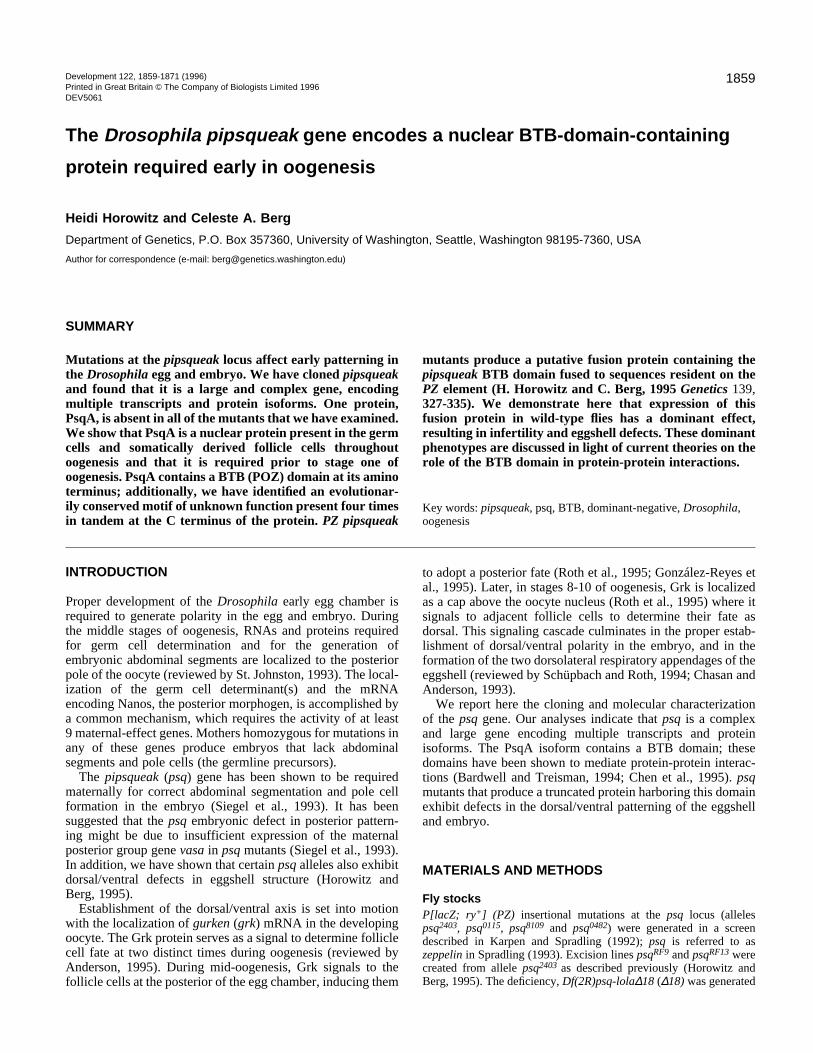
Analysis of psq mutantsMutations at the psq locus cause a variety of defects inoogenesis and embryogenesis (see Table 1 for a summary ofalleles). The phenotypes fall into three classes. The weak allelepsqfs1 disrupts posterior segmentation and pole cell formationduring embryogenesis (Siegel et al., 1993). The intermediatealleles psqHK38 and psq2403 exhibit dorsal/ventral defects in theeggshell and embryo in addition to the posterior axis defectsdescribed above (Figs 1, 2). Both of these intermediate allelesdisplay a range of phenotypes; psqHK38 is significantly weakerthan psq2403 (Figs 1, 2). Flies homozygous or transheterozy-gous for the strong alleles psq8109 and psq0115 have reducedviability, produce only a few, strongly dorsalized late stageeggs (Fig. 2), and have defects in early oogenesis. As expected,psq8109/psq2403 transheterozygotes produce eggs with strongerdorsal/ventral eggshell defects than psq2403 homozygotes (Fig.2). As is true for most mutants with eggshell defects, thenumber of embryos fertilized in all of the psq strains decreaseswith the increase in severity of the eggshell phenotypes.
Two other alleles do not fall readily into this classificationscheme. The PZ insertion allele psq0482 is lethal, but acts as aweakly moderate allele in trans to all other alleles. psq0482 alsofails to complement mutations in a nearby lethal locus (lola,E. Giniger, personal communication) that is a hotspot for Pelement insertion. In a complementation analysis betweenalleles of this lethal locus and all the psq alleles discussed here,
ueak allelesMolecular nature
P[lacZ; w+ ] insertion (Siegel et al., 1993) into first intron of psq-1.Excision allele of fs1 (Siegel et al., 1993). Deletes 5′-most exon and
promoter for psq-1.EMS allele, unknown change (Schüpbach and Wieschaus, 1991).
P[lacZ; ry+] insertion into largest psq-1 intron. Aberrant fusion protein created (Horowitz and Berg, 1995).
Excision of 2403. Removes all of P element except 3′ end (Horowitz and Berg, 1995).
Excision of 2403. Deletes entire P element and at least 40 kb surrounding insertion site (Horowitz and Berg, 1995).
P[lacZ; ry+] insertion into largest psq-1 intron. Aberrant fusion protein created. (Horowitz and Berg, 1995).
P[lacZ; ry+] insertion into largest psq-1 intron. Aberrant fusion protein created. (Horowitz and Berg, 1995).
Two P[lacZ; ry+] insertions: one in largest intron of psq-1(Horowitz and Berg, 1995), second in 5′ end of lola (E. Giniger, personal communication).
Excision of both P elements in 0482. Deletes DNA at 0482 site in largest intron of psq-1, extending through promoter for psq-1. Probable deletion of all DNA between psq and lola.
1862 H. Horowitz and C. A. Berg
Fig. 1. Larval cuticle preparations fromeggs laid by wild-type and psqmothers. Mutations in psq affectposterior segmentation. Some allelesalso disrupt dorsal/ventral axisformation. (A) Canton S. (B) psq1-30/∆18 mothers produce embryos thatlack posterior segments. Shown here isthe most severe phenotype observed.(C) Cuticle from an egg laid by apsqHK38 mother in which posteriorsegments are absent, head defects areapparent and the dorsal hairs areslightly expanded into ventral regions.psqHK38 phenotypes range from weakposterior defects to strongly dorsalized,as in psq2403 mutants. (D) Cuticle froman egg laid by a psq2403 mother. Thisanimal lacks posterior and headstructures and is twisted.
Fig. 2. Chorionic appendages of eggs produced by wild-type and psq females.(A) Canton S. (B) An egg from a psq2403 mother showing dorsal appendagesthat are fused at the base. (C) An egg from a psq2403 female showing a typical,single, spade-like dorsal appendage. (D) An egg, from a psq8109/psq2403
transheterozygote showing a broader, thicker dorsal appendage characteristicof a more dorsalized phenotype. (E) Percent of eggs having wild-type ormutant eggshell structures in eggs laid by Canton S or psq females. (a) Thevalues shown for psq8109/psq0115 represent stage 14 egg chambers dissectedfrom ovaries of transheterozygous flies, as these flies lay no eggs. (b) The X-3;psq2403 values were obtained from eggs laid by females kept for seven days at24.5°C, without heat shock.
E
only psq0482 failed to complement mutations in both Southern blot analysis demonstrates that psq0482 contaiP elements (data not shown), suggesting that both psq anearby hotspot locus contain PZ insertions.
A second allele that cannot be classified in our sim-plified groupings is the excision line psq1-30
generated by mobilization of the PW element in psqfs1
(Siegel et al., 1993). As described below, this allelecontains a deletion of the 5′ end of the psq gene. Thepsq1-30 allele in homozygous flies produces defectsearly in oogenesis (Siegel et al., 1993), yet acts likea simple posterior group allele in trans to all otherpsq alleles. We have generated a larger deletion, ∆18(see Materials and Methods) which encompasses andextends beyond deletion 1-30. psq1-30/∆18 trans-heterozygotes produce embryos with a classicposterior group phenotype (Fig. 1); no defects in earlyoogenesis or in eggshell structures (Fig. 2) areobserved. These transheterozygotes are null for thepsq 5.1 kb transcript (see below).
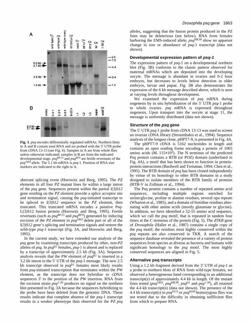
psq transcriptsTo facilitate our understanding of the psq mutant phe-notypes, we have conducted a molecular analysis ofthe gene. We previously reported the identification ofa 5.1 kb psq transcript in wild-type females and theisolation of a 1.0 kb partial cDNA, 13-13, represent-ing this message (Horowitz and Berg, 1995); we nowcall the 5.1 kb transcript psq-1. Using a 300 bpfragment from the 5′ UTR of psq-1 as a probe tonortherns of RNA from wild-type adult males,females and isolated ovaries, two transcripts arerevealed, the 5.1 kb psq-1 and a 6 kb message (Fig.3). psq-1 is highly abundant in females but below thelevel of detection in males, while the 6 kb transcriptappears to be present equally in both sexes. It shouldbe noted that probes further 3′ in the psq-1 messagedetect additional transcripts by northern analysis (seebelow).
We have found that the psq-1 transcript is reducedin amount in females from all the PZ lines and is
genes.ns twond the
replaced by an abundant 1.6 kb transcript (Horowitz and Berg,1995; and see 2403 and 0115/CyO in Fig. 3A). In a previousstudy, we demonstrated that this product results from an
1863Drosophila psq gene
Fig. 3. psq encodes differentially regulated mRNAs. Northern blotsin A and B contain total RNA and are probed with the 5′ UTR probefrom cDNA 13-13 (see Fig. 6). Samples in A are from whole fliesunless otherwise indicated; samples in B are from the indicateddevelopmental stage. psqRF13 and psqRF9 are fertile revertants of thepsq2403 allele. The 5.1 kb mRNA is psq-1. Position of RNA sizemarkers are indicated to the right in A.
aberrant splicing event (Horowitz and Berg, 1995). The PZelements in all four PZ mutant lines lie within a large intronof the psq gene. Sequences present within the partial l(3)S12gene residing on the PZ element provide a splice acceptor siteand termination signal, causing the psq-initiated transcript tobe spliced to l(3)S12 sequence in the PZ element, thentruncated. This truncated mRNA encodes a putative Psq-L(3)S12 fusion protein (Horowitz and Berg, 1995). Fertilerevertants (such as psqRF13 and psqRF9) generated by inducingexcision of the PZ element in psq2403 delete part or all of thel(3)S12 gene’s splicing and termination signals and restore thewild-type psq-1 transcript (Fig. 3A, and Horowitz and Berg,1995).
In the current study, we have extended our analysis of thepsq gene by examining transcripts produced by other, non-PZalleles of psq. In psqfs1 females, psq-1 is absent and is replacedby a transcript of approximately 2.5 kb (Fig. 3A). Sequenceanalysis reveals that the PW element of psqfs1 is inserted in a3.2 kb intron in the 5′ UTR of the psq-1 message. The new 2.5kb transcript observed in psqfs1 females most likely resultsfrom psq-initiated transcription that terminates within the PWelement, as the transcript does not hybridize to cDNAsequences 3′ to the position of the PW insertion. RNA fromthe excision strain psq1-30 produces no signal on the northernblot presented in Fig. 3A because the sequences hybridizing tothe probe have been deleted from the genomic DNA. Theseresults indicate that complete absence of the psq-1 transcriptresults in a weaker phenotype than observed for the PZ psq
alleles, suggesting that the fusion protein produced in the PZlines may be deleterious (see below). RNA from femalesharboring the EMS-induced allele, psqHK38 show no apparentchange in size or abundance of psq-1 transcript (data notshown).
Developmental expression pattern of psq-1The expression pattern of psq-1 on a developmental northernblot (Fig. 3B) conforms to the classic pattern observed formaternal mRNAs which are deposited into the developingoocyte. The message is abundant in ovaries and 0-2 hourembryos, but decreases to levels below detection in olderembryos, larvae and pupae. Fig. 3B also demonstrates theexpression of the 6 kb message described above, which is seenat varying levels throughout development.
We examined the expression of psq mRNA duringoogenesis by in situ hybridization of the 5′ UTR psq-1 probeto whole ovaries. psq mRNA is expressed throughoutoogenesis. Upon transport into the oocyte at stage 11, themessage is uniformly distributed (data not shown).
Structure of the psq geneThe 5′ UTR psq-1 probe from cDNA 13-13 was used to screenan ovarian cDNA library (Stroumbakis et al., 1994). Sequenceanalysis of the longest clone, pHPT7-9, is presented in Fig. 4A.
The pHPT7-9 cDNA is 5162 nucleotides in length andcontains an open reading frame encoding a protein of 1065amino acids (Mr 115×103). The N terminus of the predictedPsq protein contains a BTB (or POZ) domain (underlined inFig. 4A), a motif that has been shown to function in protein-protein interactions (Bardwell and Treisman, 1994; Chen et al.,1995). The BTB domain of psq has been cloned independentlyby virtue of its homology to other BTB domains in a studydesigned to isolate members of the BTB family of proteins(BTB-V in Zollman et al., 1994).
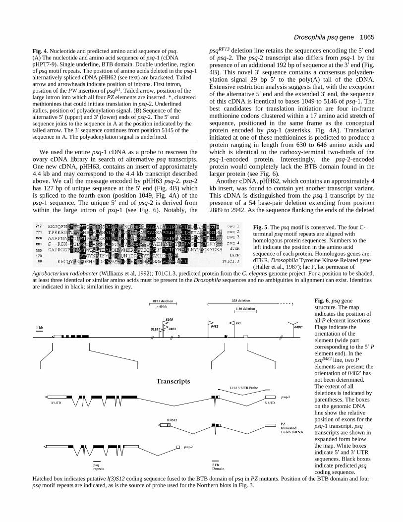
The Psq protein contains a number of repeated amino acidsequences, including multiple regions enriched forserine/glycine, proline or alanine residues, several opa repeats(Wharton et al., 1985), and a domain of histidine residues alter-nating with other amino acids (mostly glutamate or glycine).In addition, we have identified a 52-55 amino acid sequence,which we call the psq motif, that is repeated in tandem fourtimes at the C terminus of the protein (Fig. 5). The dTKR geneof Drosophila (Haller et al., 1987) contains a single copy ofthe psq motif; the residues most highly conserved within thepsq repeats are also conserved in TKR. A search of thesequence database revealed the presence of a variety of proteinsequences from species as diverse as bacteria and humans withsignificant homology to the psq motif. The most highlyconserved sequences are aligned in Fig. 5.
Alternative psq transcriptsUsing a 1.2 kb fragment derived from the 3′ UTR of psq-1 asa probe to northern blots of RNA from wild-type females, weobserved a heterogeneous band corresponding to an additionaltranscript(s) of approximately 4.4 kb in length. Of the mutantlines tested (psq2403, psqHK38, psqfs1 and psq1-30), all retainedthe 4.4 kb transcript(s) (data not shown). The presence of the4.4 kb transcript(s) in psq0115 or psq8109 homozygous flies wasnot tested due to the difficulty in obtaining suffficient fliesfrom which to prepare RNA.
1864 H. Horowitz and C. A. Berg
A
B
1865Drosophila psq gene
Fig. 4. Nucleotide and predicted amino acid sequence of psq.(A) The nucleotide and amino acid sequence of psq-1 (cDNApHPT7-9). Single underline, BTB domain. Double underline, regionof psq motif repeats. The position of amino acids deleted in the psq-1alternatively spliced cDNA pHH62 (see text) are bracketed. Tailedarrow and arrowheads indicate position of introns. First intron,position of the PW insertion of psqfs1. Tailed arrow, position of thelarge intron into which all four PZ elements are inserted. *, clusteredmethionines that could initiate translation in psq-2. Underlineditalics, position of polyadenylation signal. (B) Sequence of thealternative 5′ (upper) and 3′ (lower) ends of psq-2. The 5′ endsequence joins to the sequence in A at the position indicated by thetailed arrow. The 3′ sequence continues from position 5145 of thesequence in A. The polyadenylation signal is underlined.
We used the entire psq-1 cDNA as a probe to rescreen theovary cDNA library in search of alternative psq transcripts.One new cDNA, pHH63, contains an insert of approximately4.4 kb and may correspond to the 4.4 kb transcript describedabove. We call the message encoded by pHH63 psq-2. psq-2has 127 bp of unique sequence at the 5′ end (Fig. 4B) whichis spliced to the fourth exon (position 1049, Fig. 4A) of thepsq-1 sequence. The unique 5′ end of psq-2 is derived fromwithin the large intron of psq-1 (see Fig. 6). Notably, the
Hatched box indicates putative l(3)S12 coding sequence fused to the BTBpsq motif repeats are indicated, as is the source of probe used for the Nor
Agrobacterium radiobacter (Williams et al, 1992); T01C1.3, predicted pat least three identical or similar amino acids must be present in the Drosare indicated in black; similarities in grey.
24030115
8109
AAl(3)S12
( )RF13 deletion
psqrepeats
Transcripts
psq-2
3' UTR
1 kb
> 40 kb
psqRF13 deletion line retains the sequences encoding the 5′ endof psq-2. The psq-2 transcript also differs from psq-1 by thepresence of an additional 192 bp of sequence at the 3′ end (Fig.4B). This novel 3′ sequence contains a consensus polyaden-ylation signal 29 bp 5′ to the poly(A) tail of the cDNA.Extensive restriction analysis suggests that, with the exceptionof the alternative 5′ end and the extended 3′ end, the sequenceof this cDNA is identical to bases 1049 to 5146 of psq-1. Thebest candidates for translation initiation are four in-framemethionine codons clustered within a 17 amino acid stretch ofsequence, positioned in the same frame as the conceptualprotein encoded by psq-1 (asterisks, Fig. 4A). Translationinitiated at one of these methionines is predicted to produce aprotein ranging in length from 630 to 646 amino acids andwhich is identical to the carboxy-terminal two-thirds of thepsq-1-encoded protein. Interestingly, the psq-2-encodedprotein would completely lack the BTB domain found in thelarger protein (see Fig. 6).
Another cDNA, pHH62, which contains an approximately 4kb insert, was found to contain yet another transcript variant.This cDNA is distinguished from the psq-1 transcript by thepresence of a 54 base-pair deletion extending from position2889 to 2942. As the sequence flanking the ends of the deleted
Fig. 6. psq genestructure. The mapindicates the position ofall P element insertions.Flags indicate theorientation of theelement (wide partcorresponding to the 5′ Pelement end). In thepsq0482 line, two Pelements are present; theorientation of 0482′ hasnot been determined.The extent of alldeletions is indicated byparentheses. The boxeson the genomic DNAline show the relativeposition of exons for thepsq-1 transcript. psqtranscripts are shown inexpanded form belowthe map. White boxesindicate 5′ and 3′ UTRsequences. Black boxesindicate predicted psqcoding sequence.
domain of psq in PZ mutants. Position of the BTB domain and fourthern blots in Fig. 3.
Fig. 5. The psq motif is conserved. The four C-terminal psq motif repeats are aligned withhomologous protein sequences. Numbers to theleft indicate the position in the amino acidsequence of each protein. Homologous genes are:dTKR, Drosophila Tyrosine Kinase Related gene(Haller et al., 1987); lac F, lac permease of
rotein from the C. elegans genome project. For a position to be shaded,ophila sequences and no ambiguities in alignment can exist. Identities
0482fs1
(
(
)1-30 deletion
∆18 deletion
13-13 5' UTR Probe
BTB Domain
PZtruncated1.6 kb mRNA
psq-1
5' UTR
0482'
)
1866 H. Horowitz and C. A. Berg
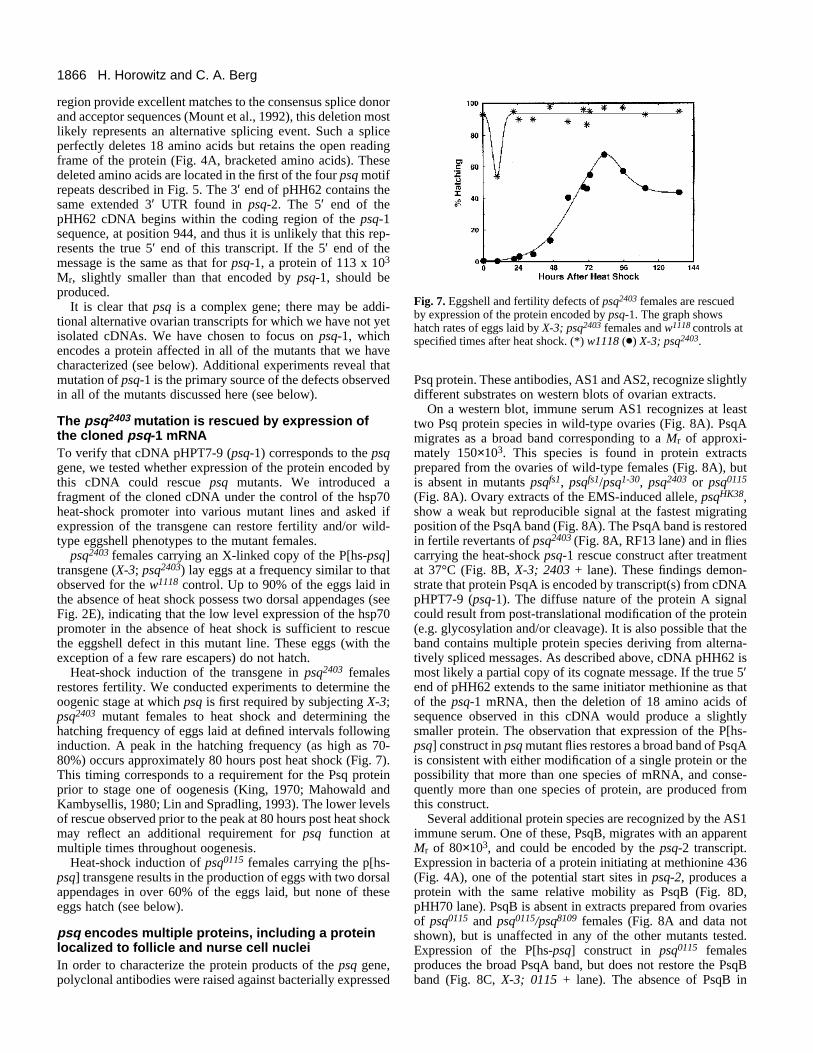
Fig. 7. Eggshell and fertility defects of psq2403 females are rescuedby expression of the protein encoded by psq-1. The graph showshatch rates of eggs laid by X-3; psq2403 females and w1118 controls atspecified times after heat shock. (*) w1118 (d) X-3; psq2403.
region provide excellent matches to the consensus splice donorand acceptor sequences (Mount et al., 1992), this deletion mostlikely represents an alternative splicing event. Such a spliceperfectly deletes 18 amino acids but retains the open readingframe of the protein (Fig. 4A, bracketed amino acids). Thesedeleted amino acids are located in the first of the four psq motifrepeats described in Fig. 5. The 3′ end of pHH62 contains thesame extended 3′ UTR found in psq-2. The 5′ end of thepHH62 cDNA begins within the coding region of the psq-1sequence, at position 944, and thus it is unlikely that this rep-resents the true 5′ end of this transcript. If the 5′ end of themessage is the same as that for psq-1, a protein of 113 x 103
Mr, slightly smaller than that encoded by psq-1, should beproduced.
It is clear that psq is a complex gene; there may be addi-tional alternative ovarian transcripts for which we have not yetisolated cDNAs. We have chosen to focus on psq-1, whichencodes a protein affected in all of the mutants that we havecharacterized (see below). Additional experiments reveal thatmutation of psq-1 is the primary source of the defects observedin all of the mutants discussed here (see below).
The psq2403 mutation is rescued by expression ofthe cloned psq-1 mRNATo verify that cDNA pHPT7-9 (psq-1) corresponds to the psqgene, we tested whether expression of the protein encoded bythis cDNA could rescue psq mutants. We introduced afragment of the cloned cDNA under the control of the hsp70heat-shock promoter into various mutant lines and asked ifexpression of the transgene can restore fertility and/or wild-type eggshell phenotypes to the mutant females.
psq2403 females carrying an X-linked copy of the P[hs-psq]transgene (X-3; psq2403) lay eggs at a frequency similar to thatobserved for the w1118 control. Up to 90% of the eggs laid inthe absence of heat shock possess two dorsal appendages (seeFig. 2E), indicating that the low level expression of the hsp70promoter in the absence of heat shock is sufficient to rescuethe eggshell defect in this mutant line. These eggs (with theexception of a few rare escapers) do not hatch.
Heat-shock induction of the transgene in psq2403 femalesrestores fertility. We conducted experiments to determine theoogenic stage at which psq is first required by subjecting X-3;psq2403 mutant females to heat shock and determining thehatching frequency of eggs laid at defined intervals followinginduction. A peak in the hatching frequency (as high as 70-80%) occurs approximately 80 hours post heat shock (Fig. 7).This timing corresponds to a requirement for the Psq proteinprior to stage one of oogenesis (King, 1970; Mahowald andKambysellis, 1980; Lin and Spradling, 1993). The lower levelsof rescue observed prior to the peak at 80 hours post heat shockmay reflect an additional requirement for psq function atmultiple times throughout oogenesis.
Heat-shock induction of psq0115 females carrying the p[hs-psq] transgene results in the production of eggs with two dorsalappendages in over 60% of the eggs laid, but none of theseeggs hatch (see below).
psq encodes multiple proteins, including a proteinlocalized to follicle and nurse cell nucleiIn order to characterize the protein products of the psq gene,polyclonal antibodies were raised against bacterially expressed
Psq protein. These antibodies, AS1 and AS2, recognize slightlydifferent substrates on western blots of ovarian extracts.
On a western blot, immune serum AS1 recognizes at leasttwo Psq protein species in wild-type ovaries (Fig. 8A). PsqAmigrates as a broad band corresponding to a Mr of approxi-mately 150×103. This species is found in protein extractsprepared from the ovaries of wild-type females (Fig. 8A), butis absent in mutants psqfs1, psqfs1/psq1-30, psq2403 or psq0115
(Fig. 8A). Ovary extracts of the EMS-induced allele, psqHK38,show a weak but reproducible signal at the fastest migratingposition of the PsqA band (Fig. 8A). The PsqA band is restoredin fertile revertants of psq2403 (Fig. 8A, RF13 lane) and in fliescarrying the heat-shock psq-1 rescue construct after treatmentat 37°C (Fig. 8B, X-3; 2403 + lane). These findings demon-strate that protein PsqA is encoded by transcript(s) from cDNApHPT7-9 (psq-1). The diffuse nature of the protein A signalcould result from post-translational modification of the protein(e.g. glycosylation and/or cleavage). It is also possible that theband contains multiple protein species deriving from alterna-tively spliced messages. As described above, cDNA pHH62 ismost likely a partial copy of its cognate message. If the true 5′end of pHH62 extends to the same initiator methionine as thatof the psq-1 mRNA, then the deletion of 18 amino acids ofsequence observed in this cDNA would produce a slightlysmaller protein. The observation that expression of the P[hs-psq] construct in psq mutant flies restores a broad band of PsqAis consistent with either modification of a single protein or thepossibility that more than one species of mRNA, and conse-quently more than one species of protein, are produced fromthis construct.
Several additional protein species are recognized by the AS1immune serum. One of these, PsqB, migrates with an apparentMr of 80×103, and could be encoded by the psq-2 transcript.Expression in bacteria of a protein initiating at methionine 436(Fig. 4A), one of the potential start sites in psq-2, produces aprotein with the same relative mobility as PsqB (Fig. 8D,pHH70 lane). PsqB is absent in extracts prepared from ovariesof psq0115 and psq0115/psq8109 females (Fig. 8A and data notshown), but is unaffected in any of the other mutants tested.Expression of the P[hs-psq] construct in psq0115 femalesproduces the broad PsqA band, but does not restore the PsqBband (Fig. 8C, X-3; 0115 + lane). The absence of PsqB in
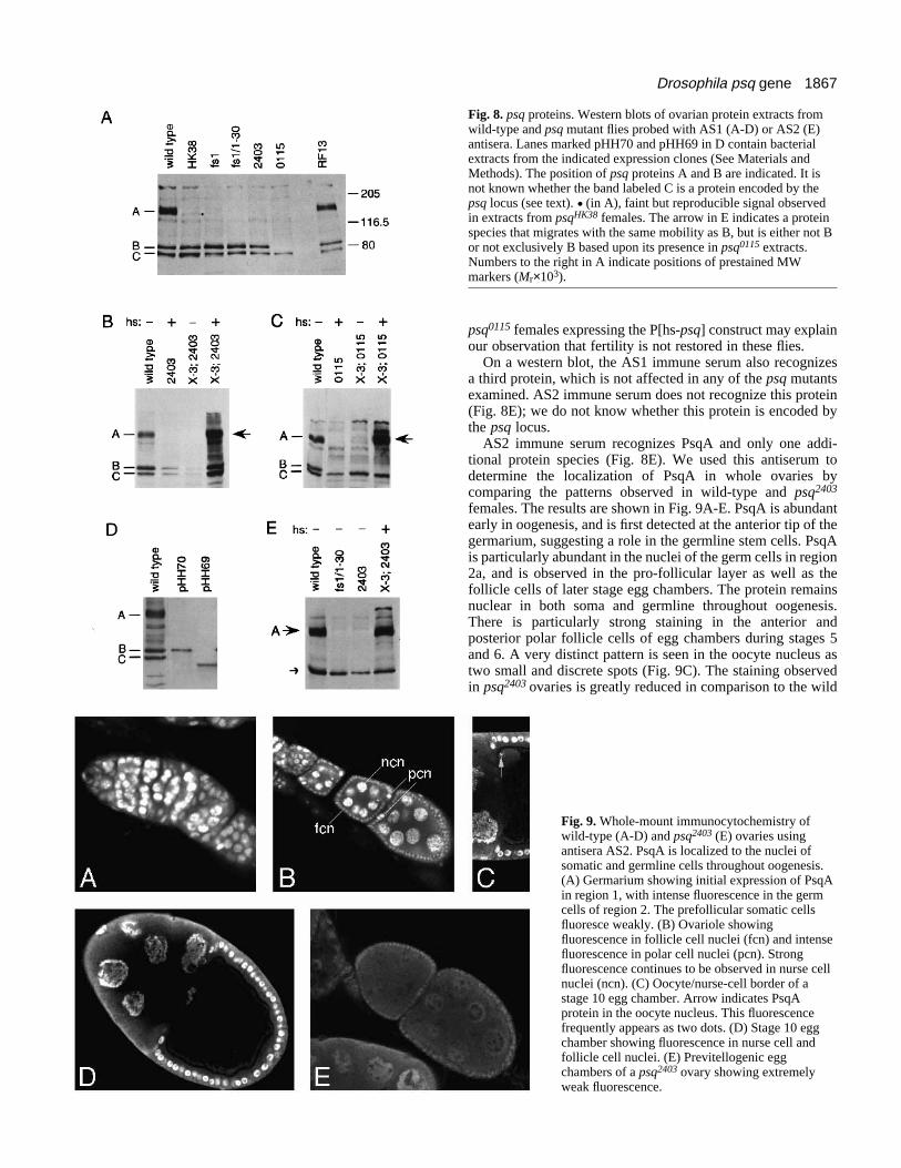
1867Drosophila psq gene
Fig. 8. psq proteins. Western blots of ovarian protein extracts fromwild-type and psq mutant flies probed with AS1 (A-D) or AS2 (E)antisera. Lanes marked pHH70 and pHH69 in D contain bacterialextracts from the indicated expression clones (See Materials andMethods). The position of psq proteins A and B are indicated. It isnot known whether the band labeled C is a protein encoded by thepsq locus (see text). . (in A), faint but reproducible signal observedin extracts from psqHK38 females. The arrow in E indicates a proteinspecies that migrates with the same mobility as B, but is either not Bor not exclusively B based upon its presence in psq0115 extracts.Numbers to the right in A indicate positions of prestained MWmarkers (Mr×103).
psq0115 females expressing the P[hs-psq] construct may explainour observation that fertility is not restored in these flies.
On a western blot, the AS1 immune serum also recognizesa third protein, which is not affected in any of the psq mutantsexamined. AS2 immune serum does not recognize this protein(Fig. 8E); we do not know whether this protein is encoded bythe psq locus.
AS2 immune serum recognizes PsqA and only one addi-tional protein species (Fig. 8E). We used this antiserum todetermine the localization of PsqA in whole ovaries bycomparing the patterns observed in wild-type and psq2403
females. The results are shown in Fig. 9A-E. PsqA is abundantearly in oogenesis, and is first detected at the anterior tip of thegermarium, suggesting a role in the germline stem cells. PsqAis particularly abundant in the nuclei of the germ cells in region2a, and is observed in the pro-follicular layer as well as thefollicle cells of later stage egg chambers. The protein remainsnuclear in both soma and germline throughout oogenesis.There is particularly strong staining in the anterior andposterior polar follicle cells of egg chambers during stages 5and 6. A very distinct pattern is seen in the oocyte nucleus astwo small and discrete spots (Fig. 9C). The staining observedin psq2403 ovaries is greatly reduced in comparison to the wild
Fig. 9. Whole-mount immunocytochemistry ofwild-type (A-D) and psq2403 (E) ovaries usingantisera AS2. PsqA is localized to the nuclei ofsomatic and germline cells throughout oogenesis.(A) Germarium showing initial expression of PsqAin region 1, with intense fluorescence in the germcells of region 2. The prefollicular somatic cellsfluoresce weakly. (B) Ovariole showingfluorescence in follicle cell nuclei (fcn) and intensefluorescence in polar cell nuclei (pcn). Strongfluorescence continues to be observed in nurse cellnuclei (ncn). (C) Oocyte/nurse-cell border of astage 10 egg chamber. Arrow indicates PsqAprotein in the oocyte nucleus. This fluorescencefrequently appears as two dots. (D) Stage 10 eggchamber showing fluorescence in nurse cell andfollicle cell nuclei. (E) Previtellogenic eggchambers of a psq2403 ovary showing extremelyweak fluorescence.

1868 H. Horowitz and C. A. Berg
Fig. 10. In situ hybridization of wild-type (A) andpsqPZ (B-C) egg chambers using grk sequences as aprobe. Lower levels of message in PZ egg chambersrequired that the color reaction proceed for twice thelength of time as was utilized for wild-type eggchambers to obtain a photographable representation ofthe localization pattern. (A) In Canton S egg
chambers, grk mRNA is localized in a tight cap above the oocyte nucleus. (B) In psq2403 egg chambers, grk message is localized to the dorsalregion of the oocyte, but sometimes appears more diffuse than in wild type. (C) In approximately 25% of psq8109/psq2403 egg chambers, grkmessage localization is abnormal.
Fig. 11. Production of the PsqA-L(3)S12 fusion protein in wild-typefemales induces a dramatic decrease in egg-laying approximately 4days following heat shock. Two samples of w1118 control animalsand six independent insertion lines carrying the P[hs-psq-l(3)S12]transgene were placed on a daily heat-shock regimen. Number of laideggs were counted at 12 hour intervals. Shown here are a weightedaverage of the two control samples (dashed line) and three of the sixexperimental lines (solid lines). Females laid more eggs at night thanduring the day, presumably due to circadian rhythms and to theeffect of the daily heat shock.
type (Fig. 9E), indicating that the patterns described above pre-dominantly reflect the distribution of PsqA.
grk localization is affected in psq mutantsThe eggshell and cuticle defects that we observed with somepsq mutant alleles suggested that dorsal/ventral patterning isdisrupted in these mutants. To determine if this effect ismediated by the gurken signal transduction pathway, weexamined the localization of grk mRNA in ovaries from psqmutants. In ovaries from wild-type females, grk mRNA islocalized to the oocyte in early egg chambers and by stage 8is seen as a tight cap on the dorsal side of the oocyte nucleus(Fig. 10A). In egg chambers from psq2403 homozygousfemales and psq2403/psq8109 transheterozygous females, theamount of grk mRNA is reduced. In psq2403 mutants, grkmRNA is localized correctly in 90% (n=46) of stage 10 eggchambers (Fig. 10B). In the remaining 10% of egg chambers,grk mRNA is found as a diffuse band in the dorsal, anteriorregion of the oocyte, or fails to be localized at all. Inpsq2403/psq8109 mutants, grk message is mislocalized in 24%(n=29) of stage 10 egg chambers. In these cases, message iseither uniformly distributed throughout the oocyte or presentin a ring at the anterior end (Fig. 10C). The combination ofreduced levels and mislocalization of grk mRNA could leadto the unusual eggshell phenotypes that we observe in PZ psqmutants (Fig. 2). It is also possible that some contribution tothe mutant phenotype is due to defects downstream of grk inthe follicle cells.
Expression of a Psq-L(3)S12 fusion protein in wild-type flies causes sterility in femalesAnalysis of the mutant phenotypes associated with the PZalleles suggests that the aberrant splicing of psq into thel(3)S12 sequences in these lines creates a fusion protein withnegative effects on oogenesis. To test this possibility, weexpressed the predicted Psq-L(3)S12 fusion protein in trans-genic flies. We generated a construct, P[hs-psq-l(3)S12], inwhich a mRNA encoding the fusion protein could be expressedby heat-shock induction from the hsp70 promoter. Six inde-pendent transgenic lines containing homozygous insertions ofthis construct were placed on a daily heat-shock regime andtheir egg-laying abilities compared to w1118 control animals.Three and a half days following the initial heat shock, all sixtransgenic lines demonstrate a dramatic drop in the number ofeggs laid (Fig. 11, and data not shown), while control animalscontinue to lay eggs at a high rate. Dissection of the ovariesfrom these animals reveals a decreased number of eggchambers at all stages. In addition, a significant proportion oflate stage egg chambers are short in length, with partially dor-salized eggshell structures. Thus, overexpression of the Psq-
L(3)S12 fusion protein is sufficient to render flies sterile, phe-nocopying weak PZ alleles.
DISCUSSION
We report here the cloning and molecular characterization ofthe Drosophila psq gene, which encodes a novel nuclearprotein involved in axis definition. Our results demonstrate thatpsq is a complex gene encoding multiple differentially splicedtranscripts and several protein isoforms. In the course of sub-mitting this manuscript, a characterization of the embryonicpsq transcripts appeared in the literature (Weber et al., 1995).A sequence comparison of the ovarian and embryonic psq tran-scripts reveals that they have distinct 5′ and 3′ ends, and thatthe proteins encoded by transcripts from the two tissues differslightly at their C termini (see Materials and Methods fordetails).
Protein structureWe have focused our efforts on the characterization of psq-1,the mRNA that encodes PsqA. This protein, which is drasti-cally reduced or absent in all of the psq mutants that we haveexamined, has a number of interesting features. At the amino
1869Drosophila psq gene
terminus, PsqA contains a BTB domain (Godt et al. 1993; alsoreferred to as a POZ domain by Bardwell and Treisman, 1994),a motif that has been shown to function in protein-protein inter-actions. Although BTB domains are often found near the Nterminus of Cys2-His2 zinc finger proteins, PsqA does notappear to contain a zinc finger. Downstream of the BTBdomain, PsqA contains 34 alternating histidine residues,(HX)n, a motif that is present in a number of other Drosophilaproteins, primarily transcription factors. It has been proposedthat these histidine repeats could mediate protein-protein inter-actions by coordinating metal ions to form a ‘histidine-metalzipper’ between two proteins containing the repeats (Janknechtet al., 1991). The presence of two potential protein-proteininteraction domains suggests that PsqA monomers mayinteract with each other or with heterologous protein species.Additionally, PsqA contains four tandem copies of a conservedsequence of unknown function at its carboxy terminus; we callthis sequence the psq motif.
We have identified another psq transcript, psq-2, whichencodes a second isoform of the Psq protein lacking both theBTB and (HX)n domains but retaining the psq motif repeats.This isoform is unlikely to be involved in the same type ofprotein-protein interactions predicted for PsqA. The finding ofBardwell and Treisman (1994) that a BTB-containing zincfinger protein was more competent to bind DNA when theBTB domain was removed raises the possibility that theisoform encoded by psq-2 could be an activated form of thePsq protein.
Dominant mutantsWe previously hypothesized that the PZ fusion protein mightact in a dominant-negative fashion, possibly by disruptingnormal protein-protein interactions mediated by the BTBdomain of the protein (Horowitz and Berg, 1995). This pre-diction was based on genetic analyses indicating that the pro-duction of a truncated protein is more harmful than productionof no protein at all. Our observation that the psq2403 alleleshows eggshell defects whereas the psqfs1 and psq1-30 allelesdo not, despite the fact that all three mutants display the sameloss of PsqA using our antibodies, provides additional evidencethat the fusion protein is deleterious.
Our experiments with P[hs-psq-l(3)S12] reveal thatexpression of an isolated BTB domain can behave in adominant manner. Expression of this construct in wild-typeflies leads to marked reduction in egg-laying. Furthermore, thepresence of two copies of the transgene causes a more severeeffect on egg production than a single copy (data not shown).These observations, along with the results of the rescue exper-iments (see below), suggest that the dominant nature of the PZmutation is sensitive to the ratio of the PZ and wild-type formsof the Psq protein, a notion that we previously forwarded,based on the absence of eggshell defects in PZ/+ flies(Horowitz and Berg, 1995).
A model for Psq-L(3)S12 actionAs no PsqA can be detected in any of the psq PZ alleles, theeggshell defects that we observe in these lines cannot be dueto interaction of the PZ fusion protein Psq-L(3)S12 with PsqA.However, if PsqA normally interacts with a heterologous BTB-containing protein, the presence in the PZ mutants of the PsqABTB domain without its associated carboxy-terminal
sequences may result in inappropriate activation or inactiva-tion of the heterologous protein.
Recent work by two groups lends support to this model forPsq-L(3)S12 action. Bardwell and Treisman (1994) and Chenet al. (1995) have demonstrated that the BTB domain canpromote dimerization or multimerization of proteins in eitherhomomeric or heteromeric complexes in vitro. Interestingly,the presence of the BTB domain is associated with a reductionin DNA binding by the associated zinc finger region in severalzinc finger BTB proteins tested (Bardwell and Treisman,1994). Of particular relevance to our work is the finding thatcoexpression of a protein fragment containing only the BTBdomain along with the full-length BTB-containing proteinrelieves the BTB-mediated inhibition of DNA-binding in vitro(Bardwell and Treisman, 1994). Thus, interaction of the BTBdomain present in the psq PZ fusion protein with that of a het-erologous BTB-containing DNA-binding protein might inap-propriately activate the binding activity of that protein.
In contrast, expression of a BTB domain without its associ-ated C terminus (eg. a BTB domain fused to heterologoussequence) may inactivate proteins that interact with the mutantBTB protein. Several examples of human cancers associatedwith translocations that may involve the joining of a BTBdomain from one protein to another protein have been identi-fied (see, for example, Chen et al., 1993). This observation hasled to the suggestion that transformation could arise by adominant-negative mechanism involving sequestration ofpartner BTB domain proteins in inactive complexes (Bardwelland Treisman, 1994; Chen et al., 1995). Our results provide thefirst direct evidence supporting the hypothesis that an abnormalBTB-containing protein can exert a dominant effect in vivo.
The dominant effect of the fusion protein is apparent in thedorsal/ventral defects of the PZ mutants. Because proper local-ization of grk message is required for dorsal/ventral polarity(reviewed by Schüpbach and Roth, 1994), we examined grkmRNA localization in the PZ lines. We find that PZ mutantswith more severe eggshell defects display a higher frequencyof grk mRNA mislocalization. Interestingly, in wild-type eggchambers, we see an abundance of PsqA in polar cells at stages6-7, a time when these cells signal to the oocyte. This signalingresults in a repolarization of the microtubules in the oocyte, aprocess required for proper anterior/posterior anddorsal/ventral axis definition (Ruohola et al., 1991; Lane andKalderon, 1994). It is possible that the mislocalization of grkmRNA in psq PZ mutants is due to disruption of the signalingbetween the polar follicle cells and the oocyte. In addition,these mutants may affect processes that occur in the dorsalfollicle cells during later stages, which could contribute to thedorsal/ventral defects observed. These effects may be producedby interaction of the mutant PZ protein with factors normallyassociated with the wild-type PsqA. Alternatively, the PZfusion protein may be interacting spuriously with proteinsinvolved in these signaling cascades.
Rescue of psq mutantsOur heat-shock rescue experiments suggest that psq is requiredvery early, prior to stage 1 in oogenesis. These results are con-sistent with the early oogenesis defects observed in the mostsevere psq mutants and with the abundant level of Psq proteindetected in the 16-cell cysts in region 2 of the germarium.Interestingly, the rate of hatching of heat-shocked X-3; psq2403
1870 H. Horowitz and C. A. Berg
flies does not drop back to the pre-shock value, even after 160hours post heat shock (data not shown). Rather, hatchingappears to plateau at a value intermediate to the pre-shock andpeak value of hatching observed. This may indicate that apermanent change is effected by expression of psq and thatcontinued expression of the gene is not necessary to maintainfertility. Our finding that PsqA is present in the very tip of thegermarium, and thus may be expressed in the stem cells, is con-sistent with this scenario. Alternatively, the PsqA proteinproduced upon heat shock may be quite stable, such thatfertility drops off slowly after the heat-shock peak is achieved.
PsqA plays a role in regulating gene expressionduring oogenesisAntibody staining of ovaries shows that PsqA is a nuclearprotein, expressed in both the somatically derived follicle cellsand in the germline. The apparent association of the PsqAprotein with chromatin in the nurse cells, and the specific fluor-escence in distinct dots in the oocyte nucleus, argue that PsqAprotein is associated, directly or indirectly, with DNA. Theseobservations, along with the results discussed below, suggestthat PsqA plays a role in regulating gene expression inoogenesis.
Previous work (Siegel et al., 1993) has indicated that ovariesfrom psq mutant females have reduced levels of vasa mRNAand protein, implicating psq in the regulation of vasaexpression. Expression of vasa is initially observed in thegermarium; we find that PsqA is both abundant in the earlygermarium and required very early in oogenesis, correlatingwith the presence of Vasa at this time.
High levels of PsqA are observed in the posterior polar cellsat stages 5-6, a time when these cells are purported to beinvolved in receiving and responding to a signal from the pos-teriorly localized Grk (reviewed by Anderson, 1995). It ispossible that PsqA is involved in regulating gene expressionas part of this complex signaling process.
The striking and specific pattern of PsqA localization in theoocyte nucleus at stages 6-10 (seen as two small bright spotsof fluorescence in Fig. 9C) suggests that PsqA may bind, eitherdirectly or indirectly, to a specific site on the DNA in theoocyte nucleus. It is intriguing to consider the possibility thatpsq may be directly affecting the expression of a particularlocus in the oocyte nucleus.
Finally, psq could also play a more general role in regulat-ing gene expression by affecting chromatin structure. Ourresults indicate that PsqA protein is associated with the nursecell chromatin; Siegel et al. (1993) found that psq mutantsfailed to undergo the normal decondensation of nurse cell DNAat stage 5. Interestingly, two other BTB-containing proteins,E(var)3-93D and GAGA, have been implicated in the modu-lation of chromatin structure, as well (Dorn et al., 1993;Croston et al., 1991). It is possible that the BTB-containingfusion protein produced in the psq PZ mutants may disrupt theprotein-protein interactions of these or other BTB-containingmodulators of chromatin structure, leading to some aspects ofthe mutant phenotypes we observe in these lines.
In conclusion, we have shown that psq encodes a nuclearprotein which may be involved in the control of geneexpression in oogenesis. The structure of PsqA and the natureof the psq PZ fusion protein suggest that these proteins interactdirectly with other proteins. These interactions could be
involved in regulating the expression of vasa, or other genesrequired to establish polarity. Future experiments designed toidentify the factors with which PsqA interacts should providevaluable insight into psq’s function.
We thank Haifan Lin, Vivian Siegel and Trudi Schüpbach forproviding mutant strains and DNA. We acknowledge the experttechnical assistance of Doreen Gillespie in whole-mount in situhybridization and immunocytochemistry and express our gratitude toPhilippa Webster and Alison Volpe for critical reading of the manu-script. We are especially indebted to members of the Berg lab for helpin preparation of this manuscript and for insightful discussions:Doreen Gillespie, Philippa Webster, Kim Rittenhouse, Alison Volpe,Jon Schnorr, and Rebecca Botham, and to Michael Gelb and MarkKot. This work was supported by the March of Dimes Basil O’ConnorResearch Program and NIH (GM45248).
The nucleotide sequence data reported in this paper are availablefrom the GenBank Nucleotide Sequence Databases under theaccession numbers U48358 (psq-1) and U48402 (psq-2).
REFERENCES
Altschul, S., Gish, Miller, W., Myers, E. and Lipman, D. (1990). Basic localalignment search tool. J. Mol. Biol. 215, 403-410.
Anderson, K. (1995). One signal, two body axes. Science 269, 489-490. Bardwell, V. and Treisman, R. (1994). The POZ domain: A conserved
protein-protein interaction motif. Genes Dev. 8, 1664-1677.Chasan, R. and Anderson, K. (1993). Maternal control of dorsal-ventral
polarity and pattern in the embryo. In The Development of Drosophilamelanogaster. (ed. M. Bate and A. Martinez-Arias), pp. 387-424. ColdSpring Harbor: Cold Spring Harbor Laboratory Press.
Chen, W., Zollman, S., Couderc, J.-L. and Laski, F. (1995). The BTBDomain of bric a brac mediates dimerization in vitro. Mol. Cell. Biol. 15,3424-3429.
Chen, Z., Brand, N., Chen, A., Chen, S., Tong, J., Wang, Z., Waxman, S.and Zelent, A. (1993). Fusion between a novel Krüppel-like zinc finger geneand the retinoic acid receptor-alpha locus due to a variant t(11;17)translocation associated with acute promyelocytic leukaemia. EMBO J. 12,1161-1167.
Cooley, L., Thompson, D. and Spradling, A. (1990). Constructing deletionswith defined endpoints in Drosophila. Proc. Natl. Acad. Sci. USA 87, 3170-3173.
Croston, G. E., Kerrigan, L. A., Lira, L. M., Marshak, D. R. andKadonaga, J. T. (1991). Sequence-specific antirepression of histone H1-mediated inhibition of basal RNA Polymerase II transcription. Science 251,643-649.
Dorn, R., Krauss, V., Reuter, G. and Saumweber, H. (1993). The enhancerof position effect variegation of Drosophila, E(var)3-93D, codes for achromatin protein containing a conserved domain common to severaltranscriptional regulators. Proc. Natl. Acad. Sci. USA 90, 11376-11380.
Dutton, F. and Chovnick, A. (1991). The l(3)S12 locus of Drosophilamelanogaster: heterochromatic position effects and stage-specificmisexpression of the gene in P element transposons. Genetics 128, 103-118.
Gillespie, D. and Berg, C. (1995). homeless is required for RNA localization inDrosophila oogenesis and encodes a new member of the DE-H family ofRNA-dependent ATPases. Genes Dev. 9, 2495-2508.
Godt, D., Couderc, J.-L., Cramton, S. and Laski, F. (1993). Patternformation in the limbs of Drosophila: bric a brac is expressed in both agradient and a wave-like pattern and is required for specification and propersegmentation of the tarsus. Development 119, 799-812.
González-Reyes, A., Elliott, H. and St. Johnston, D. (1995). Polarization ofboth major body axes in Drosophila by gurken-torpedo signalling. Nature375, 654-658.
Haller, J., Cote, S., Bronner, G. and Jäckle, H. (1987). Dorsal and neuralexpression of a tyrosine kinase-related Drosophila gene during embryonicdevelopment. Genes Dev. 1, 862-867.
Horowitz, H. and Berg, C. (1995). Aberrant splicing and transcriptiontermination caused by P element insertion into the intron of a Drosophilagene. Genetics 139, 327-335.
Janknecht, R., Sander, C. and Pongs, O. (1991). (HX)n repeats: a pH-
1871Drosophila psq gene
controlled protein-protein interaction motif of eukaryotic transcriptionfactors? Fed. Eur. Bioch. Soc. 295, 1-2.
Karpen, G. and Spradling, A. (1992). Analysis of subtelomericheterochromatin in the Drosophila minichromosome Dp1187 by single Pelement insertional mutagenesis. Genetics 132, 737-753.
King, R. (1970). Ovarian development in Drosophila melanogaster. NewYork: Academic Press.
Lane, M. and Kalderon, D. (1994). RNA localization along theanteroposterior axis of the Drosophila oocyte requires PKA-mediated signaltransduction to direct normal microtubule organization. Genes Dev. 8, 2986-2995.
Lin, H. and Spradling, A. (1993). Germline stem cell division and eggchamber development in transplanted Drosophila germaria. Dev. Biol. 159,140-152.
Mahowald, A. and Kambysellis, M. (1980). Oogenesis. In The Genetics andBiology of Drosophila. vol. 2d (ed. M. Ashburner and T. Wright). pp. 141-224. New York: Academic Press.
Mount, S., Burks, C., Hertz, G., Stormo, G., White, O. and Fields, C.(1992). Splicing signals in Drosophila: intron size, information content, andconsensus sequences. Nucl. Acids Res. 20, 4255-4262.
Roth, S., Neuman-Silberberg, F., Barcelo, G. and Schüpbach, T. (1995).cornichon and the EGF receptor signaling process are necessary for bothanterior-posterior and dorsal-ventral pattern formation in Drosophila. Cell81, 967-978.
Ruohola, H., Bermer, K., Baker, D., Swedlow, F., Jan, L. and Jan, Y.(1991). Role of neurogenic genes in establishment of follicle cell fate andoocyte polarity during oogenesis in Drosophila. Cell 66, 433-449.
Schüpbach, T. and Roth, S. (1994). Dorsoventral patterning in Drosophilaoogenesis. Curr. Opin. Genet. Dev. 4, 502-507.
Schüpbach, T. and Wieschaus, E. (1991). Female sterile mutations on thesecond chromosome of Drosophila melanogaster. II. Mutations blockingoogenesis or altering egg morphology. Genetics 129, 1119-1136.
Siegel, V., Jongens, T., Jan, L. and Jan Y. (1993). pipsqueak, an early actingmember of the posterior group of genes affects vasa level and germ cell-somatic cell interaction in the developing egg chamber. Development 119,1187-1202.
Spradling, A. (1986). P element-mediated transformation. In Drosophila aPractical Approach (ed. D. B. Roberts), pp. 175-197. Oxford: InformationPrinting Ltd.
Spradling, A. (1993). Developmental genetics of oogenesis. In TheDevelopment of Drosophila melanogaster. (ed. M. Bate and A. Martinez-Arias), pp. 1-69. Cold Spring Harbor: Cold Spring Harbor Laboratory Press.
St. Johnston, D. (1993). Pole plasm and the posterior group genes. In TheDevelopment of Drosophila melanogaster. (ed. M. Bate and A. Martinez-Arias), pp. 325-364. Cold Spring Harbor: Cold Spring Harbor LaboratoryPress.
Stroumbakis, N., Li, Z. and Tolias, P. (1994). RNA- and single-strandedDNA-binding (SSB) proteins expressed during Drosophila melanogasteroogenesis: a homolog of bacterial and eukaryotic mitochondrial SSBs. Gene143, 171-177.
Theurkauf, W., Smiley, S., Wong, M. and Alberts, B. (1992). Reorganizationof the cytoskeleton during Drosophila oogenesis: implications for axisspecification and intercellular transport. Development 115, 923-936.
Thummel, C. and Pirrotta, V. (1991). New pCaSpeR P element vectors.Drosophila Information News 2.
Verheyen, E. and Cooley, L. (1994). Looking at oogenesis. In Methods in CellBiology. vol. 44 (ed. L. Goldstein and E. Fyrberg), pp. 545-561. San Diego:Academic Press.
Weber, U., Siegel, V., and Mlodzik, M. (1995). pipsqueak encodes a novelnuclear protein required downstream of seven-up for the development ofphotoreceptors R3 and R4. The EMBO J. 14, 6247-6257.
Wharton, K., Yedvobnick, B., Finnerty, V. and Artavanis-Tsakonas, S.(1985). opa: a novel family of transcribed repeats shared by the Notch locusand other developmentally regulated loci in Drosophila melanogaster. Cell40, 515-562.
Wieschaus, E. and Nüsslein-Volhard, C. (1986). Looking at embryos. InDrosophila a Practical Approach (ed. D. B. Roberts), pp. 199-227. Oxford:Information Printing Ltd.
Williams, S., Greenwood, J. and Jones, C. (1992). Molecular analysis of thelac operon encoding the binding-protein-dependent lactose transport systemand β-galactosidase in Agrobacterium radiobacter. Molecular Microbiology6, 1755-1768.
Zollman, S., Godt, D., Privé, G., Couderc, J.-L. and Laski, F. (1994). TheBTB domain, found primarily in zinc finger proteins, defines anevolutionarily conserved family that includes several developmentallyregulated genes in Drosophila. Proc. Natl. Acad. Sci USA 91, 10717-10721.
(Accepted 15 March1996)