THE DIGESTIVE SYSTEMphysiology.nuph.edu.ua/wp-content/uploads/2018/01/... · 2018-01-23 · • The...
40
1 THE DIGESTIVE SYSTEM
Transcript of THE DIGESTIVE SYSTEMphysiology.nuph.edu.ua/wp-content/uploads/2018/01/... · 2018-01-23 · • The...

1
THE DIGESTIVE
SYSTEM

2
Digestion
Difficult physiological process in
which course the food which
has arrived in a digestive tube,
is exposed to mechanical and
chemical transformations, and
nutrients containing in it after a
depolymerization are soaked
up in a blood and a lymph.
3
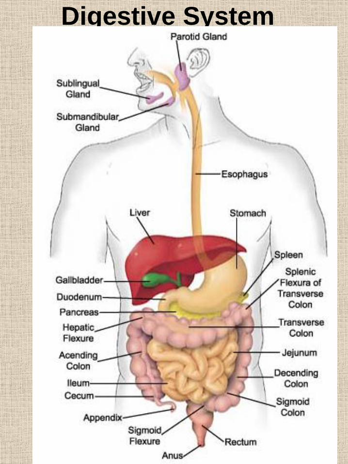
Digestive System

4
Digestive System
Provides processes physical and a chemical food
processing and its transformation into such products which
can be soaked up by vascular system for carrying over and
the further mastering by an organism.
The processes occurring in a
gastroenteric tract
(GASTROINTESTINAL TRACT)
Physical processing of nutrition
(making smaller, a ramollissement, mixing)
Nutrition chemical processing
(splitting of complex proteins, fats,
carbohydrates into monomers by the
digestive juice)
Absorption of nutrients, water, salts,
vitamins in various departments
GASTROINTESTINAL TRACT with their
subsequent entering in a blood and a lymph

5
DIGESTION IN THE ORAL
CAVITY

6
The Mouth
• Lips and cheeks enclose the
mouth.
• Taste buds on the tongue
provide the sense of taste;
skeletal muscle in the tongue
allows it to move.
• The roof of the mouth is
formed by the hard and soft
palates that separate it from
the nasal cavities.
• The soft palate ends in a
finger-shaped projection called
the uvula.

7
• Tonsils at the back sides of the
mouth protect against
infections.
• Tonsillitis results when the
tonsils become inflamed; the
infection can spread to the
middle ears.
• Three pairs of salivary glands
send saliva (containing salivary
amylase for digestion of starch
to maltose) into the mouth.

8
The Teeth
• Twenty deciduous (baby) teethare replaced by 32 adult teeth.
• Each tooth has a crown and a root.
• The crown has a layer of enamel, dentin, and an inner pulp with nerves and blood vessels that extend into the root.
• The tongue mixes the chewed food with saliva and then forms the mixture into a mass called a bolus in preparation for swallowing.

9
Adult mouth

10
Longitudinal section of
a tooth

11
TONGUETongue is an organ consisting of muscles, which
difficult movements play an essential role in such
processes, as conversation, chewing, swallowing. Its
top surface is covered by the special tissue containing
sensitive elements (papillae), distinguishing taste.

12
Taste

13
DIGESTION PROCESSES IN
THE ORAL CAVITY
1. Making smaller of a food
2. Wetting by a saliva
3. Formation of an food bolus
4. Occurrence of gustatory
sensations
The mouth, or oral cavity, is the first part of
the digestive tract. It is adapted to receive
food by ingestion, break it into small
particles by mastication, and mix it with
saliva. The lips, cheeks, and tongue form
the food bolus. The oral cavity contains the
teeth and tongue and receives the
secretions from the salivary glands.

14
Mastication
It is made by reflex
1. Irritation of receptors
2. Mastication phases:
• Rest.
• Nutrition introduction in a
mouth
• The rough
• The basic
• Formation of a food bolus
• Swallowing

15
SALIVARY GLANDS
16
SALIVARY GLANDS
• The glands are found in and around your
mouth and throat. We call the major
salivary glands the parotid,
submandibular, and sublingual glands.
• They all secrete saliva into your mouth,
the parotid through tubes that drain
saliva, called salivary ducts, near your
upper teeth, submandibular under your
tongue, and the sublingual through many
ducts in the floor of your mouth.
• Besides these glands, there are many
tiny glands called minor salivary glands
located in your lips, inner cheek area
(buccal mucosa), and extensively in other
linings of your mouth and throat. Salivary
glands produce the saliva used to
moisten your mouth, initiate digestion,
and help protect your teeth from decay.

17
SALIVARY GLANDS
Parotid (cosecrete a serous saliva, a liquid secret, doesn't contain a mucin)
Submandibular (Cosecrete an admixed saliva,
Serous-mucous secret)
Sublingual (Cosecrete an admixed saliva,
Serous-mucous secret)
A lot of tiny salivary glands that cosecreting the admixed and mucous secret rich Mucin
Water(95-99%)
The dense
rest
Mucin;
Enzyme -amylase (starch
hydrolysis);
Enzyme a maltose (maltose
hydrolysis);
Globulins;
Amino acids;
Urea;
Salts К+, Са2+.
lysocime,
IgA,
growth factors,
рН 5,8-7,4
0,5-2 l
18
SALIVA FUNCTIONS
• Digestive (hydrolysis of carbohydrates at the expense of amylase and maltose enzymes)
• Promotes occurrence of gustatory sensations (the alimentary substances dissolved by a saliva, provide nutrition influence on gustatory receptors)
• Formation of an alimentary lump (the mucin binds separate particles of nutrition)
• Excretory (as a part of a saliva metabolism products can be allocated)
• Protective (washes the irritating substances which have got to an oral cavity)
• Bactericidal action (lysocime presence)
• Stimulation of secretion a gastric juice
• Participation in the swallowing act

19
SECRETION OF THE
SALIVARY GLANDS
• Instinctive reflexes (stimuli the substances operating on receptors of an oral cavity)
• The parasympathetic department –raises secretion (a considerable quantity of a liquid saliva)
• Sympathetic – a small amount of a dense saliva
• Conditioned reflexes (stimuli the factors operating on visual, acoustical, olfactory and other receptors)

20
ESOPHAGUS
The esophagus is a muscular tube about ten inches
(25 cm.) long, extending from the hypopharynx to the
stomach. The esophagus lies posterior to the trachea
and the heart and passes through the mediastinum
and the hiatus, an opening in the diaphragm, in its
descent from the thoracic to the abdominal cavity. The
esophagus does not have serous layer; tissue around
the esophagus is called adventitia. It is used
exclusively as passage for nutrition and doesn't accept
participation in digestion and absorption process.

21
The esophagus consists of four principle layers:
• the mucosa,
• submucosa
• muscularis.
• serosa (The outermost connective tissue covering of esophagus is adventitia and it form an integral part of such organ or structure.)
22
The Wall of the
Digestive Tract
• The digestive tract wall has four
layers:
• Mucosa (mucous membrane –
secretes digestive enzymes and
mucus),
• Submucosa (loose connective
tissue – houses blood and lymph
vessels),
• Muscularis (two layers of smooth
muscle - for peristalsis), and
• Serosa (serous membrane –
secretes serous fluid to prevent
sticking).

23
• The esophagus consists of three principle layers: the
mucosa, submucosa and muscularis.
• The mucosa consists of non-keratinized stratified
squamous epithelium, lamina propria (areolar
connective tissue) and muscularis mucosa (smooth
muscle). Near the stomach the mucosa also contains
mucus glands. This provides a protective layer to the
esophagus.
• The submucosa consists of areolar connective tissue,
blood vessels and mucus glands. This provides a
loose connection between the mucosa and
muscularis.
• The muscularis consists of two layers: internal
circular muscles and external longitudinal fibers. This
provides propulsion. It should be noted that
muscularis is covered by the adventitia, an areolar
connective tissue that merges with the connective
tissue of surrounding tissues, as opposed to serosa
like the stomach, small and large intestines.
• Musculature
• The muscularis of the upper third of the esophagus is
skeletal muscle. The middle is mixed skeletal and
smooth muscle and distal is smooth muscle.
Movement is by primary peristalsis. If a food bolus
becomes lodged in the esophagus stretch receptors
stimulate secondary peristalsis causing an increase
of impulses form the swallowing center in the brain.

24
The Esophagus
• The esophagus is a muscular
tube that conducts food
through the thoracic cavity and
diaphragm into the stomach.
• Peristalsis begins in the
esophagus; this collapsed tube
moves the bolus of food
downward after swallowing
occurs.
• Heartburn is a burning pain
when acidic stomach contents
enter the esophagus.

25
• No chemical digestion occurs
in the esophagus.
• The entrance of the esophagus
to the stomach is marked by a
constriction, called a sphincter;
the sphincter must relax in
order for food to enter the
stomach.
• The sphincter prevents food
from backing up into the
esophagus.

26
Swallowing
• The air passage and food passage cross in the pharynx because the trachea is ventral to the esophagus.
• Swallowing occurs in the pharynx and is a reflex action.
• During swallowing, the air passage is usually blocked off by the soft palate and uvula, and the trachea moves under the epiglottis to cover the glottis opening to
the windpipe.

27
Swallowing
28
SWALLOWING
(or deglutition)
• Passes in 3 phases:
1. Buccal (voluntary)
2. Pharyngeal (fast involuntary)
3. The esophageal (slow involuntary)
• Regulation occurs at the expense of sympathetic and parasympathetic influences and metasympathetic nervous formations

29
Planes and stomach
areas

30
STOMACHThe stomach, which receives food from the esophagus, is
located in the upper left quadrant of the abdomen. The
stomach is divided into the fundic, cardiac, body, and pyloric
regions. The lesser and greater curvatures are on the right and
left sides, respectively, of the stomach.
31
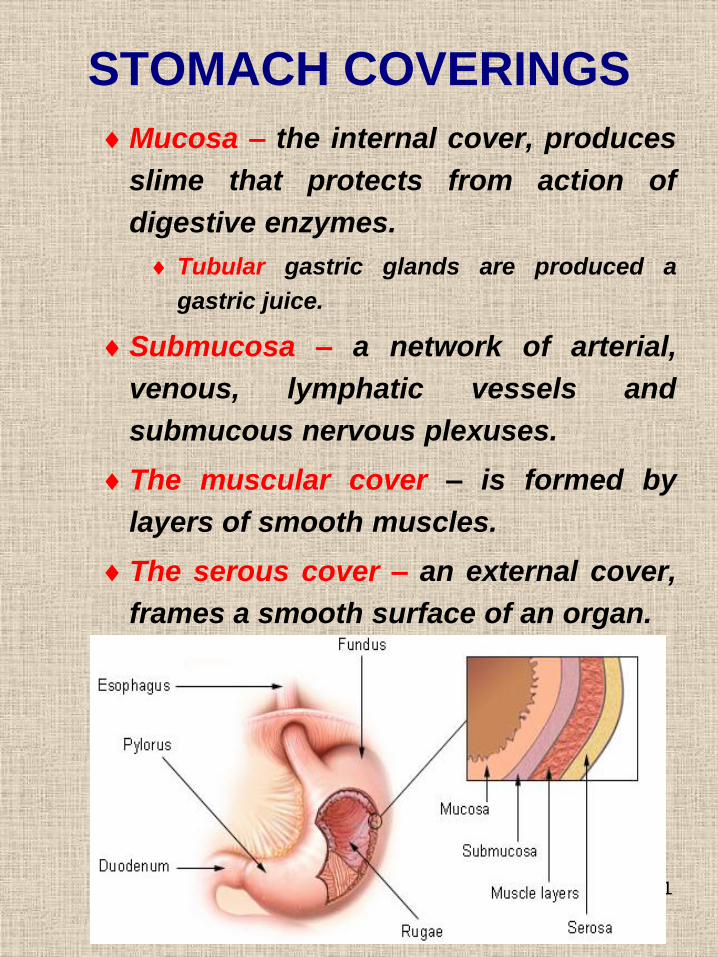
STOMACH COVERINGS
Mucosa – the internal cover, produces
slime that protects from action of
digestive enzymes.
Tubular gastric glands are produced a
gastric juice.
Submucosa – a network of arterial,
venous, lymphatic vessels and
submucous nervous plexuses.
The muscular cover – is formed by
layers of smooth muscles.
The serous cover – an external cover,
frames a smooth surface of an organ.

32
Anatomy and histology
of the stomach

33
STOMACH FUNCTIONS
Secretory – it is produced of secret by
the chief, parietal and mucous cells.
Absorbtion – promotes entering from a
stomach into an organism of water,
mineral salts, amino acids.
Motor – it is carried out at the expense of
contraction of a stomach muscular wall,
provide mixing and propulsion of chyme.
Excretory – allocation of metabolites of
proteins and carbohydrates (urea, lactic
acid) together with a gastric juice.
Incretory – secretion of some hormones
that cause a specific effect to digestion.
Bactericidal – it is provided by a
hydrochloric acid of gastric juice.
34
• Processes occurring in the
stomach:
1) Storage - the stomach allows a meal
to be consumed and the materials
released incrementally into the
duodenum for digestion. It may take
up to four hours for food from a
complete meal to clear the stomach.
2) Chemical digestion - pepsin begins
the process of protein digestion
cleaving large polypeptides into
shorter chains .
3) Mechanical digestion - the churning
action of the muscularis causes
liquefaction and mixing of the contents
to produce acid chyme.
4) Some absorption - water,
electrolytes, monosaccharides, and fat
soluble molecules including alcohol
are all absorbed in the stomach to
some degree.

35
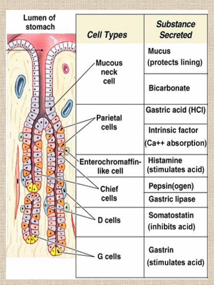
GASTRIC GLANDS
• Gastric Secretions
• The mucosal lining of the stomach is simple columnar epithelium with numerous tubular gastric glands. The gastric glands open to the surface of the mucosa through tiny holes called gastric pits. Four different types of cells make up the gastric glands:
• Mucous cells produce mucous
• Parietal cells produce H+ from reaction of CO2 and H20. HCO3 - is sent to blood in exchange for Cl-.
• Chief cells produce pepsinogen
• Endocrine cells - G) cells produce hormones such as gastrin which are secreted into the bloodstream.
• Also present, ECL cells (enterochromaffin-like cells) which produce histamine in response to gastrin and ACh.
• The secretions of the exocrine gastric glands -composed of the mucous, parietal, and chief cells - make up the gastric juice. The products of the endocrine cells are secreted directly into the bloodstream and are not a part of the gastric juice. The endocrine cells secrete the hormone gastrin, which functions in the regulation of gastric activity.
36

37
GASTRIC JUICE
STRUCTURE
• рН 0.9-1.5 2.0-2.5 lEnzymes
• Pepsin splits proteins to polypeptides
• The chemosin provides of transforming of soluble protein caseinogens in insoluble casein
• The gelatinize splits proteins of a connecting tissue gelatin
• The gastric lipase – splits emulsified milk fats
The hydrochloric acid transforms pepsinogens into pepsins, causes a denaturation of proteins and a milk coagulation, bactericidal function, control of intestine secretion.
Mucous protects an internal cover of the stomach from mechanical and chemical destruction
Inorganic substances

38
PHASES OF GASTRIC
SECRETION• Complex-reflex (cerebral) is caused by
an appearance and a flavor of food
(conditioned reflexes) and taste of
appetizing nutrition (instinctive
reflexes).
• Gastric begins after nutrition entering
into the stomach. It is regulated by
nervous, chemical and endocrine
influences on mechanoreceptors.
• Intestinal begins after the nutrition
transferring into a small intestine. It is
caused by elimination of enterogastrin
from a duodenum and a stretching of a
small intestine by chyme.

39
Regulation of a gastric
secretion
• Parasympathetic department VNS –stimulates secretion
• Sympathetic department VNS – brakes secretion
• Humoral factors:
• Gastrin - stimulates secretion
• The histamine stimulates secretion
• Digestion products - stimulate secretion
• The secretin and cholecystokinin (SIP, VIP, an enteroanthelone, a serotonin, somatostatin, etc.) – brake hydrochloric acid secretion
• Bombesin and mothilin - stimulates secretion

40
Motor function of the
stomach
• Nutrition deposition
• Hashing with a gastric juice
• Movement of gastric contents to an exit
• Portions evacuation
• PDVNS – strengthens a motility
• SDVNS – brake a motility
• Hormones:
stimulate: gastrin, motilin, serotonin, insulin;
inhibit: a secretin, cholecystokinin-pancreozymin, SIP, VIP, bulbogastron, an enteroanthelone)