Jan Bazner-Chandler CPNP, CNS, MSN, RN Respiratory Assessment.
Upload
maximilian-craigCategory
view
221download
1
The Child With a Malignancy
Jan Bazner-ChandlerCPNP,MSN, CNS, RN
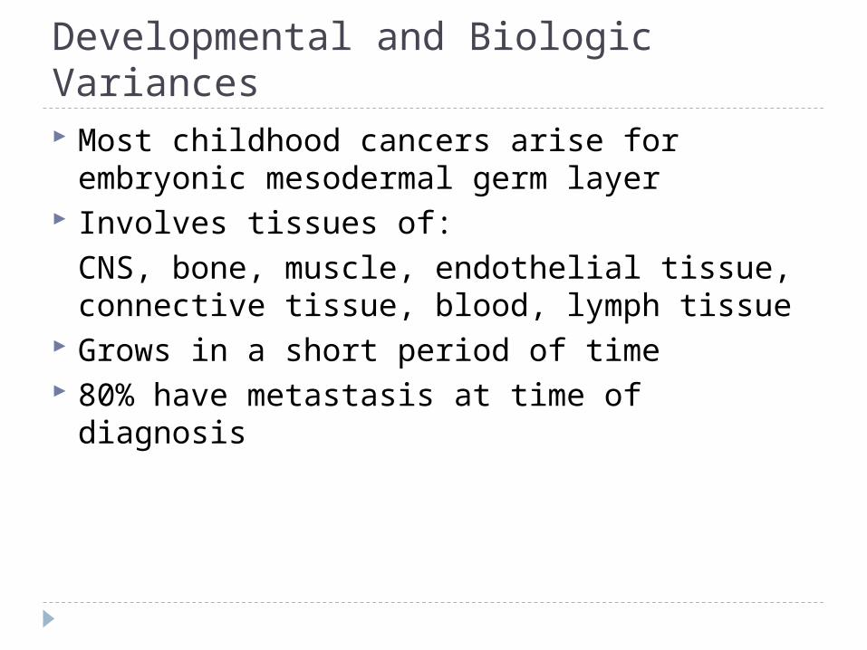
Developmental and Biologic Variances Most childhood cancers arise for embryonic
mesodermal germ layer Involves tissues of:
CNS, bone, muscle, endothelial tissue, connective tissue, blood, lymph tissue
Grows in a short period of time 80% have metastasis at time of diagnosis

Assessment Unusual mass or swelling Unexplained paleness and loss of energy Spontaneous bruising Prolonged, unexplained fever Headaches in morning Sudden eye or vision changes Excessive – rapid weight loss

Diagnostic Tests X-ray Skeletal survey CT scan Ultrasound MRI Bone marrow aspiration CBC with absolute neutrophil count Urinalysis Lumbar puncture Urine catecholamines

Treatment Modalities Determined by:
Type of cancer
Location
Extent of disease

Surgical Management The oldest form of cancer treatment Surgery plays important role in initial
diagnosis: biopsy of primary tumor. Excision of tumor when possible Facilitating treatment: insertion of catheters
for long-term treatment
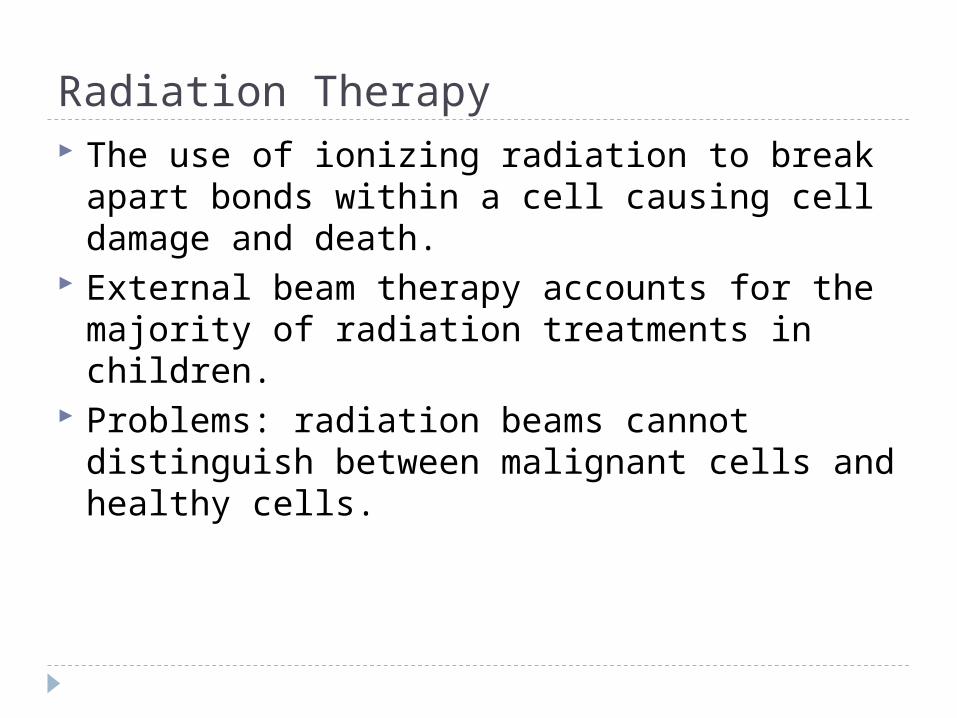
Radiation Therapy The use of ionizing radiation to break apart
bonds within a cell causing cell damage and death.
External beam therapy accounts for the majority of radiation treatments in children.
Problems: radiation beams cannot distinguish between malignant cells and healthy cells.

Chemotherapy Primary treatment modality used to cure
many pediatric cancers. Chemotherapy is the use of drugs to destroy
cancer cells. The destruction is accomplished by inhibiting
cells within the body to divide, which eventually leads to cell death.

Chemotherapy Can be given in addition to another form of
therapy such as radiation or surgery. Drugs may be administered before surgery to
reduce size of tumor. Adjuvant chemotherapy is used after surgery
or radiation therapy to prevent relapse.
Chemotherapy Combination chemotherapy is the use of more
than one class of drug. Administering different classes of chemo
drugs ensures a greater chance of achieving complete cancer cell destruction and achieving remission.

Administration Chemotherapy can be given by mouth,
subcutaneous or intramuscular injections, intravenously, or intrathecally. Oral route used if drug is well absorbed and non
irritating to the GI tract Sub-q or IM: Slow systemic release IV push, piggyback or intravenous infusion

Goals of Chemotherapy Reducing the primary tumor size Destroying cancer cells Preventing metastases and microscopic
spread of the disease
Chemotherapy Drugs Alkylating drug: attack DNA Antimetabolites: interfere with DNA
production Antitumor antibiotics: interferes with DNA
production Plant alkaloids: prevent cells from dividing Steroid hormones: slow growth of some
cancers
Bone Marrow Transplant HSCT: Hematopoietic Stem Cell Transplant:
CHLA has one of the largest program. The option of HSCT depends on the patients
disease, disease status, and general physical condition.
Involves: Umbilical cord blood Parent’s stem cells
Gene Therapy Use of gene therapy in the treatment of
childhood cancer is promising yet complex and still in early phases of clinical application.

Management Patient / family education
Begins at time of diagnosis Continues through treatment phases Maintained in post-survival years Support if death of child

Pain Management Pain caused by disease
Pain with procedures and treatments
Pain associated with side effects of treatment

Pain Management Pharmacologic
Non-Pharmacologic
Sedation or anesthetic medications EMLA cream Conscious sedation

Pain Control

Immunosuppression and Infection Children with cancer become immune impaired
from a number of causes: Lymphocyte production is altered Splenic dysfunction can prevent maturation of blood
cells and alteration is inflammatory response. Cancer therapy can decrease immunoglobulin
concentrations.

Neutropenia Significant neutropenia can develop during
chemotherapy creating an increased risk of infection in the child with cancer.
Neutropenia occurs when the absolute neutrophil count decreases below 500.

Treatment for Neutropenia Granulocyte colony stimulating factor
decreases the duration of neutropenia by stimulating the proliferation of the progenitor cells of the granulocytes, specifically the neutophils.
G-CSF: 5mcg/kg/day given subcutaneous

Varicella If an immunosuppressed child with no history
of varicella infection or varicella immunization has direct contact with an individual with chickenpox or shingles, varicella zoster immune globulin should be administered.
Acyclovir IV is used in some cases.

Varicella Immunizations Three months after chemotherapy Off prednisone Many will have already had the immunization
as a toddler since it is now a required immunization.

Central Venous Access Devices Two decades ago, CVAD were introduced as
an integral part of the pediatric oncology patient’s treatment plan.
Used to deliver chemotherapy, blood components, antibiotics, fluids, TPN, medications and blood sampling.

CVAD Infection Prevention• Teach family to report signs of catheter
infections: fever, chills, swelling, pain, drainage, or erythema.
• Aseptic technique for dressing changes and heparin flushing.
• Avoid trauma to device• Observe for catheter occlusion

Chemotherapy Side Effect Drugs affect not only the cancer cells but also
healthy cells. Cells most affected are rapidly growing cells
such as hair follicles, reproductive system, bone marrow and gastrointestinal tract.
Management of Side Effects

Malnutrition Occurs in 8 to 32% of the pediatric oncology
population Nutritional goals focus on maintaining normal
growth and development as well as preventing nutritional deficiencies.
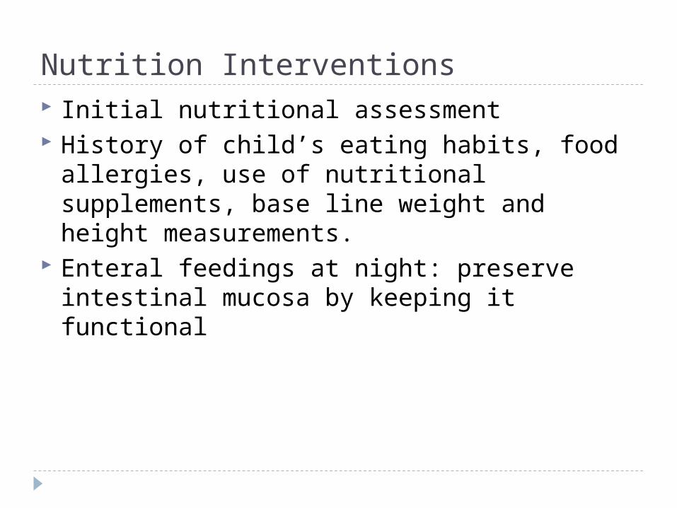
Nutrition Interventions Initial nutritional assessment History of child’s eating habits, food allergies,
use of nutritional supplements, base line weight and height measurements.
Enteral feedings at night: preserve intestinal mucosa by keeping it functional

Nausea and Vomiting Most common side effect of cancer treatment. Chemotherapy-associated vomiting is a reflex
controlled by chemoreceptor trigger zone that stimulates the vomiting center in the brain.
Tumor location Radiation therapy Anticipatory nausea

Interventions Antiemetics such as Phenothiazines:
(Trilafon), (Phenergan)and (Thorazine) block dopamine receptors from stimulating the chemoreceptor trigger zones.
Serotonin-receptor antagonist such as Granisetron (Kytril) and Ondansetron (Zofran) are very effective. (>3 years)
Antihistamines: benadryl Administer before chemotherapy
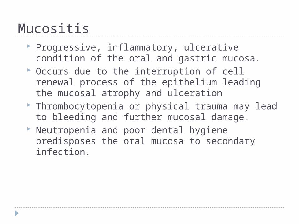
Mucositis Progressive, inflammatory, ulcerative condition of
the oral and gastric mucosa. Occurs due to the interruption of cell renewal
process of the epithelium leading the mucosal atrophy and ulceration
Thrombocytopenia or physical trauma may lead to bleeding and further mucosal damage.
Neutropenia and poor dental hygiene predisposes the oral mucosa to secondary infection.

Assessment / Interventions Baseline assessment including the oral cavity,
teeth, and gingival mucosa. History of dental exam and use of orthodontic
appliances Meticulous oral care Mouth rinses Monitor hydration status

Constipation Assess normal bowel habits Increase fiber and fluids in diet Stool softeners / colace Physical activity Avoid digital manipulation

Diarrhea Assess for signs of dehydration Record stool patterns IV fluids as needed Low-residue or lactose-free diet Good hand washing
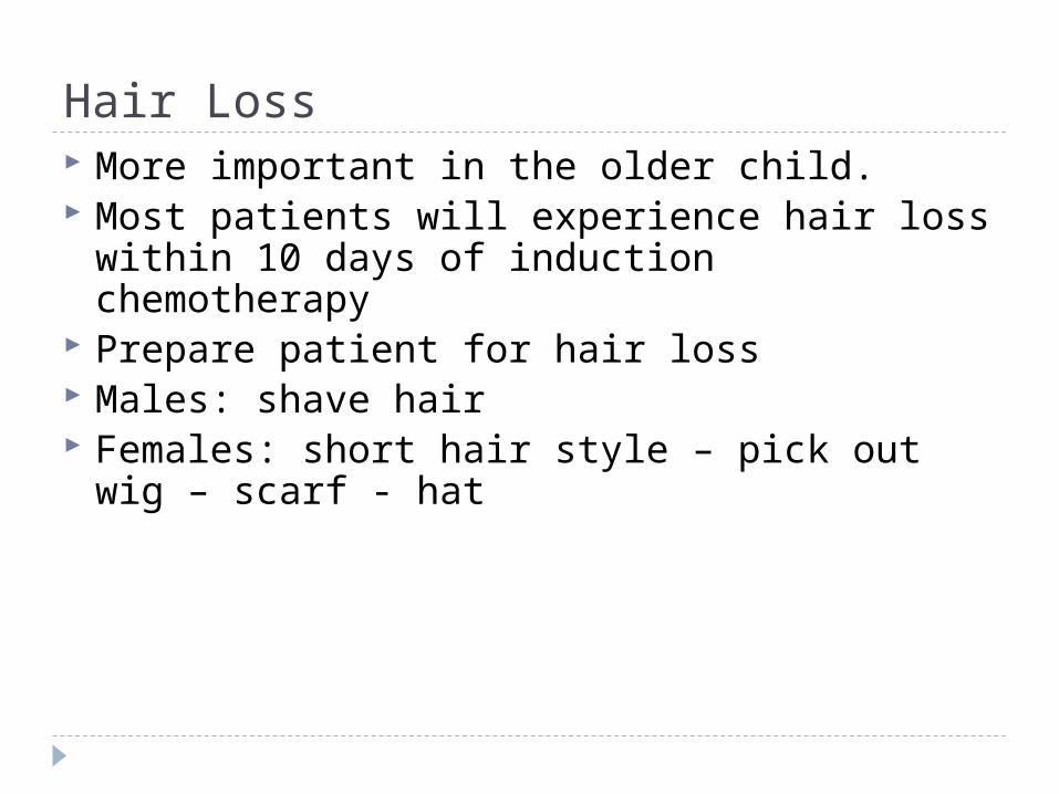
Hair Loss More important in the older child. Most patients will experience hair loss within
10 days of induction chemotherapy Prepare patient for hair loss Males: shave hair Females: short hair style – pick out wig – scarf
- hat
Psychosocial Support Support groups
Open communication
Daily contact with oncology team
Trusting relationship with nurse

Growth and Development Promote normal G & D Allow decision making Establish daily routines Play therapy Friends School attendance or tutor

Late Effects of Cancer Therapy Endocrine: sterility Thyroid Cardiovascular Musculoskeletal Vision Hearing Respiratory Gastrointestinal Genitourinary Hematopoietic

Leukemia Most common malignancy Cancer of blood or bone marrow characterized
by an abnormal proliferation of blood cells, usually white blood cells (leukocytes)
Two types Acute lymphoblastic leukemia: 78% Acute Myelogenous leukemia: 15 to 20%
High survival rate
Prognosis Initial WBC most significant The higher the count the poorer the outcomes
Greater than 100,000 WBC count = poor outcome Children under 2 years and older than 10 Girls do better than boys
Diagnosis Peripheral blood smear
Bone marrow analysis
Lumbar puncture

Assessment Pallor and fever Lethargy Anorexia Weight loss Hemorrhage / petechiae Hepatomegly / splenomegaly

3 Phase Treatment Induction: goal is to achieve remission last
about a month Consolidation: most intensive phase of
chemotherapy lasts 4 to 8 months Maintenance: last two to three years If leukemia cells are detected in bone marrow
process is started all over again.

Induction Therapy Goal of therapy is to achieve remission Leukemia cells are no longer found in the
bone marrow samples, the normal cells return and blood counts become normal.
Drugs used: L-asparaginase, vincristine and a steroid (dexamethasone), for high-risk children a fourth drug (daunorbucin) is often used

Consolidation Phase Several drugs are used in combination to
prevent remaining leukemia cells from developing resistance.
Drugs include: methotrexate and 6-mercaptopurine, vincristine and prednisone

Maintenance If leukemia continues to be in remission
maintenance therapy can be started. Two drugs: vincristine and steroids over a
brief period every 4 to 8 weeks. Duration of total therapy 2 to 3 years.

CNS Therapy CNS prophylaxis is initiated at diagnosis and is
used to reduce the risk for CNS disease. Preventive CNS is based on the premise that
the CNS provides a sanctuary site for leukemic cells that are undetected at diagnosis and reside protected from the action of systemic therapy by the brain blood barrier.
Multidisciplinary Interventions Assess for infection Monitor blood values I & O / nutrition Complications of chemotherapy Good hand washing Aseptic technique for blood draws

Leukemia Time Line 1962 cure rate for pediatric cancer is 4 %. 1971 – A combination of chemotherapy and
cranial irradiation proves it can cure at least half of all children with ALL.
1975 – A new combination of chemo drugs helps patients with reocurrence of the disease.
1991 Long-term survival rate increases to 73% with intensive induction therapy followed by two years of treatment with eight anti cancer drugs used on a rotating basis

Time Line 1997 – Bone marrow transplants from
unrelated, genetically matched donors are effective against many childhood leukemia's.
1998 – survival rate 73% to 80%. 2004 – survival rate 85% 2006 – survival rate 94%

CNS Tumors• 2nd most common malignancy• 65% have 5 year survival rate• Most common tumors:
• Astrocytomas 50%• Medulloblastomas 25%• Brain stem gliomas 10%

Assessment Classic signs and symptoms are indicative of
increased intracranial pressure. Pressure is due to tumor mass compressing
vital structure, blockage of cerebrospinal fluid flow or tumor associated edema. Gait changes / ataxia Headache with or without vomiting Blurred vision, or diplopia Forceful vomiting upon rising in the morning or
papilledema.

Multidisciplinary Interventions Surgery if tumor accessible Chemotherapy Radiation = Reserved for patient older that 2-
years of age Survival rate based on location

Chemotherapy After surgery to prevent tumor from coming
back Shrink tumor that cannot be operated on Shrink tumor so it can be operated on

Chemotherapy Blood brain barrier – natural filter within the
body that allows certain substances through from the blood to the brain tissues.
Drugs used are: temozolamide, procarbazine or lomustine
Methotrexate is injected intra-thecal Implantable wafers: drug is fixed with gel
wafer – drug is slowly released into brain over 2 to 3 weeks
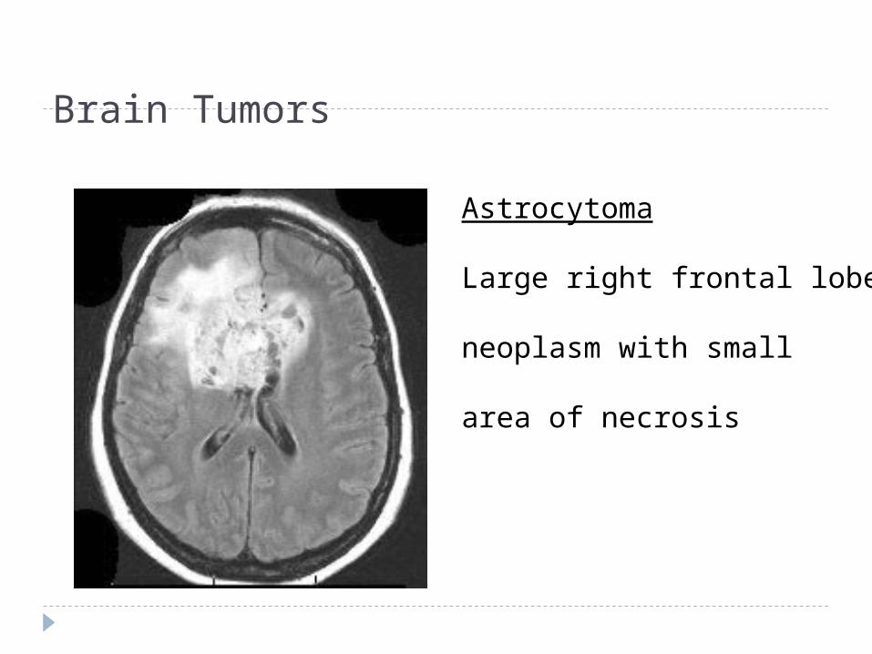
Brain Tumors
Astrocytoma
Large right frontal lobe
neoplasm with small
area of necrosis

Hodgkin's Disease 3rd most common malignancy
15 to 30 years
Three times higher in males
Excellent cure rates

Assessment Night sweats
Weight loss
Malaise
Painless, firm nodes
Treatment Radiation to nodes Chemotherapy
Combination therapy for six months Prednisone
Stem cell transplant

Neuroblastoma Approximately 600 new cases a year. Embryonic tumor Average age of diagnosis is 2 years. Poorest survival rate 50 to 60% have metastases at time of
diagnosis.

Assessment Depends on site of tumor
Diagnosis CT scan Bone scan 95% secrete catecholamines in the urine.

Multidisciplinary Interventions Determined by the stage of disease and age
of child. Children who have localized disease and
complete response to treatment are more likely to achieve a disease free state and long-term survival.
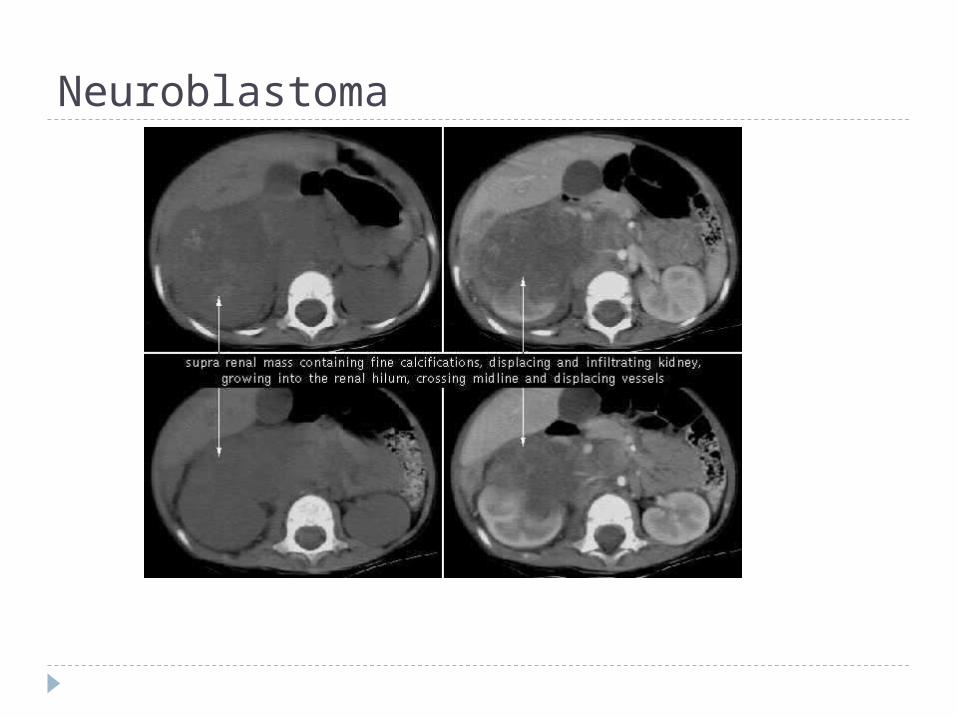
Neuroblastoma

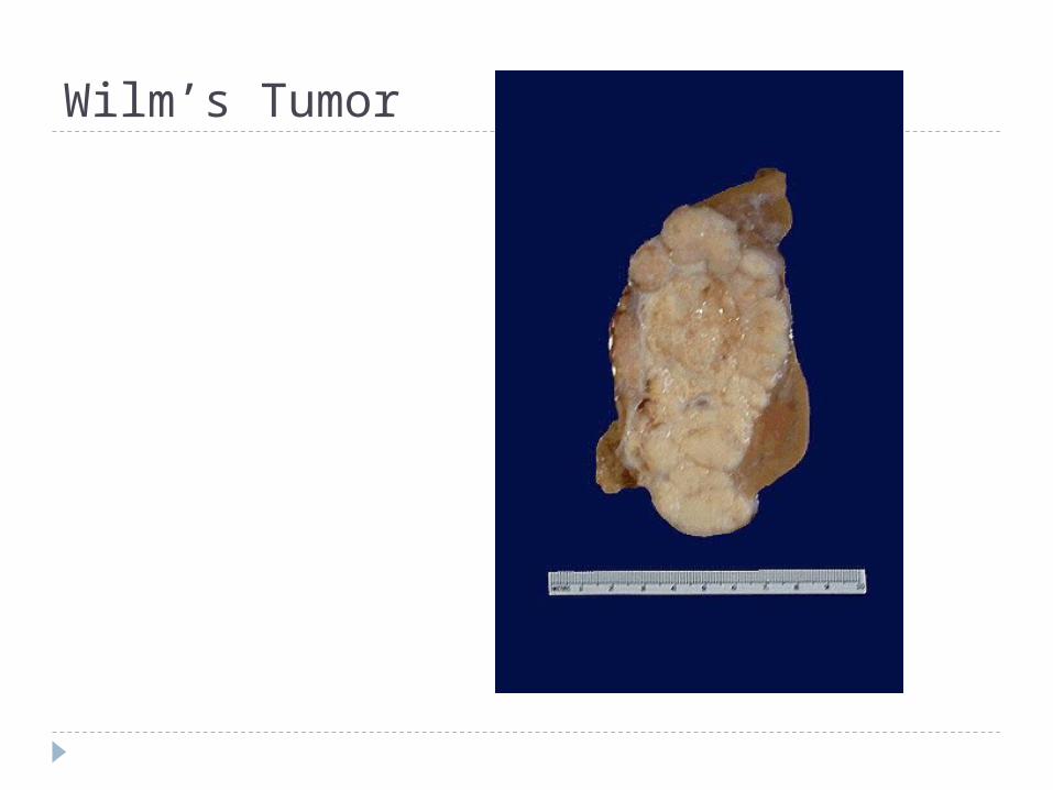
Wilm’s Tumor Most common type of renal tumor in children Approximately 460 new cases each year. Children with hypospadius or cryptorchidism
have a slightly higher incidence. African American and Females at highest risk

Assessment Firm non-tender, painless mass in abdomen
Hematuria
Hypertension
Do not palpate the abdomen

CT Scan Wilm’s Tumor
Wilm’s Tumor

Multidisciplinary Interventions Surgery
Nephrectomy Prevent rupture of capsule Sample for pathology
Chemotherapy and radiation are given based on the stage of the disease.

Osteogenic Sarcoma Malignant tumor of bone
Peak incidence between ages 10 and 20 years
Genetic predisposition
Approximately 20% have metastases at diagnosis
High rate of metastasis to lungs

Assessment Pain in affected limb that increases with
activity or weight bearing Refusal to walk and limited range of motion Tenderness and edema Diagnosis often made after traumatic injury

Diagnosis
Multidisciplinary Interventions Limb salvage
Amputation
Chemotherapy
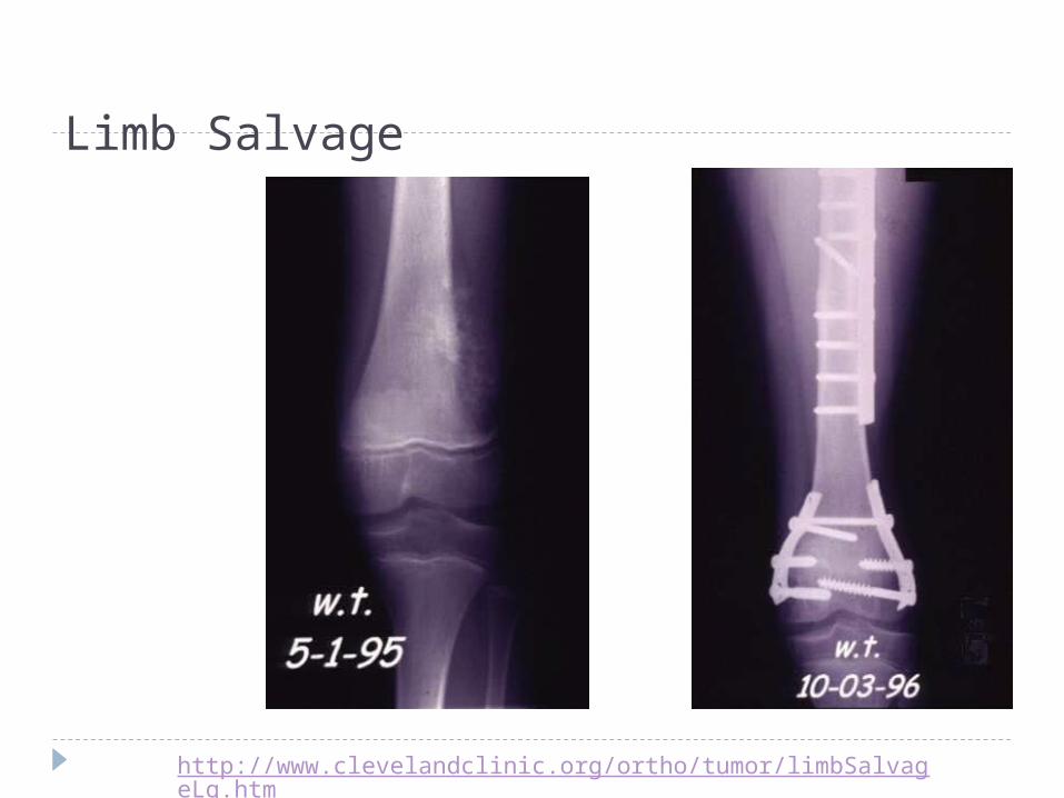
Limb Salvage
http://www.clevelandclinic.org/ortho/tumor/limbSalvageLg.htm

Ewing Sarcoma Tumor of flat bones
Pelvis, chest, vertebrae
Rare in children under 5 years
75% diagnosed by age 20

Ewing Sarcoma

Rhabdomyosarcoma Most common soft bone tissue tumor
Head and neck 40% GU 20% Extremities 20% Trunk 15%

Prognosis Location Extent of disease Children with no detectible metastases at
time of diagnosis have better outcomes Head and neck tumors have better prognosis 72% survival rate after 5 years
Multidisciplinary Interventions Biopsy of tumor mass Imaging studies Surgical removal Chemo based on tissue biopsy Radiation

Retinoblastoma Intraocular tumor composed of embryonal
retinal cells
1 in 16,000
+ family history
High incidence of malignancies

Assessment Child may initially present with strabismus Impaired vision Creamy white pupillary reflex Painful eyes from inflammation

Retinoblastoma
Pupil reflex
“Cat Eyes”

Multidisciplinary Interventions Surgical enucleation of eye
Genetic counseling
Follow-up care up to 18 Years